Embed Size (px)
Citation preview
Advances in Infectious Diseases, 2016, 6, 82-95 Published Online June 2016 in SciRes. http://www.scirp.org/journal/aid http://dx.doi.org/10.4236/aid.2016.62011
Immune Evasion by Plasmodium falciparum Parasites: Converting a Host Protection Mechanism for the Parasite’s Benefit Bismarck Dinko1*, Gabriele Pradel2 1Department of Biomedical Sciences, School of Basic and Biomedical Sciences, University of Health and Allied Sciences, Ho, Ghana 2Division of Cellular and Applied Infection Biology, Institute of Zoology, RWTH Aachen University, Aachen, Germany
Received 9 April 2016; accepted 25 June 2016; published 28 June 2016
Copyright © 2016 by authors and Scientific Research Publishing Inc. This work is licensed under the Creative Commons Attribution International License (CC BY). http://creativecommons.org/licenses/by/4.0/
Abstract Immune evasion is a strategy used by pathogenic microbes to evade the host immune system in order to ensure successful propagation. Immune evasion is particularly important for the blood stages of Plasmodium falciparum, the causative agent of the deadly disease malaria tropica. Be-cause Plasmodium blood stage parasites require human erythrocytes for replication, their ability to evade attack by the human immune system is essential for parasite survival. In order to escape immunity-induced killing, the intraerythrocytic parasites have evolved a variety of evasion me-chanisms, including expansion of plasmodial surface proteins, organ-specific sequestration of the infected red blood cells and acquisition of immune-regulatory proteins by the parasite. This re-view aims to highlight recent advances in the molecular understanding of the immune evasion strategies by P. falciparum, including antigenic variation, surface protein polymorphisms and in-vasion ligand diversification. The review will further discuss new findings on the regulatory mechan-isms applied by P. falciparum to avoid lysis by the human complement as well as killing by im-mune factors of the mosquito vector.
Keywords Malaria, Plasmodium falciparum, Immune Evasion, Infected Red Blood Cell, Merozoite, Antibody, Complement, Factor H
*Corresponding author.
How to cite this paper: Dinko, B. and Pradel, G. (2016) Immune Evasion by Plasmodium falciparum Parasites: Converting a Host Protection Mechanism for the Parasite’s Benefit. Advances in Infectious Diseases, 6, 82-95. http://dx.doi.org/10.4236/aid.2016.62011
B. Dinko, G. Pradel
1. Introduction With an estimated 214 million cases annually among 3.3 billion people at risk, the tropical disease malaria is a leading cause of death worldwide. Particularly affected are children under 5 years of age and pregnant women in sub-Saharan Africa [1]. Global efforts to roll back malaria are undermined by the spread of parasite resistance against commonly used drugs. Acute is the increasing resistance against artemisinin-based medications, which till recently were highly recommended by the WHO to treat malaria infections [2] [3]. To date, there is no pre-ventive vaccine available and besides drugs, malaria control measures are mostly based on insecticide-treated bed nets as well as spraying vector resting and breeding sites.
Malaria is caused by intracellularly living parasites of the genus Plasmodium and transmitted through the bite of a female Anopheles mosquito. Of the five Plasmodium species that are known to infect humans, P. falcipa-rum causes the majority of deaths. During a mosquito bite, sporozoites are released from the mosquito salivary glands into the human dermis, enter the bloodstream, and circulate to the liver. In the liver, a single sporozoite will undergo asexual replication within approximately 5 days to release thousands (10,000 - 30,000) of mero-zoites, which then infect red blood cells (RBCs). During erythrocytic schizogony, which takes approximately 48 hours for P. falciparum, a single merozoite grows from the ring to the trophozoite stage, eventually resulting in a schizont containing 16 - 32 daughter merozoites. These are released into circulation upon erythrocyte rupture and can each infect new erythrocytes to begin the cycle again. This erythrocytic replication phase can continue for weeks and, in the case of P. falciparum, months and is responsible for the clinical symptoms of malaria. While acute clinical symptoms of malaria include fever, headache or nausea, severe malaria results in anemia, excessive inflammation, and the sequestration of infected RBCs (iRBCs) in small blood vessels of select organs, particularly the brain. Due to stress factors like lack of nutrition or host immune pressure, a small proportion of merozoites eventually develop into sexual precursor cells, the male and female gametocytes. Within minutes af-ter their intake by the Anopheline mosquito, the gametocytes transform to male and female gametes and fertili-zation occurs in the mosquito midgut. The resulting zygote develops into an infective ookinete within the fol-lowing 20 hours, which breaks through the gut epithelium and settles down at its basal site to transform into an oocyst. Sheltered by the oocyst wall, a final round of replication occurs, during which the sporozoites are formed, which then migrate to the mosquito salivary gland to wait for the next mosquito bite [4]-[7].
The obligate intracellularly living plasmodia reside within a membrane-bound vacuole for the most part of their life-cycle. Cell invasion by the sporozoites and merozoites occurs by active penetration of the hepatocyte and the erythrocyte, respectively, resulting in the formation of a parasitophorous vacuole (PV). In order to adapt the host cell for its own purpose, Plasmodium, however, has to insert its own proteins, like receptors, transpor-ters or adhesins, into the PV membrane or the host cell plasmalemma [8] [9]. The subcellular compartmentaliza-tion is a measure of the malaria parasites to avoid the human immune system and theseparasites aim to minimize the time spent outside host cells. However, the infective sporozoite and merozoite stages are exposed to the hu-man immune system during the minutes they need to infect their target cells. Also, the modified iRBCs can be recognized by the immune components.
During the liver phase of Plasmodium, naturally acquired immunity is not very effective. It has previously been reported, however, that infected hepatocytes can be destroyed by antibody (Ab)-dependent cellular cyto-toxicity, or by the action of CD4+ and CD8+ T-cells and NK-cells, which induce final effectors such as nitric oxide [10]. The subsequent blood-stage infection, on the other hand, is characterized by a pro-inflammatory cy-tokine response, which enhances phagocytosis and killing of iRBCs by macrophages [11] [12]. Phagocytosis of the iRBCs is augmented by Ab-mediated opsonisation, which bind to plasmodial proteins that had been exported to the iRBC surface. However, these antigens are highly polymorphic and undergo clonal antigenic variation, meaning that effective opsonisation may only develop after many and varied malaria infections [13] [14]. De-spite the fact that antigens on the merozoite surface are also polymorphic, Abs against conserved or semi-con- served epitopes can develop and then either inhibit RBC invasion or result in complement-mediated killing of the merozoites [15].
In order to avoid killing by human immune components, the blood stages of P. falciparum parasites have evolved multiple immune evasion strategies in parallel. In this review, the immune evasion mechanisms em-ployed by P. falciparum to establish persistent infections and thereby increasing its chances of survival and transmission are described. Here we also focus on what is known about the underlying molecular mechanisms underpinning complement evasion in falciparum malaria and highlight the latest findings and prospects of this interesting area of research.
83
B. Dinko, G. Pradel
2. Immune Evasion by the Intraerythrocytic Parasites The success of evasion of the human immune system by the malaria parasite blood stages depends on the large repertoire of anti-genetically diverse parasite proteins displayed on the surfaces of merozoites and iRBCs. Switching in the expression of the polymorphic proteins between blood stage generations offers an efficient mechanism for the blood stages to escape Ab-mediated recognition and to thus establish chronic asymptomatic infections [16]. It also remains a possibility that immune evasion can occur at the level of gametocyte-iRBCs. Generally, the mechanisms of immune evasion by the intraerythrocytic parasites are by antigenic variation and sequestration.
2.1. Antigenic Variation In the malaria-endemic areas adults and older children develop non-sterile immunity to malaria. The acquisition of this immunity is slow and initially protects endemic individuals from susceptibility to severe malaria symp-toms. Subsequently, continuous exposure to malaria infections leads to the development of natural immunity that offers protection from clinical disease [16]-[18]. The development of clinical immunity to malaria only happens after repeated reinfections, because the parasite has evolved mechanisms to efficiently evade the human immune response via antigenic variation. This strategy is the expression of variable and distinct proteins at the different life-cycle stages of the parasites. This changes the proteins exposed to and recognized by the immune system, thereby enabling the parasite to evade immune clearance and to establish chronic infections. Despite the ability to evade immunity, some gene products involved in switching are important candidate vaccine targets because they play a role in the development of non-sterile protective immunity [16] [19] [20]. It is known that antigenic diversity develops from two mechanisms: 1) the presence of multicopy gene families encoding variant surface antigens (VSA), and 2) the presence of polymorphic alleles in the parasite population [16] [21]. While the first measure is prominent in the intraerythrocytic parasites, the second measure is better known for the me-rozoites.
After merozoite invasion of an RBC, the parasite undergoes a series of drastic morphological and biochemical changes from rings to trophozoites to schizonts [22]-[24]. It is thought that during the 24-hours lasting develop-ment of the ring stages, which are found in circulation, the machinery for RBC modifications necessary for the export and surface exposure of parasite proteins involved in immune escape is installed [25]. The resulting mod-ifications reshape the interphase between the iRBC and the host mediating their ability to sequester in micro-vasculature [16].
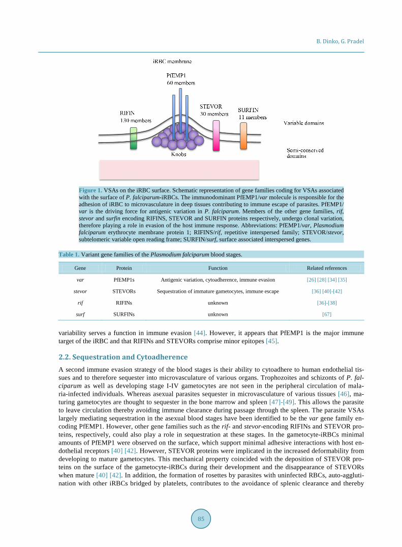
The var multigene family encodes Plasmodium falciparum erythrocyte membrane protein 1 (PfEMP1) pro-teins [26]-[28] and there are approximately 60 copies of var genes per parasite genome [29] [30]. A single var gene is expressed at one time during the ring stage while the others are silenced [31], and in the trophozoite and schizont stages, only a single antigenic variant PfEMP1 is expressed on the RBC surface (Table 1; Figure 1). However, parasites switch the expression of var genes, leading to different PfEMP1 molecules within a clonal population. Var gene silencing and switching is mediated by a variety of epigenetic mechanisms, like the in-volvement of histone modifications and the heterochromatin protein (HP1) [32]. The phenomenon of var gene switching enables the parasite to avoid immunity acquired to the already expressed PfEMP1 and this helps maintain a chronic infection via the process of clonal antigenic variation [16] [33]. The human immune response to the P. falciparum blood stages is determined mainly by recognition of the clonally variant PfEMP1 surface molecules [34] [35].
The rif and stevor gene families form part of known multigene families possibly involved in antigenic varia-tion and immune evasion during the erythrocytic stages of P. falciparum. They encode the repetitive inters-persed family (RIFIN) and sub-telomeric variable open reading frame (STEVOR) proteins, respectively, contri-buting to the iRBC surface repertoire [36] [37] (Table 1; Figure 1). Both, STEVORS and RIFINs, are expressed in a clonal fashion and undergo switches in gene expression, therefore supporting a role in antigenic variation and immune escape [38] [39] The expression of STEVOR influences the mechanical properties of the RBC membrane in asexual- and sexual-stage parasites, making them more rigid, which possibly enhances sequestra-tion of immature gametocytes and iRBCs [40]-[42]. The role of RIFINs and STEVORs in sequestration as a mechanism of immune evasion was further supported by a case report of a patient who after splenectomy expe-rienced a malaria relapse with an expansion of parasites that had lost transcription of PfEMP1, STEVOR and A-type RIFINs [43]. In volunteer infections, Abs to RIFINs were rapidly acquired, supporting the idea that their
84
B. Dinko, G. Pradel
Figure 1. VSAs on the iRBC surface. Schematic representation of gene families coding for VSAs associated with the surface of P. falciparum-iRBCs. The immunodominant PfEMP1/var molecule is responsible for the adhesion of iRBC to microvasculature in deep tissues contributing to immune escape of parasites. PfEMP1/ var is the driving force for antigenic variation in P. falciparum. Members of the other gene families, rif, stevor and surfin encoding RIFINS, STEVOR and SURFIN proteins respectively, undergo clonal variation, therefore playing a role in evasion of the host immune response. Abbreviations: PfEMP1/var, Plasmodium falciparum erythrocyte membrane protein 1; RIFINS/rif, repetitive interspersed family; STEVOR/stevor, subtelomeric variable open reading frame; SURFIN/surf, surface associated interspersed genes.
Table 1. Variant gene families of the Plasmodium falciparum blood stages.
Gene Protein Function Related references
var PfEMP1s Antigenic variation, cytoadherence, immune evasion [26] [28] [34] [35]
stevor STEVORs Sequestration of immature gametocytes, immune escape [36] [40]-[42]
rif RIFINs unknown [36]-[38]
surf SURFINs unknown [67]
variability serves a function in immune evasion [44]. However, it appears that PfEMP1 is the major immune target of the iRBC and that RIFINs and STEVORs comprise minor epitopes [45].
2.2. Sequestration and Cytoadherence A second immune evasion strategy of the blood stages is their ability to cytoadhere to human endothelial tis-sues and to therefore sequester into microvasculature of various organs. Trophozoites and schizonts of P. fal-ciparum as well as developing stage I-IV gametocytes are not seen in the peripheral circulation of mala-ria-infected individuals. Whereas asexual parasites sequester in microvasculature of various tissues [46], ma-turing gametocytes are thought to sequester in the bone marrow and spleen [47]-[49]. This allows the parasite to leave circulation thereby avoiding immune clearance during passage through the spleen. The parasite VSAs largely mediating sequestration in the asexual blood stages have been identified to be the var gene family en-coding PfEMP1. However, other gene families such as the rif- and stevor-encoding RIFINs and STEVOR pro-teins, respectively, could also play a role in sequestration at these stages. In the gametocyte-iRBCs minimal amounts of PfEMP1 were observed on the surface, which support minimal adhesive interactions with host en-dothelial receptors [40] [42]. However, STEVOR proteins were implicated in the increased deformability from developing to mature gametocytes. This mechanical property coincided with the deposition of STEVOR pro-teins on the surface of the gametocyte-iRBCs during their development and the disappearance of STEVORs when mature [40] [42]. In addition, the formation of rosettes by parasites with uninfected RBCs, auto-aggluti- nation with other iRBCs bridged by platelets, contributes to the avoidance of splenic clearance and thereby
85
B. Dinko, G. Pradel
immune escape [50]-[53].
3. Immune Evasion by Merozoites Merozoites are one of the few plasmodial stages that are extracellular and therefore directly exposed to the hu-man immunological response [54]. In order to survive, the merozoite limits the time of exposure to possible neutralization by complement-mediated lysis or opsonization by host Abs. Another strategy is to deploy a range of molecular mechanisms to evade the immune systems so as to complete the invasion process. Blocking mero-zoite invasion is an attractive vaccine target since invasion is an obligate aspect of the parasite life cycle. How-ever, any successful vaccine strategy targeting these stages would have to overcome the various types of im-mune evasion during merozoite invasion of RBCs. The mechanisms used by the merozoite to evade immunity include extensive diversity as well as complexity in the number of proteins that have been shown or hypothe-sized to be involved in the invasion process.
3.1. Polymorphism of Merozoite Surface Proteins Though the precise function of some of these proteins are not known, surface proteins of P. falciparum mero-zoites have been classed into the merozoite surface proteins (MSPs) which form a permanent structurally com-plex coat, as well as P. falciparum apical membrane antigen 1 (PfAMA1), the group of P. falciparum erythro-cyte-binding antigens (PfEBA), and the P. falciparum reticulocyte-binding proteins (PfRHs), all of which are stored in specialized apical organelles, the micronemes and the rhoptries, respectively [54]-[56].
In order to avoid immune attack, many MSPs are highly polymorphic. Further, msp genes frequently bear signatures of being under balancing selection pressure [57] [58]. These mechanisms result in immunologically distinct merozoites within a single infected individual. This distinction is made complex if a malaria patient is infected with multiple genetically distinct strains. As a result a primed immune system is less likely to effec-tively block the invasion of all merozoites [54]. Though anti-merozoite immune responses are often very strong in adults who have previously been infected with P. falciparum multiple times, this immunity is always partially effective at preventing parasite invasion [59]. The levels of MSP polymorphisms and the resulting contribution to immune evasion make them unattractive vaccine candidates. However, it is worth noting that some subdo-mains of MSPs are quite conserved and might contribute effectively towards a multivalent vaccine approach. The C-terminal domain of MSP1 is a good example of this phenomenon as it can have potent invasive inhibitory effects [15] [54].
3.2. Expansion of Invasion Ligands Besides the use of polymorphic proteins in the immune evasion process, malaria parasites have evolved func-tional redundancy in invasion ligands to evade host humoral immune response. Members of the EBA and RH families of invasion proteins are already known to be targets of host Abs [54]. Therefore if the parasite relied on a single ligand to complete the later stages of invasion, the human host could develop sterile protective immuni-ty against this ligand. Thus the parasite responds by expanding these two proteins to create paralogues, which then presents to the host a difficult task of having to block several invasion ligands. This strategy is also known as the “alternative invasion pathway” and phenotypes have been demonstrated in field isolates that have recently been adapted into culture including those in the first round of invasion [60] [61]. Another type of invasion pro-teins is AMA1, a micronemaltype I transmembrane protein that translocates to the merozoite surface [62]. AMA1 exhibits strain variability meaning that only strains encoding an AMA1 variant immunologically similar or identical to the AMA1 protein sequence variant used in a vaccine are inhibited. In addition, studies using double-knockout parasite lines provided evidence that variation in erythrocyte binding-like protein (EBL) family member expression plays a role in phenotypic variation and immune evasion [63].
Another level of antigenic variation and immune evasion in merozoites could be determined by the expression of RIFINs and STEVORs in these stages. STEVOR variants are exposed on the merozoite membrane [64]-[66] and have been reported to interact with glycophorin C as its host receptor in invasion [66]. It was also shown that Abs targeting STEVORs blocked invasion, but as STEVORs are clonally variant, the use of this avenue to evade immunity was not investigated. The surface associated interspersed gene (surf) coding for SURFINs is another gene family implicated in antigenic variation in P. falciparum merozoites [67] (Table 1). However the importance of these findings in the light of immune evasion remains to be elucidated.
86
B. Dinko, G. Pradel
4. Complement Evasion by the Blood and Sexual Stages The complement system destroys invading microbes by C3b-mediated opsonization, immune cell recruitment mediated by C3a and C5a, and the formation of a terminal complement complex (TCC) to induce targeted lysis of invading microbes. Three major pathways are known, termed the alternative, classical and lectin pathways. While the classical and lectin pathways are initiated in response to bacterial molecular patterns or anti-gen-immune-complexes, the alternative complement pathway (ACP) is active continuously at a low rate by spontaneous hydrolysis of complement factor C3.
Human complement is abundantly present in the blood and thus it is the first immune defense mechanism the malaria parasite comes into contact with, once it enters the human body. Malaria infections can induce the clas-sical pathway via the formation of antigen-Ab immune complexes or the ACP, among others via the parasite di-gestive vacuoles, which during schizont rupture are released together with the merozoites into the blood stream [68]-[73]. It is meanwhile postulated that acquired merozoite invasion-inhibitory Abs act through activation of the classical complement pathway rather than through functionally inhibiting the invading merozoites [74].
It is known that a high number of microbial pathogens are able to prevent complement recognition, among others via the recruitment of host regulators to the pathogens’ surfaces [75] [76], and increasing insights are currently gained on complement evasion by P. falciparum. While complement evasion by the sporozoites has not yet been investigated, several new studies were able to unveil the molecular mechanisms used by the blood and mosquito midgut stages of Plasmodium to avoid destruction by the ACP [77]-[79].
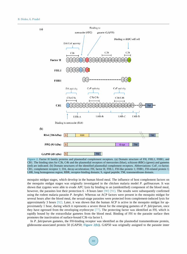
4.1. Complement Evasion by the Asexual Blood Stages In order to prevent any damage by complement, human cells use a variety of complement regulators, which ei-ther exhibit decay-acceleration and/or co-factor activity, thus they either help disassembling the C3 and C5 con-vertase complexes or support cleavage of C3b by factor I. Such inhibitors include membrane-bound regulators like complement receptor 1 (CR-1), cluster of differentiation 55 (CD55) or 59 (CD59), and central fluid-phase regulators like C4-binding protein (C4BP) and factor H (FH) [80]. As the main regulator of the ACP, FH has an important role in discriminating between self and non-self surfaces. The protein comprises 20 complement con-trol protein (CCP) modules, beta-sandwich domains containing about 60 amino acid residues. Besides FH, the FH family consists of FH-like protein (FHL1), an alternative splicing product of FH comprising CCP modules 1-7, as well as the FH-related proteins FHR1-5 (Figure 2(a)) [81]-[83].
Two new studies demonstrated that free merozoites as well as intraerythrocytic schizonts acquire FH and FHL1 to inactivate C3b attached to the iRBC surface, in consequence protecting themselves from TCC assem-bly and subsequent lysis [78] [79]. The schizont-iRBCs further bind FHR1. FH-binding to merozoites was mapped to module CCP5, while schizonts acquire FH via two contact sites, CCP5 and CCP20. Both modules are known binding-sites of bacterial microbes, CCP20 further comprises the binding motif for self-surfaces [76] [84] [85] (Figure 2(a)).
The transmembrane protein Pf92 was identified as the FH-binding receptor of merozoites [78] (Figure 2(b)). Pf92 is a member of the six-cysteine protein family, which together with other surface molecules like the ones of the MSP1 family forms multi-protein complexes on the merozoite surface. Parasites lacking Pf92 are unable to recruit FH and in consequence inefficient in C3b inactivation. Considering that Ab-mediated inactivation of FH results in significantly impaired blood stage replication [79], plasmodial receptors involved in complement eva-sion might represent highly attractive targets for malaria vaccines.
Noteworthy in this context, RBC invasion by merozoites involves CR1 (Figure 2(b)), which is recognized by Rh4 [86] [87]. The binding site of Rh4 was mapped to CCP1 of CR1, and this region is known to be involved in binding C3b and C4b to accelerate decay of the C3 and C5 convertases [88]. In consequence, Rh4 binding spe-cifically inhibits the convertase decay-acceleration activity of CR1 [89]. However, because merozoite invasion of RBCs is a rapid process, CR1 inhibition should be rather inefficient. It has thus been discussed that the high conservation of CCP1 and not its decay-acceleration activity is the reason for merozoite binding to this module [80].
4.2. Complement Evasion by the Mosquito Midgut Stages Another point of attack for the complement system to kill the malaria parasite is represented by the extracellular
87
B. Dinko, G. Pradel
Figure 2. Factor H family proteins and plasmodial complement receptors. (a) Domain structure of FH, FHL1, FHR1, and CR1. The binding sites for C3b, C4b and the plasmodial receptors of merozoites (blue), schizont-iRBCs (green) and gametes (red) are indicated. (b) Domain structures of the identified plasmodial complement receptors. Abbreviations: CoF, co-factor; CR1, complement receptor 1; DA, decay-acceleration; FH, factor H, FHL1, FH-like protein 1; FHR1, FH-related protein 1; LHR, long homogenous region; RBR, receptor-binding domain; S, signal peptide; TM, transmembrane domain. mosquito midgut stages, which develop in the human blood meal. The influence of host complement factors on the mosquito midgut stages was originally investigated in the chicken malaria model P. gallinaceum. It was shown that zygotes were able to evade APC lysis by binding to an (unidentified) component of the blood meal, however, the parasites lost their protection 6 - 8 hours later [90] [91]. The results were subsequently confirmed using the rodent malaria parasite P. berghei. Whereas rat ACP factors were present in the mosquito midgut for several hours after the blood meal, the sexual-stage parasites were protected from complement-induced lysis for approximately 3 hours [92]. Later, it was shown that the human ACP is active in the mosquito midgut for ap-proximately 1 hour, during which it represents a severe threat for the emerging gametes of P. falciparum, once they have egressed from the enveloping erythrocyte [77]. The protecting factor was identified as FH, which is rapidly bound by the extracellular gametes from the blood meal. Binding of FH to the parasite surface then promotes the inactivation of surface-bound C3b via factor I.
In P. falciparum gametes, the FH-binding receptor was identified as the plasmodial transmembrane protein, glideosome-associated protein 50 (GAP50; Figure 2(b)). GAP50 was originally assigned to the parasite inner
88
B. Dinko, G. Pradel
membrane complex (IMC), an alveolar double-membrane structure underneath the gametocyte plasmalemma, where N-terminal portion of GAP50 protrudes into the IMC lumen [93]-[95]. However, at onset of gametogene-sis the IMC disintegrates and GAP50 relocates to the plasmalemma, where it then binds the FH module CCP7 via its N-terminal portion and thus protects the extracellular gametes from attack by the human complement [77] [96].
Abs directed against GAP50 are able to reduce FH-binding to the gamete surface, leading to impaired game-togenesis and blocked transmission of the malaria parasites to the mosquito, making GAP50 a promising new candidate for transmission blocking vaccines [77]. In accordance with these data, known transmission-blocking Abs directed against prominent sexual stage surface proteins like Pfs230 or Pfs25 require active human com-plement to kill the mosquito midgut stages via the classical complement pathway [97]-[102].
5. Evasion of the Mosquito Immune System? Mosquitoes possess an innate immune system to defend microbial offenders. In this context it was previously demonstrated that Anopheles gambiae mosquitoes have the capacity to mount an immune response against plasmodia. Among others, midgut epithelial cells activate nitration reactions against Plasmodium infection, leading to reduced parasite survival by promoting lysis via thioester-containing protein 1 (TEP 1) [103]-[105]. TEP1 is an important component of the mosquito complement-like system. A recent study showed that some parasite lines from areas also endemic to A. gambiae are able to evade the mosquito immune system. However co-infection experiments revealed that parasite survival is determined by genetic differences in the various P. falciparum strains [106]. Consequently, genetic mapping, linkage group selection and functional genomics showed that P. falciparum parasites require the female gamete-specific six-cysteine protein Pfs47 to evade A. gambiae mosquito immune responses mediated by TEP1. The activity of Pfs47 may be to prevent TEP1-mediated lysis by suppressing midgut epithelial nitration responses [107].
6. Conclusion Intracellular pathogens avoid direct exposure to the immune system by infecting host cells and notably, P. fal-ciparum is not an exception to this immune evasive strategy. By infecting RBCs and liver cells, malaria para-sites eschew destruction by the host immune system, in consequence enhancing their survival and thus main-taining continuous transmission. Sequestration as an immune evasion mechanism is used by both, the asexual and sexual stages of malaria parasites. The blood stages in addition to employing VSAs and polymorphic mole-cules to escape immune attack also acquire FH to their surface to protect themselves from complement-mediated lysis. Gametes on the other hand have so far been shown only to exhibit complement evasion mechanisms by co-opting FH for protection. There is no published data as at yet demonstrating antigenic variation by gameto-cytes and/or gamete antigens, but this remains a possibility. As FH polymorphisms have already been de-scribed, investigations focusing on the potential implications of these variations on complement evasion and how the occurrence of polymorphic variants influences the incidence of malaria in the endemic setting are cur-rent pressing tasks. The findings would be of great benefit for the design of a malaria intervention with a unique opportunity to target both the asexual blood and the sexual stages, thereby preventing disease progression and transmission.
Acknowledgements This work was supported by a short term German Academic Exchange Service (DAAD) fellowship to BD. BD is also supported by the US National Institute of Health/Fogarty Global Health Fellowship as part of the Van-derbilt-Emory-Cornell-Duke consortium.GP is funded by the Heisenberg programme of the Deutsche For-schungsgemeinschaft (DFG).
References [1] WHO World Malaria Report 2015. http://www.who.int/malaria/publications/world-malaria-report-2015/report/en/ [2] Dondorp, A.M., Nosten, F., Yi, P., Das, D., Phyo, A.P., Tarning, J., Lwin, K.M., Ariey, F., Hanpithakpong, W., Lee,
S.J., Ringwald, P., Silamut, K., Imwong, M., Chotivanich, K., Lim, P., Herdman, T., An S.S., Yeung, S., Singhasiva-non, P., Day, N.P., Lindegardh, N., Socheat, D. and White, N.J. (2009) Artemisinin Resistance in Plasmodium falci-
89
B. Dinko, G. Pradel
parum Malaria. New England Journal of Medicine, 361, 455-467. http://dx.doi.org/10.1056/NEJMoa0808859 [3] Ashley, E.A., Dhorda, M., Fairhurst, R.M., Amaratunga, C., Lim, P., Suon, S., Sreng, S., Anderson, J.M., Mao, S.,
Sam, B., Sopha, C., Chuor, C.M., Nguon, C., Sovannaroth, S., Pukrittayakamee, S., Jittamala, P., Chotivanich, K., Chutasmit, K., Suchatsoonthorn, C., Runcharoen, R., Hien, T.T., Thuy-Nhien, N.T., Thanh, N.V., Phu, N.H., Htut, Y., Han, K.T., Aye, K.H., Mokuolu, O.A., Olaosebikan, R.R., Folaranmi, O.O., Mayxay, M., Khanthavong, M., Hongvan-thong, B., Newton, P.N., Onyamboko, M.A., Fanello, C.I., Tshefu, A.K., Mishra, N., Valecha, N., Phyo, A.P., Nosten, F., Yi, P., Tripura, R., Borrmann, S., Bashraheil, M., Peshu, J., Faiz, M.A., Ghose, A., Hossain, MA,., Samad, R., Rahman, M.R., Hasan, M.M., Islam, A., Miotto, O., Amato, R., MacInnis, B., Stalker, J., Kwiatkowski, D.P., Bozdech, Z., Jeeyapant, A., Cheah, P.Y., Sakulthaew, T., Chalk, J., Intharabut, B., Silamut, K., Lee, S.J., Vihokhern, B., Kunasol, C., Imwong, M., Tarning, J., Taylor, W.J., Yeung, S., Woodrow, C.J., Flegg, J.A., Das, D., Smith, J., Venkatesan, M., Plowe, C.V., Stepniewska, K., Guerin, P.J., Dondorp, A.M., Day, N.P. and White, N.J.; Tracking Resistance to Arte-misinin Collaboration (TRAC) (2014) Spread of Artemisinin Resistance in Plasmodium falciparum Malaria. New England Journal of Medicine, 371, 411-423. http://dx.doi.org/10.1056/NEJMoa1314981
[4] Mota, M.M., Hafalla, J.C. and Rodriguez, A. (2002) Migration through Host Cells Activates Plasmodium sporozoites for Infection. Nature Medicine, 8, 1318-1322. http://dx.doi.org/10.1038/nm785
[5] Frischknecht, F., Baldacci, P., Martin, B., Zimmer, C., Thiberge, S., Olivo-Marin, J.C., Shorte, S.L. and Menard, R. (2004) Imaging Movement of Malaria Parasites during Transmission by Anopheles Mosquitoes. Cellular Microbiology, 6, 687-694. http://dx.doi.org/10.1111/j.1462-5822.2004.00395.x
[6] Amino, R., Thiberge, S., Martin, B., Celli, S., Shorte, S., Frischknecht, F. and Menard, R. (2006) Quantitative Imaging of Plasmodium Transmission from Mosquito to Mammal. Nature Medicine, 12, 220-224. http://dx.doi.org/10.1038/nm1350
[7] Menard, R., Tavares, J., Cockburn, I., Markus, M., Zavala, F. and Amino, R. (2013) Looking under the Skin: The First Steps in Malarial Infection and Immunity. Nature Reviews Microbiology, 11, 701-712. http://dx.doi.org/10.1038/nrmicro3111
[8] Charpian, S. and Przyborski, J.M. (2008) Protein Transport across the Parasitophorous Vacuole of Plasmodium falci-parum: Into the Great Wide Open. Traffic, 9, 157-165.
[9] Zuccala, E.S. and Baum, J. (2011) Cytoskeletal and Membrane Remodelling during Malaria Parasite Invasion of the Human Erythrocyte. British Journal Hematology, 154, 680-689. http://dx.doi.org/10.1111/j.1365-2141.2011.08766.x
[10] Horowitz, A., Newman, K.C., Evans, J.H., Korbel, D.S., Davis, D.M. and Riley, E.M. (2010) Cross-Talk between T Cells and NK Cells Generates Rapid Effect or Responses to Plasmodium falciparum-Infected Erythrocytes. Journal of Immunology, 184, 6043-6052. http://dx.doi.org/10.4049/jimmunol.1000106
[11] Arese, P., Turrini, F. and Ginsburg, H. (1991) Erythrophagocytosis in Malaria: Host Defence or Menace to the Macro-phage? Parasitology Today, 7, 25-28. http://dx.doi.org/10.1016/0169-4758(91)90082-Y
[12] Malaguarnera, L. and Musumeci, S. (2002) The Immune Response to Plasmodium falciparum Malaria. Lancet Infec-tious Diseases, 2, 472-478. http://dx.doi.org/10.1016/S1473-3099(02)00344-4
[13] Bull, P.C., Lowe, B.S., Kortok, M., Molyneux, C.S., Newbold, C.I. and Marsh, K. (1998) Parasite Antigens on the In-fected Red Cell Surface Are Targets for Naturally Acquired Immunity to Malaria. Nature Medicine, 4, 358-360. http://dx.doi.org/10.1038/nm0398-358
[14] Ofori, M.F., Dodoo, D., Staalsoe, T., Kurtzhals, J.A., Koram, K., Theander, T.G., Akanmori, B.D. and Hviid, L. (2002) Malaria-Induced Acquisition of Antibodies to Plasmodium falciparum Variant Surface Antigens. Infection and Immun-ity, 70, 2982-2988. http://dx.doi.org/10.1128/IAI.70.6.2982-2988.2002
[15] Moss, D.K., Remarque, E.J., Faber, B.W., Cavanagh, D.R., Arnot, D.E., Thomas, A.W. and Holder, A.A. (2012) Plasmodium falciparum 19-Kilodalton Merozoite Surface Protein 1 (MSP1)-Specific Antibodies That Interfere with Parasite Growth in Vitro Can Inhibit MSP1 Processing, Merozoite Invasion, and Intracellular Parasite Development. Infection and Immunity, 80, 1280-1287. http://dx.doi.org/10.1128/IAI.05887-11
[16] Petter, M. and Duffy, M.F. (2015) Antigenic Variation in Plasmodium falciparum. Results and Problems in Cell Diffe-rentiation, 57, 47-90. http://dx.doi.org/10.1007/978-3-319-20819-0_3
[17] Bruce-Chwatt, L.J. (1963) A Longitudinal Survey of Natural Malaria Infection in a Group of West African Adults. West African Medical Journal, 12, 199-217.
[18] McGregor, I.A. (1974) Mechanisms of Acquired Immunity and Epidemiological Patterns of Antibody Responses in Malaria in Man. Bulletin of the World Health Organization, 50, 259-266.
[19] Chen, Q. (2007) The Naturally Acquired Immunity in Severe Malaria and Its Implication for a PfEMP-1 Based Vac-cine. Microbes and Infection, 9, 777-783. http://dx.doi.org/10.1016/j.micinf.2007.02.009
[20] Hviid, L. (2007) Development of Vaccines against Plasmodium falciparum Malaria: Taking Lessons from Naturally Acquired Protective Immunity. Microbes and Infection, 9, 772-776. http://dx.doi.org/10.1016/j.micinf.2007.02.008
90
B. Dinko, G. Pradel
[21] Ferreira, M.U., da Silva Nunes, M. and Wunderlich, G. (2004) Antigenic Diversity and Immune Evasion by Malaria Parasites. Clinical and Diagnostic Laboratory Immunology, 11, 987-995. http://dx.doi.org/10.1128/cdli.11.6.987-995.2004
[22] Bannister, L.H., Hopkins, J.M., Fowler, R.E., Krishna, S. and Mitchell, G.H. (2000) A Brief Illustrated Guide to the Ultrastructure of Plasmodium falciparum Asexual Blood Stages. Parasitology Today, 16, 427-433. http://dx.doi.org/10.1016/S0169-4758(00)01755-5
[23] Grüring, C., Heiber, A., Kruse, F., Ungefehr, J., Gilberger, T.W. and Spielmann, T. (2011) Development and Host Cell Modifications of Plasmodium falciparum Blood Stages in Four Dimensions. Nature Communication, 2, Article Num-ber: 165. http://dx.doi.org/10.1038/ncomms1169
[24] Bannister, L.H., Hopkins, J.M., Margos, G., Dluzewski, A.R. and Mitchell, G.H. (2004) Three-Dimensional Ultra- structure of the Ring Stage of Plasmodium falciparum: Evidence for Export Pathways. Microscopy and Microanalysis, 10, 551-562. http://dx.doi.org/10.1017/S1431927604040917
[25] Spielmann, T., Hawthorne, P.L., Dixon, M.W., Hannemann, M., Klotz, K., Kemp, D.J., Klonis, N., Tilley, L., Tren-holme, K.R. and Gardiner, D.L. (2006) A Cluster of Ring Stage-Specific Genes Linked to a Locus Implicated in Cy-toadherence in Plasmodium falciparum Codes for PEXEL-Negative and PEXEL-Positive Proteins Exported into the Host Cell. Molecular Biology and the Cell, 17, 3613-3624. http://dx.doi.org/10.1091/mbc.E06-04-0291
[26] Baruch, D.I., Pasloske, B.L., Singh, H.B, Bi, X., Ma, X.C., Feldman, M., Taraschi, T.F. and Howard, R.J. (1995) Cloning the P. falciparum Gene Encoding PfEMP1, a Malarial Variant Antigen and Adherence Receptor on the Sur-face of Parasitized Human Erythrocytes. Cell, 82, 77-87. http://dx.doi.org/10.1016/0092-8674(95)90054-3
[27] Smith, J.D., Chitnis, C.E., Craig, A.G., Roberts, D.J., Hudson-Taylor, D.E., Peterson, D.S., Pinches, R., Newbold, C.I. and Miller, L.H. (1995) Switches in Expression of Plasmodium falciparum Var Genes Correlate with Changes in An-tigenic and Cytoadherent Phenotypes of Infected Erythrocytes. Cell, 82, 101-110. http://dx.doi.org/10.1016/0092-8674(95)90056-X
[28] Su, X.Z., Heatwole, V.M., Wertheimer, S.P., Guinet, F., Herrfeldt, J.A., Peterson, D.S., Ravetch, J.A. and Wellems, T.E. (1995) The large Diverse Gene Family Var Encodes Proteins Involved in Cytoadherence and Antigenic Variation of Plasmodium falciparum-Infected Erythrocytes. Cell, 82, 89-100. http://dx.doi.org/10.1016/0092-8674(95)90055-1
[29] Gardner, M.J., Hall, N., Fung, E., White, O., Berriman, M., Hyman, R.W., Carlton, J.M., Pain, A., Nelson, K.E., Bowman, S., Paulsen, I.T., James, K., Elsen, J.A., Rutherford, K., Saizberg, S.L., Craig, A., Kyes, S., Chan, M.-S., Nene, V., Shallom, S.J., Suh, B., Peterson, J., Angluoll, S., Pertea, M., Allen, J., Selengut, J., Haft, D., Mather, M.W., Valdya, A.B., Martin, D.M.A., Fairlamb, A.H., Fraunholz, M.J., Roos, D.S., Ralph, S.A., McFadden, G.I., Cummings, L.D., Subramanlan, G.M., Mungall, C., Venter, J.C., Carucci, D.J., Hoffman, S.L., Newbold, C., Davis, R.W., Fraser, C.M. and Barrell, B. (2002) Genome Sequence of the Human Malaria Parasite Plasmodium falciparum. Nature, 419, 498-511. http://dx.doi.org/10.1038/nature01097
[30] Rask, T.S., Hansen, D.A., Theander, T.G., Pedersen, A.G. and Lavstsen, T. (2010) Plasmodium falciparum Erythro-cyte Membrane Protein 1 Diversity in Seven Genomes—Divide and Conquer. PLoS Computational Biology, 6, e1000933. http://dx.doi.org/10.1371/journal.pcbi.1000933
[31] Tonkin, C.J., Carret, C.K., Duraisingh, M.T., Voss, T.S., Ralph, S.A., Hommel, M., Duffy, M.F., Silva, L.M., Scherf, A., Ivens, A., Speed, T.P., Beeson, J.G. and Cowman, A.F. (2009) Sir2 Paralogues Cooperate to Regulate Virulence Genes and Antigenic Variation in Plasmodium falciparum. PLoS Biology, 7, e1000084. http://dx.doi.org/10.1371/journal.pbio.1000084
[32] Guizetti, J. and Scherf, A. (2013) Silence, Activate, Poise and Switch! Mechanisms of Antigenic Variation in Plasmo-dium falciparum. Cellular Microbiology, 15, 718-726. http://dx.doi.org/10.1111/cmi.12115
[33] Biggs, B.A., Goozé, L., Wycherley, K., Wollish, W., Southwell, B., Leech, J.H. and Brown, G.V. (1991) Antigenic Variation in Plasmodium falciparum. Proceedings of the National Academy of Sciences of the United States of America, 88, 9171-9174. http://dx.doi.org/10.1073/pnas.88.20.9171
[34] Leech, J.H., Barnwell, J.W., Miller, L.H. and Howard, R.J. (1984) Identification of a Strain-Specific Malarial Antigen Exposed on the Surface of Plasmodium falciparum-Infected Erythrocytes. Journal of Experimental Medicine, 159, 1567-1575. http://dx.doi.org/10.1084/jem.159.6.1567
[35] Scherf, A., Lopez-Rubio, J.J. and Riviere, L. (2008) Antigenic Variation in Plasmodium falciparum. Annual Review of Microbiology, 62, 445-470. http://dx.doi.org/10.1146/annurev.micro.61.080706.093134
[36] Cheng, Q., Cloonan, N., Fischer, K., Thompson, J., Waine, G., Lanzer, M. and Saul, A. (1998) Stevor and Rif Are Plasmodium falciparum Multicopy Gene Families Which Potentially Encode Variant Antigens. Molecular and Bio-chemical Parasitology, 97, 161-176. http://dx.doi.org/10.1016/S0166-6851(98)00144-3
[37] Kyes, S.A., Rowe, J.A., Kriek, N. and Newbold, C.I. (1999) Rifins: A Second Family of Clonally Variant Proteins Ex-pressed on the Surface of Red Cells Infected with Plasmodium falciparum. Proceedings of the National Academy of Sciences of the United States of America, 96, 9333-9338. http://dx.doi.org/10.1073/pnas.96.16.9333
91
B. Dinko, G. Pradel
[38] Fernandez, V., Hommel, M., Chen, Q., Hagblom, P. and Wahlgren, M. (1999) Small, Clonally Variant Antigens Ex-pressed on the Surface of the Plasmodium falciparum-Infected Erythrocyte Are Encoded by the Rif Gene Family and Are the Target of Human Immune Responses. Journal of Experimental Medicine, 190, 1393-1404. http://dx.doi.org/10.1084/jem.190.10.1393
[39] Lavazec, C., Sanyal, S. and Templeton, T.J. (2007) Expression Switching in the Stevor and Pfmc-2TM Superfamilies in Plasmodium falciparum. Molecular Microbiology, 64, 1621-1634. http://dx.doi.org/10.1111/j.1365-2958.2007.05767.x
[40] Sanyal, S., Egee, S., Bouyer, G., Perrot, S., Safeukui, I., Bischoff, E., Buffet, P., Deitsch, K.W., Mercereau-Puijalon, O., David, P.H., Templeton, T.J. and Lavazec, C. (2012) Plasmodium falciparum STEVOR Proteins Impact Erythro-cyte Mechanical Properties. Blood, 119, e1-e8. http://dx.doi.org/10.1182/blood-2011-08-370734
[41] Tibúrcio, M., Niang, M., Deplaine, G., Perrot, S., Bischoff, E., Ndour, P.A., Silvestrini, F., Khattab, A., Milon, G., Da-vid, P.H., Hardeman, M., Vernick, K.D., Sauerwein, R.W., Preiser, P.R., Mercereau-Puijalon, O., Buffet, P., Alano, P. and Lavazec, C. (2012) A Switch in Infected Erythrocyte Deformability at the Maturation and Blood Circulation of Plasmodium falciparum Transmission Stages. Blood, 119, e172-e180. http://dx.doi.org/10.1182/blood-2012-03-414557
[42] Tibúrcio, M., Sauerwein, R., Lavazec, C. and Alano, P. (2015) Erythrocyte Remodeling by Plasmodium falciparum Gametocytes in the Human Host Interplay. Trends in Parasitology, 31, 270-278. http://dx.doi.org/10.1016/j.pt.2015.02.006
[43] Bachmann, A., Esser, C., Petter, M., Predehl, S., von Kalckreuth, V., Schmiedel, S., Bruchhaus, I. and Tannich, E. (2009) Absence of Erythrocyte Sequestration and Lack of Multicopy Gene Family Expression in Plasmodium falcipa-rum from a Splenectomized Malaria Patient. PLoS ONE, 4, e7459. http://dx.doi.org/10.1371/journal.pone.0007459
[44] Turner, L., Wang, C.W., Lavstsen, T., Mwakalinga, S.B., Sauerwein, R.W., Hermsen, C.C. and Theander, T.G. (2011) Antibodies against PfEMP1, RIFIN, MSP3 and GLURP Are Acquired during Controlled Plasmodium falciparum Ma-laria Infections in Naive Volunteers. PLoS ONE, 6, e29025. http://dx.doi.org/10.1371/journal.pone.0029025
[45] Chan, J.-A., Howell, K.B., Reiling, L., Ataide, R., Mackintosh, C.L., Fowkes, F.J.I., Petter, M., Chesson, J.M., Langer, C., Warimwe, G.M., Duffy, M.F., Rogerson, S.J., Bull, P.C., Cowman, A.F., Marsh, K. and Beeson, J.G. (2012) Tar-gets of Antibodies against Plasmodium falciparum-Infected Erythrocytes in Malaria Immunity. Journal of Clinical In-vestigations, 122, 3227-3238. http://dx.doi.org/10.1172/JCI62182
[46] Miller, L.H., Baruch, D.I., Marsh, K. and Doumbo, O.K. (2002) The Pathogenic Basis of Malaria. Nature, 415, 673- 679. http://dx.doi.org/10.1038/415673a
[47] Smalley, M.E., Abdalla, S. and Brown, J. (1980) The Distribution of Plasmodium falciparum in the Peripheral Blood and Bone Marrow of Gambian Children. Transactions of the Royal Society of Tropical Medicine and Hygiene, 75, 103-105. http://dx.doi.org/10.1016/0035-9203(81)90019-5
[48] Farfour, E., Charlotte, F., Settegrana, C., Miyara, M. and Buffet, P. (2012) The Extravascular Compartment of the Bone Marrow: A Niche for Plasmodium falciparum Gametocyte Maturation? Malaria Journal, 11, 285. http://dx.doi.org/10.1186/1475-2875-11-285
[49] Joice, R., Nilsson, S.K., Montgomery, J., Dankwa, S., Egan, E., Morahan, B., Seydel, K.B., Bertuccini, L., Alano, P., Williamson, K.C., Duraisingh, M.T., Taylor, T.E., Milner, D.A. and Marti, M. (2014) Plasmodium falciparum Trans-mission Stages Accumulate in the Human Bone Marrow. Science Translational Medicine, 6, 244re5. http://dx.doi.org/10.1126/scitranslmed.3008882
[50] Carlson, J., Helmby, H., Hill, A.V., Brewster, D., Greenwood, B.M. and Wahlgren, M. (1990) Human Cerebral Mala-ria: Association with Erythrocyte Rosetting and Lack of Anti-Rosetting Antibodies. Lancet, 336, 1457-1460. http://dx.doi.org/10.1016/0140-6736(90)93174-N
[51] Rowe, J.A., Moulds, J.M., Newbold, C.I. and Miller, L.H. (1997) P. falciparum Rosetting Mediated by a Para-site-Variant Erythrocyte Membrane Protein and Complement-Receptor 1. Nature, 388, 292-295.
[52] Roberts, D.J., Pain, A., Kai, O., Kortok, M. and Marsh, K. (2000) Autoagglutination of Malaria-Infected Red Blood Cells and Malaria Severity. Lancet, 355, 1427-1428. http://dx.doi.org/10.1016/S0140-6736(00)02143-7
[53] Pain, A., Ferguson, D.J., Kai, O., Urban, B.C., Lowe, B., Marsh, K. and Roberts, D.J. (2001) Platelet-Mediated Clumping of Plasmodium falciparum-Infected Erythrocytes Is a Common Adhesive Phenotype and Is Associated with Severe Malaria. Proceedings of the National Academy of Sciences of the United States of America, 98, 1805-1810. http://dx.doi.org/10.1073/pnas.98.4.1805
[54] Harvey, K.L., Gilson, P.R. and Crabb, B.S. (2012) A Model for the Progression Ofreceptor-Ligand Interactions during Erythrocyte Invasion by Plasmodium falciparum. International Journal for Parasitology, 42, 567-573. http://dx.doi.org/10.1016/j.ijpara.2012.02.011
[55] Tham, W.H., Healer, J. and Cowman, A.F. (2012) Erythrocyte and Reticulocyte Binding-Like Proteins of Plasmodium falciparum. Trends in Parasitology, 28, 23-30. http://dx.doi.org/10.1016/j.pt.2011.10.002
92
B. Dinko, G. Pradel
[56] Wright, G.J. and Rayner, J.C. (2014) Plasmodium falciparum Erythrocyte Invasion: Combining Function with Immune Evasion. PLoS Pathogens, 10, e1003943. http://dx.doi.org/10.1371/journal.ppat.1003943
[57] Amambua-Ngwa, A., Tetteh, K.K., Manske, M., Gomez-Escobar, N., Stewart, L.B., Deerhake, M.E., Cheeseman, I.H., Newbold, C.I., Holder, A.A., Knuepfer, E., Janha, O., Jallow, M., Campino, S., Macinnis, B., Kwiatkowski, D.P. and Conway, D.J. (2012) Population Genomic Scan for Candidate Signatures of Balancing Selection to Guide Antigen Characterization in Malaria Parasites. PLoS Genetics, 8, e1002992. http://dx.doi.org/10.1371/journal.pgen.1002992
[58] Rovira-Graells, N., Gupta, A.P., Planet, E., Crowley, V.M., Mok, S., Ribas de Pouplana, L., Preiser, P.R., Bozdech, Z. and Cortés, A. (2012) Transcriptional Variation in the Malaria Parasite Plasmodium falciparum. Genome Research, 22, 925-938. http://dx.doi.org/10.1101/gr.129692.111
[59] Fowkes, F.J., Richards, J.S., Simpson, J.A. and Beeson, J.G. (2010) The Relationship between Anti-Merozoite Antibo-dies and Incidence of Plasmodium falciparum Malaria: A Systematic Review and Meta-Analysis. PLoS Medicine, 7, e1000218. http://dx.doi.org/10.1371/journal.pmed.1000218
[60] Bei, A.K., Membi, C.D., Rayner, J.C., Mubi, M., Ngasala, B., Sultan, A.A., Premji, Z. and Duraisingh, M.T. (2007) Variant Merozoite Protein Expression Is Associated with Erythrocyte Invasion Phenotypes in Plasmodium falciparum Isolates from Tanzania. Molecular and Biochemical Parasitology, 153, 66-71. http://dx.doi.org/10.1016/j.molbiopara.2007.01.007
[61] Gomez-Escobar, N., Amambua-Ngwa, A., Walther, M., Okebe, J., Ebonyi, A. and Conway, D.J. (2010) Erythrocyte Invasion and Merozoite Ligand Gene Expression in Severe and Mild Plasmodium falciparum Malaria. Journal of In-fectious Diseases, 201, 444-452. http://dx.doi.org/10.1086/649902
[62] Narum, D.L. and Thomas, A.W. (1994) Differential Localization of Full-Length Andprocessed Forms of PF83/AMA-1 an Apical Membrane Antigen of Plasmodium falciparum Merozoites. Molecular and Biochemical Parasitology, 67, 59-68. http://dx.doi.org/10.1016/0166-6851(94)90096-5
[63] Persson, K.E., Fowkes, F.J., McCallum, F.J., Gicheru, N., Reiling, L., Richards, J.S., Wilson, D.W., Lopaticki, S., Cowman, A.F., Marsh, K. and Beeson, J.G. (2013) Erythrocyte-Binding Antigens of Plasmodium falciparum Are Tar-gets of Human Inhibitory Antibodies and Function to Evade Naturally Acquired Immunity. Journal of Immunology, 191, 785-794. http://dx.doi.org/10.4049/jimmunol.1300444
[64] McRobert, L., Preiser, P., Sharp, S., Jarra, W., Kaviratne, M., Taylor, M.C., Renia, L. and Sutherland, C.J. (2004) Dis-tinct Trafficking and Localization of STEVOR Proteins in Three Stages of the Plasmodium falciparum Life Cycle. In-fection and Immunity, 72, 6597-6602. http://dx.doi.org/10.1128/IAI.72.11.6597-6602.2004
[65] Khattab, A. and Meri, S. (2011) Exposure of the Plasmodium falciparum Clonally Variant STEVOR Proteins on the Merozoite Surface. Malaria Journal, 10, 58. http://dx.doi.org/10.1186/1475-2875-10-58
[66] Niang, M., Bei, A.K., Madnani, K.G., Pelly, S., Dankwa, S., Kanjee, U., Gunalan, K., Amaladoss, A., Yeo, K.P., Bob, N.S., Malleret, B., Duraisingh, M.T. and Preiser, P.R. (2014) STEVOR Is a Plasmodium falciparum Erythrocyte Binding Protein that Mediates Merozoite Invasion and Rosetting. Cell Host and Microbe, 16, 81-93. http://dx.doi.org/10.1016/j.chom.2014.06.004
[67] Winter, G., Kawai, S., Haeggstrom, M., Kaneko, O., von Euler, A., Kawazu, S., Palm, D., Fernandez, V. and Wahlgren, M. (2005) SURFIN Is a Polymorphic Antigen Expressed on Plasmodium falciparum Merozoites and Infected Eryt-hrocytes. Journal of Experimental Medicine, 201, 1853-1863. http://dx.doi.org/10.1084/jem.20041392
[68] Adam, C., Geniteau, M., Gougerot-Pocidalo, M., Verroust, P., Lebras, J., Gibert, C. and Morel Maroger, L. (1981) Cryoglobulins, Circulating Immune Complexes, and Complement Activation in Cerebral Malaria. Infection and Im-munity, 31, 530-535.
[69] Jhaveri, K.N., Ghosh, K., Mohanty, D., Parmar, B.D., Surati, R.R., Camoens, H.M., Joshi, S.H., Iyer, Y.S., Desai, A. and Badakere, S.S. (1997) Autoantibodies, Immunoglobulins, Complement and Circulating Immune Complexes in Acute Malaria. The National Medical Journal of India, 10, 5-7.
[70] Stoute, J.A., Odindo, A.O., Owuor, B.O., Mibei, E.K., Opollo, M.O. and Waitumbi, J.N. (2003) Loss of Red Blood Cell Complement Regulatory Proteins and Increased Levels of Circulating Immune Complexes Are Associated with Severe Malarial Anemia. Journal of Infectious Diseases, 187, 522-525. http://dx.doi.org/10.1086/367712
[71] Dasari, P., Heber, S.D., Beisele, M., Torzewski, M., Reifenberg, K., Orning, C., Fries, A., Zapf, A.L., Baumeister, S., Lingelbach, K., Udomsangpetch, R., Bhakdi, S.C. and Reiss, K. (2012) Digestive Vacuole of Plasmodium falciparum Released during Erythrocyte Rupture Dually Activates Complement and Coagulation. Blood, 119, 4301-4310. http://dx.doi.org/10.1182/blood-2011-11-392134
[72] Dasari, P., Fries, A., Heber, S.D., Salama, A., Blau, I.W., Lingelbach, K., Bhakdi, S.C., Udomsangpetch, R., Torzews-ki, M., Reiss, K. and Bhakdi, S. (2014) Malarial Anemia: Digestive Vacuole of Plasmodium falciparum Mediates Complement Deposition on Bystander Cells to Provoke Hemophagocytosis. Medical Microbiology and Immunology, 203, 383-393. http://dx.doi.org/10.1007/s00430-014-0347-0
93
B. Dinko, G. Pradel
[73] Biryukov, S. and Stoute, J.A. (2014) Complement Activation in Malaria: Friend or Foe? Trends of Molecular Medicine, 20, 293-301. http://dx.doi.org/10.1016/j.molmed.2014.01.001
[74] Boyle, M.J., Reiling, L., Feng, G., Langer, C., Osier, F.H., Aspeling-Jones, H., Cheng, Y.S., Stubbs, J., Tetteh, K.K., Conway, D.J., McCarthy, J.S., Muller, I., Marsh, K. anders, R.F. and Beeson, J.G..(2015) Human Antibodies Fix Complement to Inhibit Plasmodium falciparum Invasion of Erythrocytes and Are Associated with Protection against Malaria. Immunity, 42, 580-590. http://dx.doi.org/10.1016/j.immuni.2015.02.012
[75] Blom, A.M., Hallström, T. and Riesbeck, K. (2009) Complement Evasion Strategies of Pathogens-Acquisition of Inhi-bitors and Beyond. Molecular Immunology, 46, 2808-2817. http://dx.doi.org/10.1016/j.molimm.2009.04.025
[76] Zipfel, P.F., Hallström, T. and Riesbeck, K. (2013) Human Complement Control and Complement Evasion by Patho-genic Microbes-Tipping the Balance. Molecular Immunology, 56, 152-160. http://dx.doi.org/10.1016/j.molimm.2013.05.222
[77] Simon, N., Lasonder, E., Scheuermayer, M., Kuehn, A., Tews, S., Fischer, R., Zipfel, P.F., Skerka, C. and Pradel, G. (2013) Malaria Parasites Co-Opt Human Factor H to Prevent Complement-Mediated Lysis in the Mosquito Midgut. Cell Host and Microbe, 13, 29-41. http://dx.doi.org/10.1016/j.chom.2012.11.013
[78] Kennedy, A.T., Schmidt, C.Q., Thompson. J.K., Weiss, G.E., Taechalertpaisarn, T., Gilson, P.R., Barlow, P.N., Crabb, B.S., Cowman, A.F. and Tham, W.H. (2016) Recruitment of Factor H as a Novel Complement Evasion Strategy for Blood-Stage Plasmodium falciparum Infection. Journal of Immunology, 196, 1239-1248. http://dx.doi.org/10.4049/jimmunol.1501581
[79] Rosa, T.F., Flammersfeld, A., Ngwa, C.J., Kiesow M1, Fischer, R., Zipfel, P.F., Skerka, C. and Pradel, G. (2016) The Plasmodium falciparum Blood Stages Acquire Factor H Family Proteins to Evade Destruction by Human Complement. Cellular Microbiology, 18, 573-590. http://dx.doi.org/10.1111/cmi.12535
[80] Schmidt, C.Q., Kennedy, A.T. and Tham, W.H. (2015) More Than just Immune Evasion: Hijacking Complement by Plasmodium falciparum. Molecular Immunology, 67, 71-84. http://dx.doi.org/10.1016/j.molimm.2015.03.006
[81] Józsi, M. and Zipfel, P.F. (2008) Factor H Family Proteins and Human Diseases. Trends in Immunology, 29, 380-387. http://dx.doi.org/10.1016/j.it.2008.04.008
[82] Skerka, C., Chen, Q., Fremeaux-Bacchi, V. and Roumenina, L.T. (2013) Complement Factor H Related Proteins (CFHRs). Molecular Immunology, 56, 170-180. http://dx.doi.org/10.1016/j.molimm.2013.06.001
[83] Józsi, M., Tortajada, A., Uzonyi, B., Goicoechea de Jorge, E. and Rodríguez de Córdoba, S. (2015) Factor H-Related Proteins Determine Complement-Activating Surfaces. Trends in Immunology, 36, 374-384. http://dx.doi.org/10.1016/j.it.2015.04.008
[84] Zipfel, P.F., Würzner, R. and Skerka, C. (2007) Complement Evasion of Pathogens: Common Strategies Are Shared by Diverse Organisms. Molecular Immunology, 44, 3850-3857. http://dx.doi.org/10.1016/j.molimm.2007.06.149
[85] Zipfel, P.F., Hallström, T., Hammerschmidt, S. and Skerka, C. (2008) The Complement Fitness Factor H: Role in Hu-man Diseases and for Immune Escape of Pathogens, Like Pneumococci. Vaccine, 26, I67-I74. http://dx.doi.org/10.1016/j.vaccine.2008.11.015
[86] Spadafora, C., Awandare, G.A., Kopydlowski, K.M., Czege, J., Moch, J.K., Finberg, R.W., Tsokos, G.C. and Stoute, J.A. (2010) Complement Receptor 1 Is a Sialic Acid-Independent Erythrocyte Receptor of Plasmodium falciparum. PLoS Pathogens, 6, e1000968. http://dx.doi.org/10.1371/journal.ppat.1000968
[87] Tham, W.H., Wilson, D.W., Lopaticki, S., Schmidt, C.Q., Tetteh-Quarcoo, P.B., Barlow, P.N., Richard, D., Corbin, J.E., Beeson, J.G. and Cowman, A.F. (2010) Complement Receptor 1 Is the Host Erythrocyte Receptor for Plasmo-dium falciparum PfRh4 Invasion Ligand. Proceedings of the National Academy of Sciences of the United States of America, 107, 17327-17332. http://dx.doi.org/10.1073/pnas.1008151107
[88] Park, H.J., Guariento, M., Maciejewski, M., Hauhart, R., Tham, W.H., Cowman, A.F., Schmidt, C.Q., Mertens, H.D., Liszewski, M.K., Hourcade, D.E., Barlow, P.N. and Atkinson, J.P. (2014) Using Mutagenesis and Structural Biology to Map the Binding Site for the Plasmodium falciparum Merozoite Protein PfRh4 on the Human Immune Adherence Receptor. Journal of Biological Chemistry, 289, 450-463. http://dx.doi.org/10.1074/jbc.M113.520346
[89] Tham, W.H., Schmidt, C.Q., Hauhart, R.E., Guariento, M., Tetteh-Quarcoo, P.B., Lopaticki, S., Atkinson, J.P., Barlow, P.N. and Cowman, A.F. (2011) Plasmodium falciparum Uses a Key Functional Site in Complement Receptor Type-1 for Invasion of Human Erythrocytes. Blood, 118, 1923-1933. http://dx.doi.org/10.1182/blood-2011-03-341305
[90] Grotendorst, C.A., Carter, R., Rosenberg, R. and Koontz, L.C. (1986) Complement Effects on the Infectivity of Plas-modium gallinaceum to Aedes aegypti Mosquitoes. I. Resistance of Zygotes to the Alternative Pathway of Complement. Journal of Immunology, 136, 4270-4274.
[91] Grotendorst, C.A. and Carter, R. (1987) Complement Effects of the Infectivity of Plasmodium gallinaceum to Aedes aegypti Mosquitoes. II. Changes in Sensitivity to Complement-Like Factors during Zygote Development. Journal of Parasitology, 73, 980-984. http://dx.doi.org/10.2307/3282521
94
B. Dinko, G. Pradel
[92] Margos, G., Navarette, S., Butcher, G., Davies, A., Willers, C., Sinden, R.E. and Lachmann, P.J. (2001). Interaction between Host Complement and Mosquito-Midgut-Stage Plasmodium berghei. Infection and Immunity, 69, 5064-5071. http://dx.doi.org/10.1128/IAI.69.8.5064-5071.2001
[93] Bosch, J., Paige, M.H., Vaidya, A.B., Bergman, L.W. and Hol, W.G. (2012) Crystal Structure of GAP50, the Anchor of the Invasion Machinery in the Inner Membrane Complex of Plasmodium falciparum. Journal of Structural Biology, 178, 61-73. http://dx.doi.org/10.1016/j.jsb.2012.02.009
[94] Dearnley, M.K., Yeoman, J.A., Hanssen, E., Kenny, S., Turnbull, L., Whitchurch, C.B., Tilley, L. and Dixon, M.W. (2012) Origin, Composition, Organization and Function of the Inner Membrane Complex of Plasmodium falciparum Gametocytes. Journal of Cell Science, 125, 2053-2063. http://dx.doi.org/10.1242/jcs.099002
[95] Kono, M., Herrmann, S., Loughran, N.B., Cabrera, A., Engelberg, K., Lehmann, C., Sinha, D., Prinz, B., Ruch, U., Heussler, V., Spielmann, T., Parkinson, J. and Gilberger, T.W. (2012) Evolution and Architecture of the Inner Mem-brane Complex in Asexual and Sexual Stages of the Malaria Parasite. Molecular Biology and Evolution, 29, 2113-2132. http://dx.doi.org/10.1093/molbev/mss081
[96] Sologub, L., Kuehn, A., Kern, S., Przyborski, J., Schillig, R. and Pradel, G. (2011) Malaria Proteases Mediate In-side-Out Egress of Gametocytes from Red Blood Cells Following Parasite Transmission to the Mosquito. Cellular Mi-crobiology, 13, 897-912. http://dx.doi.org/10.1111/j.1462-5822.2011.01588.x
[97] Read, D., Lensen, A.H., Begarnie, S., Haley, S., Raza, A. and Carter, R. (1994) Transmission-Blocking Antibodies against Multiple, Non-Variant Target Epitopes of the Plasmodium falciparum Gamete Surface Antigen Pfs230 Are All Complement-Fixing. Parasite Immunology, 16, 511-519. http://dx.doi.org/10.1111/j.1365-3024.1994.tb00305.x
[98] Williamson, K.C., Keister, D.B., Muratova, O. and Kaslow, D. C. (1995) Recombinant Pfs230, a Plasmodium falcipa-rum Gametocyte Protein, Induces Antisera that Reduce the Infectivity of Plasmodium falciparum to Mosquitoes. Mo-lecular and Biochemical Parasitology, 75, 33-42. http://dx.doi.org/10.1016/0166-6851(95)02507-3
[99] Healer, J., McGuinness, D., Hopcroft, P., Haley, S., Carter, R. and Riley, E. (1997) Complement-Mediated Lysis of Plasmodium falciparum Gametes by Malaria-Immune Human Sera Is Associated with Antibodies to the Gamete Sur-face Antigen Pfs230. Infection and Immunity, 65, 3017-3023.
[100] Beiss, V., Spiegel, H., Boes, A., Kapelski, S., Scheuermayer, M., Edgue, G., Sack, M., Fendel, R., Reimann, A., Schillberg, S., Pradel, G. and Fischer, R. (2015) Heat-Precipitation Allows the Efficient Purification of a Functional Plant-Derived Malaria Transmission-Blocking Vaccine Candidate Fusion Protein. Biotechnology and Bioengineering, 112, 1297-1305. http://dx.doi.org/10.1002/bit.25548
[101] Boes, A., Spiegel, H., Voepel, N., Edgue, G., Beiss, V., Kapelski, S., Fendel, R., Scheuermayer, M., Pradel, G., Bol-scher, J.M., Behet, M.C., Dechering, K.J., Hermsen, C.C., Sauerwein, R.W., Schillberg, S., Reimann, A. and Fischer, R. (2015) Analysis of a Multi-Component Multi-Stage Malaria Vaccine Candidate—Tackling the Cocktail Challenge. PLoS ONE, 10, e0131456. http://dx.doi.org/10.1371/journal.pone.0131456
[102] Pradel, G. (2007) Proteins of the Malaria Parasite Sexual Stages: Expression, Function and Potential for Transmission Blocking Strategies. Parasitology, 134, 1911-1929. http://dx.doi.org/10.1017/S0031182007003381
[103] Blandin, S., Shiao, S.H., Moita, L.F., Janse, C.J., Waters, A.P., Kafatos, F.C. and Levashina, E.A. (2004) Comple-ment-Like Protein TEP1 Is a Determinant of Vectorial Capacity in the Malaria Vector Anopheles gambiae. Cell, 116, 661-670. http://dx.doi.org/10.1016/S0092-8674(04)00173-4
[104] Kumar, S., Gupta, L., Han, Y.S. and Barillas-Mury, C. (2004) Inducible Peroxidases Mediate Nitration of Anopheles Midgut Cells Undergoing Apoptosis in Response to Plasmodium Invasion. Journal of Biological Chemistry, 279, 53475-53482.
[105] Oliveira, Gde A., Lieberman, J. and Barillas-Mury C. (2012) Epithelial Nitration by a Peroxidase/NOX5 System Me-diates Mosquito Antiplasmodial Immunity. Science, 335, 856-859. http://dx.doi.org/10.1126/science.1209678
[106] Molina-Cruz, A., DeJong, R.J., Ortega, C., Haile, A., Abban, E., Rodrigues, J., Jaramillo-Gutierrez, G. and Barillas- Mury C. (2012) Some Strains of Plasmodium falciparum, a Human Malaria Parasite, Evade the Complement-Like System of Anopheles gambiae Mosquitoes. Proceedings of the National Academy of Sciences of the United States of America, 109, E1957-E1962. http://dx.doi.org/10.1073/pnas.1121183109
[107] Molina-Cruz, A., Garver, L.S., Alabaster, A., Bangiolo, L., Haile, A., Winikor, J., Ortega, C., van Schaijk, B.C., Sauerwein, R.W., Taylor-Salmon, E. and Barillas-Mury C. (2013) The Human Malaria Parasite Pfs47 Gene Mediates Evasion of the Mosquito Immune System. Science, 340, 984-987. http://dx.doi.org/10.1126/science.1235264
95