Embed Size (px)
Citation preview
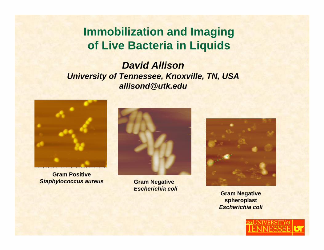
David AllisonUniversity of Tennessee, Knoxville, TN, USA
Immobilization and Imaging of Live Bacteria in Liquids
Gram PositiveStaphylococcus aureus Gram Negative
Escherichia coliGram Negative
spheroplastEscherichia coli
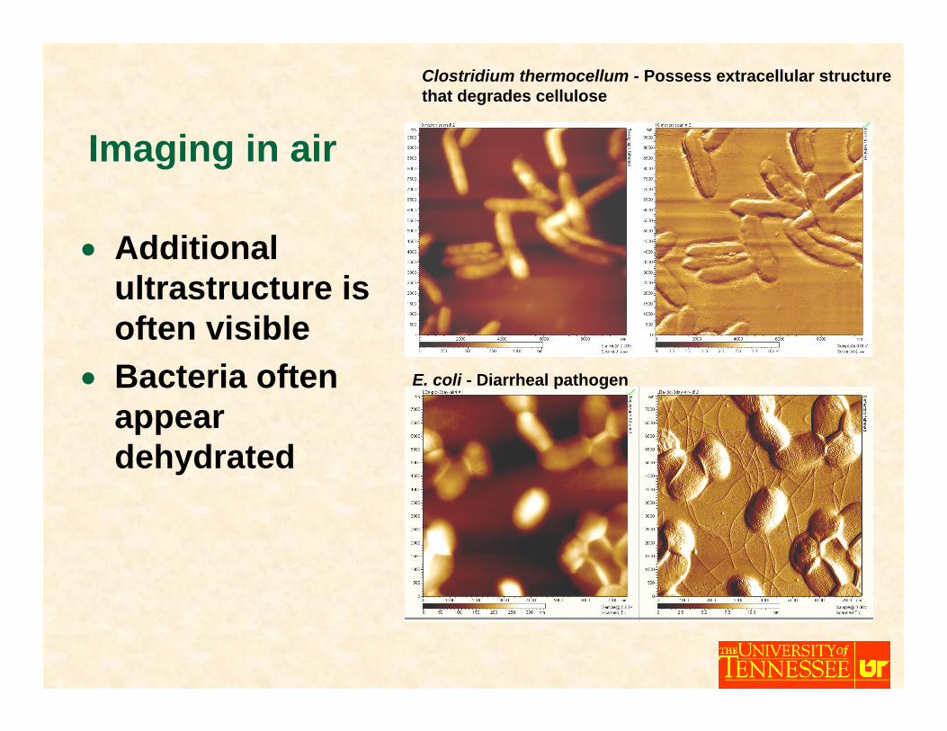
Clostridium thermocellum - Possess extracellular structure that degrades cellulose
Imaging in air
• Additional ultrastructure is often visible
• Bacteria often appear dehydrated
E. coli - Diarrheal pathogen
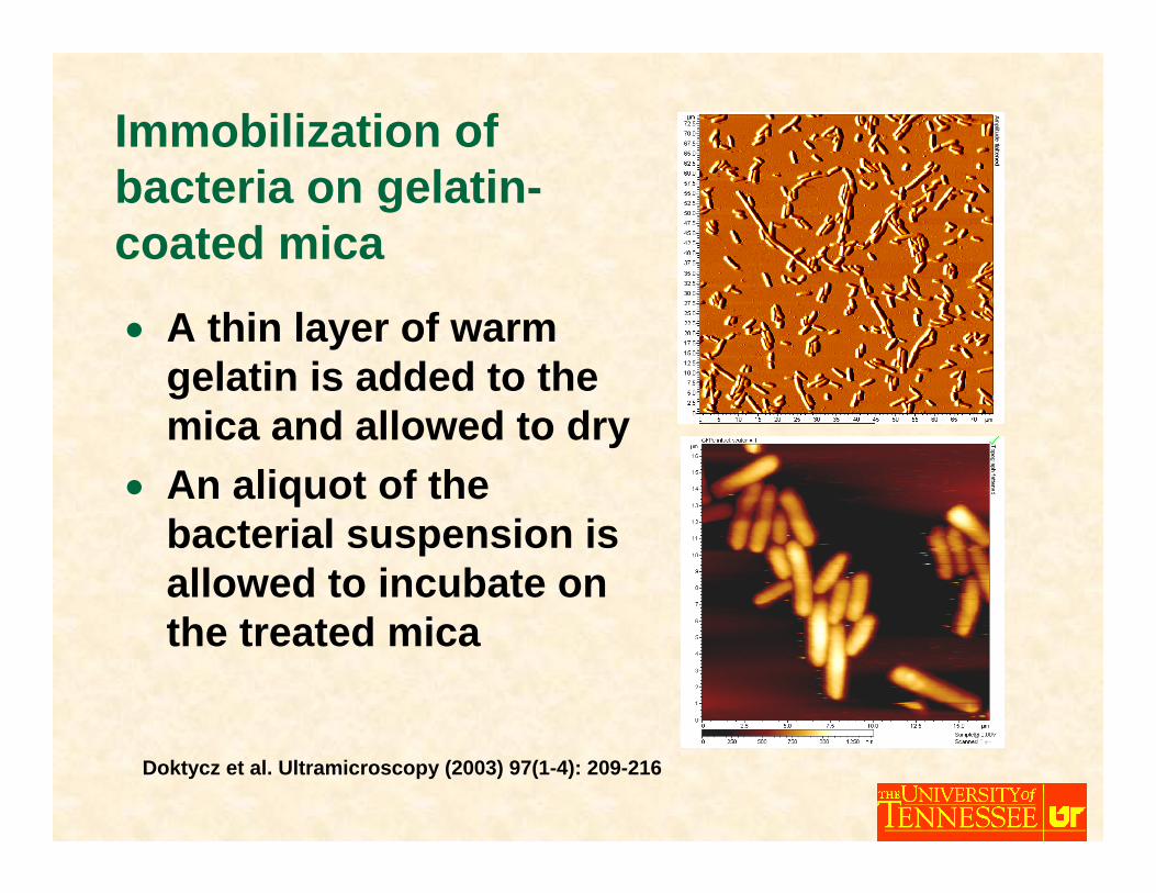
Immobilization of bacteria on gelatin-coated mica
• A thin layer of warm gelatin is added to the mica and allowed to dry
• An aliquot of the bacterial suspension is allowed to incubate on the treated mica
Doktycz et al. Ultramicroscopy (2003) 97(1-4): 209-216
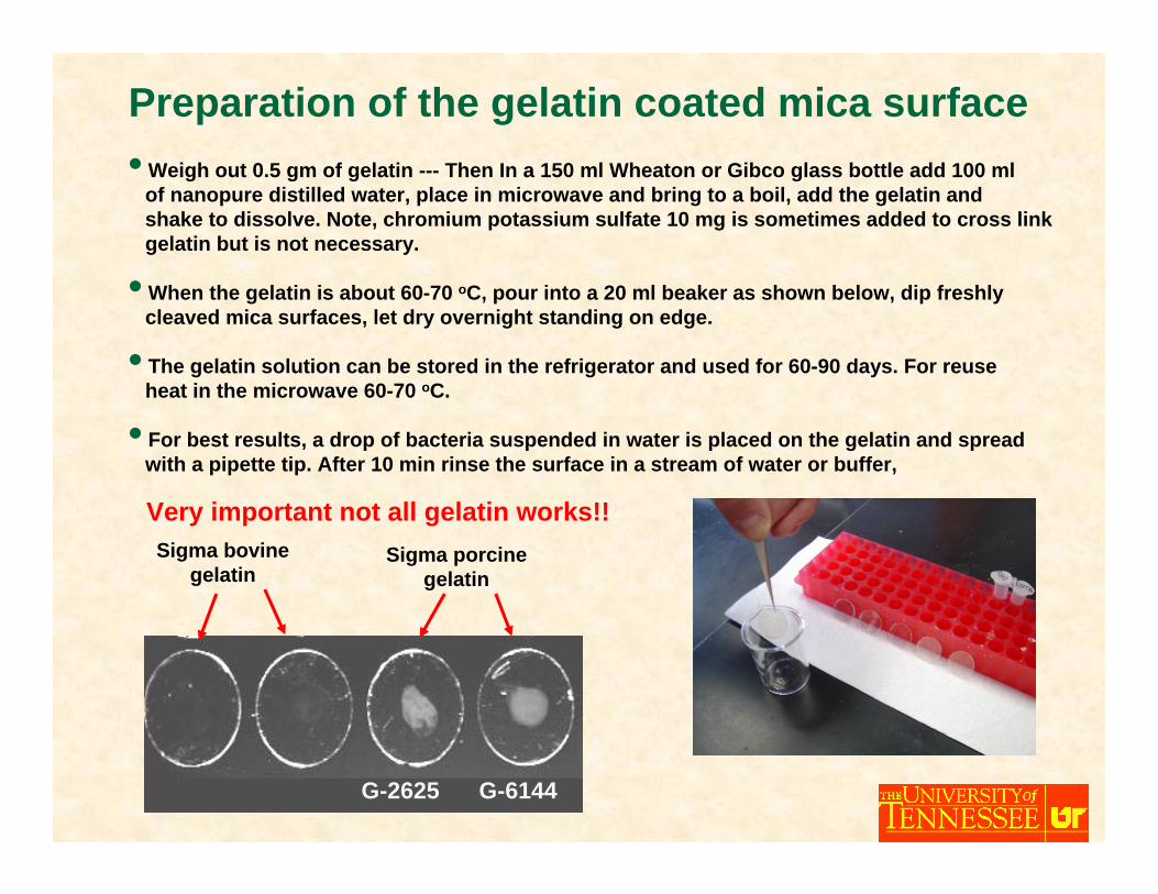
Preparation of the gelatin coated mica surface• Weigh out 0.5 gm of gelatin --- Then In a 150 ml Wheaton or Gibco glass bottle add 100 ml
of nanopure distilled water, place in microwave and bring to a boil, add the gelatin and shake to dissolve. Note, chromium potassium sulfate 10 mg is sometimes added to cross link gelatin but is not necessary.
• When the gelatin is about 60-70 oC, pour into a 20 ml beaker as shown below, dip freshly cleaved mica surfaces, let dry overnight standing on edge.
• The gelatin solution can be stored in the refrigerator and used for 60-90 days. For reuse heat in the microwave 60-70 oC.
• For best results, a drop of bacteria suspended in water is placed on the gelatin and spreadwith a pipette tip. After 10 min rinse the surface in a stream of water or buffer,
Very important not all gelatin works!!Sigma bovine
gelatinSigma porcine
gelatin
G-2625 G-6144
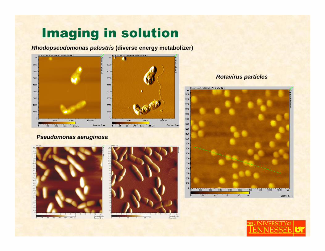
Rhodopseudomonas palustris (diverse energy metabolizer)
Imaging in solution
Pseudomonas aeruginosa
Rotavirus particles
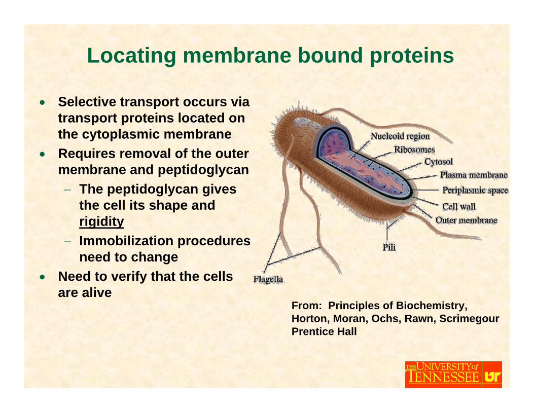
Locating membrane bound proteins
• Selective transport occurs via transport proteins located on the cytoplasmic membrane
• Requires removal of the outer membrane and peptidoglycan− The peptidoglycan gives
the cell its shape and rigidity
− Immobilization procedures need to change
• Need to verify that the cells are alive
From: Principles of Biochemistry, Horton, Moran, Ochs, Rawn, ScrimegourPrentice Hall
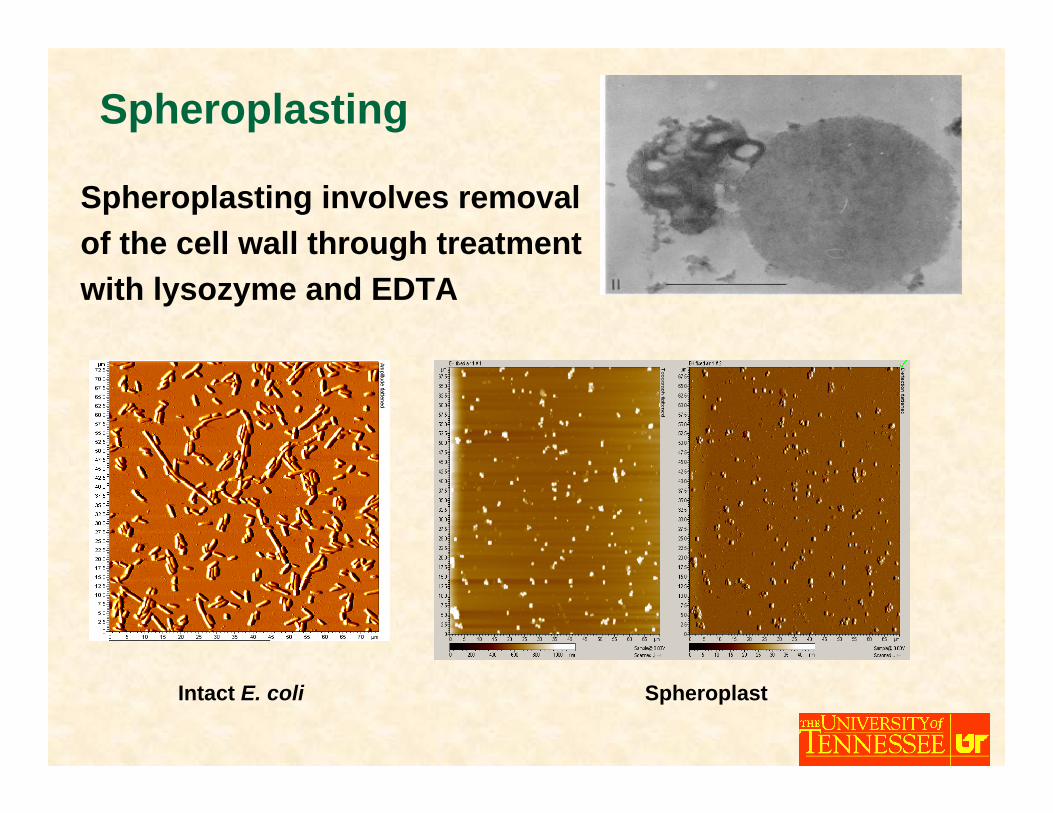
Spheroplasting
Spheroplasting involves removal of the cell wall through treatment with lysozyme and EDTA
Intact E. coli Spheroplast
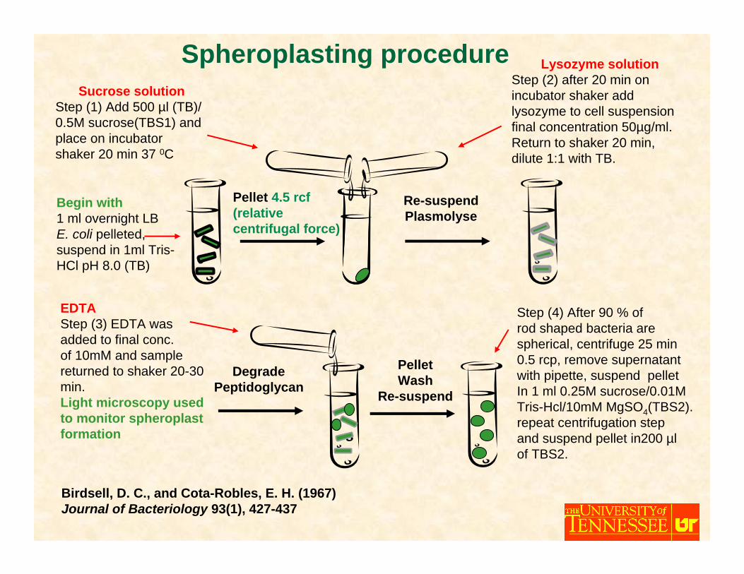
Lysozyme solutionStep (2) after 20 min on incubator shaker add lysozyme to cell suspension final concentration 50µg/ml. Return to shaker 20 min, dilute 1:1 with TB.
Sucrose solutionStep (1) Add 500 µl (TB)/ 0.5M sucrose(TBS1) and place on incubator shaker 20 min 37 0C
Pellet 4.5 rcf(relative centrifugal force)
Re-suspendPlasmolyse
EDTAStep (3) EDTA wasadded to final conc.of 10mM and sample returned to shaker 20-30 min.Light microscopy used to monitor spheroplast formation
DegradePeptidoglycan
PelletWash
Re-suspend
Birdsell, D. C., and Cota-Robles, E. H. (1967) Journal of Bacteriology 93(1), 427-437
Begin with1 ml overnight LBE. coli pelleted, suspend in 1ml Tris-HCl pH 8.0 (TB)
Step (4) After 90 % ofrod shaped bacteria arespherical, centrifuge 25 min0.5 rcp, remove supernatantwith pipette, suspend pelletIn 1 ml 0.25M sucrose/0.01M Tris-Hcl/10mM MgSO4(TBS2).repeat centrifugation step and suspend pellet in200 µlof TBS2.
Spheroplasting procedure

• Pretreat mica with aminopropyltriethoxysilane and glutaraldehyde (APTES/glut)− Previously shown to successfully
immobilize chromatin on mica (Wang, H.D., et al., Biophysical Journal, 2002. 83(6): 3619-3625)
• Incubate the spheroplast suspension on the treated mica− Immobilization results from interactions
between the proteins and the substrate − Conceivably, only the surface in contact
with the substrate is affected, leaving the exposed surface in its native state and accessible to the tip
• Rinse and image in sucrose buffer
Topograph
Amplitude
Immobilizing Spheroplasts
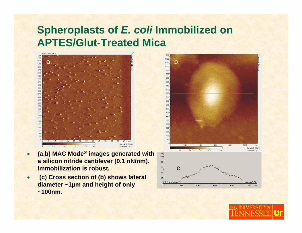
Spheroplasts of E. coli Immobilized on APTES/Glut-Treated Mica
• (a,b) MAC Mode® images generated with a silicon nitride cantilever (0.1 nN/nm). Immobilization is robust.
• (c) Cross section of (b) shows lateral diameter ~1µm and height of only ~100nm.
a. b.
c.
a. b.
c.
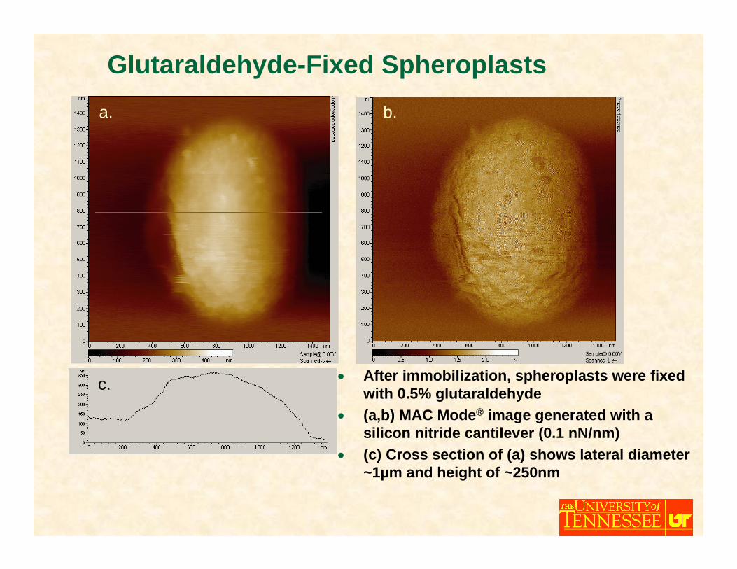
Glutaraldehyde-Fixed Spheroplasts
• After immobilization, spheroplasts were fixed with 0.5% glutaraldehyde
• (a,b) MAC Mode® image generated with a silicon nitride cantilever (0.1 nN/nm)
• (c) Cross section of (a) shows lateral diameter ~1µm and height of ~250nm
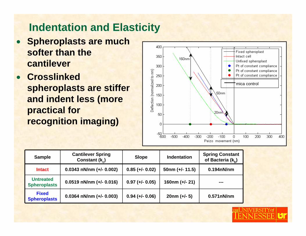
Indentation and Elasticity
Sample Cantilever Spring Constant (kc)
Slope Indentation Spring Constant of Bacteria (kb)
Intact 0.0343 nN/nm (+/- 0.002) 0.85 (+/- 0.02) 50nm (+/- 11.5) 0.194nN/nm
Untreated Spheroplasts 0.0519 nN/nm (+/- 0.016) 0.97 (+/- 0.05) 160nm (+/- 21) ---
Fixed Spheroplasts 0.0364 nN/nm (+/- 0.003) 0.94 (+/- 0.06) 20nm (+/- 5) 0.571nN/nm
• Spheroplasts are much softer than the cantilever
• Crosslinked spheroplasts are stiffer and indent less (more practical for recognition imaging)
mica control
Summary
• Due to forces exerted by the AFM tip imaging samples with scanning probe microscopes require immobilizationtechniques
• Gelatin coated mica surfaces can be effectively used toimmobilize both gram positive and gram negative bacteriafor AFM imaging
• Immobilization of spheroplasts cannot be accomplished with gelatin coated mica surfaces
• Spheroplast immobilization can be accomplished by a technique using a mica surface treated with APTES andglutaraldehyde

Http://ispm.bris.ac.uk10th Anniversary Meeting