Embed Size (px)
Citation preview

Imaging modalities in prostate cancer
Bahjat moussa PGY4 urologyDr Georges Assaf Moderator
24-04-14
PET in PC patients
• Role of functional imaging – not well established yet
• The aim of this review – to offer an overview about the main applications
of choline PET in PC patients

Detection of intra-prostatic cancer

• Use of choline PET/CT for initial diagnosis and local staging of prostate cancer– not recommended as a first line screening method
• The only potential application of PET/CT– increase the detection rate of cancer on repeated
TRUS-guided biopsies– in patients in which at least 2 inconclusive TRUS-
guided biopsy have been already performed

Staging
• The use of choline PET/CT for preoperative LN staging
– showed very contradictory results
– However good specificity and PPV
– limited to patients with very high risk for LN positive status according to nomograms
• At the present time
– routine clinical use of choline PET/CT cannot be recommended in staging patients with PC

• A negative Choline PET/CT – is not sufficient to rule out a lymph-adenectomy
• PET could be useful to exclude from surgery– patients with high surgical risk in which the
presence of LN lesions were assessed by PET (high PPV)
• PET/CT showed
– sensitivity 60%
– a much better specificity 97%

Restaging

• Imaging should be able to find the site of recurrence
– distinguish between local failure and distant metastasis
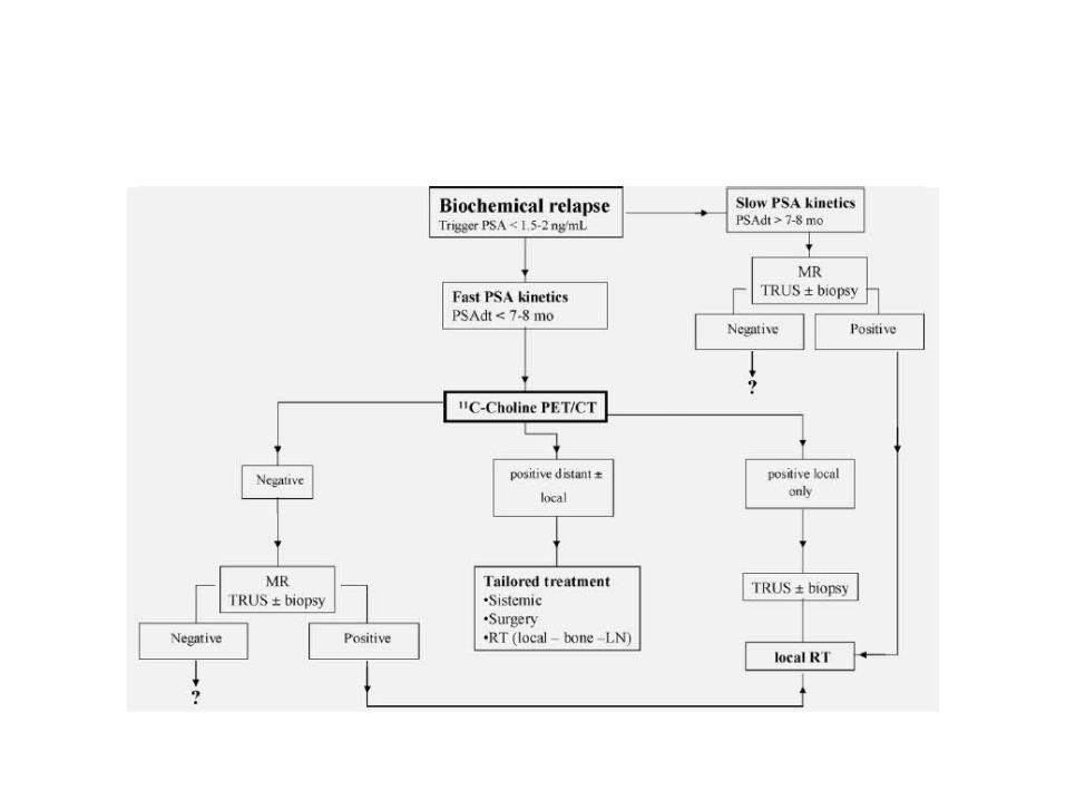
Detection of LN and distant recurrence in PC patients with biochemical recurrence
– significantly high detection rate
– relationship between detection rate and Trigger PSA values
– a relationship between detection rate and PSA kinetics • a crucial role as first diagnostic procedure in patients
who demonstrate a fast growing PSA kinetics and low Trigger PSA
• In case of slow growing PSA kinetics
– sensitivity of PET does not seems to be so high
– questionable if a PET/CT should be performed as first imaging procedure

• In case of local relapse
– TRUS and/or pelvic endorectal MR remain the first procedures
– choline PET/CT could have only a complementary role to exclude the presence of distant metastasis, before a local RT salvage treatment
Conclusion
• Use of choline PET/CT for initial diagnosis and staging – is not recommended as a first-line method
• Most important application of choline PET/CT– restaging of the disease in case of biochemical
relapse for the detection of LN and distant recurrence
Conclusion
• Choline PET/CT– could play a crucial role as first diagnostic
procedure in PC patients who show a fast growing PSA kinetics


• The diagnostic evidence is stronger in restaging than in staging settings
• Proper patient selection– PSA level
– PSA doubling time
– initial tumor stage
is the key to avoiding FN results up front

• The use of choline PET/CT scanning– May accurately provide the localisation of the site
of prostate recurrence in a single step
• Choline PET/CT’s detection rate of recurrences rises together with the increase in PSA serum value
• According to the current available data
– the routine use of choline PET/CT scanning cannot be commonly recommended for PSA values <1 ng/ml
• Independent predictors of positive choline PET/CT– PSA DT
– previous biochemical failure
– locally advanced tumour
– pathologic lymph node disease at initial staging
• Can choline positron emission tomography/computed tomography help individualise treatment decisions?
• Confirmatory data are still needed
• Choline PET/CT imaging has recently been proposed to allow new opportunities for individualised treatment on recurrent lesions after radical treatment for PCa
• Patients with local recurrence after RP – best treated by salvage RT when the PSA serum
level is <0.5 ng/ml
• Choline PET/CT scanning is not commonly useful in this scenario – low detection rate for PSA serum values <1 ng/ml
• Choline PET/CT scanning, providing whole-body information on Pca spread– may be useful in selecting patients to be referred
to local treatment
– by distinguishing those patients with local recurrences from those who present with distant metastases

Salvage lymphadenectomy
• Choline PET/CT scanning – very useful for indicating the presence of lymph
nodal involvement
• in patients who present with a progressive PSA increase after radical treatment
• it provides a basis for further treatment decisions

Role of MRI
According to the guidelines
PSA increase over a threshold of 0.2 ng/ml later than 6 to 12 months after radical prostatectomy • suggests treatment failure with a high risk of local
recurrence
increase within a shorter period• correlates with distant metastasis
For EBRT; biochemical failure • increasing PSA level after a nadir level
Transrectal ultrasound-guided biopsy
• The current reference standard for the detection of local recurrence in patients with biochemical failure
• Invasive
• may fail to depict some tumours because only a small fraction of the gland is sampled
Computed tomography
• Not widely used for the detection of local recurrence
– low accuracy in the differentiation of local recurrence from postsurgical scarring
MRI
• MRI can accurately detect local recurrences after EBRT and radical prostatectomy– DCE MRI is particularly accurate
• The addition of 1H-MRSI to DCE MRI– significantly improve the diagnostic accuracy of
local prostate cancer recurrence
MRI
– usually used for local staging in intermediate and high risk patient groups
– useful in low risk patients as well
– sensitivity and specificity 75% and 95% respectively
• Functional MRI techniques – diffusion-weighted magnetic resonance (DW-MR)
– dynamic contrast-enhanced (DCE-MR)
– MR spectroscopy

• Conventional MRI – only able to diagnose metastatic lymph nodes
bigger than 10 mm
• A newly invented MRI technique lymphotropic superparamagnetic nanoparticles– detect occult lymph node metastasis smaller than
10 mm– 100% sensitivity and 95.7% specificity
MR Spectroscopy
• Measures the level of specific metabolites in the prostate gland– Combination of choline and creatine is measured
in MRS
– The other metabolite that MRS measures is citrate• accumulate in peripheral zone • high in normal prostate tissue but decreases in
malignant tissues
MR Spectroscopy
• The ratio of Cho+Cr/Ci – used for evaluation of prostate cancer
• Higher ratio– in favor of higher risk of malignancy– more than 0.75 is considered as significant and is
consistent with prostate cancer
MR Spectroscopy
• More accurate in detecting prostate cancers with high grade of malignancy
– in low grade cancers its accuracy is limited

Dynamic Contrast Study
• Works based on neo angiogenesis in tumor cells
• Angiogenesis rate is high– newly made vessels have low integrity in their wall– more permeable than normal vessels
Dynamic Contrast Study
• Gadolinium contrast agent is injected– then serial 3D T1- weighted images are obtained
• Fast leakage of contrast agent from leaky tumoral vasculature– early enhancement of tumoral tissue in T1 -
weighted MRI – early wash out of contrast agent are seen in
prostate cancer

Diffusion Weighted Imaging
• Works based on water molecules movements– Water molecules movement decrease in a high
cellular environment– so diffusion become lower
• Sensitivity and specificity of DWI when added to T2-Weighted MRI for detecting prostate cancer is about 84% and 87% respectively

MRI Ability to Detection Bony Metastasis
• The most sensitive and specific technique in detecting bony metastasis
Whole-body DW imaging
• The most newly MRI technique
• Very helpful in detection of prostate cancer and its metastasis as well as post cancer therapy fallow up

Local Staging of Prostate Cancer
• High resolution MR images– especially with the use of endorectal coil
– can show with high accuracy • whether the tumor is confined to prostate gland or
there is extra capsular extension
• The gold standard approach for:– Diagnosis– Staging and management of prostate cancer
Is using 1.5 T MR machines with both endorectal and pelvic phased-array coils
Evaluation of Local Recurrence After Treatment
• MR spectroscopy detects recurrence after radical prostatectomy – 84% and 88% sensitivity and specificity
respectively• DWMRI – capable to detect cancer recurrence after radical
prostatectomy in patients that conventional MRI has missed recurrence

• DW-MR imaging alone shows low sensitivity in cancer recurrence detection after radiotherapy (25%)
• In combination with T2-Weighted MRI– sensitivity increases to 62%
– Specificity in both condition is acceptable (92% vs 97%)
High resolution Multiparametric MR imaging
• includes:– regular T1 weighted and T2 weighted images
– dynamic contrast-enhanced MRI
– diffusion weighted imaging
– MR spectroscopy
High resolution Multiparametric MR imaging
• Obtained in 1.5 T MR machines with simultaneous use of pelvic and endorectal coils – best imaging modality in prostate cancer
• useful for– detection and local staging of prostate cancer – follow-up of patients after radical prostatectomy or radiation
therapy – detection of skeletal metastasis – targeting biopsies in patients highly suspicious of prostate
cancer but with previous negative TRUS guided biopsies

References