Embed Size (px)
Citation preview
4/13/2021
1
Ultrasound and Management of the Axilla
Lisa M. Zorn, MD, MPH
Division of Breast Imaging
Jefferson Health
Disclosures
None
Objectives
Anatomy
Normal vs. abnormal sonographic features
Sampling technique
Management of patient with abnormal axilla
and no known primary
Management of axilla in breast cancer
patient
Imaging axilla
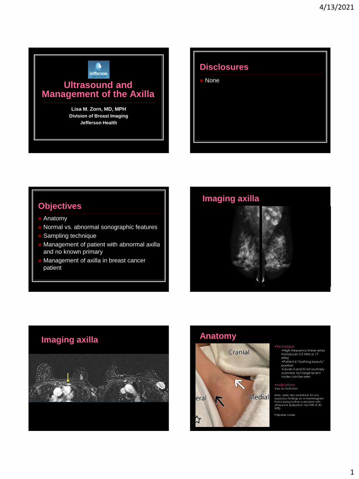
Imaging axilla Anatomy•Technique
•High-frequency linear-array
transducer (12 MHz or 17
MHz)
•Patient in "bathing beauty"
position
•Levels II and III not routinely
scanned, but large level II nodes can be seen
•IndicationsVary by institution
Here, axilla also examined for any
suspicious findings on a mammogram
that is being further evaluated with
ultrasound (palpation has FNR of 30-
50%)
Palpable nodes
4/13/2021
2
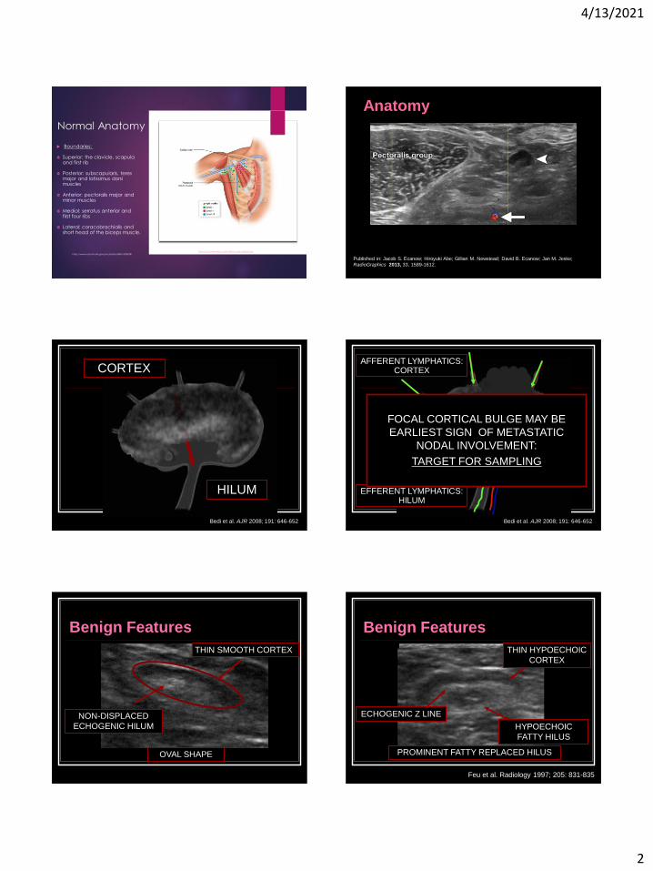
Normal Anatomy
Boundaries:
Superior: the clavicle, scapula and first rib
Posterior: subscapularis, teres major and latissimus dorsi muscles
Anterior: pectoralis major and minor muscles
Medial: serratus anterior and first four ribs
Lateral: coracobrachialis and short head of the biceps muscle.
https://oncohemakey.com/axillary-node-clearance/
https://www.ncbi.nlm.nih.gov/pmc/articles/PMC4376818/
Anatomy
Published in: Jacob S. Ecanow; Hiroyuki Abe; Gillian M. Newstead; David B. Ecanow; Jan M. Jeske;
RadioGraphics 2013, 33, 1589-1612.
HILUM
Bedi et al. AJR 2008; 191: 646-652
CORTEXAFFERENT LYMPHATICS:
CORTEX
EFFERENT LYMPHATICS:HILUM
Bedi et al. AJR 2008; 191: 646-652
FOCAL CORTICAL BULGE MAY BE
EARLIEST SIGN OF METASTATIC
NODAL INVOLVEMENT:
TARGET FOR SAMPLING
Benign Features
THIN SMOOTH CORTEX
NON-DISPLACED
ECHOGENIC HILUM
OVAL SHAPE
Benign Features
Feu et al. Radiology 1997; 205: 831-835
HYPOECHOIC
FATTY HILUS
ECHOGENIC Z LINE
THIN HYPOECHOIC
CORTEX
PROMINENT FATTY REPLACED HILUS
4/13/2021
3
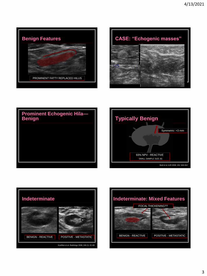
Benign Features
PROMINENT FATTY REPLACED HILUS
CASE: “Echogenic masses”
Prominent Echogenic Hila—Benign
93% NPV - REACTIVE
**SMALL SAMPLE SIZE (6)
Bedi et al. AJR 2008; 191: 646-652
Symmetric: >3 mm
Typically Benign
Indeterminate
BENIGN - REACTIVE POSITIVE - METASTATIC
Koelliker et al. Radiology 2008; 246 (1): 81-89
Indeterminate: Mixed Features
BENIGN - REACTIVE POSITIVE - METASTATIC
FOCAL THICKENING??
4/13/2021
4
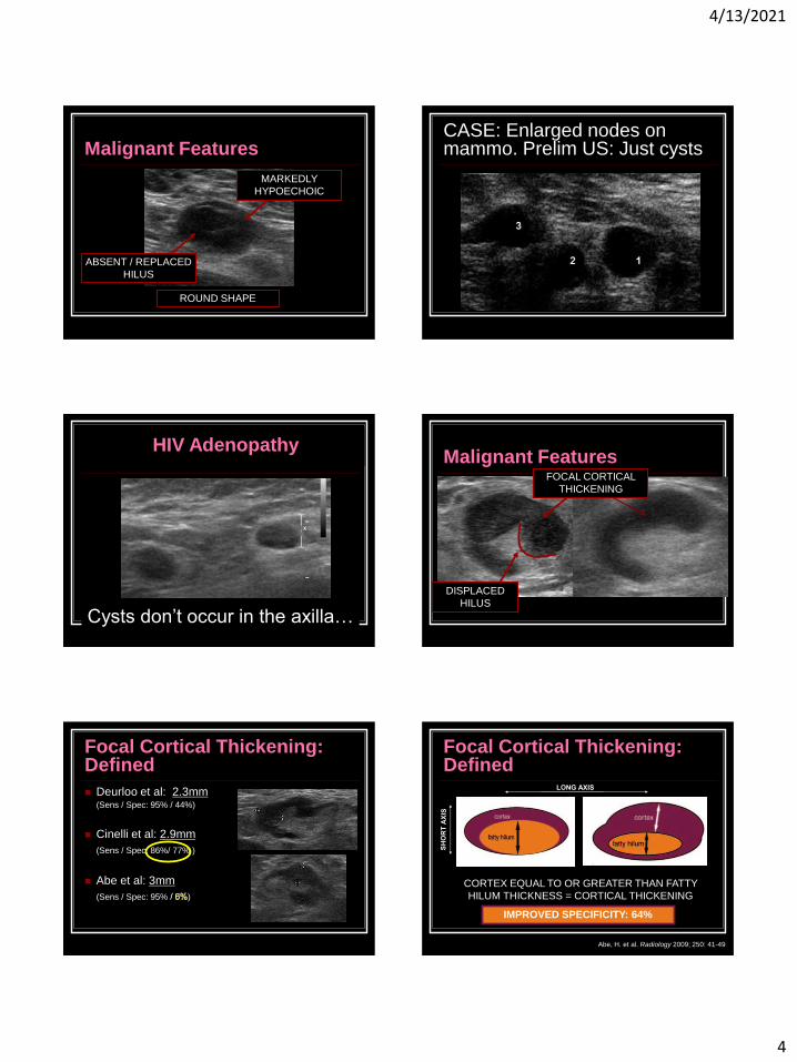
Malignant Features
ROUND SHAPE
ABSENT / REPLACED
HILUS
MARKEDLY
HYPOECHOIC
CASE: Enlarged nodes on mammo. Prelim US: Just cysts
HIV Adenopathy
Cysts don’t occur in the axilla…
Malignant Features
DISPLACED
HILUS
FOCAL CORTICAL
THICKENING
Focal Cortical Thickening: Defined
Deurloo et al: 2.3mm(Sens / Spec: 95% / 44%)
Cinelli et al: 2.9mm
(Sens / Spec: 86%/ 77% )
Abe et al: 3mm
(Sens / Spec: 95% )
Focal Cortical Thickening: Defined
Abe, H. et al. Radiology 2009; 250: 41-49
CORTEX EQUAL TO OR GREATER THAN FATTY
HILUM THICKNESS = CORTICAL THICKENING
IMPROVED SPECIFICITY: 64%
4/13/2021
5
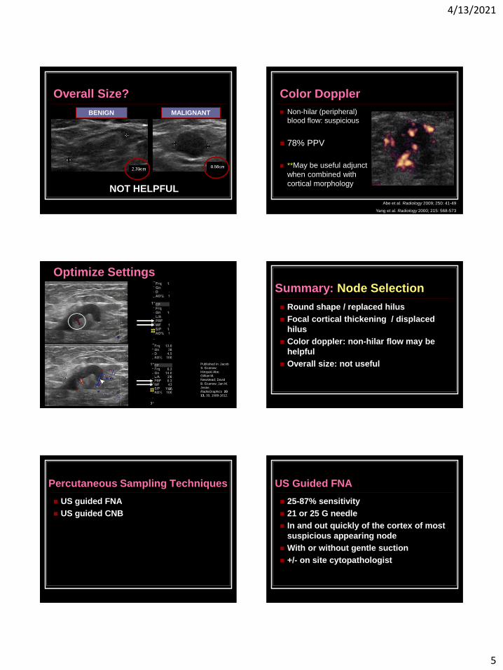
Overall Size?
BENIGN MALIGNANT
NOT HELPFUL
Color Doppler
Non-hilar (peripheral)
blood flow: suspicious
78% PPV
**May be useful adjunct
when combined with
cortical morphology
Abe et al. Radiology 2009; 250: 41-49
Yang et al. Radiology 2000; 215: 568-573
Optimize Settings
Published in: Jacob
S. Ecanow;
Hiroyuki Abe;
Gillian M.
Newstead; David
B. Ecanow; Jan M.
Jeske;
RadioGraphics 20
13, 33, 1589-1612.
Summary: Node Selection
Round shape / replaced hilus
Focal cortical thickening / displaced
hilus
Color doppler: non-hilar flow may be
helpful
Overall size: not useful
Percutaneous Sampling Techniques
US guided FNA
US guided CNB
US Guided FNA
25-87% sensitivity
21 or 25 G needle
In and out quickly of the cortex of most
suspicious appearing node
With or without gentle suction
+/- on site cytopathologist
4/13/2021
6
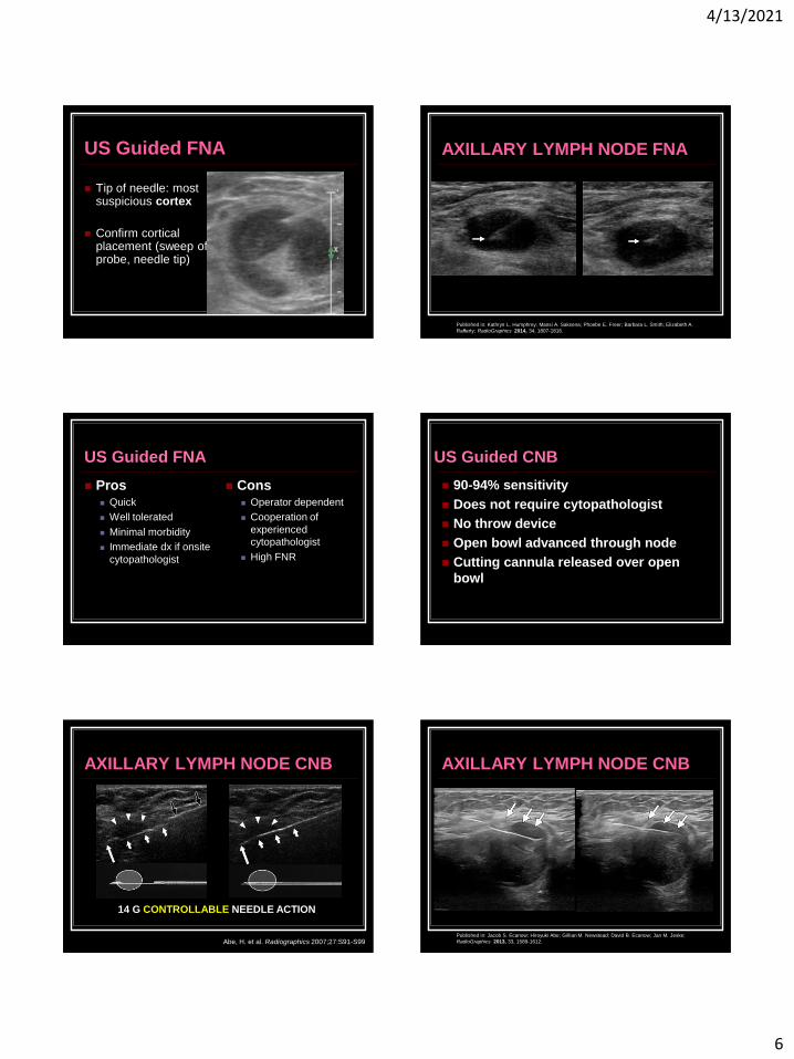
Tip of needle: most suspicious cortex
Confirm cortical placement (sweep of probe, needle tip)
US Guided FNA AXILLARY LYMPH NODE FNA
Published in: Kathryn L. Humphrey; Mansi A. Saksena; Phoebe E. Freer; Barbara L. Smith; Elizabeth A.
Rafferty; RadioGraphics 2014, 34, 1807-1816.
US Guided FNA
Pros Quick
Well tolerated
Minimal morbidity
Immediate dx if onsite
cytopathologist
Cons Operator dependent
Cooperation of
experienced
cytopathologist
High FNR
US Guided CNB
90-94% sensitivity
Does not require cytopathologist
No throw device
Open bowl advanced through node
Cutting cannula released over open
bowl
AXILLARY LYMPH NODE CNB
Abe, H. et al. Radiographics 2007;27:S91-S99
14 G CONTROLLABLE NEEDLE ACTION
AXILLARY LYMPH NODE CNB
Published in: Jacob S. Ecanow; Hiroyuki Abe; Gillian M. Newstead; David B. Ecanow; Jan M. Jeske;
RadioGraphics 2013, 33, 1589-1612.
4/13/2021
7
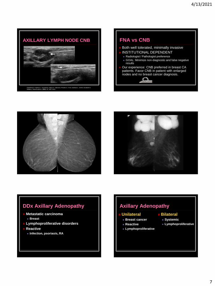
AXILLARY LYMPH NODE CNB
Published in: Kathryn L. Humphrey; Mansi A. Saksena; Phoebe E. Freer; Barbara L. Smith; Elizabeth A.
Rafferty; RadioGraphics 2014, 34, 1807-1816.
FNA vs CNB
Both well tolerated, minimally invasive
INSTITUTIONAL DEPENDENT Radiologist / Pathologist preference
GOAL: Minimize non-diagnostic and false negative results
Our experience: CNB preferred in breast CA patients. Favor CNB in patient with enlarged nodes and no breast cancer diagnosis.
DDx Axillary Adenopathy
Metastatic carcinoma
Breast
Lymphoproliferative disorders
Reactive
Infection, psoriasis, RA
Axillary Adenopathy
Unilateral Breast cancer
Reactive
Lymphoproliferative
Bilateral Systemic
Lymphoproliferative
4/13/2021
8
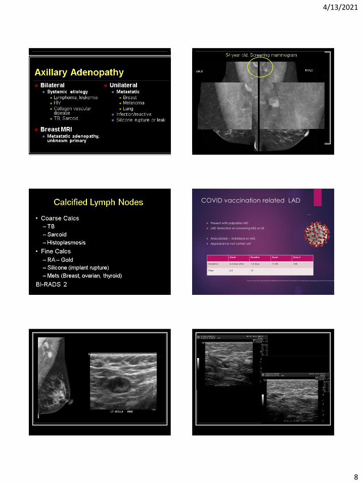
Axillary Adenopathy
COVID vaccination related LAD
Present with palpable LAD
LAD detected on screening MG or US
Anecdotally – Unilateral on MG
Appearance not certain yet
Onset Duration Dose1 Dose 2
Moderna 2-4 days after 1-2 days 11.6% 16%
Pfizer 2-4 10
https://www.sbi-online.org/Portals/0/Position%20Statements/2021/SBI-recommendations-for-managing-axillary-adenopathy-post-COVID-vaccination.pdf
4/13/2021
9
SBI Recommendations
COVID-19 vaccination status, timing and side (left vs. right arm) of
vaccination.
BI-RADS category 0 assessment to allow for further assessment of
the ipsilateral breast and documentation of medical history,
including COVID-19 vaccination.
Consider a short term follow up exam in 4-12 weeks (BI-RADS
category 3) following the second vaccine dose.
If adenopathy persists after short term follow up, consider
sampling
Scheduling screening exams prior to the first dose of a COVID-19
vaccination or 4-6 weeks following the second dose of a COVID-
19 vaccination.
https://www.sbi-online.org/Portals/0/Position%20Statements/2021/SBI-recommendations-for-managing-axillary-adenopathy-post-COVID-vaccination.pdf
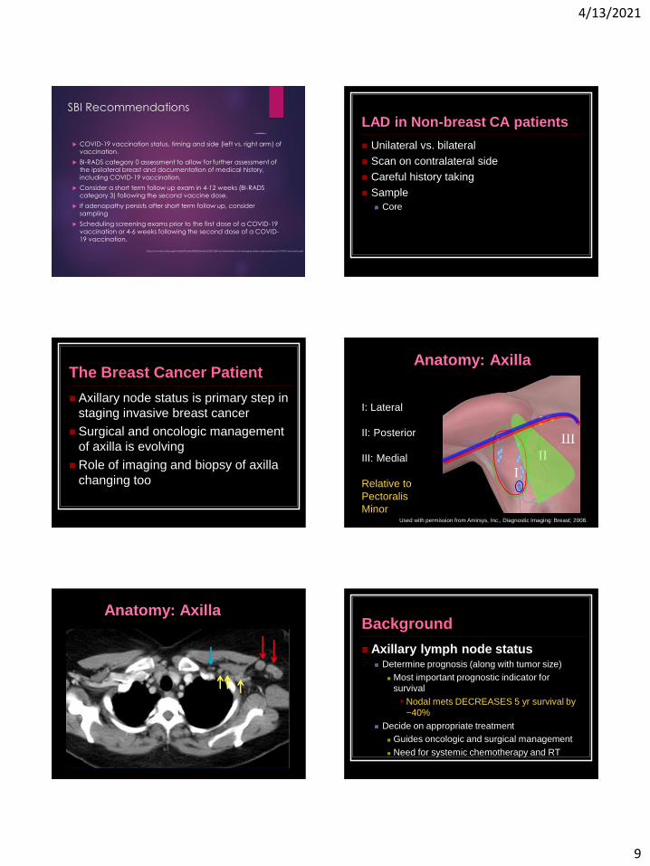
LAD in Non-breast CA patients
Unilateral vs. bilateral
Scan on contralateral side
Careful history taking
Sample
Core
The Breast Cancer Patient
Axillary node status is primary step in
staging invasive breast cancer
Surgical and oncologic management
of axilla is evolving
Role of imaging and biopsy of axilla
changing too
Anatomy: Axilla
Used with permission from Amirsys, Inc., Diagnostic Imaging: Breast; 2008.
I: Lateral
II: Posterior
III: Medial
Relative to
Pectoralis
Minor
Anatomy: AxillaBackground
Axillary lymph node status Determine prognosis (along with tumor size)
Most important prognostic indicator for
survival
Nodal mets DECREASES 5 yr survival by
~40%
Decide on appropriate treatment
Guides oncologic and surgical management
Need for systemic chemotherapy and RT

4/13/2021
10
Axillary Nodes in BreastCancer
https://epomedicine.com/medical-students/tnm-staging-breast-cancer-simplified/
Intramammary nodes in the axillary tail are not
considered to represent axillary metastases.
Background
-1975 Radical mastectomy
1975-1995 BCT and ALND
1995-2011 SLNB
2011-
1974: NSABP B-04
Randomized radical (AD) vs. total + nodal XRT
vs. total mastectomy
No difference in survival at 10 years if nodes
were clinically negative
BUT…axillary dissection continued as standard
procedure for prognostic info and for
management decisions
N Engl J Med. 1985 Mar 14;312(11):674-81
Background
Traditional staging: ALND Definitive method, Level I and II nodes
Significant morbidity
As screening mammo increased detection, more
T0, T1 lesions found
Fewer women with ax LN mets
ALND 80-85% neg in T0, T1 Ca
Led to SLNB in 1990s
1995-2011 Surgical Staging
SLNB replaced ALND as initial method of
staging the axilla
If SLNB +, ALND performed for complete
staging and local control
US axilla with FNA/core bx of abnormal
nodes = straight to ALND if +
4/13/2021
11
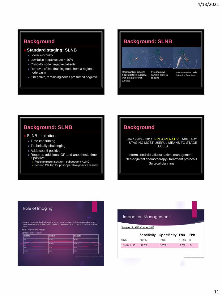
Background
Standard staging: SLNB Lower morbidity
Low false negative rate ~ 10%
Clinically node negative patients
Removal of first draining node from a regional
node basin
If negative, remaining nodes presumed negative.
Background: SLNB
Radionuclide injection
hours before surgery:
Peri-areolar or Peri-
tumoral
Pre-operative
gamma camera
imaging
Intra-operative node
detection / excision
Background: SLNB
SLNB Limitations
Time consuming
Technically challenging
Adds cost if positive
Requires additional OR and anesthesia time if positive Positive frozen section - subsequent ALND
Second OR trip for post-operative positive results
Background
Late 1990’s - 2011: PRE-OPERATIVE AXILLARY STAGING MOST USEFUL MEANS TO STAGE
AXILLA
Informs (individualizes) patient management:
Neo-adjuvant chemotherapy / treatment protocols
Surgical planning
Role of Imaging
Imaging - preoperatively determine lymph node involvement in non-palpable lymph nodes to determine upfront which patients may need ALND and obviate SLNB in these cases.
Assess response to therapy
Sample, mark, localize
Modality Sensitivity Specificity
MG 66.9% 80.8%
US 87-94% 53-97%
MRI 77% 90%
PET/CT 64% 93%
https://www.ncbi.nlm.nih.gov/pmc/articles/PMC7011661/pdf/ONCO-25-e231.pdf
Impact on Management
Sensitivity Specificity FNR FPR
SLNB 88.7% 100% 11.3% 0
USNB+SLNB 97.2% 100% 2.8% 0
Wang et al., BMC Cancer, 2015
https://www.ncbi.nlm.nih.gov/pmc/articles/PMC4435774/
4/13/2021
12
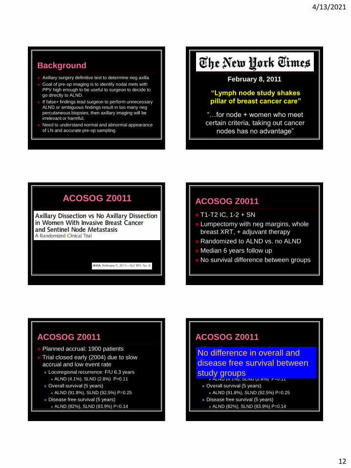
Background
Axillary surgery definitive test to determine neg axilla
Goal of pre-op imaging is to identify nodal mets with
PPV high enough to be useful to surgeon to decide to
go directly to ALND.
If false+ findings lead surgeon to perform unnecessary
ALND or ambiguous findings result in too many neg
percutaneous biopsies, then axillary imaging will be
irrelevant or harmful.
Need to understand normal and abnormal appearance
of LN and accurate pre-op sampling
February 8, 2011
“Lymph node study shakes
pillar of breast cancer care”
“…for node + women who meet
certain criteria, taking out cancer
nodes has no advantage”
ACOSOG Z0011 ACOSOG Z0011
T1-T2 IC, 1-2 + SN
Lumpectomy with neg margins, whole
breast XRT, + adjuvant therapy
Randomized to ALND vs. no ALND
Median 6 years follow up
No survival difference between groups
Planned accrual: 1900 patients
Trial closed early (2004) due to slow
accrual and low event rate
Locoregional recurrence: F/U 6.3 years
ALND (4.1%), SLND (2.8%) P=0.11
Overall survival (5 years)
ALND (91.8%), SLND (92.5%) P=0.25
Disease free survival (5 years)
ALND (82%), SLND (83.9%) P=0.14
ACOSOG Z0011
No difference in overall and
disease free survival between
study groups
4/13/2021
13
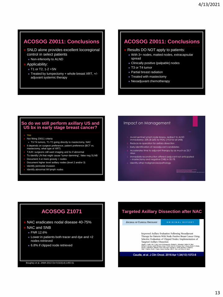
SNLD alone provides excellent locoregional
control in select patients
Non-inferiority to ALND
Applicability:
T1 or T2, 1-2 +SN
Treated by lumpectomy + whole breast XRT, +/-
adjuvant systemic therapy
ACOSOG Z0011: Conclusions
Results DO NOT apply to patients:
With 3+ nodes, matted nodes, extracapsular
spread
Clinically positive (palpable) nodes
T3 or T4 tumor
Partial breast radiation
Treated with mastectomy
Neoadjuvant chemotherapy
ACOSOG Z0011: Conclusions
So do we still perform axillary US and US bx in early stage breast cancer?
Yes
Not fitting Z0011 criteria
T3-T4 tumors, T1-T2 going directly to mastectomy, NAC
It depends on surgeon preference, patient preference (BCT vs.
mastectomy, what type of XRT)
TJUH: surgeons still want imaging and bx if abnormal
To identify LN that might cause “tumor damming”, false neg SLNB
Document 3 or more grossly + nodes
Document higher level axillary nodes (level 2 and/or 3)
Identify perinodal invasion
Identify abnormal IM lymph nodes
Impact on Management
1. Avoid sentinel lymph node biopsy, redirect to ALND
immediately: 33% (8-26% for FNA) (19.8 for all UNB)
2. Reduce re-operation for axillary dissection
3. Early identification of neoadjuvant candidates
4. Accelerates time to adjuvant therapy by as much as 25.7
days
5. Immediate reconstruction offered (adjuvant not anticipated
– mastectomy and negative CNB) in 35.1%
6. Identify other malignancies/pathology
https://www.ncbi.nlm.nih.gov/pmc/articles/PMC4069800/
https://pubmed.ncbi.nlm.nih.gov/20047809/
https://hal.archives-ouvertes.fr/hal-00668067/documenthttps://www.sciencedirect.com/science/article/pii/S2211568412002392#bib0175
ACOSOG Z1071
NAC eradicates nodal disease 40-75%
NAC and SNB
FNR 12.6%
Lower in patients both tracer and dye and >2
nodes retrieved
6.8% if clipped node retrieved
Boughey et al, JAMA 2013 Oct 9;310(14):1455-61
Targeted Axillary Dissection after NAC
4/13/2021
14
Targeted Axillary Dissection
Localization of the clipped node for TAD
reduced the FNR of SLNB in setting of
NAC
Radiograph axillary specimen to identify
clipped node
Decrease FNR compared to SLNB alone
SLNB 10.1% vs clipped node 4.2%
TAD 2% (both SLNB and retrieve clipped node)
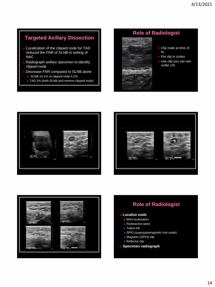
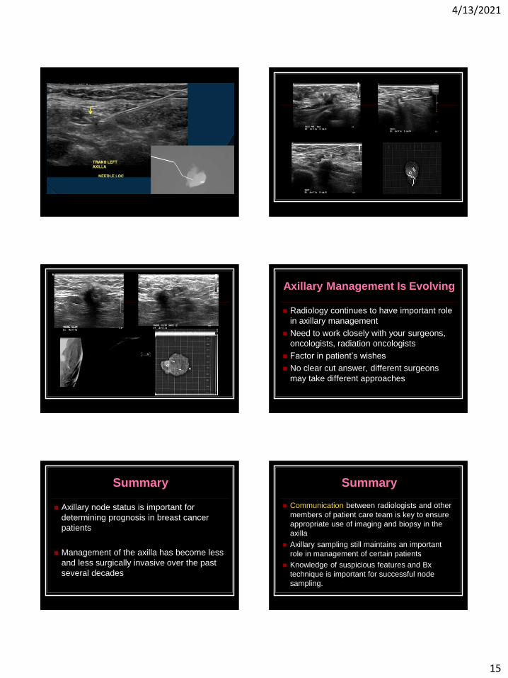
Role of Radiologist
Clip node at time of
bx
Put clip in cortex
Use clip you can see
under US
Role of Radiologist
Localize node
Wire localization
Radioactive seed
Tattoo ink
SPIO (superparamagnetic iron oxide)
Magnetic (SPIO) clip
Reflector clip
Specimen radiograph
4/13/2021
15
Axillary Management Is Evolving
Radiology continues to have important role
in axillary management
Need to work closely with your surgeons,
oncologists, radiation oncologists
Factor in patient’s wishes
No clear cut answer, different surgeons
may take different approaches
Summary
Axillary node status is important for
determining prognosis in breast cancer
patients
Management of the axilla has become less
and less surgically invasive over the past
several decades
Summary
Communication between radiologists and other
members of patient care team is key to ensure
appropriate use of imaging and biopsy in the
axilla
Axillary sampling still maintains an important
role in management of certain patients
Knowledge of suspicious features and Bx
technique is important for successful node
sampling.
4/13/2021
16
Thank you!
Questions?