Embed Size (px)
Citation preview
Case ReportIdiopathic Scrotal Hematoma in Neonate:A Case Report and Review of the Literature
Bioku Muftau Jimoh,1 Eziechila Bessie Chinwe,2 Adewumi Oluwafemi Adebisi,3
Igwilo Chinwe Ifeoma,3 Maduka Ogechi,2 and Aiyekomogbon Joshua Oluwafemi4
1 Department of Surgery, Urology Division, Federal Staff Medical Centre, Abuja, Nigeria2 Department of Pediatrics, Federal Staff Medical Centre, Abuja, Nigeria3 Department of Obstetrics and Gynecology, Federal Staff Medical Centre, Abuja, Nigeria4Department of Radiology, Federal Staff Medical Centre, Abuja, Nigeria
Correspondence should be addressed to Bioku Muftau Jimoh; [email protected]
Received 29 March 2014; Accepted 15 May 2014; Published 27 May 2014
Academic Editor: Apul Goel
Copyright © 2014 Bioku Muftau Jimoh et al. This is an open access article distributed under the Creative Commons AttributionLicense, which permits unrestricted use, distribution, and reproduction in any medium, provided the original work is properlycited.
Neonatal scrotal hematoma is a rare genitourinary emergency. Some cases have underlining aetiologic factors such as testiculartorsion, adrenal hemorrhage, or birth trauma, and others are idiopathic. Previously, immediate scrotal exploration was consideredimperative for diagnosis and treatment. With good imaging techniques, some patients are managed nonoperatively. We report acase of idiopathic scrotal hematoma in a neonate. He was managed conservatively with clinical and radiological follow-up. Therewas complete resolution of hematoma within two months, thus, avoiding unnecessary exploration.
1. Introduction
Scrotal hematoma in the neonate, though a rare condition,warrants prompt diagnosis and urgent intervention. It com-monly results from testicular torsion, adrenal hemorrhage,and birth trauma.
However, in some cases no cause may be discernable.Since Putnam recorded his first experience in 1989 [1],few other cases of neonatal scrotal hematoma have beendocumented in the literature. We report a case of idiopathicleft hemiscrotal hematoma in a newborn which is managednonoperatively in our hospital. It reechoes the significanceof neonatal scrotal and abdominal ultrasonography, thuspreventing unnecessary scrotal exploration.
2. Case Presentation
A term male neonate with vertex presentation was deliveredspontaneously per vaginuum to a 45-year-old P
7
+3 seamstressin a maternity home. The birth weight was 3 kg. Antenatalcare was inadequate. Painless left hemiscrotal swelling was
noticed 72 hours after birth by the pediatricians managinghim for neonatal jaundice and sepsis. There was no bleedingfrom any body orifice.
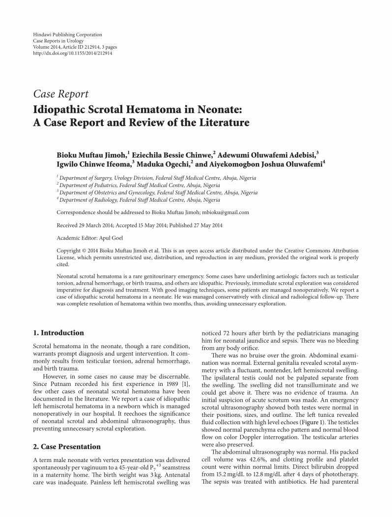
There was no bruise over the groin. Abdominal exami-nation was normal. External genitalia revealed scrotal asym-metry with a fluctuant, nontender, left hemiscrotal swelling.The ipsilateral testis could not be palpated separate fromthe swelling. The swelling did not transilluminate and wecould get above it. There was no evidence of trauma. Aninitial suspicion of acute scrotum was made. An emergencyscrotal ultrasonography showed both testes were normal intheir positions, sizes, and outline. The left tunica revealedfluid collection with high level echoes (Figure 1).The testiclesshowed normal parenchyma echo pattern and normal bloodflow on color Doppler interrogation. The testicular arterieswere also preserved.
The abdominal ultrasonography was normal. His packedcell volume was 42.6%, and clotting profile and plateletcount were within normal limits. Direct bilirubin droppedfrom 15.2mg/dL to 12.8mg/dL after 4 days of phototherapy.The sepsis was treated with antibiotics. He had parenteral
Hindawi Publishing CorporationCase Reports in UrologyVolume 2014, Article ID 212914, 3 pageshttp://dx.doi.org/10.1155/2014/212914
2 Case Reports in Urology
Figure 1
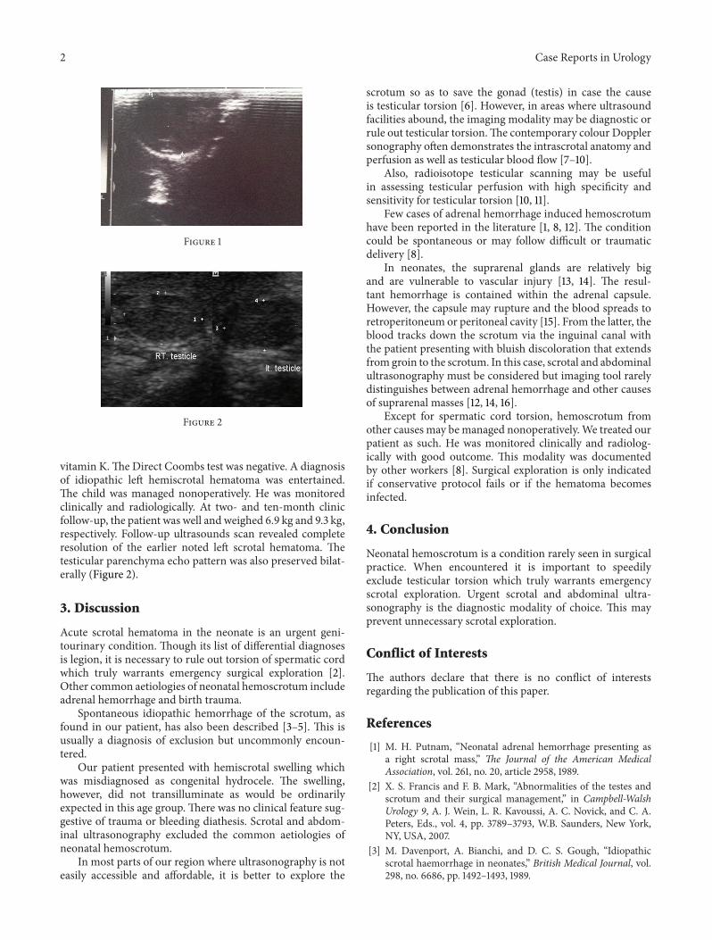
Figure 2
vitamin K.The Direct Coombs test was negative. A diagnosisof idiopathic left hemiscrotal hematoma was entertained.The child was managed nonoperatively. He was monitoredclinically and radiologically. At two- and ten-month clinicfollow-up, the patient was well and weighed 6.9 kg and 9.3 kg,respectively. Follow-up ultrasounds scan revealed completeresolution of the earlier noted left scrotal hematoma. Thetesticular parenchyma echo pattern was also preserved bilat-erally (Figure 2).
3. Discussion
Acute scrotal hematoma in the neonate is an urgent geni-tourinary condition. Though its list of differential diagnosesis legion, it is necessary to rule out torsion of spermatic cordwhich truly warrants emergency surgical exploration [2].Other common aetiologies of neonatal hemoscrotum includeadrenal hemorrhage and birth trauma.
Spontaneous idiopathic hemorrhage of the scrotum, asfound in our patient, has also been described [3–5]. This isusually a diagnosis of exclusion but uncommonly encoun-tered.
Our patient presented with hemiscrotal swelling whichwas misdiagnosed as congenital hydrocele. The swelling,however, did not transilluminate as would be ordinarilyexpected in this age group. There was no clinical feature sug-gestive of trauma or bleeding diathesis. Scrotal and abdom-inal ultrasonography excluded the common aetiologies ofneonatal hemoscrotum.
In most parts of our region where ultrasonography is noteasily accessible and affordable, it is better to explore the
scrotum so as to save the gonad (testis) in case the causeis testicular torsion [6]. However, in areas where ultrasoundfacilities abound, the imaging modality may be diagnostic orrule out testicular torsion.The contemporary colour Dopplersonography often demonstrates the intrascrotal anatomy andperfusion as well as testicular blood flow [7–10].
Also, radioisotope testicular scanning may be usefulin assessing testicular perfusion with high specificity andsensitivity for testicular torsion [10, 11].
Few cases of adrenal hemorrhage induced hemoscrotumhave been reported in the literature [1, 8, 12]. The conditioncould be spontaneous or may follow difficult or traumaticdelivery [8].
In neonates, the suprarenal glands are relatively bigand are vulnerable to vascular injury [13, 14]. The resul-tant hemorrhage is contained within the adrenal capsule.However, the capsule may rupture and the blood spreads toretroperitoneum or peritoneal cavity [15]. From the latter, theblood tracks down the scrotum via the inguinal canal withthe patient presenting with bluish discoloration that extendsfrom groin to the scrotum. In this case, scrotal and abdominalultrasonography must be considered but imaging tool rarelydistinguishes between adrenal hemorrhage and other causesof suprarenal masses [12, 14, 16].
Except for spermatic cord torsion, hemoscrotum fromother causes may bemanaged nonoperatively.We treated ourpatient as such. He was monitored clinically and radiolog-ically with good outcome. This modality was documentedby other workers [8]. Surgical exploration is only indicatedif conservative protocol fails or if the hematoma becomesinfected.
4. Conclusion
Neonatal hemoscrotum is a condition rarely seen in surgicalpractice. When encountered it is important to speedilyexclude testicular torsion which truly warrants emergencyscrotal exploration. Urgent scrotal and abdominal ultra-sonography is the diagnostic modality of choice. This mayprevent unnecessary scrotal exploration.
Conflict of Interests
The authors declare that there is no conflict of interestsregarding the publication of this paper.
References
[1] M. H. Putnam, “Neonatal adrenal hemorrhage presenting asa right scrotal mass,” The Journal of the American MedicalAssociation, vol. 261, no. 20, article 2958, 1989.
[2] X. S. Francis and F. B. Mark, “Abnormalities of the testes andscrotum and their surgical management,” in Campbell-WalshUrology 9, A. J. Wein, L. R. Kavoussi, A. C. Novick, and C. A.Peters, Eds., vol. 4, pp. 3789–3793, W.B. Saunders, New York,NY, USA, 2007.
[3] M. Davenport, A. Bianchi, and D. C. S. Gough, “Idiopathicscrotal haemorrhage in neonates,” British Medical Journal, vol.298, no. 6686, pp. 1492–1493, 1989.

Case Reports in Urology 3
[4] M.-L. Yeh, C.-J. Chang, and S.-C. Mu, “Neonatal idiopathicscrotal hemorrhage: patient reports,” Clinical Pediatrics, vol. 39,no. 8, pp. 493–494, 2000.
[5] S.-C. Wu and C.-Y. Chou, “Neonatal idiopathic scrotal hemor-rhage: report of one case,” Clinical Neonatology, vol. 12, no. 2,pp. 64–66, 2005.
[6] A. O. Omisanjo, S. O. Ikuerowo, M. J. Bioku, and J. O. Esho,“Torsion of the testis in Lagos: a five year experience,” NigerianJournal of Urology, vol. 1, no. 1-2, pp. 24–27, 2012.
[7] B. Karmazyn, R. Steinberg, L. Kornreich et al., “Clinical andsonographic criteria of acute scrotum in children: a retrospec-tive study of 172 boys,” Pediatric Radiology, vol. 35, no. 3, pp.302–310, 2005.
[8] L. Lai, L. Chen, M. Tseng, C. Chang, C. Lu, and P. Chu, “Neona-tal adrenal hemorrhage associated with scrotal hematoma:an unusual case report and literature review,” Pediatrics andNeonatology, vol. 53, no. 3, pp. 210–212, 2012.
[9] P. Pavlica and L. Barozzi, “Imaging of the acute scrotum,”European Radiology, vol. 11, no. 2, pp. 220–228, 2001.
[10] W. W. C. Lam, T. L. Yap, A. S. Jacobsen, and H. J. Teo, “Colourdoppler ultrasonography replacing surgical exploration foracute scrotum:myth or reality?” Pediatric Radiology, vol. 35, no.6, pp. 597–600, 2005.
[11] K. Al-Khawaldeh, T. Bisheh, and F. Jarad, “The value of Tc99m-pertechnitate testicular scintigraphy in diagnosis of acutetesticular torsion in pediatric and adult patients,” Journal ofTropical Urology, vol. 5, no. 1, pp. 48–50, 2007.
[12] G. Renata, A. Allan, F. F. A. Rubia, and A. F. Claudia, “Scrotalhematoma as a sign of adrenal hemorrhage in newborns,” SaoPaulo Medical Journal, vol. 129, no. 2, pp. 113–115, 2011.
[13] T. Tulassay, I. Seri, and J. Evans, “Renal vascular disease in thenewborn,” in Schaffers and Avery’s Diseases of Newborn, H. W.Taeugah, R. A. Ballard, andM. E. Avery, Eds., pp. 1177–1187,W.B.Saunders, Philadelphia, Pa, USA, 7th edition, 1998.
[14] J. M. O’Neil, G. M. Hendry, and G. A. MacKinlay, “An unusualpresentation of neonatal adrenal hemorrhage,” European Jour-nal of Ultrasound, vol. 16, no. 3, pp. 261–264, 2003.
[15] G. Bergami, S.Malena,M.DiMario, andG. Fariello, “L’ecografianel follow-up dell’emorragia surrenalica in eta neonatale. Pre-sentazione di 14 casi,” La Radiologia Medica, vol. 79, no. 5, pp.474–478, 1990.
[16] O. Adorisio, R. Mattei, E. Ciardini, N. Centonze, and B.Noccioli, “Neonatal adrenal hemorrhage mimicking an acutescrotum,” Journal of Perinatology, vol. 27, no. 2, pp. 130–132,2007.