Embed Size (px)
Citation preview
Identification and Characterization of a Type III PolyketideSynthase Involved in Quinolone Alkaloid Biosynthesis fromAegle marmelos Correa*
Received for publication, October 24, 2012, and in revised form, January 16, 2013 Published, JBC Papers in Press, January 17, 2013, DOI 10.1074/jbc.M112.429886
Mohankumar Saraladevi Resmi‡, Priyanka Verma§, Rajesh S. Gokhale§¶, and Eppurathu Vasudevan Soniya‡1
From the ‡Plant Molecular Biology Division, Rajiv Gandhi Centre for Biotechnology, Thycaud (P.O), Thiruvananthapuram, 695 014Kerala, India, the §National Institute of Immunology, Aruna Asaf Ali Marg, New Delhi 110067, India, and the ¶Systems BiologyGroup, Council of Scientific and Industrial Research, Institute of Genomics and Integrative Biology, Mall Road, Delhi 110 007, India
Background: Type III polyketide synthase is hypothesized to produce quinolones, but no such enzyme has been identifiedso far.Results: QNS, a type III PKS from Aegle marmelos synthesizes diketide 4-hydroxy-1-methyl-2H-quinolone using a uniquesubstrate binding site.Conclusion:QNS is a novel 4-hydroxy-1-methyl-2H-quinolone synthase.Significance: This is the first report of a gene involved in quinolone biosynthesis from plants.
Quinolone alkaloids, found abundantly in the roots of bael(Aegle marmelos), possess various biological activities and haverecently gained attention as potential lead molecules for noveldrug designing. Here, we report the characterization of a novelType III polyketide synthase, quinolone synthase (QNS), fromA. marmelos that is involved in the biosynthesis of quinolonealkaloid. Using homology-based structural modeling, we iden-tify two crucial amino acid residues (Ser-132 andAla-133) at theputative QNS active site. Substitution of Ser-132 to Thr andAla-133 to Ser apparently constricted the active site cavityresulting in production of naringenin chalcone from p-couma-royl-CoA. Measurement of steady-state kinetic parametersdemonstrates that the catalytic efficiency of QNS was several-fold higher for larger acyl-coenzymeA substrates as comparedwith smaller precursors.Ourmutagenic studies suggest that thisproteinmight have evolved froman evolutionarily relatedmem-ber of chalcone synthase superfamily by mere substitution oftwo active site residues. The identification and characterizationof QNS offers a promising target for gene manipulation studiestoward the production of novel alkaloid scaffolds.
Quinolones are anthranilic acid-derived alkaloids that arelargely or completely restricted to plants of Rutaceae (1). Someof these naturally occurring quinolones have profound medic-inal properties, whereas others have served as lead structuresand provided inspiration for the design of synthetic quinolonesas useful drugs (2, 3). Quinoline alkaloids such as quinine, chlo-roquin, mefloquine, and amodiaquine are used as efficientdrugs for the treatment of malaria (4). Skimmianine is a poten-tial quinolone alkaloid derived from the roots ofmany rutacean
plants and is reported to possess various biological activitiessuch as inhibitory effect on spontaneous motor function (5),cardiovascular effect (6), antiplatelet aggregation activity (7),cytotoxic in HeLa cell line (8), and also as antagonists of 5-hy-droxytryptamine receptors (9). Because of these properties,quinolone biosynthesis is a subject of great interest. Recently, areport by Abe et al. (10) demonstrated the ability of benzalac-etone synthase (BAS),2 a type III PKS to accept non-physiolog-ical substrate N-methylanthraniloyl CoA and catalyze forma-tion of 4-hydroxy-2(1H)-quinolones. However, until now, notype III PKS dedicated to biosynthesis of quinolone alkaloid hasbeen identified. In this report, we characterize a novel type IIIPKS from Aegle marmelos Corr. and demonstrate its remarka-ble ability to catalyze formation of quinolones scaffold. Strik-ingly, A. marmelos Correa belongs to the Rutaceae family,which is widely recognized for the diversity of the associatedquinolone alkaloid (11, 12).Type III polyketide synthases (PKS) are homodimeric
enzymes where the single active site in each monomer itera-tively catalyzes the priming, extension, and cyclization reac-tions to generate the polyketide product. Despite their struc-tural simplicity, type III PKS produces a variety of compounds,including pyrones, resorcinols, acridones, chalcones, stilbenes,and phloroglucinols (13, 14). The diversity observed with typeIII PKS arises from its ability to accept variety of acyl-CoA thio-esters for priming the reaction, its capability to extend thisstarter with variable number of extender units and also performdifferent cyclizations of the poly-�-keto linear polyketide inter-mediate. Although type III PKS have been studied in plants fordecades, genome sequencing projects has resulted in identifi-cation of type III PKS homologues from several bacteria, fungi,and lower eukaryotes (15). Interestingly, the first report of atype III PKS accepting long chain acyl-CoA substrates wasreported from a bacterium,Mycobacterium tuberculosis (16).
* This work was supported by Department of Science and Technology, NewDelhi, India, and a senior research fellowship from Council of Scientific andIndustrial Research (New Delhi, India).
1 To whom correspondence should be addressed: Plant Molecular BiologyDiv., Rajiv Gandhi Centre for Biotechnology, Thycaud(P.O), Thiruvanantha-puram, 695 014 Kerala, India. Tel.: 91-471-252-9454; Fax: 91-471-234-8096;E-mail: [email protected].
2 The abbreviations used are: BAS, benzalacetone synthase; PKS, polyketidesynthase(s); QNS, quinolone synthase; RACE, rapid amplification of cDNAends; ACS, acridone synthase.
THE JOURNAL OF BIOLOGICAL CHEMISTRY VOL. 288, NO. 10, pp. 7271–7281, March 8, 2013© 2013 by The American Society for Biochemistry and Molecular Biology, Inc. Published in the U.S.A.
MARCH 8, 2013 • VOLUME 288 • NUMBER 10 JOURNAL OF BIOLOGICAL CHEMISTRY 7271
by guest on August 27, 2019
http://ww
w.jbc.org/
Dow
nloaded from

In this report, we describe cloning and characterization of anovel type III PKS from A. marmelos, which is named quin-olone synthase (QNS) on the basis of its activity. The reactioninvolves decarboxylative condensation of malonyl-CoA withN-methylanthraniloyl-CoA to form an intermediate, whichspontaneously cyclize by amide formation to yield 4-hydroxy-2(1H)-quinolone. Kinetic analysis indicated that the catalyticefficiency of QNS protein to accept larger acyl-CoA substrate isseveralfold higher than that for smaller substrates. Molecularmodeling studies suggested that QNS might have emerged bythe gain of function (by the substitution of simply two active siteresidues) mutation from a structurally homologous CHS typeIII protein. The catalytic and structural importance of activesite residues, as predicted by our structural model, was investi-gated by performing site-directed mutagenesis. The modelingand mutagenesis studies provide an insight into the structuralmechanism for the enzyme that could be used to generate phar-maceutically important products.
EXPERIMENTAL PROCEDURES
Materials—[2-14C]Malonyl-CoA (58.40 mCi/mmol) waspurchased from PerkinElmer Life Sciences, and nonradioactiveacyl-CoA starter substrates such as malonyl-CoA, n-butyryl-CoA, isovaleryl-CoA, n-hexanoyl-CoA, benzoyl CoA, narin-genin, 4-hydroxy 1-methyl quinolone, and p-hydroxy-benzala-cetone were purchased from Sigma. p-Coumaroyl-CoA,feruloyl-CoA, and N-methylanthraniloyl-CoA were purchasedfrom TransMIT.Cloning, Expression, and Purification of QNS—5� and 3�
RACE experiments using gene-specific primers were per-formed for obtaining the full-length cDNA of QNS from RNAprepared using First Choice RNA Ligase Mediated RACE kit(Ambion) as per manufacturer’s protocol. For 3� RACE, 5�-TAGTGGGTCAGGCTCTTTTTGC-3� and 5�-GCATCCAT-TGAGCGTCCATTGT-3� and for 5� RACE, 5�-AATGAACT-GTTAGACCCGCCTC-3� and 5�-CCATCAGAATCAGGCA-GAAGCG-3� were used along with adaptor specific primersprovided with the kit. Full-length QNS was isolated using for-ward primer, 5�-ATGGTAACCATGGAGGAGATTAGA-3�and reverse primer, 5�-TCAAGCTTCGATGGGGACACTG-CG-3�. Using the full-length information, QNS was also ampli-fied from genomic DNA using the same primers for full-lengthQNS cDNA amplification. The QNS was expressed in the pro-karyotic system as a fusion protein with His6 tag using theexpression vector of the series pET32 (Novagen). QNS was in-frame cloned into the EcoRI site of pET32b vector and wastransformed into competent Escherichia coli BL21(DE3) cells.The inducibility was monitored at regular interval of 1 h inpresence of 0.4 mM isopropyl-1-thio-�-galactopyranoside andconfirmed byWestern blotting using anti-His tag antibody. Forthe purification of protein, the transformed BL21(DE3) cellswere grown to A600 � 0.5 and induced in 0.4 mM isopropyl-1-thio-�-galactopyranoside at 28 °C for 6 h. The bacterial pelletwas then resuspended in protein buffer A (50 mM KPO4 buffer,pH 8.0, 100 mM NaCl, 0.7 mg/ml lysosyme, 40 mM imidazole)and sonicated in ice at 50% duty cycle at intermediate power.The lysate was analyzed by SDS-PAGE and subjected to nickel-nitrilotriacetic acid-agarose column chromatography (Invitro-
gen). After washing with protein buffer B (50 mM KPO4 buffer,pH 7.9, 500 mM NaCl, and 40 mM imidazole), QNS was elutedwith protein buffer C (15 mM KPO4 buffer, pH 7.5, 150 mM
imidazole, and 10% glycerol) and analyzed by SDS-PAGE.Site-directed Mutagenesis—A. marmelos QNS mutants
MSD1 andMSD2were constructed using theQuikChange site-directed mutagenesis kit (Stratagene) and a pair of primers asfollows (mutated codons are underlined): MSD1 (S132T/A133S) (5�-CTCATTTTCTGCACAACTTCAGGCGTCGA-CATG-3� and 5�-CATGTCGACGCCTGAAGTTGTGCAGA-AAATGAG-3�)) and MSD2 (V265F in addition to S132T/A133S) (5�)-GAGGCGGGTCTAACATTTCATTTGAAGA-AAGAC-3� and 5�-GTCTTTCTTCAAATGAAATGTTAGA-CCCGCCTC- 3�). Mutagenesis was confirmed by automatedDNA sequencing. The mutant proteins were expressed andtried to purify analogous to the wild-type QNS protein. Themutants expressed as inclusion bodies and could not beexpressed in soluble form when similar conditions as wild-typeQNS protein were used. The expression of mutant protein wasmodulated to soluble form by inducing cultures with 0.4 mM
isopropyl-1-thio-�-D-galactopyranoside concentration at18 °C for 24 h.Enzyme Assay and Product Characterization Using Mass
Spectrometry—For the in vitro enzyme assay, the standardreaction conditions involved 200�M startermolecule (n-butyr-yl-CoA, isovaleryl-CoA, n-hexanoyl-CoA, benzoyl-CoA, cin-namoyl-CoA, p-coumaroyl-CoA, feruloyl-CoA, and N-methy-lanthraniloyl-CoA) and 100 �Mmalonyl-CoA (inclusive of 9.12�M [2-14C]malonyl-CoA (58.40 mCi/mmol)). Reactions werecarried out with 45 �g of protein at 30 °C for 60 min andquenched with 5% acetic acid. The products were extractedwith 2� 300�l of ethyl acetate anddried under vacuum.Radio-labeled products were resolved on silica gel 60 F254 TLC plates(Merck) in ethyl acetate/hexanes/acetic acid (63:27:5, v/v/v).Polyketide products were characterized using nanospray elec-trospray ionization MS (API QSTAR Pulsar i MS/MS; AppliedBiosystems).Determination of the Kinetic Parameters—A standard reac-
tion contained 100 mM potassium phosphate buffer (pH 7.0),200 �M malonyl-CoA, N-methylanthraniloyl-CoA (0.5–200�M), and 40 �g of QNS in a total volume of 100 �l for determi-nation of kinetic values forN-methylanthraniloyl-CoA, and 100mM potassium phosphate buffer (pH 7.0), 200 �M malonyl-CoA, varied starter concentrations (2.5–200 �M), and 40 �g ofQNS in a total volume of 100 �l for determination of kineticvalues for all other starter CoAs. After the reactionmixture hadbeen preincubated at 37 °C for 2 min, reactions were initiatedby adding the substrate and continued for 20 min. The reac-tions were stopped with 5 �l of 5% acetic acid, and the materialin themixture was extractedwith 2� ethyl acetate. The organiclayer was collected and evaporated. The residualmaterials weredissolved in 20 �l of methanol for Radio-TLC analysis. Thekinetic parameters were calculated for formation of majorproduct of the enzyme reactions.Homology Modeling and Docking Studies—Structural mod-
eling was done based on the homology using the SWISS-MODEL workspace (17–19), which is an integrated web-basedmodeling expert system. To get the suitable template for QNS,
Quinolone Synthase from Aegle marmelos
7272 JOURNAL OF BIOLOGICAL CHEMISTRY VOLUME 288 • NUMBER 10 • MARCH 8, 2013
by guest on August 27, 2019
http://ww
w.jbc.org/
Dow
nloaded from

we did Blastp search (www.ncbi.nlm.nih.gov/blast) against theProtein Data Bank using an E value cut-off: 0.001. Based on theresult Medicago sativa CHS (Protein Data Bank code 1BI5,chain A; resolution, 1.56; 74% identity) was selected as tem-plate. Calculation of cavity volumes (Connolly’s surface vol-umes) was then performed with the CASTP program (availableonline) (20). To find out the preferred substrates for theenzyme, we tried to dock substrate molecules like p-couma-royl-CoA and N-methylanthraniloyl-CoA to the QNS wild-type and mutant proteins. For docking studies, HEX (version4.5, 21) was used with default parameters. The substrates wereobtained from PubChem database.
RESULTS
Sequence Analysis and Homology Modeling—The full-lengthcDNAof�1200 bp (Fig. 1A) was obtained from young leaves ofA. marmelos via amplification through RNA Ligase MediatesRACE experiments (the details of the method are describedunder “Experimental Procedures”). To study the genomic orga-nization of QNS, the full-length gene was also isolated andcloned from A. marmelos genomic DNA, which was �1305 bpin length (GenBank ID, JX679860) (Fig. 1B). The sequencingresults showed that the coding region of QNS is interrupted byone intervening sequence of 127 bp (Fig. 1C) as reported inalmost all plant-specific type III PKS genes (22–24) that islocated between the first and second nucleotides of Cys-60-encoding triplet (numbering of M. sativa CHS2). The primarycDNA sequence of A. marmelosQNS (GenBank ID JX679859)exhibits 60–75% identity to CHS superfamily enzymes fromother plant species, including M. sativa CHS and Gerberahybrida 2-PS. The closest homologue to QNS is chalcone syn-thase from Dictamus albus (81% amino acid identity) and acri-done synthase (ACS) from Ruta graveolens (79% amino acididentity). An alignment was made, and a phylogenetic tree wasconstructed with others type III PKS in the database, includingthe highly homologous ones to study the evolutionary relation-ship of QNS (Fig. 2, A and B). Phylogenetic analysis revealedthat QNS groups with non-chalcone forming PKS and clusterwith R. graveolens ACS (Fig. 2B). Comparative analysis of pro-
tein sequences of plants PKS superfamily of enzymes have pre-viously led to the clustering of these proteins based on theirfunction and not based on species-specific nature (25, 26).A. marmelos QNS maintains an almost identical CoA bindingsite with the reported type III PKS and a conserved catalytictriad of Cys-164, His-303, and Asn-336 (numbering inM. sativa CHS) (Table 1). In addition, the active site residues,including Leu-214, Ile-254, Gly-256, Thr-197, Leu-263, andSer-338 ofM. sativa CHS are well conserved in QNS (Table 1).Strikingly, A. marmelosQNS lacks active site Phe-265, the sec-ond “gatekeeper Phe” in type III PKS of plant and bacterialorigin and is replaced by Val-265. Additionally the active siteresidues, Thr-132 and Ser-133 ofM. sativaCHS are replaced bySer-132 and Ala-133 residues (Table 1). Similar type of T132S,S133A, and F265V substitutions are present in both ACS iso-forms from R. graveolens and are reported to play key role instarter substrate specificity (27).Structure-based homology modeling and docking studies
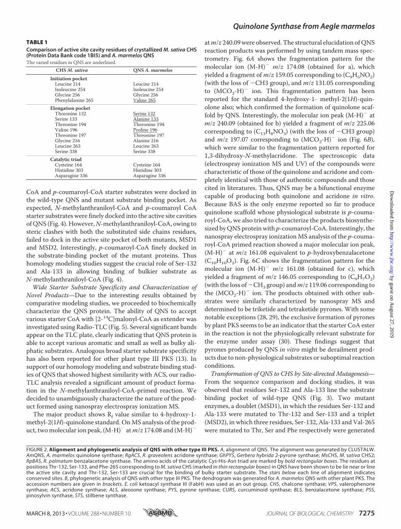
were carried out to get an insight into the role of active siteresidues in QNS substrate binding activity. Homology modelsof QNS wild-type and mutants, MSD1 (S132T/A133S), andMSD2 (S132T/A133S/V265F) were generated based onM. sativa CHS (Protein Data Bank code 1BI5) as template (Fig.3). The homology model of A. marmelos QNS wild-typeshowed that it has the same overall fold asM. sativa CHS, withcavity volume estimated to be 1196 Å3, which is much largerthan that ofM. sativa CHS (1019 Å3). This also suggested thatthe active site cavity of QNS is easily large enough to accommo-date bulkier starter substrate thanCHS. The three residues Ser-132, Ala-133, and Val-265 lie near or line the active cavity. Themutant MSD1 andMSD2 homology models shows remarkableconstriction in the active site cavity with a volume estimated tobe 1188 and 1168 Å3, respectively (Fig. 3). We therefore pre-dicted that the steric bulk from three active site residues sub-stitution, S132T, A133S, and V265F, in mutants significantlyaffect the active site volume and might hinder the binding ofbulkier starter substrates in mutants. To improve our predict-ability for MSD1 and MSD2 mutants, N-methylanthraniloyl-
FIGURE 1. Amplification of QNS from A. marmelos leaves. A and B, PCR amplification of QNS from cDNA ad genomic DNA, respectively. 1, 1-kb DNA ladder;2, PCR product; 3, control reaction. C, organization of intron in QNS gene. The arrow indicates the predicted intron position. The diagrammatic representationof QNS genome organization was also shown below.
Quinolone Synthase from Aegle marmelos
MARCH 8, 2013 • VOLUME 288 • NUMBER 10 JOURNAL OF BIOLOGICAL CHEMISTRY 7273
by guest on August 27, 2019
http://ww
w.jbc.org/
Dow
nloaded from

Quinolone Synthase from Aegle marmelos
7274 JOURNAL OF BIOLOGICAL CHEMISTRY VOLUME 288 • NUMBER 10 • MARCH 8, 2013
by guest on August 27, 2019
http://ww
w.jbc.org/
Dow
nloaded from
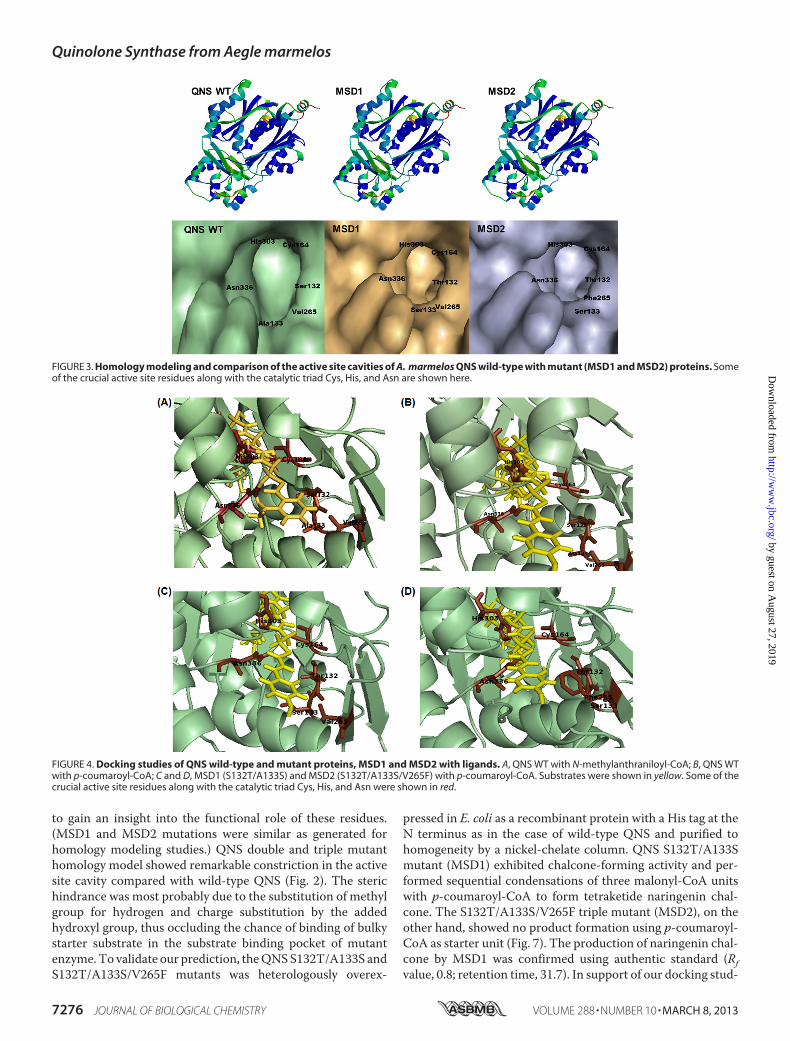
CoA and p-coumaroyl-CoA starter substrates were docked inthe wild-type QNS and mutant substrate binding pocket. Asexpected, N-methylanthraniloyl-CoA and p-coumaroyl CoAstarter substrates were finely docked into the active site cavitiesofQNS (Fig. 4).However,N-methylanthraniloyl-CoA, owing tosteric clashes with both the substituted side chains residues,failed to dock in the active site pocket of both mutants, MSD1and MSD2. Interestingly, p-coumaroyl-CoA finely docked inthe substrate-binding pocket of the mutant proteins. Thushomology modeling studies suggest the crucial role of Ser-132and Ala-133 in allowing binding of bulkier substrate asN-methylanthraniloyl-CoA (Fig. 4).Wide Starter Substrate Specificity and Characterization of
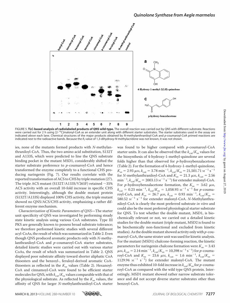
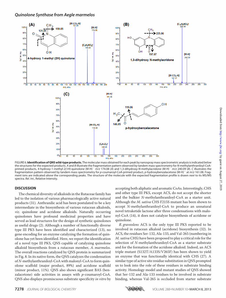
Novel Products—Due to the interesting results obtained bycomparative modeling studies, we proceeded to biochemicallycharacterize the QNS protein. The ability of QNS to acceptvarious starter CoA with [2-14C]malonyl-CoA as extender wasinvestigated using Radio-TLC (Fig. 5). Several significant bandsappear on the TLC plate, clearly indicating that QNS protein isable to accept various aromatic and small as well as bulky ali-phatic substrates. Analogous broad starter substrate specificityhas also been reported for other plant type III PKS (13). Insupport of our homologymodeling and substrate binding stud-ies of QNS that showed highest similarity with ACS, our radio-TLC analysis revealed a significant amount of product forma-tion in the N-methylanthraniloyl-CoA-primed reaction. Wedecided to unambiguously characterize the nature of the prod-uct formed using nanospray electrospray ionization MS.The major product shows Rf value similar to 4-hydroxy-1-
methyl-2(1H)-quinolone standard.OnMS analysis of the prod-uct, twomolecular ion peak, (M-H)� atm/z 174.08 and (M-H)�
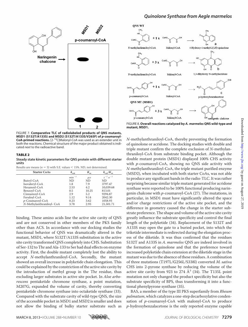
atm/z 240.09were observed. The structural elucidation ofQNSreaction products was performed by using tandem mass spec-trometry. Fig. 6A shows the fragmentation pattern for themolecular ion (M-H)� m/z 174.08 (obtained for a), whichyielded a fragment ofm/z 159.05 corresponding to (C9H5NO2)(with the loss of �CH3 group), andm/z 131.05 correspondingto (MCO2-H)� ion. This fragmentation pattern has beenreported for the standard 4-hydroxy-1- methyl-2(1H)-quin-olone also; which confirmed the formation of quinolone scaf-fold by QNS. Interestingly, the molecular ion peak (M-H)� atm/z 240.09 (obtained for b) yielded a fragment of m/z 225.06corresponding to (C13H8NO3) (with the loss of �CH3 group)and m/z 197.07 corresponding to (MCO2-H)� ion (Fig. 6B),which were similar to the fragmentation pattern reported for1,3-dihydroxy-N-methylacridone. The spectroscopic data(electrospray ionization MS and UV) of the compounds werecharacteristic of those of the quinolone and acridone and com-pletely identical with those of authentic compounds and thosecited in literatures. Thus, QNS may be a bifunctional enzymecapable of producing both quinolone and acridone in vitro.Because BAS is the only enzyme reported so far to producequinolone scaffold whose physiological substrate is p-couma-royl-CoA, we also tried to characterize the products biosynthe-sized byQNSproteinwith p-coumaroyl-CoA. Interestingly, thenanospray electrospray ionizationMS analysis of the p-couma-royl-CoA primed reaction showed a major molecular ion peak,(M-H)� at m/z 161.08 equivalent to p-hydroxybenzalacetone(C10H10O2). Fig. 6C shows the fragmentation pattern for themolecular ion (M-H)� m/z 161.08 (obtained for c), whichyielded a fragment of m/z 146.05 corresponding to (C9H7O2)(with the loss of�CH3 group) andm/z 119.06 corresponding tothe (MCO2-H)� ion. The products obtained with other sub-strates were similarly characterized by nanospray MS anddetermined to be triketide and tetraketide pyrones. With somenotable exceptions (28, 29), the exclusive formation of pyronesby plant PKS seems to be an indicator that the starter CoA esterin the reaction is not the physiologically relevant substrate forthe enzyme under assay (30). These findings suggest thatpyrones produced by QNS in vitro might be derailment prod-ucts due to non-physiological substrates or suboptimal reactionconditions.Transformation of QNS to CHS by Site-directed Mutagenesis—
From the sequence comparison and docking studies, it wasobserved that residues Ser-132 and Ala-133 line the substratebinding pocket of wild-type QNS (Fig. 3). Two mutantenzymes, a doublet (MSD1), in which the residues Ser-132 andAla-133 were mutated to Thr-132 and Ser-133 and a triplet(MSD2), in which three residues, Ser-132, Ala-133 and Val-265were mutated to Thr, Ser and Phe respectively were generated
FIGURE 2. Alignment and phylogenetic analysis of QNS with other type III PKS. A, alignment of QNS. The alignment was generated by CLUSTALW.AmQNS, A. marmelos quinolone synthase; RgACS, R. graveolens acridone synthase; GhPYS, Gerbera hybrida 2-pyrone synthase; MsCHS, M. sativa CHS2;RpBAS, R. palmatum benzalacetone synthase. The amino acids of the catalytic Cys-His-Asn triad are marked by bold rectangular boxes. The residues atpositions Thr-132, Ser-133, and Phe-265 corresponding to M. sativa CHS (marked in thin rectangular boxes) in QNS have been shown to be lie near or linethe active site cavity and Thr-132, Ser-133 are crucial for the binding of bulky starter substrate. The stars below each line of alignment indicatesconserved sites. B, phylogenetic analysis of QNS with other type III PKS. The dendrogram was generated for A. marmelos QNS with other plant PKS. Theaccession numbers are given in brackets. E. coli ketoacyl synthase III (FabH) was used as an out group. CHS, chalcone synthase; VPS, valerophenonesynthase; ACS, acridone synthase; ALS, aleosone synthase; PYS, pyrone synthase; CURS, curcuminoid synthase; BLS, benzalacetone synthase; PSS,pinosylvin synthase; STS, stilbene synthase.
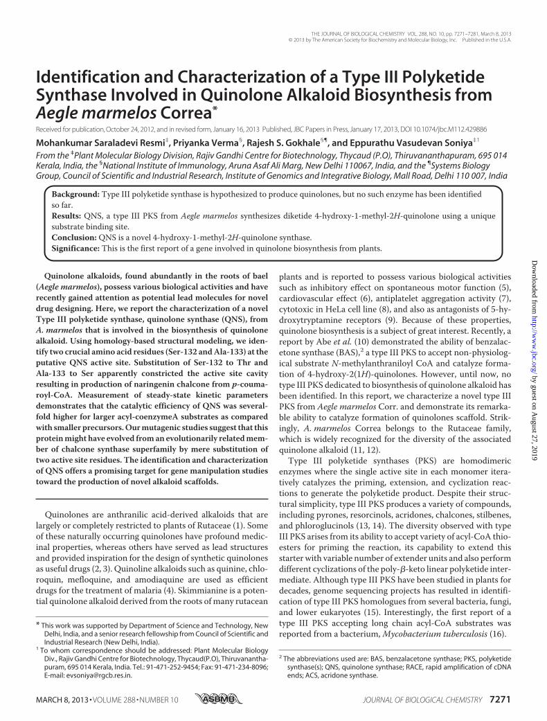
TABLE 1Comparison of active site cavity residues of crystallized M. sativa CHS(Protein Data Bank code 1BI5) and A. marmelos QNSThe varied residues in QNS are underlined.
CHSM. sativa QNS A. marmelos
Initiation pocketLeucine 214 Leucine 214Isoleucine 254 Isoleucine 254Glycine 256 Glycine 256Phenylalanine 265 Valine 265
Elongation pocketThreonine 132 Serine 132Serine 133 Alanine 133Threonine 194 Threonine 194Valine 196 Proline 196Threonine 197 Threonine 197Glycine 216 Alanine 216Leucine 263 Leucine 263Serine 338 Serine 338
Catalytic triadCysteine 164 Cysteine 164Histidine 303 Histidine 303Asparagine 336 Asparagine 336
Quinolone Synthase from Aegle marmelos
MARCH 8, 2013 • VOLUME 288 • NUMBER 10 JOURNAL OF BIOLOGICAL CHEMISTRY 7275
by guest on August 27, 2019
http://ww
w.jbc.org/
Dow
nloaded from
to gain an insight into the functional role of these residues.(MSD1 and MSD2 mutations were similar as generated forhomology modeling studies.) QNS double and triple mutanthomology model showed remarkable constriction in the activesite cavity compared with wild-type QNS (Fig. 2). The sterichindrance was most probably due to the substitution of methylgroup for hydrogen and charge substitution by the addedhydroxyl group, thus occluding the chance of binding of bulkystarter substrate in the substrate binding pocket of mutantenzyme.To validate our prediction, theQNSS132T/A133S andS132T/A133S/V265F mutants was heterologously overex-
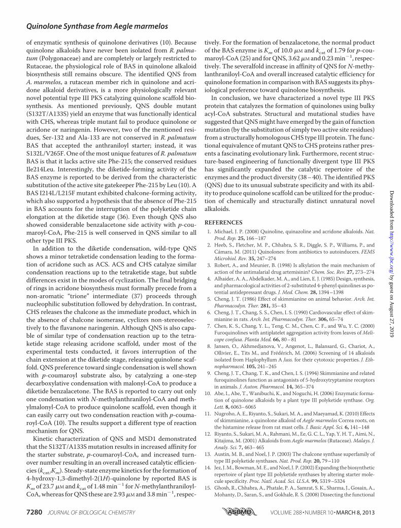
pressed in E. coli as a recombinant protein with a His tag at theN terminus as in the case of wild-type QNS and purified tohomogeneity by a nickel-chelate column. QNS S132T/A133Smutant (MSD1) exhibited chalcone-forming activity and per-formed sequential condensations of three malonyl-CoA unitswith p-coumaroyl-CoA to form tetraketide naringenin chal-cone. The S132T/A133S/V265F triple mutant (MSD2), on theother hand, showed no product formation using p-coumaroyl-CoA as starter unit (Fig. 7). The production of naringenin chal-cone by MSD1 was confirmed using authentic standard (Rfvalue, 0.8; retention time, 31.7). In support of our docking stud-
FIGURE 3. Homology modeling and comparison of the active site cavities of A. marmelos QNS wild-type with mutant (MSD1 and MSD2) proteins. Someof the crucial active site residues along with the catalytic triad Cys, His, and Asn are shown here.
FIGURE 4. Docking studies of QNS wild-type and mutant proteins, MSD1 and MSD2 with ligands. A, QNS WT with N-methylanthraniloyl-CoA; B, QNS WTwith p-coumaroyl-CoA; C and D, MSD1 (S132T/A133S) and MSD2 (S132T/A133S/V265F) with p-coumaroyl-CoA. Substrates were shown in yellow. Some of thecrucial active site residues along with the catalytic triad Cys, His, and Asn were shown in red.
Quinolone Synthase from Aegle marmelos
7276 JOURNAL OF BIOLOGICAL CHEMISTRY VOLUME 288 • NUMBER 10 • MARCH 8, 2013
by guest on August 27, 2019
http://ww
w.jbc.org/
Dow
nloaded from
ies, none of the mutants formed products with N-methylan-thraniloyl-CoA. Thus, the two amino acid substitution, S132Tand A133S, which were predicted to line the QNS substratebinding pocket in the mutant MSD1, considerably shifted thestarter substrate preference to p-coumaroyl-CoA and hencetransformed the enzyme completely to a functional CHS pro-ducing naringenin (Fig. 7). Our results correlate with thereported transformation ofACS toCHSby triplemutation (27).The triple ACS mutant (S132T/A133S/V265F) retained �35%ACS activity with an overall 10-fold increase in specific CHSactivity. Interestingly, although the double mutant protein(S132T/A133S) displayed 100% CHS activity, the triple mutantshowed no QNS/ACS/CHS activity, emphasizing a rather dif-ferent enzyme mechanism.Characterization of Kinetic Parameters of QNS—The starter
unit specificity of QNS was investigated by performing steadystate kinetic analysis using various CoA substrates. Type IIIPKS are generally known to possess broad substrate tolerance;we therefore performed kinetic studies with several differentacyl-CoAs, the result ofwhichwas summarized inTable 2. Eventhough QNS produced aromatic products only with N-methy-lanthraniloyl-CoA and p-coumaroyl-CoA starter substrates,detailed kinetic studies were carried out with various starterCoAs, the result of which was summarized in Table 2. QNSdisplayed poor substrate affinity toward shorter aliphatic CoAthioesters and the benzoyl-, feruloyl-derived aromatic CoA-thioesters as reflected in the Km values (Table 2). Hexanoyl-CoA and cinnamoyl-CoA were found to be efficient startermolecules forQNS,with kcat/Km values comparablewith that ofthe physiological substrate. As reflected by the Km values, theaffinity of QNS for larger N-methylanthraniloyl-CoA starter
was found to be higher compared with p-coumaroyl-CoAstarter units. It can also be observed that the kcat/Km values forthe biosynthesis of 4-hydroxy-1-methyl-quinolone are severalfolds higher than that observed for p-hydroxybenzalacetone(Table 2). For the formation of 4-hydroxy-1-methyl-quinolone,Km � 2.93 �M, kcat � 3.78 min�1, kcat/Km � 21,501.71 M�1 s�1
for N-methylanthraniloyl-CoA and Km � 21.3 �M, kcat � 2.56min�1, kcat/Km � 2003.13 M�1 s�1) for extender malonyl-CoA.For p-hydroxybenzalacetone formation, the Km � 3.62 �M,kcat � 0.23 min�1, kcat/Km � 1,058.93 M�1 s�1 for p-couma-royl-CoA, and Km � 26.7 �M, kcat � 0.93 min�1, kcat/Km �580.52 M�1 s�1 for extender malonyl-CoA. N-Methylanthra-niloyl-CoA is clearly the most preferred substrate in vitro andcould also be the most preferred physiological starter substratefor QNS. To test whether the double mutant, MSD1, is bio-chemically relevant or not, we carried out a detailed kineticstudies for the double mutant (triple mutant, MSD2 is found tobe biochemically non-functional and excluded from kineticstudies). As the doublemutant showed activity onlywithp-cou-maroyl-CoA, the same strater unit was used for kinetic analysis.For the mutant (MSD1) chalcone-forming reaction, the kineticparameters for naringenin chalcone formation were Km � 3.43�M, kcat � 2.14min�1, kcat/Km � 10,398 M�1 s�1) for p-couma-royl-CoA and Km � 23.6 �M, kcat � 1.6 min�1, kcat/Km �1129.94 M�1 s�1) for extender malonyl-CoA. The mutantenzyme thus exhibited 10-fold increase in kcat/Km for p-couma-royl-CoA as compared with the wild type QNS protein. Inter-estingly, MSD1 mutant showed rather narrow substrate toler-ance and did not accept diverse starter substrates other thanbenzoyl-CoA.
FIGURE 5. TLC-based analysis of radiolabeled products of QNS wild-type. The overall reaction was carried out by QNS with different substrate. Reactionswere carried out for 2 h using [2-14C]malonyl-CoA as an extender unit along with different starter substrates. The starter substrates used in the assay areindicated above each lane. Chemical structures of the major products obtained by N-methylanthraniloyl-CoA and p-coumaroyl-CoA primed reactions areindicated next to the radioactive bands. Because the Rf value of 1,3-dihydroxy-N-methylacridone was not known, it was not shown.
Quinolone Synthase from Aegle marmelos
MARCH 8, 2013 • VOLUME 288 • NUMBER 10 JOURNAL OF BIOLOGICAL CHEMISTRY 7277
by guest on August 27, 2019
http://ww
w.jbc.org/
Dow
nloaded from
DISCUSSION
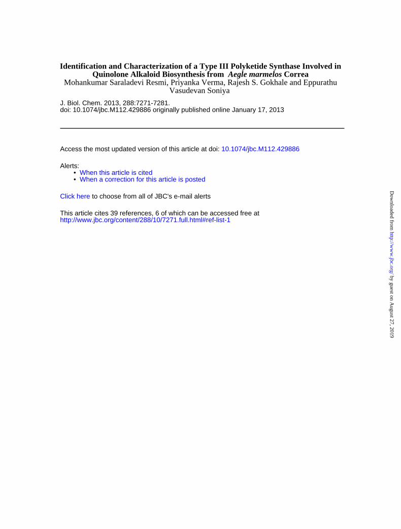
The chemical diversity of alkaloids in the Rutaceae family hasled to the isolation of various pharmacologically active naturalproducts (31). Anthranilic acid has been postulated to be a keyintermediate in the biosynthesis of various rutacean alkaloids,viz. quinolone and acridone alkaloids. Naturally occurringquinolones have profound medicinal properties and haveserved as lead structures for the design of synthetic quinolonesas useful drugs (2). Although a number of functionally diversetype III PKS have been identified and characterized (13), nogene encoding for an enzyme catalyzing the formation of quin-olone has yet been identified. Here, we report the identificationof a novel type III PKS, QNS capable of catalyzing quinolonealkaloid biosynthesis from a rutacean member, A. marmelos.The overall reactions catalyzed by QNS protein is summarizedin Fig. 8. In its native form, theQNS catalyzes the condensationofN-methylanthraniloyl-CoAwithmalonyl-CoA to formquin-olone scaffold (major product, 89%) and acridone scaffold(minor product, 11%). QNS also shows significant BAS (ben-zalacetone) side activities in assays with p-coumaroyl-CoA.QNS also displays promiscuous substrate specificity in vitro by
accepting both aliphatic and aromaticCoAs. Interestingly, CHSand other type III PKS, except ACS, do not accept the shorterand the bulkier N-methylanthraniloyl-CoA as a starter unit.Although theM. sativa CHS F215S mutant has been shown toaccept N-methylanthraniloyl-CoA to produce an unnaturalnovel tetraketide lactone after three condensations with malo-nyl-CoA (14), it does not catalyze biosynthesis of acridone orquinolone.R. graveolens ACS is the only type III PKS reported to be
involved in rutacean alkaloid (acridone) biosynthesis (32). InACS, the residues Ser-132, Ala-133, andVal-265 (numbering inM. sativaCHS) have been proposed to play a critical role for theselection of N-methylanthraniloyl-CoA as a starter substrateand for the formation of the acridone alkaloid. Indeed, an ACStriple mutant (S132T/A133S/V265F) has been shown to yieldan enzyme that was functionally identical with CHS (27). Asimilar type of active site residue substitution inQNSpromptedus to look into the role of these residues in substrate bindingactivity. Homology model and mutant studies of QNS showedthat Ser-132 and Ala-133 residues to be involved in substratebinding, whereas Val-265 is occluded from starter substrate
FIGURE 6. Identification of QNS wild-type products. The molecular mass obtained for each peak by nanospray mass spectrometric analysis is indicated belowthe structures for the expected products. A and B illustrate the fragmentation pattern observed by tandem mass spectrometry for N-methylanthraniloyl-CoA-primed products, 4-hydroxy-1-methyl-2(1H)-quinolone (M-H)� m/z 174.08 (A) and 1,3-dihydroxy-N-methylacridone (M-H)� m/z 240.09 (B). C illustrates thefragmentation pattern observed by tandem mass spectrometry for p-coumaroyl-CoA-primed product, p-hydroxybenzalacetone (M-H)� at m/z 161.08. Frag-ment ions are indicated above the corresponding peaks. The structure of the molecule with the expected fragmentation profile is drawn next to its MS/MSspectra. Rel. Int., Relative Intensity.
Quinolone Synthase from Aegle marmelos
7278 JOURNAL OF BIOLOGICAL CHEMISTRY VOLUME 288 • NUMBER 10 • MARCH 8, 2013
by guest on August 27, 2019
http://ww
w.jbc.org/
Dow
nloaded from
binding. These amino acids line the active site cavity of QNSand are not conserved in other members of the PKS familyother than ACS. In accordance with our docking studies thefunctional behavior of QNS was dramatically altered in themutant, MSD1, where S132T/A133S substitution in the activesite cavity transformedQNS completely intoCHS. Substitutionof Ser-132 toThr andAla-133 to Ser had dual effects on enzymeactivity. First, the double mutant completely lost the ability toaccept N-methylanthraniloyl-CoA. Secondly, the mutantshowed an overall increase in polyketide chain elongation. Thiscould be explained by the constriction of the active site cavity bythe introduction of methyl group in the Thr residue, thusexcluding larger substrates in active site pocket. In Aloe arbo-rescens pentaketide chromone synthase, a point mutation,M207G, expanded the volume of cavity, thereby convertingpentaketide chromone synthase into octaketide synthase (33).Compared with the substrate cavity of wild-type QNS, the sizeof the accessible pocket inMSD1 andMSD2 is smaller and doesnot allow the binding of bulky starter substrate such as
N-methylanthraniloyl-CoA, thereby preventing the formationof quinolone or acridone. The docking studies with double andtriple mutant confirm the complete exclusion of N-methylan-thraniloyl-CoA from substrate binding pocket. Although thedouble mutant protein (MSD1) displayed 100% CHS activitywith p-coumaroyl-CoA, showing no QNS side activity withN-methylanthraniloyl-CoA, the triple mutant purified enzyme(MSD2), when incubated with both starter CoAs, was not ableto produce any significant bands in the radio-TLC. It was rathersurprising because similar triplemutant generated for acridonesynthase were reported to be 100% functional producing narin-genin chalcone with p-coumaroyl-CoA (27). The mutations, inparticular, in MSD1 must have significantly altered the spaceand/or charge restrictions of the active site pocket, and thedifference in geometry caused the change in the starter sub-strate preference. The shape and volume of the active site cavitygreatly influence the substrate specificity and control the finallength of the polyketide (34). Replacement of the S132T andA133S may open the gate to a buried pocket, into which thetriketide intermediate is redirected during the elongation proc-ess of the diketide. It was thus confirmed that the residuesS132T and A133S in A. marmelos QNS are indeed involved inthe formation of quinolone and that the preference towardincreased polyketide chain extension at the diketide stage in themutantwas due to the absence of these residues. A combinationof three mutations (T197L/G256L/S338I) converted M. sativaCHS into 2-pyrone synthase by reducing the volume of theactive site cavity from 923 to 274 Å3 (34). The T135L pointmutation not only changed the product specificity but also thesubstrate specificity of BPS, thus transforming it into a func-tional phenylpyrone synthase (35).BAS, a member of the type III PKS superfamily from Rheum
palmatum, which catalyzes a one-step decarboxylative conden-sation of p-coumaroyl-CoA with malonyl-CoA to producep-hydroxybenzalacetone is the only reported enzyme capable
FIGURE 7. Comparative TLC of radiolabeled products of QNS mutants,MSD1 (S132T/A133S) and MSD2 (S132T/A133S/V265F) of p-coumaroyl-CoA-primed reactions. [2-14C]Malonyl-CoA was used as an extender unit inboth the reactions. Chemical structure of the major product obtained is indi-cated next to the radioactive band.
TABLE 2Steady state kinetic parameters for QNS protein with different starterunitsResults are means (n � 3) with S.E. values � 15%. ND, not determined.
Starter CoAs kcat Km kcat/Km
min�1 �M s�1 M�1
Butryl-CoA ND ND NDIsovaleryl-CoA 1.8 7.9 3797.47Hexanoyl-CoA 2.53 4.2 10,039.68Benzoyl-CoA 0.5 10.25 813.01Cinnamoyl-CoA 2.9 5.2 9294.87Feruloyl-CoA 1.12 9.14 2042.30p-Coumaroyl-CoA 0.23 3.62 1058.93N-Methylanthraniloyl- CoA 3.78 2.93 21,501.71
FIGURE 8. Overall reactions catalyzed by A. marmelos QNS wild-type andmutant, MSD1.
Quinolone Synthase from Aegle marmelos
MARCH 8, 2013 • VOLUME 288 • NUMBER 10 JOURNAL OF BIOLOGICAL CHEMISTRY 7279
by guest on August 27, 2019
http://ww
w.jbc.org/
Dow
nloaded from
of enzymatic synthesis of quinolone derivatives (10). Becausequinolone alkaloids have never been isolated from R. palma-tum (Polygonaceae) and are completely or largely restricted toRutaceae, the physiological role of BAS in quinolone alkaloidbiosynthesis still remains obscure. The identified QNS fromA. marmelos, a rutacean member rich in quinolone and acri-done alkaloid derivatives, is a more physiologically relevantnovel potential type III PKS catalyzing quinolone scaffold bio-synthesis. As mentioned previously, QNS double mutant(S132T/A133S) yield an enzyme that was functionally identicalwith CHS, whereas triple mutant fail to produce quinolone oracridone or naringenin. However, two of the mentioned resi-dues, Ser-132 and Ala-133 are not conserved in R. palmatumBAS that accepted the anthraniloyl starter; instead, it wasS132L/V265F. One of themost unique features ofR. palmatumBAS is that it lacks active site Phe-215; the conserved residuesIle214Leu. Interestingly, the diketide-forming activity of theBAS enzyme is reported to be derived from the characteristicsubstitution of the active site gatekeeper Phe-215 by Leu (10). ABAS I214L/L215F mutant exhibited chalcone-forming activity,which also supported a hypothesis that the absence of Phe-215in BAS accounts for the interruption of the polyketide chainelongation at the diketide stage (36). Even though QNS alsoshowed considerable benzalacetone side activity with p-cou-maroyl-CoA, Phe-215 is well conserved in QNS similar to allother type III PKS.In addition to the diketide condensation, wild-type QNS
shows a minor tetraketide condensation leading to the forma-tion of acridone such as ACS. ACS and CHS catalyze similarcondensation reactions up to the tetraketide stage, but subtledifferences exist in the modes of cyclization. The final bridgingof rings in acridone biosynthesis must formally precede from anon-aromatic “trione” intermediate (37) proceeds throughnucleophilic substitution followed by dehydration. In contrast,CHS releases the chalcone as the immediate product, which inthe absence of chalcone isomerase, cyclizes non-stereoselec-tively to the flavanone naringenin. Although QNS is also capa-ble of similar type of condensation reaction up to the tetra-ketide stage releasing acridone scaffold, under most of theexperimental tests conducted, it favors interruption of thechain extension at the diketide stage, releasing quinolone scaf-fold. QNS preference toward single condensation is well shownwith p-coumaroyl substrate also, by catalyzing a one-stepdecarboxylative condensation with malonyl-CoA to produce adiketide benzalacetone. The BAS is reported to carry out onlyone condensation with N-methylanthraniloyl-CoA and meth-ylmalonyl-CoA to produce quinolone scaffold, even though itcan easily carry out two condensation reaction with p-couma-royl-CoA (10). The results support a different type of reactionmechanism for QNS.Kinetic characterization of QNS and MSD1 demonstrated
that the S132T/A133Smutation results in increased affinity forthe starter substrate, p-coumaroyl-CoA, and increased turn-over number resulting in an overall increased catalytic efficien-cies (kcat/Km). Steady-state enzyme kinetics for the formation of4-hydroxy-1,3-dimethyl-2(1H)-quinolone by reported BAS isKm of 23.7�M and kcat of 1.48min�1 forN-methylanthraniloyl-CoA,whereas forQNS these are 2.93�Mand 3.8min�1, respec-
tively. For the formation of benzalacetone, the normal productof the BAS enzyme is Km of 10.0 �M and kcat of 1.79 for p-cou-maroyl-CoA (25) and forQNS, 3.62�Mand 0.23min�1, respec-tively. The severalfold increase in affinity of QNS forN-methy-lanthraniloyl-CoA and overall increased catalytic efficiency forquinolone formation in comparisonwithBAS suggests its phys-iological preference toward quinolone biosynthesis.In conclusion, we have characterized a novel type III PKS
protein that catalyzes the formation of quinolones using bulkyacyl-CoA substrates. Structural and mutational studies havesuggested thatQNSmight have emerged by the gain of functionmutation (by the substitution of simply two active site residues)froma structurally homologousCHS type III protein. The func-tional equivalence ofmutant QNS to CHS proteins rather pres-ents a fascinating evolutionary link. Furthermore, recent struc-ture-based engineering of functionally divergent type III PKShas significantly expanded the catalytic repertoire of theenzymes and the product diversity (38–40). The identified PKS(QNS) due to its unusual substrate specificity and with its abil-ity to produce quinolone scaffold can be utilized for the produc-tion of chemically and structurally distinct unnatural novelalkaloids.
REFERENCES1. Michael, J. P. (2008) Quinoline, quinazoline and acridone alkaloids. Nat.
Prod. Rep. 25, 166–1872. Heeb, S., Fletcher, M. P., Chhabra, S. R., Diggle, S. P., Williams, P., and
Cámara, M. (2011) Quinolones: from antibiotics to autoinducers. FEMSMicrobiol. Rev. 35, 247–274
3. Robert, A., and Meunier, B. (1998) Is alkylation the main mechanism ofaction of the antimalarial drug artemisinin? Chem. Soc. Rev. 27, 273–274
4. Alhaider, A. A., Abdelkader,M. A., and Lien, E. J. (1985) Design, synthesis,and pharmacological activities of 2-substituted 4-phenyl quinolines as po-tential antidepressant drugs. J. Med. Chem. 28, 1394–1398
5. Cheng, J. T. (1986) Effect of skimmianine on animal behavior. Arch. Int.Pharmacodyn. Ther. 281, 35–43
6. Cheng, J. T., Chang, S. S., Chen, I. S. (1990) Cardiovascular effect of skim-mianine in rats. Arch. Int. Pharmacodyn. Ther. 306, 65–74
7. Chen, K. S., Chang, Y. L., Teng, C. M., Chen, C. F., and Wu, Y. C. (2000)Furoquinolines with antiplatelet aggregation activity from leaves ofMeli-cope confusa. Planta Med. 66, 80–81
8. Jansen, O., Akhmedjanova, V., Angenot, L., Balansard, G., Chariot, A.,Ollivier, E., Tits M., and Frédérich, M. (2006) Screening of 14 alkaloidsisolated from Haplophyllum A Juss. for their cytotoxic properties. J. Eth-nopharmacol. 105, 241–245
9. Cheng, J. T., Chang, T. K., and Chen, I. S. (1994) Skimmianine and relatedfuroquinolines function as antagonists of 5-hydroxytryptamine receptorsin animals. J. Auton. Pharmacol. 14, 365–374
10. Abe, I., Abe, T.,Wanibuchi, K., and Noguchi, H. (2006) Enzymatic forma-tion of quinolone alkaloids by a plant type III polyketide synthase. Org.Lett. 8, 6063–6065
11. Nugroho,A. E., Riyanto, S., Sukari,M.A., andMaeyamad, K. (2010) Effectsof skimmianine, a quinolone alkaloid of Aegle marmelos Correa roots, onthe histamine release from rat mast cells. J. Basic Appl. Sci. 6, 141–148
12. Riyanto, S., Sukari,M.A., Rahmani,M., Ee, G. C. L., Yap, Y.H. T., Aimi, N.,Kitajima, M. (2001) Alkaloids from Aegle marmelos (Rutaceae).Malays. J.Analy. Sci. 7, 463–465
13. Austin, M. B., and Noel, J. P. (2003) The chalcone synthase superfamily oftype III polyketide synthases. Nat. Prod. Rep. 20, 79–110
14. Jez, J.M., Bowman,M. E., andNoel, J. P. (2002) Expanding the biosyntheticrepertoire of plant type III polyketide synthases by altering starter mole-cule specificity. Proc. Natl. Acad. Sci. U.S.A. 99, 5319–5324
15. Ghosh, R., Chhabra, A., Phatale, P. A., Samrat, S. K., Sharma, J., Gosain, A.,Mohanty, D., Saran, S., andGokhale, R. S. (2008) Dissecting the functional
Quinolone Synthase from Aegle marmelos
7280 JOURNAL OF BIOLOGICAL CHEMISTRY VOLUME 288 • NUMBER 10 • MARCH 8, 2013
by guest on August 27, 2019
http://ww
w.jbc.org/
Dow
nloaded from

role of polyketide synthases in Dictyostelium discoideum: biosynthesis ofthe differentiation regulating factor 4-methyl-5-pentylbenzene-1,3-diol.J. Biol. Chem. 283, 11348–11354
16. Saxena, P., Yadav, G.,Mohanty, D., andGokhale, R. S. (2003) A new familyof type III polyketide synthases in Mycobacterium tuberculosis. J. Biol.Chem. 278, 44780–44790
17. Arnold, K., Bordoli, L., Kopp, J., and Schwede, T. (2006) The SWISS-MODEL workspace: a web-based environment for protein structure ho-mology modelling. Bioinformatics 22, 195–201
18. Kiefer, F., Arnold, K., Künzli, M., Bordoli, L., and Schwede, T. (2009) TheSWISS-MODEL Repository and associated resources. Nucleic Acids Res.37, D387–92
19. Peitsch, M. C. (1995) Protein modeling by E-mail Bio/Technology. Nat.Biotechnol. 13, 658–660
20. Liang, J., Edelsbrunner, H., Woodward, C. (1998) Anatomy of proteinpockets and cavities: Measurement of binding site geometry and implica-tions for ligand design. Protein Sci. 7, 1884–1897
21. Ritchie, D. W., and Kemp, G. J. (2000) Protein docking using sphericalpolar Fourier correlations. Proteins 39, 178–194
22. Durbin,M. L., McCaig, B., and Clegg,M. T. (2000)Molecular evolution ofthe chalcone synthase multigene family in the morning glory genome.Plant Mol. Biol. 42, 79–92
23. Schroder, J. (1997) A family of plant-specific polyketide synthases: Factsand predictions. Trends Plant Sci. 2, 373–378
24. Zheng, D., Schröder, G., Schröder, J., and Hrazdina, G. (2001) Molecularand biochemical characterization of three aromatic polyketide synthasegenes from Rubus idaeus. Plant Mol. Biol. 46, 1–15
25. Abe, I., Takahashi, Y., Morita, H., Noguchi, H. (2001) Benzalacetone syn-thase. A novel polyketide synthase that plays a crucial role in the biosyn-thesis of phenylbutanones in Rheum palmatum. Eur. J. Biochem. 268,3354–3359
26. Resmi, M. S., and Soniya, E. V. (2012) Molecular cloning and differentialexpressions of two cDNA encoding Type III polyketide synthase in differ-ent tissues of Curcuma longa L. Gene 491, 278–283
27. Lukacin, R., Schreiner, S., and Matern, U. (2001) Transformation of acri-done synthase to chalcone synthase. FEBS Lett. 508, 413–417
28. Akiyama, T., Shibuya,M., Liu, H.M., and Ebizuka, Y. (1999) p-Coumaroyltriaceticacid synthase, a new homologue of chalcone synthase, from Hy-drangea macrophylla var. thunbergii. Eur. J. Biochem. 263, 834–839
29. Eckermann, S., Schroder, G., Schmidt, J., Strack, D., Edrada, R. A., Helar-iutta, Y., Elomaa, P., Kotilainen, M., Kilpelainen, I., Proksch, P., Teeri,T. H., and Schroder, J. (1998) New pathway to polyketides in plants. Na-ture 396, 387–390
30. Samappito, S., Page, J. E., Schmidt, J., De-Eknamkul, W., and Kutchan,T. M. (2003) Aromatic and pyrone polyketides synthesized by a stilbenesynthase from Rheum tataricum. Phytochemistry 62, 313–323
31. Tillequin, F. (2007) Rutaceous alkaloids as models for the design of novelantitumor drugs. Phytochem. Rev. 6, 65–79
32. Junghanns, K. T., Kneusel, R. E., Baumert, A., Maier, W., Gröger, D., andMatern, U. (1995) Molecular cloning and heterologous expression of ac-ridone synthase from elicited Ruta graveolens L. cell suspension cultures.Plant Mol. Biol. 27, 681–692
33. Morita,H., Kondo, S.,Oguro, S., Noguchi,H., Sugio, S., Abe, I., andKohno,T. (2007) Structural insight into chain-length control and product speci-ficity of pentaketide chromone synthase from Aloe arborescens. Chem.Biol. 14, 359–369
34. Jez, J. M., Austin, M. B., Ferrer, J., Bowman, M. E., Schröder, J., Noel, J. P.(2000) Structural control of polyketide formation in plant-specificpolyketide synthases. Chem. Biol. 7, 919–930
35. Klundt, T., Bocola, M., Lütge, M., Beuerle, T., Liu, B., and Beerhues, L.(2009) A Single Amino Acid Substitution Converts Benzophenone Syn-thase into Phenylpyrone Synthase. J. Biol. Chem. 284, 30957–30964
36. Abe, I., Sano, Y., Takahashi, Y., and Noguchi, H. (2003) Site-directed mu-tagenesis of benzalacetone synthase. The role of the Phe215 in plant typeIII polyketide synthases. J. Biol. Chem. 278, 25218–25226
37. Dewick, P. M. (1997) Medicinal Natural Products. A Biosynthetic Ap-proach, pp. 123–124, Wiley, New York
38. Abe, I., Watanabe, T., Morita, H., Kohno, T., Noguchi, H. (2006) Engi-neered biosynthesis of plant polyketides: manipulation of chalcone syn-thase. Org. Lett. 8, 499–502
39. Shi, S. P., Wanibuchi, K., Morita, H., Endo, K., Noguchi, H., and Abe, I.(2009) Enzymatic formation of unnatural novel chalcone, stilbene, andbenzophenone scaffolds by plant type III polyketide synthase. Org. Lett.11, 551–554
40. Morita, H., Yamashita, M., Shi, S. P., Wakimoto, T., Kondo, S., Kato, R.,Sugio, S., Kohno, T., and Abe, I. (2011) Synthesis of unnatural alkaloidscaffolds by exploiting plant polyketide synthase. Proc. Natl. Acad. Sci.U.S.A. 108, 13504–13509
Quinolone Synthase from Aegle marmelos
MARCH 8, 2013 • VOLUME 288 • NUMBER 10 JOURNAL OF BIOLOGICAL CHEMISTRY 7281
by guest on August 27, 2019
http://ww
w.jbc.org/
Dow
nloaded from
Vasudevan SoniyaMohankumar Saraladevi Resmi, Priyanka Verma, Rajesh S. Gokhale and Eppurathu
CorreaAegle marmelosQuinolone Alkaloid Biosynthesis from Identification and Characterization of a Type III Polyketide Synthase Involved in
doi: 10.1074/jbc.M112.429886 originally published online January 17, 20132013, 288:7271-7281.J. Biol. Chem.
10.1074/jbc.M112.429886Access the most updated version of this article at doi:
Alerts:
When a correction for this article is posted•
When this article is cited•
to choose from all of JBC's e-mail alertsClick here
http://www.jbc.org/content/288/10/7271.full.html#ref-list-1
This article cites 39 references, 6 of which can be accessed free at
by guest on August 27, 2019
http://ww
w.jbc.org/
Dow
nloaded from