Embed Size (px)
Citation preview

Biochemical Pharmacology 79 (2010) 1516–1525
Identification of metabolic pattern and bioactive form of resveratrol in humanmedulloblastoma cells
Xiao-Hong Shu, Hong Li, Zheng Sun, Mo-Li Wu, Jing-Xin Ma, Jian-Min Wang, Qian Wang, Yuan Sun,Yuan-Shan Fu, Xiao-Yan Chen, Qing-You Kong, Jia Liu *
Liaoning Laboratory of Cancer Genomics and Department of Cell Biology, College of Basic Medical Sciences, Dalian Medical University, Dalian 116044, China
A R T I C L E I N F O
Article history:
Received 14 December 2009
Received in revised form 18 January 2010
Accepted 19 January 2010
Keywords:
Resveratrol
Medulloblastoma
Drug metabolism
Sulfotransferase
Chemosensitivity
A B S T R A C T
Cancer preventive reagent trans-resveratrol is intracellularly biotransformed to different metabolites.
However, it is still unclear whether trans-resveratrol exerts its biological effects directly or through its
metabolite(s). This issue was addressed here by identifying the metabolic pattern and the bioactive form
of resveratrol in a resveratrol-sensitive human medulloblastoma cell line, UW228-3. The cell lysates and
condition media of UW228-3 cells with or without 100 mM resveratrol treatment were analyzed by
HPLC and LC/MS which revealed (1) that resveratrol was chemically unstable and the spontaneous
generation of cis-resveratrol reduced resveratrol’s anti-medulloblastoma efficacy and (2) that
resveratrol monosulfate was the major metabolite of the cells. To identify the bioactive form of
resveratrol, a mixture-containing approximately half fraction of resveratrol monosulfate was prepared
by incubating trans-resveratrol with freshly prepared rat brain lysates. Medulloblastoma cells treated by
100 mM of this mixture showed attenuated cell crisis. The overall levels of the three brain-associated
sulfotransferases (SULT1A1, 1C2 and 4A1) were low in medulloblastoma cells in vivo and in vitro in
comparison with that in human noncancerous and rat normal cerebella; resveratrol could more or less
up-regulate the production of these enzymes in UW228-3 cells but their overall level was still lower than
that in normal cerebellum tissue. Our study thus demonstrated for the first time that trans-resveratrol is
the bioactive form in medulloblastoma cells in which the expression of brain-associated SULTs was
down-regulated, resulting in the increased intracellular bioavailability and anti-medulloblastoma
efficacy of trans-resveratrol.
� 2010 Elsevier Inc. All rights reserved.
Contents lists available at ScienceDirect
Biochemical Pharmacology
journa l homepage: www.e lsev ier .com/ locate /b iochempharm
1. Introduction
Resveratrol (3,5,40-trihydroxy-trans-stilbene, Res) is a plantpolyphenol existing in grapes and many other natural foods [1,2],which possesses a wide range of biological activities such as
Abbreviations: Resveratrol, Res or R; HPLC, high performance liquid chromatogra-
phy; MS, mass spectrum; LC/MS, liquid chromatography coupled with tandem mass
spectrum; SULT, sulfotransferases; MB, medulloblastoma; DMEM, Dulbecco’s
modified Eagle’s essential medium; ICC, immunocytochemical staining; TUNEL,
terminal deoxynucleotidyl transferase mediated nick end labeling; PBS, phosphate
buffered saline solution; SPE, solid phase extraction; ESI, electrospray ionisation;
FWHM, full width at half maximum; TMA, the tissue microarray; IHC,
immunohistochemical staining; DAB, 3,30-diaminobenzidine tetrahydrochloride;
SDS-PAGE, sodium dodecylsulfate polyacrylamide gel electrophoresis; N2, nebu-
lizing gas; LC/MS-IT-TOF, ion trap-time-of-flight hybrid mass spectrometer; HRMS,
high resolution mass spectrometry; Mops, 3-[N-morpholino] propanesulfonic acid;
PAPS, 30-phosphoadenosine 50-phosphosulfate; DTT, dithiothreitol; STAT3, signal
transducer and activator of transcription 3; RT-PCR, reverse transcription-
polymerase chain reaction; TIC, total ion chromatogram; CID, collision induced
dissociation.
* Corresponding author. Tel.: +86 411 86110316; fax: +86 411 86110278.
E-mail address: [email protected] (J. Liu).
0006-2952/$ – see front matter � 2010 Elsevier Inc. All rights reserved.
doi:10.1016/j.bcp.2010.01.022
cardiovascular protection [3,4], antioxidative activity [5], anti-inflammatory property [6,7], and cancer preventive and therapeu-tic effects [8,9]. It was found that resveratrol affected carcinogenicprocess by inhibiting cancer-associated gene expression [10,11],altering multiple signaling pathways and/or modulating epigenet-ic machineries [12,13]. More importantly, this compound has littlecytotoxic effect and is able to penetrate blood–brain barrier [14],suggesting its potential therapeutic values in the management ofbrain malignancies especially those occur at childhood.
It has been recognized that resveratrol, as a polyphenolcompound, can be biotransformed intracellularly by multiplemetabolic enzymes. Although the chemotherapeutic value andmolecular effects of resveratrol have been demonstrated in manytypes of cancers [11,15–19], it is still unclear whether trans-resveratrol exerts its anticancer effects directly or through itsmetabolite(s) [20]. So far, no direct evidence has been availableconcerning the anticancer activity of the parent and conjugatedresveratrol because of the difficulty to maintain resveratrolunmetabolized in vivo and to fully transform resveratrol to anidentical conjugate in vitro. As an alternative approach, it would beworthwhile to elucidate the molecular bases and potential

X.-H. Shu et al. / Biochemical Pharmacology 79 (2010) 1516–1525 1517
therapeutic implications of resveratrol metabolism using a reliableresveratrol-sensitive cancer cells.
Medulloblastoma (MB) is originated from primitive neuralprecursor cells in the external germinal layer of the developingcerebellum and accounts for more than 25% of cancer-relateddeath among child patients [21]. Great majority of MB patients haspoor prognosis and suffers from direct surgical damages, long-term side effects and developmental defects [22]. Therefore, amore effective and less toxic therapeutic approach is urgentlyrequired for better treatment of MBs. It has been recognized thatMBs maintain the potential for further differentiation whenexposed to differentiation promoters [23]. According to ourresults obtained from four human MB cell lines [11,15,16,24],resveratrol possessed anti-medulloblastoma capacity throughpromoting both neuronal-like differentiation and apoptosis byarresting cell cycle at G1 phase and down-regulating a panel ofcancer-associated gene expression in MB cells presumablythrough altering the activities of several signaling pathways.The resveratrol-sensitive feature of MB cells thus offers us an idealmodel to shed light on the metabolic pattern and the bioactiveform of resveratrol in cancer cells.
2. Materials and methods
2.1. Cell culture and treatments
Human medulloblastoma cell line UW228-3 [25] werecultured in Dulbecco’s modified Eagles medium (DMEM;Invitrogen Co., Grand Island, NY, USA) containing 10% fetalbovine serum (Invitrogen Co., Grand Island, NY, USA) under37 8C and 5% CO2 condition. The cells (5 � 104/mL) were platedto 100 mm dishes (Nunc A/S, Roskilde, Denmark) and incubatedfor 24 h before further experiments. For morphologic evaluationand ICC staining, the coverslips were put into the dishes beforeinitial cell seeding and collected in 6 h intervals during theexperiments.
Stock solution of 100 mM trans-resveratrol (Sigma Chem Co.,St. Louis, MO, USA) was prepared in dimethylsulfoxide (DMSO;Sigma Chem Co., St. Louis, MO, USA), wrapped in aluminium foilfor protection against light and stored at �20 8C. cis-Resveratrolwas prepared by exposure of trans-resveratrol-containing solu-tion to natural light for 48 h [26]. Resveratrol were diluted withculture medium to an optimal working concentration (100 mM;11) just before cell treatment. The cells incubated with the sameconcentration of DMSO (0.2%) were used as background control.After being treated by 100 mM resveratrol for 48 h, the cells aswell as their culture media were collected respectively andanalyzed by HPLC and LC/MS. The remaining cells were used forparalleled protein and RNA preparations and flow cytometryanalysis. The total numbers and the viability of cells with orwithout 100 mM resveratrol treatment were determined bystaining the single-cell suspensions with 0.25% Trypan Blue(Sigma Chem Co., St. Louis, MO, USA) and counting the stained andunstained cells with the hemocytometers by two independentresearchers. Cell-bearing coverslips were harvested at 48 h timepoint and fixed properly for morphologic examination, immuno-cytochemical (ICC) staining and terminal deoxynucleotidyltransferase mediated nick end labeling (TUNEL; Promega Corp.,Madison, WI, USA) by the methods described elsewhere [16]. Eachof experimental groups was set in triplicate, and the experimentswere repeated at least three times to establish confidentialconclusions. The antibodies used for ICC staining were the mouseanti-human synaptophysin antibody (Santa Cruz Inc., CA, USA:1:120) and the rabbit anti-human SULT1A1, 1C2 and 4A1antibodies (1:120, 1:100 and 1:120; Protein Tech Group, Inc.,Chicago, USA).
2.2. Sample preparation for HPLC and LC/MS analyses
After resveratrol treatment, the cells were scraped off, washedthree times with PBS (pH 7.4), and lysed by sonication. Meanwhile,cell-free media were harvested directly by the end of 48-hresveratrol treatment or after an additional 24-h normal culture.The collected media and cell lysates were centrifuged at 10,000 � g
for 5 min, and the supernatants were purified by SPE [27]. Briefly,Cleanert PEP-SPE cartridges (60 mg; Agela Technol Inc. PA, USA)were conditioned with 5 mL of methanol (Fisher Sci, Fair Lawn, NJ,USA) and then with 5 mL of ultrapure water purified with a Milli-Qwater purification system (Millipore, Bedford, MA, USA). Thesamples were loaded onto the cartridges, and the cartridges werewashed with 5 mL of ultrapure water after absorption of thesamples on the solid phase. The samples were finally eluted with3 mL of methanol, and the eluate was dried by nitrogen spraying.The residues were dissolved in 500 mL methanol and a 10 mLaliquot was injected onto the liquid chromatography column forHPLC and LC/MS analysis.
2.3. HPLC analysis
Four kinds of samples were prepared from UW228-3 cell linesas Sample 1, the culture medium treated with 100 mM resveratrolfor 48 h; Sample 2, 24-h culture medium after 100 mM resveratroltreatment for 48 h; Sample 3, the cells treated with 100 mMresveratrol for 48 h; Sample 4, resveratrol-containing medium asbackground control.
The HPLC system (Waters Co., Milford, MA, USA) is consisted ofa Waters 1525 binary pump and 2487 dual wavelength UV–visdetector. The detection was carried out at 306 nm [28].Chromatographic separation of resveratrol and its metabolite(s)was performed on a Hypersil BDS-C18 column (5 mm, 4.6 mm �250 mm, Elite, Dalian, China), preceded by a C18 guard column(5 mm, 4.6 mm � 10 mm), using a column oven set at 35 8C with aflow rate of 1 mL/min. The mobile phase consisted of 5 mMammonium acetate (mobile phase A, Alfa Aesar, A Johnson MattheyCompany, Ward Hill, MA, USA), and methanol (mobile phase B).These solutions were degassed by sonication for 15 min at roomtemperature prior to use. A gradient elution was carried out asfollows [29]: 0 min, 10% B; 4 min, 20% B; 7 min, 80% B; 16 min, 55%B; 18 min, 55% B; 19 min, 90% B; 25 min, 90% B. Subsequently, thepercentage of methanol was decreased within 5 min to 10% inorder to equilibrate the column for 10 min before application of thenext injection. Samples were filtered through a 0.45 mm filter(Millipore, Bedford, MA, USA) and a 10 mL aliquot was injected.
2.4. Structural identification of resveratrol metabolite(s)
Chromatography was performed on a Agilent 1200 liquidchromatography series (Agilent Technol Inc., Santa Clara, CA, USA)coupled to an Applied Biosystems API 3200 QTrap tandem massspectrometer (Applied Biosystem/MDS SCIEX, Foster City, CA, USA)equipped with an ESI source, and operated by Applied Biosystem/MDS SCIEX analyst software (Version 1.4.1) to obtain the MS andMS/MS data. The separation was carried out using a C18 reversephase Hypersil BDS-C18 column (5 mm, 4.6 mm � 250 mm, Elite,Dalian, China) with a guard column. 5 mM ammonium acetate wasused as solvent A, and methanol as solvent B, at a flow rate of0.5 mL/min with the following gradient: 10–20% B linear (0–4 min), 20–80% B linear (4–7 min), 80–55% B linear (7–16 min),55–90% B (18–19 min), 90–10% B (25–30 min). This was followedby a 10-min equilibrium period with initial conditions prior toinjection of the next sample. After being filtered in the filter(0.45 mM, Millipore), 10 mL of the sample was directly injectedinto the column.
Table 1Primer sequences used for RT-PCR.
Parameters Primer sequence (50 !30) Annealing temperature Product (bp)
SULT1A1 Forward 50-GCAACGCAAAGGATGTGGCA-30 60 8C 122
Reverse 50-TCCGTAGGACACTTCTCCGA-30
SULT1C2 Forward 50-GGTTTGGGGTTCCTGGTTTGAC-30 58 8C 460
Reverse 50-GGCTGGGACTGAAGGATTGAAG-30
SULT4A1 Forward 50-AGATTCCTGGGGGTGTCCT-30 58 8C 250
Reverse 50-GTGAGCATGCAGGTTGTTGT-30
b-Actin Forward 50-GCATGGAGTCCTGTGGCAT-30 58 8C 326
Reverse 50-CATGAAGCATTTGCGGTGG-30
X.-H. Shu et al. / Biochemical Pharmacology 79 (2010) 1516–15251518
The MS analysis was performed in a negative ion mode. Prior touse, the ion spray interface and mass spectrometric parameterswere optimized to obtain maximum sensitivity at unit resolution.The source temperature was set at 250 8C with a curtain gaspressure (N2) of 25 psi, a N2 pressure of 30 psi, an auxiliary heat gaspressure of 40 psi. The ion sprayer voltage was set at 5000 V fornegative ion mode with a declustering potential of 45 V. Thecollision energy was set at 10 V and collision gas (N2) was set athigh. Full-scan data acquisition was performed by scanning fromm/z 100 to 600 in profile mode [30], using a cycle time of 2 s and apause between scans of 2 ms. For MS/MS, a product ion scan wasused at a cycle time of 2 s. To identify the characteristic ions foreach compound, samples were injected into the LC/MS-system innegative ionisation mode.
Further identification and confirmation of the metabolites wasperformed on a Shimadzu LCMS-IT-TOF (Shimadzu Co., Kyoto,Japan) instrument equipped with an ESI source in negative ionmode at a resolution of 10,000 FWHM. Accurate masses werecorrected by using the standard sample sodium trifluoroacetate.10 mL sample was injected onto a Shim-pack VP-ODS column(5 mm, 2.0 mm � 150 mm; Shimadzu Co., Kyoto, Japan), then waseluted (0.2 mL/min) with a gradient of 5–65% acetonitrile (AlfaAesar, A Johnson Matthey Company, Ward Hill, MA, USA) in 5 mMammonium, over 25 min with column temperature maintained at40 8C. To avoid the cross contamination, the blank run was insertedbetween the consecutive samples. HRMS analytical conditionswere as follows: acquisition m/z, 100–800; interface voltage,4.50 kV; IT area vacuum, 1.2 � 10�2 Pa; TOF area vacuum,1.2 � 10�4 Pa; equipment temperature, 40 8C; N2 flow, 1.5 L/min; drying gas (N2) pressure, 0.2 MPa; CDL and heater blocktemperature, 200 8C; interface voltage, 4.50 kV; ion accumulation,20 ms. MS data were processed with LC/MS solution ver. 3.4software (Shimadzu, Japan).
2.5. Immunohistochemical staining for sulfotransferases (SULTs)
Surgical specimens were incised from 37 medulloblastomacases in the range from 9- to 23-year-old. After getting theconsents from the patients and/or their parents, the freshspecimens of the tumor mass and, where possible, tumor-surrounding tissues were collected from the operation rooms ofthe First Affiliated Hospital, Dalian Medical University and Sheng-Jing Hospital, China Medical University at Shenyang within 2 h,fixed in 10% formaldehyde and treated conventionally forpreparing paraffin-embedded tissue blocks. The tissue microarrays(TMAs) in a density of 42 tissue spots/cm2 was constructed usingthe target tissues cored from the tumors, the surroundingcerebellum regions and rat normal cerebella. Three members ofSULT1A1, SULT1C2 and SULT4A1 were selected for TMA-basedimmunohistochemical staining (IHC) because of their preferentialexpression in cerebellum [31–33]. The sections without first
antibody incubation were used as background control. Accordingto the labeling intensity, the staining results were evaluated by twoindependent researchers and scored as negative (�) if no immuno-labeling was observed in target cells, weakly positive (+) if thelabeling was faint, and moderately to strongly positive (>++) whenthe labeling was stronger or distinctly stronger than (+). Beforeundertaking the above study, the experimental protocol wasreviewed and approved by the ethnic committee of Dalian MedicalUniversity for protection of human subjects.
2.6. RNA Isolation and RT-PCR
Total cellular RNA was isolated from each of experimentalgroup using Trizol solution (Life Technol., Grand Island, NY, USA).Reverse transcription (RT) was performed on RNA samples,followed by polymerase chain reaction (PCR) amplification. ForRT, 0.5 mg of the RNA sample was added to 20 mL of RT reactionmixture (Takara, Inc., Ltd., Dalian, China) containing 4 mL of MgCl2,2 mL of 10� RNA PCR buffer, 9.5 mL of RNase-free distilled H2O,2 mL of dNTP mixture, 0.5 mL of RNase inhibitor, 1 mL of AMVreverse transcriptase, and 1 mL of oligo dT-adaptor primer. Thereaction was carried out by treating the samples at 55 8C for30 min, at 99 8C for 5 min, and at 5 8C for 5 min. PCR was conductedusing the primers specific for each of the target genes (Table 1).Briefly, 2.5 mL of RT products were mixed with 16 mL of PCR-gradewater, then with 6.5 mL of PCR working solution containing 1� PCRbuffer, 1 mL of dNTP, 2.5 units of Taq DNA polymerase, and 50 pMupstream and downstream primers for SULT 1A1, SULT 1C2 andSULT 4A1, respectively. The PCR for individual genes wereperformed as follows: After initial denaturation for 5 min at95 8C, samples were subjected to 35 cycles of 95 8C for 25 s;annealing temperature was 58 8C or 60 8C for 20 s and 72 8C for20 s, with a final extension time of 2 min at 72 8C, and at 5 8C for5 min. The PCR products were resolved on 1% agarose gelcontaining ethidium bromide (0.5 mg/mL). The bands werevisualized and photographed using UVP Biospectrum ImagingSystem (UVP, Inc, Upland, CA, USA). The PCR products generatedfrom the same RT solution by a pair of b-actin primers were citedas internal quantitative controls.
2.7. Protein preparation and Western blot analyses
Total cellular proteins were prepared from UW228-3 cellsunder different culture conditions by the method describedpreviously [16]. Because of the impossibility to obtain normalbrain tissues of humans, the corresponding tissues of rats wereused as positive controls for SULTs. For Western blot analyses, thesample proteins (50 mg/lane) were separated by electrophoresis in10% SDS-PAGE, transferred to polyvinylidene difluoride membrane(Amersham Biosci, Buckinghamshire, UK). The membrane wasblocked with 5% skimmed milk in TBS-T (10 mM Tris–Cl, pH 8.0,
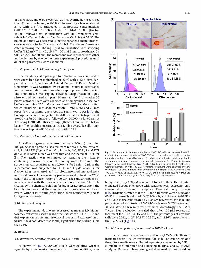
Fig. 1. Evaluation of chemosensitivities of UW228-3 cells to resveratrol. (A) To
evaluate the chemosensitivity of UW228-3 cells, the cells were collected after
incubation without (normal) or with 100 mM resveratrol for 48 h, and subjected to
synaptophysin-oriented immunocytochemical staining and TUNEL apoptosis assay
(shown in the small blocks of Fig. 1A). (B) After being cultured for 48 h, the cells
without (normal) or with 100 mM resveratrol treatment were analyzed by flow
cytometry. (C) Cells number was determined by Trypan Blue exclusion after
100 mM resveratrol incubation for 0, 12, 24, 36 and 48 h, respectively. Data are
expressed as means � S.D. (n = 3; *p < 0.01; **p < 0.001 vs. normal).
X.-H. Shu et al. / Biochemical Pharmacology 79 (2010) 1516–1525 1519
150 mM NaCl, and 0.5% Tween 20) at 4 8C overnight, rinsed threetimes (10 min each time) with TBS-T, followed by 2 h incubation at37 8C with the first antibodies in appropriate concentrations(SULT1A1, 1:1200; SULT1C2, 1:500; SULT4A1, 1:600; b-actin,1:3000) followed by 1 h incubation with HRP-conjugated anti-rabbit IgG (Zymed Lab Inc., San Francisco, CA, USA) at 37 8C. Thebound antibody was detected using the enhanced chemilumines-cence system (Roche Diagnostics GmbH, Mannheim, Germany).After removing the labeling signal by incubation with strippingbuffer (62.5 mM Tris–HCl, pH 6.7, 100 mM 2-mercaptoethanol, 2%SDS) at 55 8C for 30 min, the membrane was reprobed with otherantibodies one by one by the same experimental procedures untilall of the parameters were examined.
2.8. Preparation of SULT-containing brain lysate
One female specific pathogen free Wistar rat was cultured inwire cages in a room maintained at 22 8C with a 12-h light/darkperiod at the Experimental Animal Center of Dalian MedicalUniversity. It was sacrificed by an animal expert in accordancewith approved Ministerial procedures appropriate to the species.The brain tissue was rapidly obtained, snap frozen in liquidnitrogen and sectioned in 4 mm thickness at �40 8C; altogether 50pieces of frozen slices were collected and homogenised in ice-coldbuffer containing 250 mM sucrose, 1 mM DTT, 1� Mops buffer,which including 8 mM sodium acetate, 1 mM EDTA and 20 mMMops (pH 7.0; Sigma Chem Co., St. Louis, MO, USA) [34]. Thehomogenates were subjected to differential centrifugation at10,000 � g for 20 min at 4 8C, followed by 100,000 � g for 60 min at1 8C using CP100MX ultracentrifuge (Hitachi Koki Co. Ltd., Tokyo,Japan). The resulting supernatant containing cytosolic sulfotrans-ferase was kept at �80 8C and used within 24 h.
2.9. Resveratrol biotransformation and cell treatment
For sulfonating trans-resveratrol, a mixture (200 mL) containing100 mL cytosolic proteins isolated from rat brain, 5 mM resvera-trol, 2 mM PAPS (Sigma Chem Co., St. Louis, MO, USA), 1 mM DTTand 20 mM Mops buffer was prepared and incubated at 37 8C for2 h. The reaction was terminated by standing the mixture-containing thin-wall tube on the boiling water for 5 min. Thesuspension was centrifuged at 10,000 � g for 5 min. 10 mL of thesupernatant was subjected to HPLC and LC/MS analysis forfractionating resveratrol and its biotransformed metabolite(s),and the aliquots of the remaining part were used to treat UW228-3cells in the total concentration of 100 mM. The cellular response(s)were checked with the parameters mentioned above. The cellstreated by the chemical solution for brain lysate preparation, thebrain lysate alone and the combination of resveratrol and brainlysate (without PAPS supplementation) were respectively used asbackground controls.
2.10. Statistical analyses
The experimental data were expressed as mean � S.D. Mann–Whitney tests were used to analyze the statuses of SULT1A1, 1C2 and4A1 expression in different histological groups and expressed in p-values. It was considered statistically significant if the p-value is lessthan 0.05.
3. Results
3.1. Resveratrol-sensitive features of UW228-3 cells
As shown in Fig. 1A, UW228-3 cells were elliptical withoutsynaptophysin expression under normal culture condition; after
being treated by 100 mM resveratrol for 48 h, the cells exhibitedelongated fibrous phenotype with synaptophysin expression andshowed distinct signs of apoptosis. Flow cytometry analyses(Fig. 1B) demonstrated that the G1 and S fractions were 48.50% and45.97% in normally cultured UW228-3 cells, and changed to 97.62%and 1.26% in the cells treated by 100 mM resveratrol for 48 h. Thepercentages of apoptosis in UW228-3 cells were 3.67% before and31.56% after 48-h resveratrol treatment. Accordingly, the 0.25%Trypan Blue evaluation revealed that after 100 mM resveratroltreatment for 0, 12, 24, 36, and 48 h, the percentages of unviablecells were 0.01%, 11.2%, 30.89%, 35.36%, and 42.86% respectively inthe UW228-3 (Fig. 1C).
3.2. Metabolic pattern of resveratrol in UW228-3 cells
For identifying the resveratrol metabolites, UW228-3 cells wereincubated with 100 mM resveratrol for 48 h. The cell pellets andthe culture media were collected separately, cleaned up by SPE toeliminate the interferer and subjected to HPLC and LC–MS/MSanalyses. Resveratrol-containing cell-free medium was used as
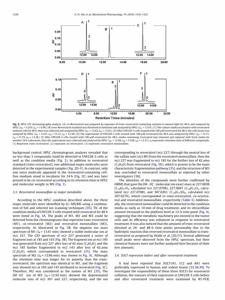
Fig. 2. HPLC-UV chromatography analysis. (A) cis-Resveratrol was prepared by exposure of trans-resveratrol-containing solution to natural light for 48 h, and analyzed by
HPLC (tR1 = 12.63, tR2 = 13.09). (B) trans-Resveratrol standard was dissolved in methanol and analyzed by HPLC (tR1 = 12.61). (C) The culture media incubation with resveratrol
without cells for 48 h, then was collected and analyzed by HPLC (tR1 = 12.62, tR2 = 13.01). (D) After UW228-3 cells treated with 100 mM resveratrol for 48 h, the cells lysate was
analyzed by HPLC (tR1 = 12.67, tR2 = 13.12, tR3 = 12.38). (E) The supernatant of UW228-3 cells treated with 100 mM resveratrol for 48 h was analyzed by HPLC (tR1 = 12.71,
tR2 = 13.19, tR3 = 12.38). (F) After UW228-3 cells treated with 100 mM resveratrol for 48 h, media containing resveratrol was removed and replaced with fresh media for
another 24 h cultivation, then the supernatant was collected and analyzed by HPLC (tR1 = 12.69, tR2 = 13.09, tR3 = 12.31). tR represents retention time of different compounds.
(1) Represent trans-resveratrol; (2) represent cis-resveratrol; (3) represent resveratrol monosulfate.
X.-H. Shu et al. / Biochemical Pharmacology 79 (2010) 1516–15251520
background control. HPLC chromatogram analyses revealed thatno less than 3 compounds could be detected in UW228-3 cells aswell as the condition media (Fig. 2). In addition to resveratrolstandard (trans-resveratrol), two additional major molecules weredetected in the experimental samples (Fig. 2D–F). In contrast, onlyone extra molecule appeared in the resveratrol-containing cell-free medium stood in incubator for 24 h (Fig. 2C) and was laterproved to be cis-resveratrol according to its retention time in HPLCand molecular weight in MS (Fig. 3).
3.3. Resveratrol monosulfate as major metabolite
According to the HPLC condition described above, the threemajor molecules were identified by LC–MS/MS using a combina-tion of full and selected ion scanning techniques [35]. TIC of thecondition media of UW228-3 cells treated with resveratrol for 48 hwere listed in Fig. 3A. The peaks of M1, M2 and M3 could bedetected from the chromatograms that represent trans-resveratrol(M1), cis-resveratrol (M2) and resveratrol monosulfate (M3),respectively. As illustrated in Fig. 3B, the negative ion massspectrum of M1 (tR = 13.61 min) showed a stable molecular ion atm/z 227. The CID spectrum of m/z 227 generated a series offragment ions at 185 and 143 (Fig. 3B). The fragment ion at m/z 185was generated from m/z 227 after loss of 42 amu (C2H2O), and them/z 185 further fragmented to m/z 143 after loss of 42 amu(C2H2O), which corresponded to resveratrol [35]. The massspectrum of M2 (tR = 13.86 min) was shown in Fig. 3C. Althoughthe retention time was longer for its polarity than the trans-resveratrol, the spectrum was identical to M1, and the fragmentions showed m/z at 185 and 143 attributed to resveratrol (Fig. 3C).Therefore, M2 was considered as the isomer of M1 [35]. The[M�H]� ion of M3 (tR = 12.92 min) showed the deprotonatedmolecule ions of m/z 307 and 227, respectively, and the ion
corresponding to resveratrol (m/z 227) through the neutral loss ofthe sulfate unit (m/z 80) from the resveratrol monosulfate, then them/z 227 was fragmented to m/z 185 for the further loss of 42 amu(C2H2O) from resveratrol (Fig. 3D), which is proven to be the maincharacteristic fragmentation pathway [35], and the structure of M3was concluded to resveratrol monosulfate as reported by otherinvestigators [36].
The identities of the compounds were further confirmed byHRMS that gave the [M�H]�molecular ion exact mass as 227.0698(C14H11O3, calculated m/z 227.0708), 227.0697 (C14H11O3, calcu-lated m/z 227.0708), and 307.0261 (C14H11SO6, calculated m/z307.0276), which corresponded to trans-resveratrol, cis-resvera-trol and resveratrol monosulfate, respectively (Table 3). Addition-ally, the resveratrol monosulfate could be detected in the conditionmedia as early as 10 min of drug treatment, and its intracellularamount increased to the platform level at 12-h time point (Fig. 4),suggesting that the metabolic machinery pre-existed in the tumorcells and its efficiency was enhanced in response to resveratroltreatment. It was also noticed that the amounts of trans-resveratrolelevated at 24- and 48-h time points presumably due to thehydrolytic reaction that reversed resveratrol monosulfate to trans-resveratrol as proposed by Walle et al. [20,37]. Several additionalsmall peaks were observed from the HPLC spectrum, but theirchemical features were not further analyzed here because of theirlow amounts.
3.4. SULT expression before and after resveratrol treatment
It had been reported that SULT1A1, 1C2 and 4A1 werepreferably expressed in human and rodent brains [32,38,39]. Toinvestigate the responsibility of these three SULTs for resveratrolsulfation, the statuses of their expression in UW228-3 cells beforeand after resveratrol treatment were examined by RT-PCR,
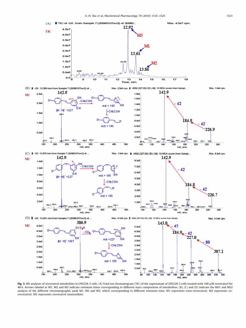
Fig. 3. MS analyses of resveratrol metabolites in UW228-3 cells. (A) Total ion chromatogram (TIC) of the supernatant of UW228-3 cells treated with 100 mM resveratrol for
48 h. Arrows labeled as M1, M2 and M3 indicate retention times corresponding to different mass composition of metabolites. (B), (C) and (D) indicate the MS1 and MS2
analysis of the different chromatographic peak M1, M2 and M3, which corresponding to different retention time. M1 represents trans-resveratrol; M2 represents cis-
resveratrol; M3 represents resveratrol monosulfate.
X.-H. Shu et al. / Biochemical Pharmacology 79 (2010) 1516–1525 1521

Fig. 4. Resveratrol metabolites in UW228-3 cells at different time points. The
supernatant of UW228-3 cells were collected and purified by SPE respectively for
HPLC analysis after 100 mM resveratrol treatment for (A) 10 min; (B) 20 min; (C)
30 min; (D) 60 min; (E) 6 h; (F) 12 h; (G) 24 h; (H) 48 h. Resveratrol monosulfate
could be detected as early as 10 min of drug treatment, and its intracellular amount
reached to the highest level at 12-h time point.
X.-H. Shu et al. / Biochemical Pharmacology 79 (2010) 1516–15251522
immunohistochemical staining and Western blotting. As shown inFig. 5, the three brain-associated SULTs were constitutivelyexpressed in normal rat cerebella and cerebrum. In UW228-3cells, SULT1A1 was expressed in relatively normal level, whereasSULT 1C2 was expressed in low and SULT4A1 in extremely lowlevels. After resveratrol treatment, the level of SULT1A1 remainedalmost unchanged, while both SULT4A1 and 1C2 were enhanced,although their levels were still lower than that in the normalcerebella.
3.5. SULT down-regulation in medulloblastoma tissues
To elucidate whether the in vitro findings were hold true invivo, IHC staining was performed on the tissue microarrayconstructed with human medulloblastomas and control samples.It was found that the overall expression levels of SULT1A1, 1C2 and4A1 in the tumors were lower than that in tumor-surroundingnoncancerous tissues and the rat normal brain (cerebella andcerebra) that were referred as normal controls for SULTs due to theimpossibility to obtain fresh normal human brain tissues ([32];Fig. 5B and Table 2). The results revealed that SULT1A1, 1C2 and4A1 were absent in 24.3%, 10.8% and 8.1% and down-regulated in43.2%, 64.9% and 29.7% of medulloblastoma cases, respectively. Itwas also noticed that the down-regulation patterns of the threeSULTs were not identical in different cases irrespective to thesimilarity of their morphology (Fig. 5B).
3.6. Reduced effects of cis-resveratrol and resveratrol monosulfate
cis-Resveratrol was prepared by standing 100 mM trans-resveratrol under natural light for 48 h [26]. After confirming50% transition of cis-resveratrol in the stock solution by HPLC andLC/MS analysis (Fig. 6B and Table 3), this trans- and cis-resveratrolmixture was used to treat UW228-3 cells for 48 h in a finalconcentration of 100 mM. Unlike the original reagent, the cis-resveratrol-enriched solution exhibited little influence on anti-medulloblastoma activity in terms of growth inhibition, neuron-like differentiation and apoptosis induction (Fig. 6C and D). On the
other hand, trans-resveratrol was incubated in the brain lysates for2 h and then analyzed by HPLC and LC/MS. It was also revealed thatthe monosulfate was the major metabolite and about 1/2 of parenttrans-resveratrol was biotransformed to resveratrol monosulfate(Fig. 6B). In difference with the situation of 100 mM trans-resveratrol treatment, UW228-3 cells treated by the sameconcentration of this mixture for 48 h showed neither distinctgrowth suppression nor the signs of neuron-oriented differentia-tion and apoptosis (Fig. 6C and D). As shown in Fig. 6C, PAPS-containing brain lysate alone (B) exerted little effected on cellgrowth and the anti-medulloblastoma efficacy of resveratrol wasnot influenced by brain lysate without PAPS supplementation(B + R). Similarly, no observable cellular change could be found inthe cells treated by 10 mL of the chemical solution for brain lysatepreparation (data not shown).
4. Discussion
An ideal cancer therapeutic agent should have minimalcytotoxicity to normal tissues, meanwhile, exerts crucial effectson cancer cells. Resveratrol is such candidate, because of itsnontoxic property and anticancer activities in a variety of humanand rodent cancers [40]. It has been recognized that resveratrolundergoes intracellular enzymatic biotransformation that gener-ates one or more metabolites [36,41]. However, the pharmaceuti-cal potentials of those metabolic products have not yet been wellascertained. Some researchers proposed that resveratrol metabo-lite(s) such as resveratrol sulfates or resveratrol glucuronides werethe bioactive forms because of the low bioavailability of parenttrans-resveratrol in vivo [37], while others considered that trans-resveratrol by itself was sufficient to cause vital cellular andmolecular consequences in the treated cancer cells [40,42,43].Apparently, determination of the bioactive form(s) of resveratrol inindividual cell and cancer types becomes a fundamental issue forthe successful application of resveratrol in preventive and clinicalmedicine. In this context, a comparison of resveratrol metabolicpattern in resveratrol-sensitive cancer cells would be helpful infiguring out this point. According to our previous observation,several medulloblastoma cell lines including UW228-3 weresensitive to resveratrol in terms of growth arrest, neuronal-oriented differentiation and apoptosis [11]. Therefore, those cellsmay serve as an ideal experimental model to identify theanticancer form(s) of resveratrol.
The main approach for identification of drug metabolites is thecombined use of HPLC, MS and LC–MS/MS techniques, which isbased on the different retention time, the polarity and m/z ratio ofthe molecules [44,45]. We therefore adopted them for profiling theresveratrol metabolites in resveratrol-sensitive UW228-3 cells. Asthe first finding, we detected cis-resveratrol in the condition mediaof UW228-3 cells. Further analysis revealed that cis-resveratrolcould also be generated by standing the cell-free resveratrol-containing medium in incubator for 48 h. These phenomenaindicated that cis-resveratrol was not a metabolic product but aspontaneously formed isomer of trans-resveratrol during theexperiment [46]. Unlike trans-resveratrol, the data about theanticancer aspect of cis-resveratrol were quite limited. To addresspotential effect of cis-resveratrol on medulloblastoma cells, amixture-containing high fraction (about 50%) of cis-resveratrolwas prepared by exposing 100 mM trans-resveratrol mediumunder natural light at room temperature for 48 h [26], and thenused to treat UW228-3 cells. The results revealed an obviousreduction of anti-medulloblastoma efficacy of this mixture interms of less differentiation and apoptosis tendencies of UW228-3cells. This result thus suggests, at least for medulloblastoma cells,that trans-resveratrol rather than its cis-counterpart possessespowerful anticancer capacity. In consideration of the unstable
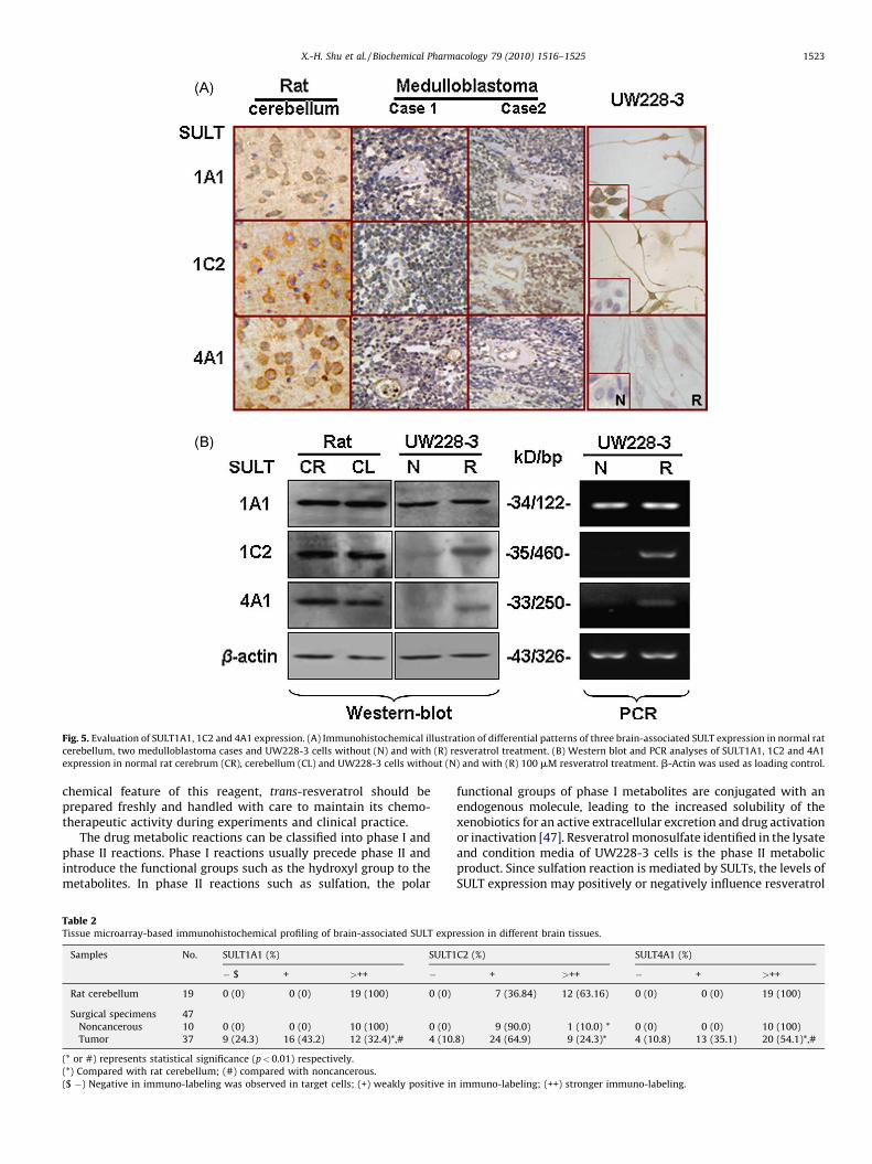
Fig. 5. Evaluation of SULT1A1, 1C2 and 4A1 expression. (A) Immunohistochemical illustration of differential patterns of three brain-associated SULT expression in normal rat
cerebellum, two medulloblastoma cases and UW228-3 cells without (N) and with (R) resveratrol treatment. (B) Western blot and PCR analyses of SULT1A1, 1C2 and 4A1
expression in normal rat cerebrum (CR), cerebellum (CL) and UW228-3 cells without (N) and with (R) 100 mM resveratrol treatment. b-Actin was used as loading control.
X.-H. Shu et al. / Biochemical Pharmacology 79 (2010) 1516–1525 1523
chemical feature of this reagent, trans-resveratrol should beprepared freshly and handled with care to maintain its chemo-therapeutic activity during experiments and clinical practice.
The drug metabolic reactions can be classified into phase I andphase II reactions. Phase I reactions usually precede phase II andintroduce the functional groups such as the hydroxyl group to themetabolites. In phase II reactions such as sulfation, the polar
Table 2Tissue microarray-based immunohistochemical profiling of brain-associated SULT expr
Samples No. SULT1A1 (%) SULT1
� $ + >++ �
Rat cerebellum 19 0 (0) 0 (0) 19 (100) 0 (0)
Surgical specimens 47
Noncancerous 10 0 (0) 0 (0) 10 (100) 0 (0)
Tumor 37 9 (24.3) 16 (43.2) 12 (32.4)*,# 4 (10.
(* or #) represents statistical significance (p<0.01) respectively.
(*) Compared with rat cerebellum; (#) compared with noncancerous.
($ �) Negative in immuno-labeling was observed in target cells; (+) weakly positive in
functional groups of phase I metabolites are conjugated with anendogenous molecule, leading to the increased solubility of thexenobiotics for an active extracellular excretion and drug activationor inactivation [47]. Resveratrol monosulfate identified in the lysateand condition media of UW228-3 cells is the phase II metabolicproduct. Since sulfation reaction is mediated by SULTs, the levels ofSULT expression may positively or negatively influence resveratrol
ession in different brain tissues.
C2 (%) SULT4A1 (%)
+ >++ � + >++
7 (36.84) 12 (63.16) 0 (0) 0 (0) 19 (100)
9 (90.0) 1 (10.0) * 0 (0) 0 (0) 10 (100)
8) 24 (64.9) 9 (24.3)* 4 (10.8) 13 (35.1) 20 (54.1)*,#
immuno-labeling; (++) stronger immuno-labeling.
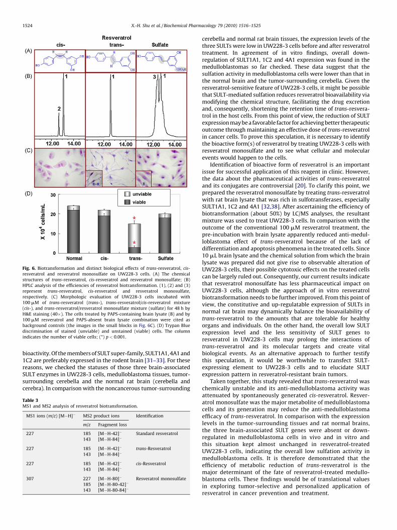
Fig. 6. Biotransformation and distinct biological effects of trans-resveratrol, cis-
resveratrol and resveratrol monosulfate on UW228-3 cells. (A) The chemical
structures of trans-resveratrol, cis-resveratrol and resveratrol monosulfate; (B)
HPLC analysis of the efficiencies of resveratrol biotransformation. (1), (2) and (3)
represent trans-resveratrol, cis-resveratrol and resveratrol monosulfate,
respectively. (C) Morphologic evaluation of UW228-3 cells incubated with
100 mM of trans-resveratrol (trans-), trans-resveratrol/cis-resveratrol mixture
(cis-), and trans-resveratrol/resveratrol monosulfate mixture (sulfate) for 48 h by
H&E staining (40�). The cells treated by PAPS-containing brain lysate (B) and by
100 mM resveratrol and PAPS-absent brain lysate combination were cited as
background controls (the images in the small blocks in Fig. 6C). (D) Trypan Blue
discrimination of stained (unviable) and unstained (viable) cells. The column
indicates the number of viable cells; (*) p < 0.001.
X.-H. Shu et al. / Biochemical Pharmacology 79 (2010) 1516–15251524
bioactivity. Of the members of SULT super-family, SULT1A1, 4A1 and1C2 are preferably expressed in the rodent brain [31–33]. For thesereasons, we checked the statuses of those three brain-associatedSULT enzymes in UW228-3 cells, medulloblastoma tissues, tumor-surrounding cerebella and the normal rat brain (cerebella andcerebra). In comparison with the noncancerous tumor-surrounding
Table 3MS1 and MS2 analysis of resveratrol biotransformation.
MS1 ions (m/z) [M�H]� MS2 product ions Identification
m/z Fragment loss
227 185 [M�H-42]� Standard resveratrol
143 [M�H-84]�
227 185 [M�H-42]� trans-Resveratrol
143 [M�H-84]�
227 185 [M�H-42]� cis-Resveratrol
143 [M�H-84]�
307 227 [M�H-80]� Resveratrol monosulfate
185 [M�H-80-42]�
143 [M�H-80-84]�
cerebella and normal rat brain tissues, the expression levels of thethree SULTs were low in UW228-3 cells before and after resveratroltreatment. In agreement of in vitro findings, overall down-regulation of SULT1A1, 1C2 and 4A1 expression was found in themedulloblastomas so far checked. These data suggest that thesulfation activity in medulloblastoma cells were lower than that inthe normal brain and the tumor-surrounding cerebella. Given theresveratrol-sensitive feature of UW228-3 cells, it might be possiblethat SULT-mediated sulfation reduces resveratrol bioavailability viamodifying the chemical structure, facilitating the drug excretionand, consequently, shortening the retention time of trans-resvera-trol in the host cells. From this point of view, the reduction of SULTexpression may be a favorable factor for achieving better therapeuticoutcome through maintaining an effective dose of trans-resveratrolin cancer cells. To prove this speculation, it is necessary to identifythe bioactive form(s) of resveratrol by treating UW228-3 cells withresveratrol monosulfate and to see what cellular and molecularevents would happen to the cells.
Identification of bioactive form of resveratrol is an importantissue for successful application of this reagent in clinic. However,the data about the pharmaceutical activities of trans-resveratroland its conjugates are controversial [20]. To clarify this point, weprepared the resveratrol monosulfate by treating trans-resveratrolwith rat brain lysate that was rich in sulfotransferases, especiallySULT1A1, 1C2 and 4A1 [32,38]. After ascertaining the efficiency ofbiotransformation (about 50%) by LC/MS analyses, the resultantmixture was used to treat UW228-3 cells. In comparison with theoutcome of the conventional 100 mM resveratrol treatment, thepre-incubation with brain lysate apparently reduced anti-medul-loblastoma effect of trans-resveratrol because of the lack ofdifferentiation and apoptosis phenomena in the treated cells. Since10 mL brain lysate and the chemical solution from which the brainlysate was prepared did not give rise to observable alteration ofUW228-3 cells, their possible cytotoxic effects on the treated cellscan be largely ruled out. Consequently, our current results indicatethat resveratrol monosulfate has less pharmaceutical impact onUW228-3 cells, although the approach of in vitro resveratrolbiotransformation needs to be further improved. From this point ofview, the constitutive and up-regulatable expression of SULTs innormal rat brain may dynamically balance the bioavailability oftrans-resveratrol to the amounts that are tolerable for healthyorgans and individuals. On the other hand, the overall low SULTexpression level and the less sensitivity of SULT genes toresveratrol in UW228-3 cells may prolong the interactions oftrans-resveratrol and its molecular targets and create vitalbiological events. As an alternative approach to further testifythis speculation, it would be worthwhile to transfect SULT-expressing element to UW228-3 cells and to elucidate SULTexpression pattern in resveratrol-resistant brain tumors.
Taken together, this study revealed that trans-resveratrol waschemically unstable and its anti-medulloblastoma activity wasattenuated by spontaneously generated cis-resveratrol. Resver-atrol monosulfate was the major metabolite of medulloblastomacells and its generation may reduce the anti-medulloblastomaefficacy of trans-resveratrol. In comparison with the expressionlevels in the tumor-surrounding tissues and rat normal brains,the three brain-associated SULT genes were absent or down-regulated in medulloblastoma cells in vivo and in vitro andthis situation kept almost unchanged in resveratrol-treatedUW228-3 cells, indicating the overall low sulfation activity inmedulloblastoma cells. It is therefore demonstrated that theefficiency of metabolic reduction of trans-resveratrol is themajor determinant of the fate of resveratrol-treated medullo-blastoma cells. These findings would be of translational valuesin exploring tumor-selective and personalized application ofresveratrol in cancer prevention and treatment.
X.-H. Shu et al. / Biochemical Pharmacology 79 (2010) 1516–1525 1525
Acknowledgments
We gratefully acknowledge Hui Wang, Jing-Ping Cao andYing Gao for their assistance with the HPLC and LC/MSdetection. We thank Drs. Xin-Feng Zhao and Peng Gao for theirassistance with the HRMS analysis and Professor Xiang-HongYang for providing human medulloblastoma specimens. Thiswork is supported by the grants from National Natural ScienceFoundation of China (Nos. 30527002, 30670946 and 30971038)and by the special grants of Liaoning Department of Educationfor the key laboratory (20060193) and for the creative researchteam (2007-7-26 and 2008T028).
References
[1] Jang M, Cai L, Udeani GO, Slowing KV, Thomas CF, Beecher CW, et al. Cancerchemopreventive activity of resveratrol, a natural product derived fromgrapes. Science 1997;275:218–20.
[2] Dixon RA. Natural products and plant disease resistance. Nature 2001;411:843–7.
[3] Rivera L, Moron R, Zarzuelo A, Galisteo M. Long-term resveratrol administra-tion reduces metabolic disturbances and lowers blood pressure in obeseZucker rats. Biochem Pharmacol 2009;77:1053–63.
[4] Burstein B, Maguy A, Clement R, Gosselin H, Poulin F, Ethier N, et al. Effects ofresveratrol (trans-3,5,40-trihydroxystilbene) treatment on cardiac remodelingfollowing myocardial infarction. J Pharmacol Exp Ther 2007;323:916–23.
[5] Olas B, Wachowicz B. Resveratrol and vitamin C as antioxidants in bloodplatelets. Thromb Res 2002;106:143–8.
[6] Zhu J, Yong W, Wu X, Yu Y, Lv J, Liu C, et al. Anti-inflammatory effect ofresveratrol on TNF-alpha-induced MCP-1 expression in adipocytes. BiochemBiophys Res Commun 2008;369:471–7.
[7] Shakibaei M, Csaki C, Nebrich S, Mobasheri A. Resveratrol suppresses inter-leukin-1beta-induced inflammatory signaling and apoptosis in human articu-lar chondrocytes: potential for use as a novel nutraceutical for the treatment ofosteoarthritis. Biochem Pharmacol 2008;76:1426–39.
[8] Hudson TS, Hartle DK, Hursting SD, Nunez NP, Wang TT, Young HA, et al.Inhibition of prostate cancer growth by muscadine grape skin extract andresveratrol through distinct mechanisms. Cancer Res 2007;67:8396–405.
[9] Aziz MH, Reagan-Shaw S, Wu J, Longley BJ, Ahmad N. Chemoprevention of skincancer by grape constituent resveratrol: relevance to human disease? FASEB J2005;19:1193–5.
[10] Boissy P, Andersen TL, Abdallah BM, Kassem M, Plesner T, Delaisse JM.Resveratrol inhibits myeloma cell growth, prevents osteoclast formation,and promotes osteoblast differentiation. Cancer Res 2005;65:9943–52.
[11] Zhang P, Li H, Wu ML, Chen XY, Kong QY, Wang XW, et al. c-Myc down-regulation: a critical molecular event in resveratrol-induced cell cyclearrest and apoptosis of human medulloblastoma cells. J Neurooncol2006;80:123–31.
[12] Danz ED, Skramsted J, Henry N, Bennett JA, Keller RS. Resveratrol preventsdoxorubicin cardiotoxicity through mitochondrial stabilization and the Sirt1pathway. Free Radic Biol Med 2009;46:1589–97.
[13] Venkatesan B, Ghosh-Choudhury N, Das F, Mahimainathan L, Kamat A, Kasi-nath BS, et al. Resveratrol inhibits PDGF receptor mitogenic signaling inmesangial cells: role of PTP1B. FASEB J 2008;22:3469–82.
[14] Wang Q, Xu J, Rottinghaus GE, Simonyi A, Lubahn D, Sun GY, et al. Resveratrolprotects against global cerebral ischemic injury in gerbils. Brain Res 2002;958:439–47.
[15] Wang Q, Li H, Liu N, Chen XY, Wu ML, Zhang KL, et al. Correlative analyses ofnotch signaling with resveratrol-induced differentiation and apoptosis ofhuman medulloblastoma cells. Neurosci Lett 2008;438:168–73.
[16] Yu LJ, Wu ML, Li H, Chen XY, Wang Q, Sun Y, et al. Inhibition of STAT3expression and signaling in resveratrol-differentiated medulloblastoma cells.Neoplasia 2008;10:736–44.
[17] Bishayee A. Cancer prevention and treatment with resveratrol: from rodentstudies to clinical trials. Cancer Prev Res 2009;2:409–18.
[18] Li Y, Backesjo CM, Haldosen LA, Lindgren U. Resveratrol inhibits proliferationand promotes apoptosis of osteosarcoma cells. Eur J Pharmacol 2009;609:13–8.
[19] Woo KJ, Lee TJ, Lee SH, Lee JM, Seo JH, Jeong YJ, et al. Elevated gadd153/chopexpression during resveratrol-induced apoptosis in human colon cancer cells.Biochem Pharmacol 2007;73:68–76.
[20] Gescher AJ, Steward WP. Relationship between mechanisms, bioavailibility,and preclinical chemopreventive efficacy of resveratrol: a conundrum. CancerEpidemiol Biomarkers Prev 2003;12:953–7.
[21] Ellison DW, Onilude OE, Lindsey JC, Lusher ME, Weston CL, Taylor RE, et al.Beta-catenin status predicts a favorable outcome in childhood medulloblas-toma: the United Kingdom Children’s Cancer Study Group Brain TumourCommittee. J Clin Oncol 2005;23:7951–7.
[22] Marino S. Medulloblastoma: developmental mechanisms out of control.Trends Mol Med 2005;11:17–22.
[23] Liu J, Guo L, Luo Y, Li JW, Li H. All trans-retinoic acid suppresses in vitro growthand down-regulates LIF gene expression as well as telomerase activity ofhuman medulloblastoma cells. Anticancer Res 2000;20:2659–64.
[24] Wang Q, Li H, Wang XW, Wu DC, Chen XY, Liu J. Resveratrol promotesdifferentiation and induces Fas-independent apoptosis of human medullo-blastoma cells. Neurosci Lett 2003;351:83–6.
[25] Keles GE, Berger MS, Srinivasan J, Kolstoe DD, Bobola MS, Silber JR. Establish-ment and characterization of four human medulloblastoma-derived cell lines.Oncol Res 1995;7:493–503.
[26] Chen X, He H, Wang G, Yang B, Ren W, Ma L, et al. Stereospecific determinationof cis- and trans-resveratrol in rat plasma by HPLC: application to pharmaco-kinetic studies. Biomed Chromatogr 2007;21:257–65.
[27] Mercolini L, Addolorata Saracino M, Bugamelli F, Ferranti A, Malaguti M, HreliaS, et al. HPLC-F analysis of melatonin and resveratrol isomers in wine using anSPE procedure. J Sep Sci 2008;31:1007–14.
[28] Qian G, Leung SY, Lu G, Leung KS. Optimization and validation of a chro-matographic method for the simultaneous quantification of six bioactivecompounds in Rhizoma et Radix Polygoni Cuspidati. J Pharm Pharmacol2008;60:107–13.
[29] Boocock DJ, Patel KR, Faust GE, Normolle DP, Marczylo TH, Crowell JA, et al.Quantitation of trans-resveratrol and detection of its metabolites in humanplasma and urine by high performance liquid chromatography. J Chromatogr B2007;848:182–7.
[30] Urpi-Sarda M, Zamora-Ros R, Lamuela-Raventos R, Cherubini A, Jauregui O, dela Torre R, et al. HPLC-tandem mass spectrometric method to characterizeresveratrol metabolism in humans. Clin Chem 2007;53:292–9.
[31] Salman ED, Kadlubar SA, Falany CN. Expression and localization of cytosolicsulfotransferase (SULT) 1A1 and SULT1A3 in normal human brain. Drug MetabDispos 2009;37:706–9.
[32] Liyou NE, Buller KM, Tresillian MJ, Elvin CM, Scott HL, Dodd PR, et al. Locali-zation of a brain sulfotransferase, SULT4A1, in the human and rat brain: animmunohistochemical study. J Histochem Cytochem 2003;51:1655–64.
[33] Allali-Hassani A, Pan PW, Dombrovski L, Najmanovich R, Tempel W, Dong A,et al. Structural and chemical profiling of the human cytosolic sulfotrans-ferases. PLoS Biol 2007;5:1063–78.
[34] Hui Y, Yasuda S, Liu MY, Wu YY, Liu MC. On the sulfation and methylation ofcatecholestrogens in human mammary epithelial cells and breast cancer cells.Biol Pharm Bull 2008;31:769–73.
[35] Wang DG, Hang TJ, Wu CY, Liu WY. Identification of the major metabolites ofresveratrol in rat urine by HPLC-MS/MS. J Chromatogr B 2005;829:97–106.
[36] Murias M, Miksits M, Aust S, Spatzenegger M, Thalhammer T, Szekeres T, et al.Metabolism of resveratrol in breast cancer cell lines: impact of sulfotransfer-ase 1A1 expression on cell growth inhibition. Cancer Lett 2008;261:172–81.
[37] Walle T, Hsieh F, DeLegge MH, Oatis JE, Walle UK. High absorption but very lowbioavailability of oral resveratrol in humans. Drug Metab Dispos 2004;32:1377–82.
[38] Alnouti Y, Klaassen CD. Tissue distribution and ontogeny of sulfotransferaseenzymes in mice. Toxicol Sci 2006;93:242–55.
[39] Lindsay J, Wang LL, Li Y, Zhou SF. Structure, function and polymorphism ofhuman cytosolic sulfotransferases. Curr Drug Metab 2008;39:99–105.
[40] Baur JA, Sinclair DA. Therapeutic potential of resveratrol: the in vivo evidence.Nat Rev Drug Discov 2006;5:493–506.
[41] Lancon A, Hanet N, Jannin B, Delmas D, Heydel JM, Lizard G, et al. Resveratrol inhuman hepatoma HepG2 cells: metabolism and inducibility of detoxifyingenzymes. Drug Metab Dispos 2007;35:699–703.
[42] Howitz KT, Bitterman KJ, Cohen HY, Lamming DW, Lavu S, Wood JG, et al. Smallmolecule activators of sirtuins extend Saccharomyces cerevisiae life span.Nature 2003;425:191–6.
[43] Lee SK, Zhang W, Sanderson BJ. Selective growth inhibition of human leukemiaand human lymphoblastoid cells by resveratrol via cell cycle arrest andapoptosis induction. J Agric Food Chem 2008;56:7572–7.
[44] Holcapek M, Kolarova L, Nobilis M. High-performance liquid chromatography-tandem mass spectrometry in the identification and determination of phase Iand phase II drug metabolites. Anal Bioanal Chem 2008;391:59–78.
[45] Yang J, Zhao XJ, Liu XL, Wang C, Gao P, Wang JS, et al. High performance liquidchromatography-mass spectrometry for metabonomics: potential biomarkersfor acute deterioration of liver function in chronic hepatitis B. J Proteome Res2006;5:554–61.
[46] Pervaiz S. Resveratrol: from grapevines to mammalian biology. FASEB J2003;17:1975–85.
[47] Liska D, Lyon M, Jones DS. Detoxification and biotransformational imbalances.Explore (NY) 2006;2:122–40.


![Medulloblastoma: [Print] - eMedicine Neurology · accounts for approximately 7-8% of all intracranial tumors and 30% of ... Incidence of medulloblastoma is 1.5-2 cases per ... Medulloblastoma:](https://img.dokumen.tips/doc/110x75/5b7fc2317f8b9ae6088caa0e/medulloblastoma-print-emedicine-accounts-for-approximately-7-8-of-all.jpg)


![Medulloblastoma: [Print] - eMedicine Neurology · emedicine.medscape.com eMedicine Specialties > Neurology > Pediatric Neurology Medulloblastoma George I Jallo, MD, Associate Professor](https://img.dokumen.tips/doc/110x75/5d472c3c88c993527c8b60e5/medulloblastoma-print-emedicine-neurology-emedicinemedscapecom-emedicine.jpg)


