Embed Size (px)
Citation preview
CellCell Communication
IBS 8102 – Cell, Molecular, and Developmental Biology
January 29, 2008
Communicate What?
Why do cells communicate? To govern or modify each other for the benefit of the organism
differentiate multiply perform specialized physiology; e.g. secrete contract
die (i.e. undergo apoptosis)
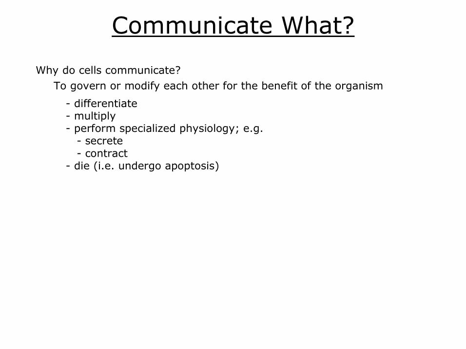
Mechanism I. Signaling molecules
intercellular intracellular A. Longdistance
neurotransmitters hormones
B. Shortdistance paracrine factors juxtacrine factors autocrine factors ECM components
D. Receptors membranebound cytoplasmic cytosolic nuclear
II. Cellular interaction
B. Ion channels
A. Diffusion lipophilic molecules may diffuse through membrane NO steroid hormones
C. Gap junctions
III. Intracellular signaling proteins (Signal Transduction components) distribute external signals often enzymatic phosphatases kinases GTP binding/hydrolysis
IV. Target proteins transcriptional regulators ion channels metabolic pathways cytoskeletal components etc.
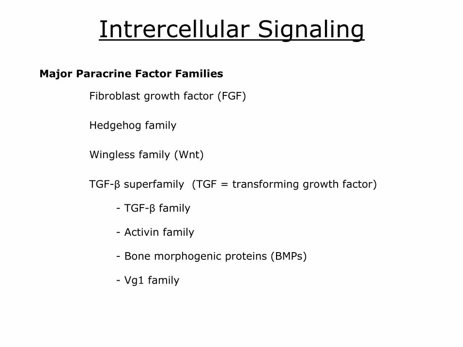
Intrercellular Signaling
Fibroblast growth factor (FGF)
Hedgehog family
Wingless family (Wnt)
TGFβ superfamily (TGF = transforming growth factor)
TGFβ family
Activin family
Bone morphogenic proteins (BMPs)
Vg1 family
Major Paracrine Factor Families
Signal Transduction Extracellular signals are transduced to the cytoplasm at the cell membrane
external signal is transmitted into the interior of the cell
e.g. receptor tyrosine kinase (RTK)
(kinase = protein phosphorylating enzyme)
Signal transduction cascades most intercellular and intracellular signals are part of larger set of pathways activated products or intermediates trigger other pathways
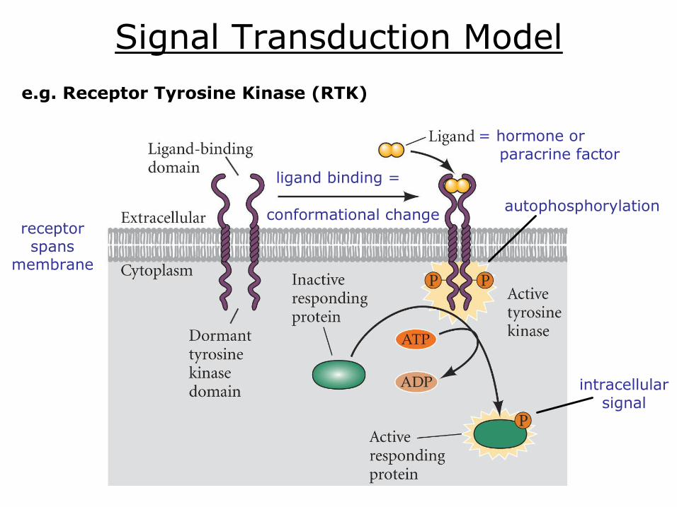
Signal Transduction Model
= hormone or paracrine factor
autophosphorylation
intracellular signal
receptor spans
membrane
ligand binding =
conformational change
e.g. Receptor Tyrosine Kinase (RTK)
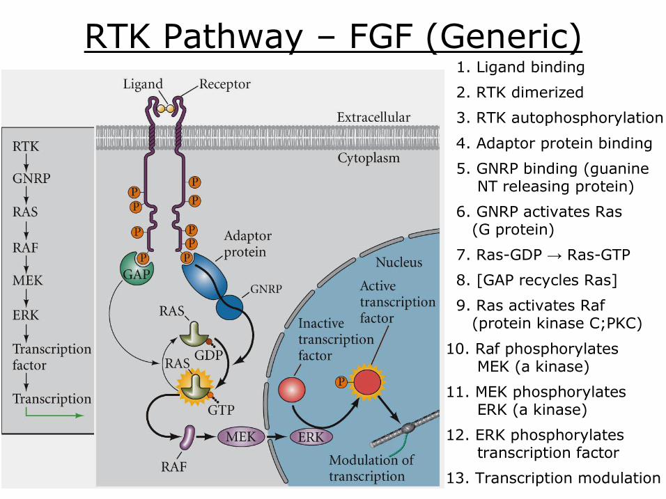
RTK Pathway – FGF (Generic) 2. RTK dimerized
1. Ligand binding
3. RTK autophosphorylation
4. Adaptor protein binding
5. GNRP binding (guanine NT releasing protein)
6. GNRP activates Ras (G protein)
7. RasGDP → RasGTP
8. [GAP recycles Ras]
9. Ras activates Raf (protein kinase C;PKC)
10. Raf phosphorylates MEK (a kinase)
11. MEK phosphorylates ERK (a kinase)
12. ERK phosphorylates transcription factor
13. Transcription modulation
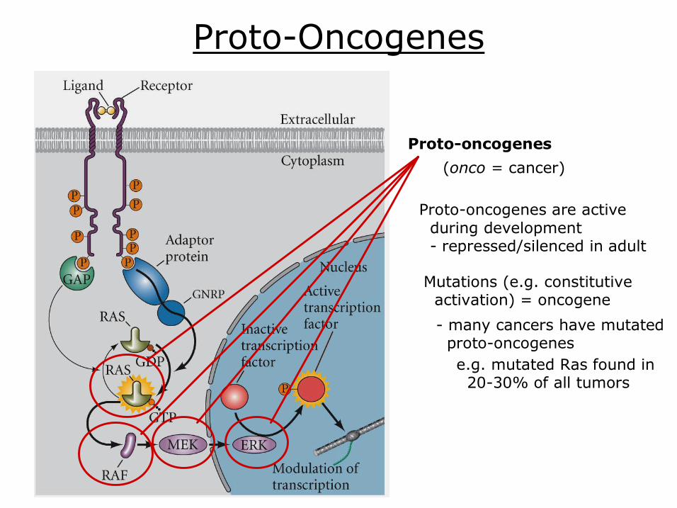
ProtoOncogenes
Protooncogenes
(onco = cancer)
e.g. mutated Ras found in 2030% of all tumors
Protooncogenes are active during development repressed/silenced in adult
many cancers have mutated protooncogenes
Mutations (e.g. constitutive activation) = oncogene
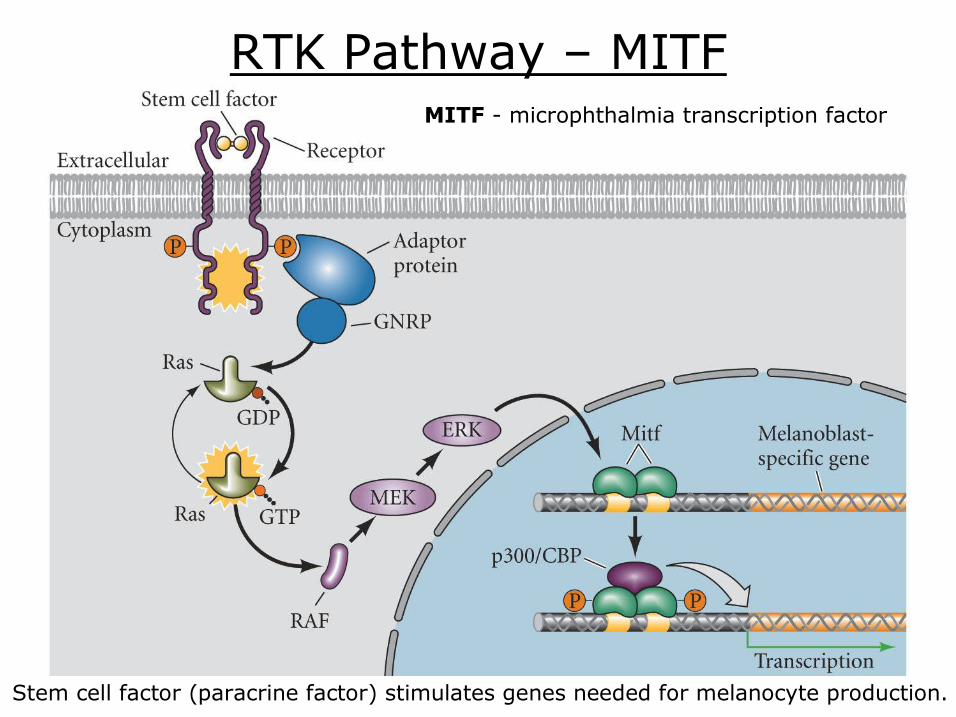
RTK Pathway – MITF
Stem cell factor (paracrine factor) stimulates genes needed for melanocyte production.
MITF microphthalmia transcription factor

JAKSTAT Pathway
JAK – Janus kinase nonreceptor tyrosine kinase
STAT – Signal Transducers and Activators of Transcription transcription factor
Pathway activators: prolactin cytokines, growth hormones; cell proliferation differentiation apoptosis
NOTE – STATs can be activated independently of JAKs RTK; e.g. EGF receptor nonreceptor tyrosine kinases; e.g. csrc
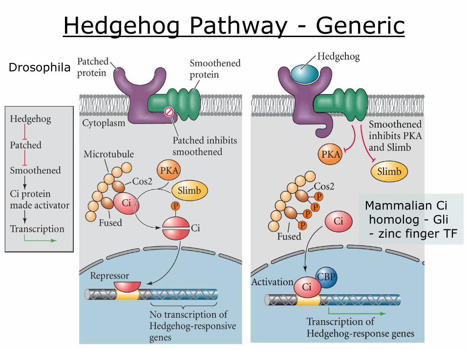
Hedgehog Pathway Generic
Drosophila
Mammalian Ci homolog Gli zinc finger TF
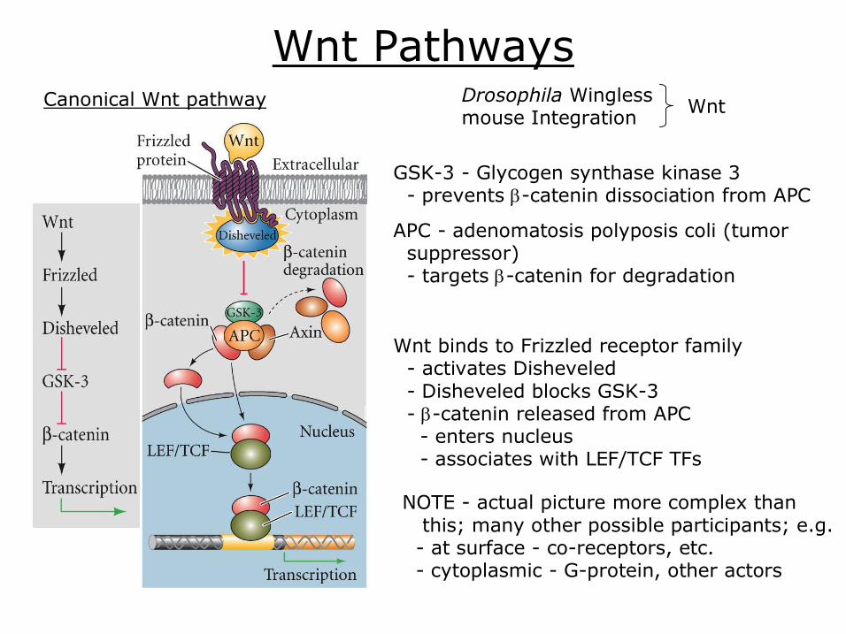
Wnt Pathways Canonical Wnt pathway
GSK3 Glycogen synthase kinase 3 prevents βcatenin dissociation from APC
APC adenomatosis polyposis coli (tumor suppressor) targets βcatenin for degradation
Wnt binds to Frizzled receptor family activates Disheveled Disheveled blocks GSK3 βcatenin released from APC enters nucleus associates with LEF/TCF TFs
NOTE actual picture more complex than this; many other possible participants; e.g. at surface coreceptors, etc. cytoplasmic Gprotein, other actors
Drosophila Wingless mouse Integration Wnt
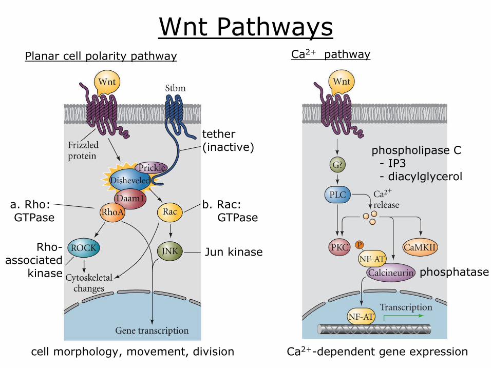
Wnt Pathways Planar cell polarity pathway
cell morphology, movement, division Ca 2+ dependent gene expression
Ca 2+ pathway
phosphatase
phospholipase C IP3 diacylglycerol
b. Rac: GTPase
Jun kinase
a. Rho: GTPase
Rho associated
kinase
tether (inactive)
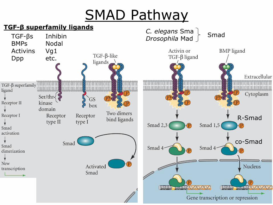
SMAD Pathway TGFβ superfamily ligands TGFβs BMPs Activins Dpp
Inhibin Nodal Vg1 etc.
RSmad
coSmad
C. elegans Sma Drosophila Mad Smad

Apoptosis
Apoptosis – programmed cell death
Developmental: embryonic neural growth embryonic brain produces 3X neurons found at birth
hand and foot webbing between digits
teeth middle ear space vaginal opening male mammary tissue frog tails (at metamorphosis)
Adult: most cells and tissues
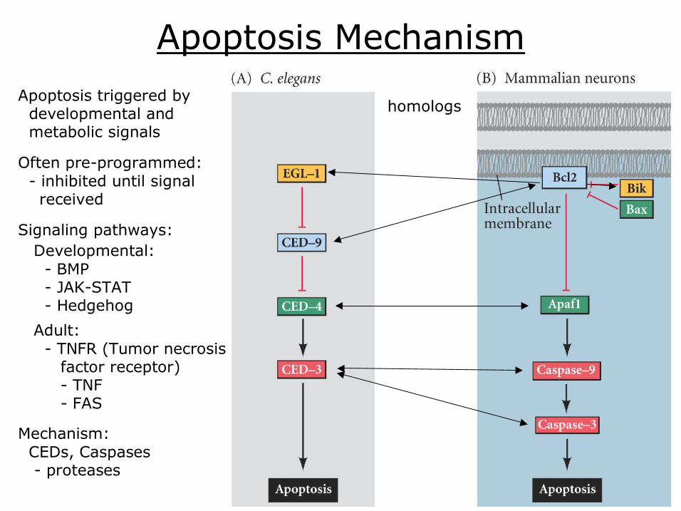
Apoptosis Mechanism
homologs
Mechanism: CEDs, Caspases proteases
Apoptosis triggered by developmental and metabolic signals
Often preprogrammed: inhibited until signal received
Signaling pathways:
Adult: TNFR (Tumor necrosis factor receptor) TNF FAS
Developmental: BMP JAKSTAT Hedgehog

Notch Pathway Juxtacrine signaling: Proteins from the inducing cell interact with receptors from adjacent responding cells without diffusing from the cell producing them.
(Serrate) (Jagged)
Delta protein bind Notch protease cleaves both outer and inner Notch portions
inner portion moves to nucleus displaces repressor recruits p300 HAT
activates transcription
outer portion remains with ligand endocytosed into ligandexpressing cell may act as signal
e.g. Notch/Delta, Ephrin/EphR, EGF/EGFR, IL15/IL15Rα, etc.
Extracellular Matrix
ECM – macromolecules secreted by cells into their immediate environment
form a region of noncellular material in the intersticies between the cells
cell adhesion, migration, formation of epithelial sheets and tubes
proteoglycans: e.g. heparan sulfate, chondroitin sulfate, keratan sulfate
polysaccharides; e.g. hyaluronic acid
proteins: e.g. collagen, fibronectin, elastin, laminin
ECM Function
intracellular signaling
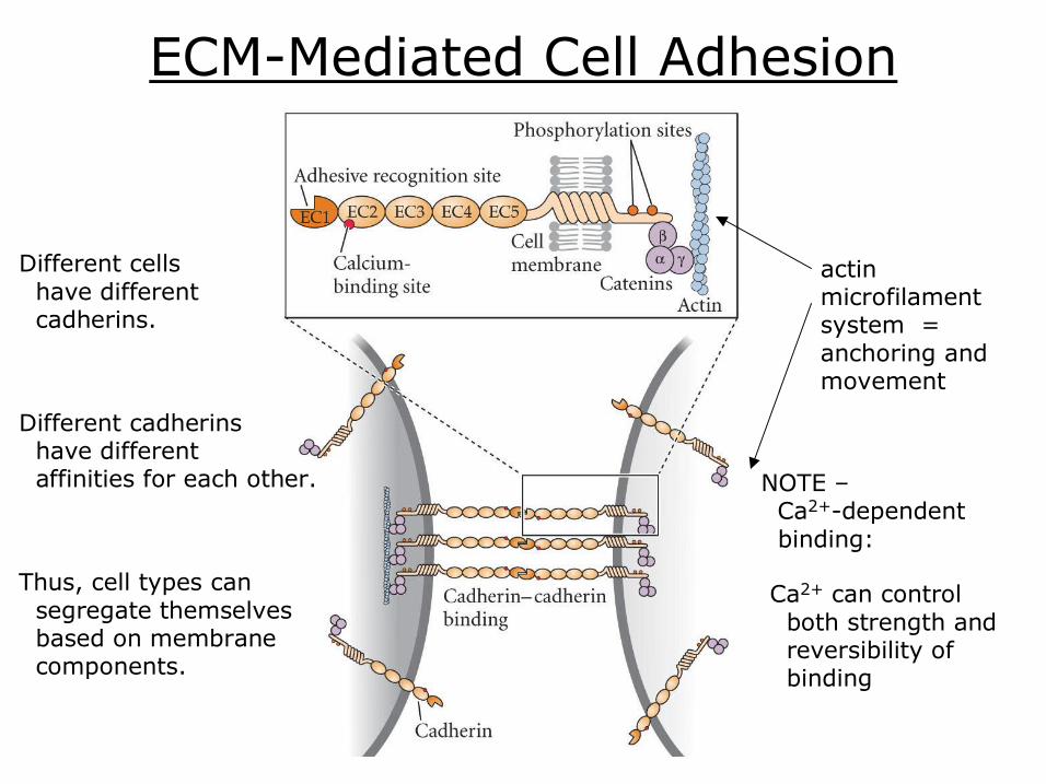
ECMMediated Cell Adhesion
actin microfilament system = anchoring and movement
Different cells have different cadherins.
Different cadherins have different affinities for each other.
Thus, cell types can segregate themselves based on membrane components.
NOTE – Ca 2+ dependent binding:
Ca 2+ can control both strength and reversibility of binding

Extracellular Matrix Signals
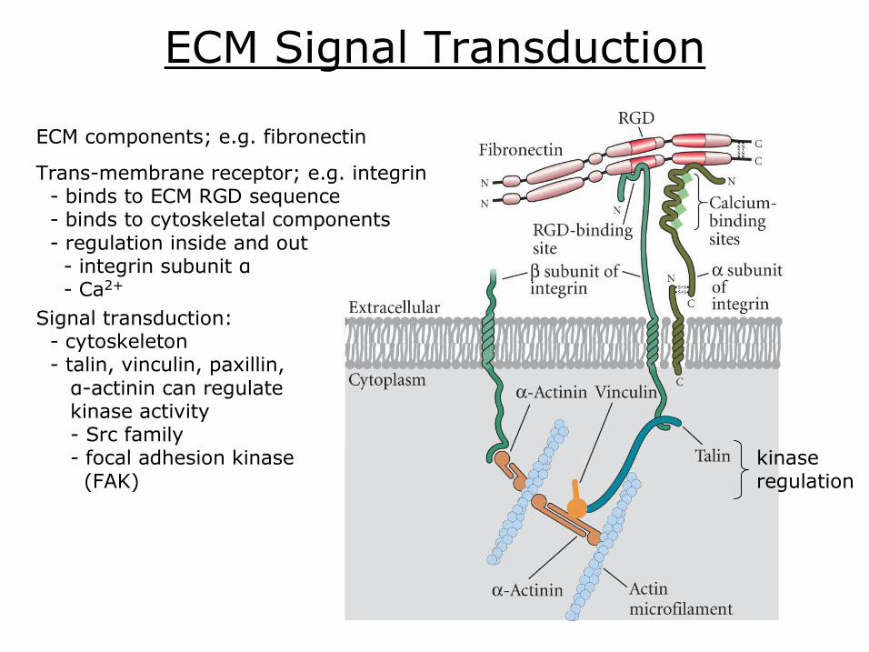
ECM Signal Transduction
ECM components; e.g. fibronectin
Transmembrane receptor; e.g. integrin binds to ECM RGD sequence binds to cytoskeletal components regulation inside and out integrin subunit α Ca 2+
kinase regulation
Signal transduction: cytoskeleton talin, vinculin, paxillin, αactinin can regulate kinase activity Src family focal adhesion kinase (FAK)
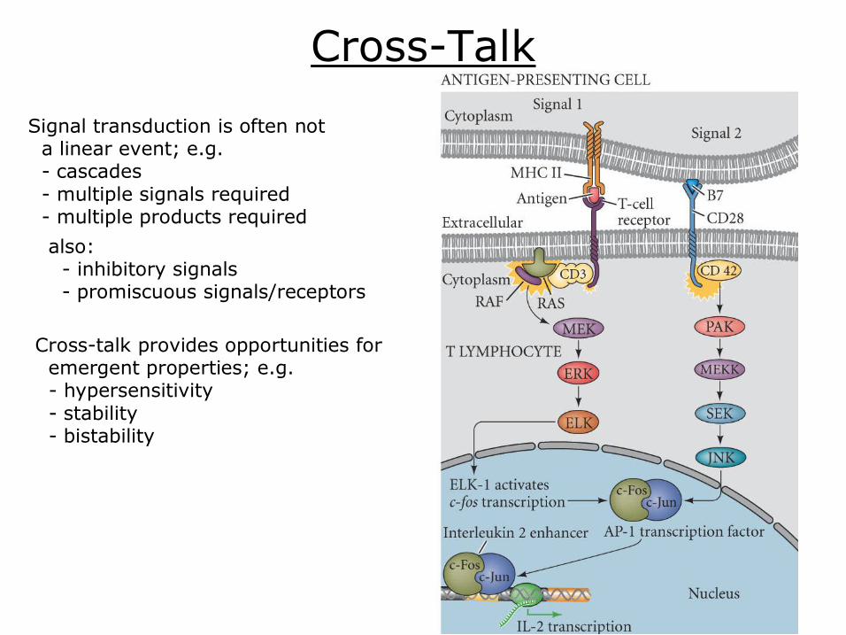
CrossTalk
Signal transduction is often not a linear event; e.g. cascades multiple signals required multiple products required
also: inhibitory signals promiscuous signals/receptors
Crosstalk provides opportunities for emergent properties; e.g. hypersensitivity stability bistability