Embed Size (px)
Citation preview
General rights Copyright and moral rights for the publications made accessible in the public portal are retained by the authors and/or other copyright owners and it is a condition of accessing publications that users recognise and abide by the legal requirements associated with these rights.
• Users may download and print one copy of any publication from the public portal for the purpose of private study or research. • You may not further distribute the material or use it for any profit-making activity or commercial gain • You may freely distribute the URL identifying the publication in the public portal
If you believe that this document breaches copyright please contact us providing details, and we will remove access to the work immediately and investigate your claim.
Downloaded from orbit.dtu.dk on: May 29, 2018
Hydrophobins as aqueous lubricant additive for a soft sliding contact
Lee, Seunghwan; Røn, Troels; Pakkanen, Kirsi I.; Linder, Markus
Published in:Colloids and Surfaces B: Biointerfaces
Link to article, DOI:10.1016/j.colsurfb.2014.10.044
Publication date:2015
Document VersionPeer reviewed version
Link back to DTU Orbit
Citation (APA):Lee, S., Røn, T., Pakkanen, K. I., & Linder, M. (2015). Hydrophobins as aqueous lubricant additive for a softsliding contact. Colloids and Surfaces B: Biointerfaces, 125, 264–269. DOI: 10.1016/j.colsurfb.2014.10.044
Accepted Manuscript
Title: Hydrophobins as aqueous lubricant additive for a softsliding contact
Author: Seunghwan Lee Troels Røn Kirsi I. PakkanenMarkus Linder
PII: S0927-7765(14)00594-3DOI: http://dx.doi.org/doi:10.1016/j.colsurfb.2014.10.044Reference: COLSUB 6708
To appear in: Colloids and Surfaces B: Biointerfaces
Received date: 1-7-2014Revised date: 7-10-2014Accepted date: 22-10-2014
Please cite this article as: S. Lee, T. Ron, K.I. Pakkanen, M. Linder, Hydrophobinsas aqueous lubricant additive for a soft sliding contact, Colloids and Surfaces B:Biointerfaces (2014), http://dx.doi.org/10.1016/j.colsurfb.2014.10.044
This is a PDF file of an unedited manuscript that has been accepted for publication.As a service to our customers we are providing this early version of the manuscript.The manuscript will undergo copyediting, typesetting, and review of the resulting proofbefore it is published in its final form. Please note that during the production processerrors may be discovered which could affect the content, and all legal disclaimers thatapply to the journal pertain.
Page 1 of 24
Accep
ted
Man
uscr
ipt
1
Hydrophobins as aqueous lubricant additive for a soft sliding contact
Seunghwan Lee,1* Troels Røn,1 Kirsi I. Pakkanen,1 and Markus Linder2,3
1Department of Mechanical Engineering, Technical University of Denmark, DK-2800, Kgs. Lyngby,
Denmark
2Technical Research Centre of Finland, VTT Biotechnology, FIN-02044 VTT, Finland3Department of Biotechnology and Chemical Technology, Aalto University, 00076 Aalto, Finland
*Corresponding author: [email protected]
Key words: hydrophobin, amphiphilic, FpHYD5, HFBI, lubrication
Page 2 of 24
Accep
ted
Man
uscr
ipt
2
Abstract
Two type II fungal hydrophobins, HFBI and FpHYD5, have been studied as aqueous lubricant
additive at a nonpolar, compliant sliding contact (self-mated poly(dimethylsiloxane) (PDMS)
contact) at two different concentrations, 0.1 mg/mL and 1.0 mg/mL. The two hydrophobins are
featured as non-glycosylated (HFBI, m.w. ca. 7 kDa) vs glycosylated (FpHYD5, m.w. ca. 10 kDa)
proteins. Far UV CD spectra of the two hydrophobins were very similar, suggesting overall
structural similarity, but showed a noticeable difference according to the concentration. This is
proposed to be related to the formation of multimers at 1.0 mg/mL. Despite ten-fold difference in
the bulk concentration, the adsorbed masses of the hydrophobins onto PDMS surface obtained from
the two solutions (0.1 and 1.0 mg/mL) were nearly identical, suggesting that a monolayer of the
hydrophobins are formed from 0.1 mg/mL solution. PDMS-PDMS sliding interface was effectively
lubricated by the hydrophobin solutions, and showed a reduction in the coefficient of friction by as
much as ca. two orders of magnitude. Higher concentration solution (1.0 mg/mL) provided a
superior lubrication, particularly in low-speed regime, where boundary lubrication characteristic is
dominant via ‘self-healing’ mechanism. FpHYD5 revealed a better lubrication than HFBI
presumably due to the presence of glycans and improved hydration of the sliding interface. Two
type II hydrophobins function more favorably compared to a synthetic amphiphilic copolymer,
PEO-PPO-PEO, with a similar molecular weight. This is ascribed to higher amount of adsorption of
the hydrophobins to hydrophobic surfaces from aqueous solution.
Page 3 of 24
Accep
ted
Man
uscr
ipt
3
1. Introduction
Hydrophobins are small (m.w. 7-10 kDa) amphiphilic proteins originating from filamentous
fungi displaying a variety of biological functions in their growth and morphogenesis [1-7]. Based
on the comparison of amino acid residue sequence, hydrophobins are classified into two groups,
type I and type II [1-7]. Surface-active properties of hydrophobins have drawn particular interests in
self-assembled adsorption behavior of hydrophobins at air/water [8-10], water/oil [11-14], and
water/solid interfaces [15-22]. This, in turn, sparked intensive researches to utilize hydrophobins as
coating materials for biomedical, technical, and personal care products [23-28].
Hydrophobins’ surface-active properties have recently started to draw attention for tribological
applications as well [29-31]. Nanotribological studies of a layer of Sc3 from schizophylum
commune on polymeric surfaces with atomic force microscopy (AFM) showed a reduction of
friction forces in ambient condition [29]. More recent studies of type II hydrophobins, namely HFBI
and FpHYD5, have shown a potential as boundary lubricant additive to lubricate stainless steel in
aqueous environment [30,31]. Despite different environment and substrates, the efficacy of
hydrophobins as lubricant additive in these studies is commonly based upon a strong adsorption
onto material surfaces.
In this study, we have investigated two type II hydrophobins, namely HFBI and FpHYD5, as
boundary lubricant additive for a sliding contact of elastomeric, hydrophobic interface by
employing a self-mated poly(dimethylsiloxane) (PDMS) pair in aqueous environment. Based on
distinct amphiphilicity of hydrophobins, it is hypothesized that they are ideally suited to hydrate and
lubricate hydrophobic interfaces in aqueous environment. While various synthetic [32-34] and
biopolymeric [35-37] amphiphiles have shown facile lubricating effects for hydrophobic interfaces,
comparative studies of the two hydrophobins in this study are particularly interesting in the
following viewpoints. Firstly, the molecular weight of the hydrophobins in this study, ca. 7 − 10
Page 4 of 24
Accep
ted
Man
uscr
ipt
4
kDa, is much smaller than those of other amphiphilic biomacromolecules that are related to
biological lubrication, such as lubricin (in the range of ca. 250 kDa [38,39]) or mucins (0.5 to 20
MDa [40]), and is rather comparable to those of synthetic amphiphilic polymers that have been used
as aqueous lubricant additive [32]. Thus, it is of interest to study whether small biomolecules as
hydrophobins can also display as effective lubricating capabilities as much larger ones. Secondly, in
the same context, it is also of interest to compare the lubricating properties of hydrophobins with
synthetic copolymers, especially those showing comparable molecular weights. Thirdly, despite the
differences in the fungi of origin, structural and sequence homology of hydrophobins are very high
[1-7]; both hydrophobins contain eight cystein residues, i.e. four disulfide bonds, one -helix and
two strands, and a number of aliphatic hydrophobic residues. A major structural difference
between the two hydrophobins is N-glycosylation (ca. 1.7 kDa) of FpHYD5 [31,41], and thus its
influence on the lubricating properties can be studied. Lastly, it is well known that hydrophobins
form multimers as a result of self-assembly in bulk aqueous solution [42-44], which may have an
influence on the surface adsorption and boundary lubricating properties too; for this reason, all the
experiments were performed at two concentrations, namely at 0.1 and 1 mg/mL.
2. Materials and Methods
2.1 Hydrophobins and hydrophobin solutions
Two type II hydrophobins, namely HFBI from T. reesei [5-9] and FpHYD5 from Fusarium
poae [41], were employed. Details on culture, extraction, and purification processes of the two
hydrophobins are found in literature [8,9,41]. Molecular weights of HFBI and FpHYD5 are 7.54
kDa [31] and 9.21 kDa [31,41], respectively. For FpHYD5, a molecular weight of ca. 1.7 kDa is
indebted from N-glycosylation at N-37 site (2 N-acetyl glycosamines and 7 hexoses [41]). For
comparison, a triblock copolymer, poly(ethylene oxide)-block-poly(propylene oxide)-block-
Page 5 of 24
Accep
ted
Man
uscr
ipt
5
poly(ethylene oxide) (PEO-PPO-PEO) with a similar molecular weight with HFBI, namely
Synperonic® PE P105 (m.w. ca. 6.5kDa, abbreviated as “P105” hereafter, Sigma-Aldrich Denmark
ApS, Broendby, Denmark), was employed.
Hydrophobin and P105 solutions were prepared by dissolving in sodium acetate buffer (pH 5)
at 0.1 mg/mL and 1.0 mg/mL, respectively. A slightly acidic buffer solution was selected based on
the previous studies, where an optimum adsorption [45] and lubrication [31] were observed in this
buffer, especially for HFBI.
2.2 Circular Dichroism Spectroscopy
Far UV circular dichroism (CD) spectra of the hydrophobin solutions were acquired with a
Chirascan spectrophotometer (Applied Photophysics Ltd., Surrey, UK) at room temperature (ca. 22
°C). A cylindrical quartz cuvette with 10 mm path length (Hellma GmbH & Co. KG, Müllheim,
Germany) was used. The wavelength range was selected from 280 to 190 nm with step size of 2 nm
and bandwidth of 1 nm. The far-UV CD signals of the buffer background were subtracted from the
data. The presented data are average of three independent measurements, each averaged of three
scans.
2.3 Optical Waveguide Lightmode Spectroscopy (OWLS)
OWLS (Microvacuum, OWLS model 210, BioSense software version 2.6.10, Hungary) is an
optical, non-labeling technique to monitor the adsorption characteristics of macromolecules from
liquid to interfacing solid surfaces. OWLS is based on the in-coupling of incident linear polarized
laser light (He-Ne, 633 nm) with diffraction grating waveguides. Upon adsorption of
macromolecules onto or at the vicinity of the waveguide surface, specific incidence angle, where
total internal reflectance occurs, is changing due to the changes in refractive index at the interface.
Page 6 of 24
Accep
ted
Man
uscr
ipt
6
Adsorbed mass on the waveguide surface can be deduced using de Feijter equation [46]. Refractive
index increment values, dn/dc, for the two hydrophobins and P105 were assumed to be 0.182 cm3/g
and 0.150 cm3/g [47], respectively. OWLS experiments started from exposing waveguides to the
buffer solution until a stable baseline was obtained. Then, hydrophobin or P105 copolymer solution
was injected into the flow cell by means of a programmable syringe pump. Upon initiation of
adsorption, the pump was stopped and the adsorption was allowed to proceed under static condition
for 1 h. Since the signal at this stage includes the contribution from not only adsorbed polymers but
also from the change in refractive index at the vicinity of the surface, the adsorbed mass was
assessed after rinsing the flow cell with buffer solution, presumably leaving only strongly bound
polymers on the surface.
In order to emulate the tribopair surface (see the section 2.4), the waveguides for OWLS
adsorption experiments were coated with a thin layer of PDMS. To this end, waveguides were
ultrasonicated in EtOH for 10 minutes and spin-coated with a Sylgard® 184 PDMS kit mixture
(base component and crosslinker 3:1 wt. ratio dissolved in heptane to give a spin coating solution of
0.5 wt. %) at 2 000 rpm for 60 s. After spin coating, the waveguides were cured overnight at 70 °C.
The reference thickness of the spin-coated PDMS layer as measured on silicon wafers by
ellipsometry was 16.4 ± 0.17 nm [34].
2.4 Pin-on-disk tribometry
The lubricating properties of hydrophobin or P105 solutions have been assessed by acquiring
the coefficient of friction vs. speed plots with a pin-on-disk tribometer (CSM Instruments, software
version 4.4 M, Switzerland). In this approach, a loaded pin is placed on disk surface, and the disk
was allowed to rotate over a defined sliding track using a motor underneath the disk. Dead weights
were employed to apply external load. The friction forces were detected by strain gauge on the arm
Page 7 of 24
Accep
ted
Man
uscr
ipt
7
holding the pin. Coefficient of friction, µ, is defined as Ff/L, where Ff is friction force and L is load,
under a fixed load (5 N). This corresponds to the Hertzian contact pressure of 0.36 MPa. Variation
of speed, from 0.25 mm/s to 100 mm/s, gives µ vs. speed plots.
PDMS discs and pins were prepared with the PDMS kit mentioned above. Base and crosslinker
were mixed at 10:1 wt. ratio. Dispersed foams generated during mixing were removed by vacuum.
The mixture was then poured into molds and cured overnight at 70 °C. Home-machined aluminum
was used for disc mold (diameter; 30 mm, thickness; 5 mm), and Nunc™ U96 MicroWell™ plates
(Thermo Scientific, Denmark) were used for pin (radius; 3.0 mm) mold. The roughness of the
PDMS discs and pins was measured by AFM tapping mode. The root-mean-square roughness (Rq)
was measured to be 1.34 nm and 4.62 nm for discs and pins, respectively, over a 2 µm 2 µm area.
Water contact angle on PDMS surfaces were 105.6 ± 2.2° (tested with Millipore water, standard
deviation from 5 measurements).
3. Results & Discussion
3.1 Secondary structures of hydrophobins
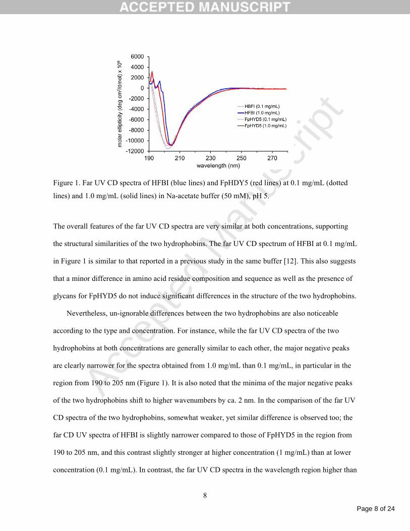
The far UV CD spectra obtained from HFBI and FpHYD5 at 0.1 mg/mL and 1.0 mg/mL are
presented in Figure 1.
Page 8 of 24
Accep
ted
Man
uscr
ipt
8
Figure 1. Far UV CD spectra of HFBI (blue lines) and FpHDY5 (red lines) at 0.1 mg/mL (dotted
lines) and 1.0 mg/mL (solid lines) in Na-acetate buffer (50 mM), pH 5.
The overall features of the far UV CD spectra are very similar at both concentrations, supporting
the structural similarities of the two hydrophobins. The far UV CD spectrum of HFBI at 0.1 mg/mL
in Figure 1 is similar to that reported in a previous study in the same buffer [12]. This also suggests
that a minor difference in amino acid residue composition and sequence as well as the presence of
glycans for FpHYD5 do not induce significant differences in the structure of the two hydrophobins.
Nevertheless, un-ignorable differences between the two hydrophobins are also noticeable
according to the type and concentration. For instance, while the far UV CD spectra of the two
hydrophobins at both concentrations are generally similar to each other, the major negative peaks
are clearly narrower for the spectra obtained from 1.0 mg/mL than 0.1 mg/mL, in particular in the
region from 190 to 205 nm (Figure 1). It is also noted that the minima of the major negative peaks
of the two hydrophobins shift to higher wavenumbers by ca. 2 nm. In the comparison of the far UV
CD spectra of the two hydrophobins, somewhat weaker, yet similar difference is observed too; the
far CD UV spectra of HFBI is slightly narrower compared to those of FpHYD5 in the region from
190 to 205 nm, and this contrast slightly stronger at higher concentration (1 mg/mL) than at lower
concentration (0.1 mg/mL). In contrast, the far UV CD spectra in the wavelength region higher than
Page 9 of 24
Accep
ted
Man
uscr
ipt
9
ca. 203 nm are much closer to each other despite the variation of the type or concentration of
hydrophobins.
We propose that the concentration-dependent changes of far UV CD spectra of the
hydrophobins are related to the self-assembly to form multimers in high concentration [42-44].
Previous studies have shown that HFBI molecules tend to assemble and form dimers/tetramers at
high concentration for its amphiphilicity in aqueous environment [43, 48]; a threshold to display the
presence of multimer was reported to be ca. 0.15 mg/mL [43]. Thus, in this study too, monomers
are likely to be the dominant form of the hydrophobins at 0.1 mg/mL solutions, whereas multimers
are more dominant species at 1.0 mg/mL solutions. As the formation of dimer/tetramer is essentially
driven by the interaction between surface hydrophobic patches, major secondary structures, such as
-helix or -strand, are expected to be preserved in this process. Thus, the fact that only minor
changes occur in the far UV CD upon increasing the concentration by 10 times (Figure 1) is
consistent with the scenario of multimerization without disturbing the major protein structural
features of the hydrophobins. In the same context, a slight difference between the two hydrophobins
shown at 1.0 mg/mL can also be related to bulkier structure of FpHYD5 than HFBI due to the
presence of glycans, and consequent alteration in the conformation of the assembled multimers.
Nevertheless, it is more important to emphasize that all these changes are minor in magnitude, and
the far UV CD spectra shown in Figure 1 mainly support the structural resemblance of the two
hydrophobins in this study.
3.2 Adsorption of hydrophobins onto surface
Upon exposure of the PDMS-coated waveguides to the solutions of hydrophobin or P105, a
rapid surface adsorption, followed by saturation was observed; more than 90% of saturated
adsorption signals were achieved within the first 5 min (representative adsorption profiles shown in
Page 10 of 24
Accep
ted
Man
uscr
ipt
10
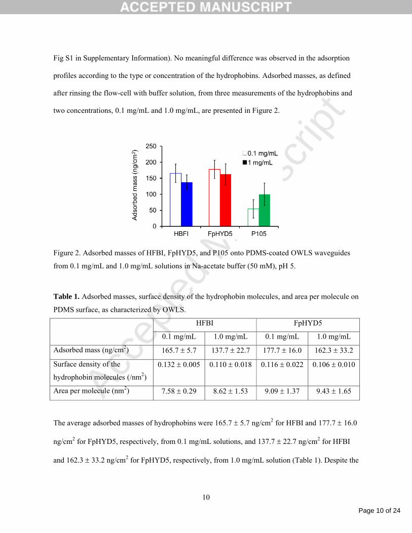
Fig S1 in Supplementary Information). No meaningful difference was observed in the adsorption
profiles according to the type or concentration of the hydrophobins. Adsorbed masses, as defined
after rinsing the flow-cell with buffer solution, from three measurements of the hydrophobins and
two concentrations, 0.1 mg/mL and 1.0 mg/mL, are presented in Figure 2.
Figure 2. Adsorbed masses of HFBI, FpHYD5, and P105 onto PDMS-coated OWLS waveguides
from 0.1 mg/mL and 1.0 mg/mL solutions in Na-acetate buffer (50 mM), pH 5.
Table 1. Adsorbed masses, surface density of the hydrophobin molecules, and area per molecule on
PDMS surface, as characterized by OWLS.
HFBI FpHYD5
0.1 mg/mL 1.0 mg/mL 0.1 mg/mL 1.0 mg/mL
Adsorbed mass (ng/cm2) 165.7 5.7 137.7 22.7 177.7 16.0 162.3 33.2
Surface density of the
hydrophobin molecules (/nm2)
0.132 0.005 0.110 0.018 0.116 0.022 0.106 0.010
Area per molecule (nm2) 7.58 0.29 8.62 1.53 9.09 1.37 9.43 1.65
The average adsorbed masses of hydrophobins were 165.7 5.7 ng/cm2 for HFBI and 177.7 16.0
ng/cm2 for FpHYD5, respectively, from 0.1 mg/mL solutions, and 137.7 22.7 ng/cm2 for HFBI
and 162.3 33.2 ng/cm2 for FpHYD5, respectively, from 1.0 mg/mL solution (Table 1). Despite the

Page 11 of 24
Accep
ted
Man
uscr
ipt
11
10-fold difference in the concentration of bulk solution, the adsorbed masses of the hydrophobins
from 0.1 mg/mL and 1.0 mg/mL solutions were statistically indistinguishable. This means that the
hydrophobin solutions at 0.1 mg/mL provide a saturated monolayer on PDMS surface, and an
increase of the concentration of bulk solution to 1.0 mg/mL does not contribute to further surface
adsorption, via e.g. multilayer formation. On the other hand, at even further lower concentration, e.g.
at 0.01 mg/mL, the adsorbed masses of both HFBI and FpHYD5 were much smaller than those at
0.1 or 1 mg/mL (< 70 ng/cm2, data not shown). Lastly, the adsorbed masses of P105 at 0.1 and 1
mg/mL concentration were 64.0 35.3 ng/cm2 and 100.0 35.3 ng/cm2, respectively. Based on the
molecular weights of the hydrophobins and the adsorbed masses, the number of hydrophobin
molecules per unit area (1 nm2), and in turn, the area occupied per hydrophobin molecule can be
estimated under the assumption of random close packing. The results are shown in Figure 3 and
Table 1; 7.58 0.29 nm2 (0.1 mg/mL) and 8.62 1.53 (1.0 mg/mL) for HFBI, and 9.09 1.37 nm2
(0.1 mg/mL) and 9.43 1.65 nm2 for FpHYD5 (1.0 mg/mL), respectively. Again, the area per
molecule was statistically indistinguishable for the two hydrophobins at both concentrations.
Figure 3. Estimated areas per hydrophobin molecule from the adsorbed masses (Figure 2) and the
molecular weights of HFBI and FpHYD5 from 0.1 mg/mL and 1 mg/mL solutions.
Page 12 of 24
Accep
ted
Man
uscr
ipt
12
Many previous experimental studies have shown facile adsorption of HFBI or HFBII onto a
variety of hydrophobic substrates, including graphite [8], alkylated gold [23], polystyrene (PS) [16],
and PDMS [18,20] from aqueous solution for its distinct amphiphilicity. An MD simulation study
also confirmed that the binding of HFBI onto PDMS surface is energetically most favorable when
the adsorption occurs exclusively through the interaction of hydrophobic patches with PDMS
substrate [15]. As FpHYD5 is relatively a new molecule, its surface adsorption has been studied to a
much less extent to date. Based on structural homology though, a similar adsorption mechanism and
conformation with HFBI, i.e. aliphatic hydrophobic patches on the protein surface acting as
anchoring units, is expected in the adsorption onto PDMS surfaces. Additionally, one N-
glycosylation (1695 Da) residing on the opposite side of hydrophobic patches [41] further improves
the amphiphilicity of FpHYD5.
3.3 Aqueous lubricating properties
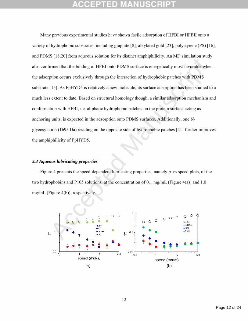
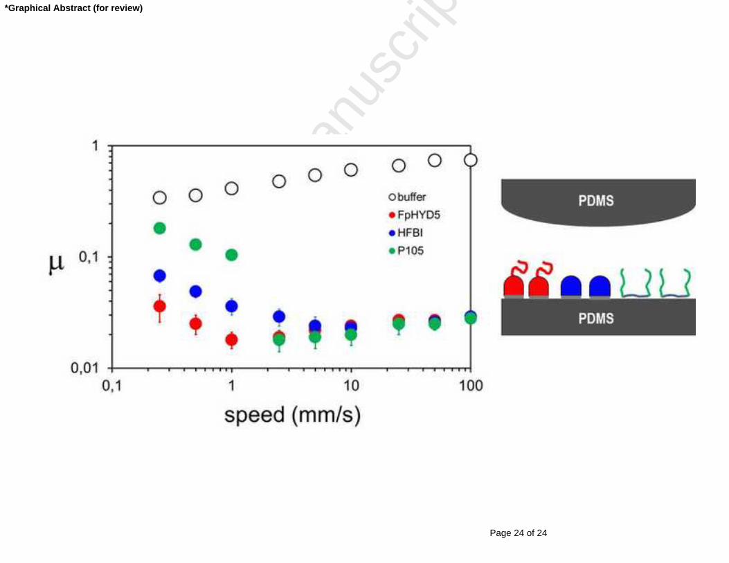
Figure 4 presents the speed-dependent lubricating properties, namely µ-vs-speed plots, of the
two hydrophobins and P105 solutions, at the concentration of 0.1 mg/mL (Figure 4(a)) and 1.0
mg/mL (Figure 4(b)), respectively.
Page 13 of 24
Accep
ted
Man
uscr
ipt
13
Figure 4. µ vs speed plots for HFBI, FpHYD5, and P105 from (a) 0.1 mg/mL and (b) 1.0 mg/mL,
respectively.
The µ-vs-speed plots of additive-free acetate buffer solution are also presented as reference. The µ
values of PDMS-PDMS sliding contacts lubricated either by the hydrophobins or P105 solutions
were lower than those of buffer solutions, yet to different extents depending on the type of additive,
concentration, and speed range.
Between the two hydrophobins, FpHYD5 showed clearly superior lubricating capabilities,
especially at low-speed (< 10 mm/s) and low-concentration (0.1 mg/mL) regime, where boundary
lubrication mechanism is dominant. This can be firstly linked to the slightly higher adsorbed mass
of FpHYD5 than HFBI (Figure 2). But, as this difference is very small, and the standard deviations
are larger than the differences at both concentrations, it cannot account for the large difference in
the µ values between them. Instead, the superior lubricating properties of FpHYD5 in this regime
can be related to the presence of glycosylated region in it and consequently more effective
hydration, as confirmed by QCM-D study in a previous study [41]. Glycosylation is a common
strategy for biomacromolecules, such as mucins [35,36] and lubricins [38,39], to enhance the
hydrophilicity and entrainment of base lubricant, i.e. water, at the gliding interface. The present
study with hydrophobins further confirms the significance of glycosylation for aqueous lubricating
properties of biopolymeric additives. Judging from the µ vs speed plots obtained in this study,
overall aqueous lubricating properties of FpHYD5 can be assessed fairly comparable to those by
much larger biomacromolecules, such as mucins at the same tribopair [49,50]. Both hydrophobins
showed superior lubricating behavior compared to P105 as aqueous lubricant additive for this
tribopair. This is expected from that the adsorbed masses of the two hydrophobins are higher than
that of P105 at the same concentrations (Figure 2). With increasing speed, however, the difference
in values for the two hydrophobins, as well as with P105, started to disappear. This difference is
Page 14 of 24
Accep
ted
Man
uscr
ipt
14
much smaller in high-concentration regime (1.0 mg/mL, Figure 4(b)). This may be an indication
that fluid-films started to form at the interface in this speed regime [32,33].
Direct comparison of each additive at different concentrations is not shown, as all of them
showed clearly superior lubricating effects (i.e. lower µ values) at high concentration (1.0 mg/mL)
than at low concentration (0.1 mg/mL), with the difference being larger in low-speed regime. A
superior lubricating effect of hydrophobins at 1 mg/mL concentration than at 0.1 mg/mL is not
directly related to the surface adsorption properties (Figure 2), according to which the adsorbed
masses from 1.0 mg/mL solutions are comparable to those from 0.1 mg/mL solutions. In other
words, higher concentration of the hydrophobin molecules in 1.0 mg/mL solution contributes to
lubrication, even though they do not contribute to higher surface adsorption. This is, however, not
due to an increase in viscosity; proteins or glycoproteins at 1.0 mg/mL concentration are virtually
identical with water in viscosity [36]. Furthermore, the improvement of lubricating properties at
high concentration is evident in slow-speed regime, where boundary lubrication is most active,
rather than in high-speed regime, where viscosity plays a significant role due to a higher likelihood
of forming fluid-films. The improved lubrication at high concentration of hydrophobins can be
related to the more effective recovery of the lubricating film under cyclic tribological stress in pin-
on-disk tribometry [51]; hydrophobins on PDMS surface are easily rubbed away from the
tribostress whereas excess proteins in bulk solution can readily reform the lubricating film as well
due to non-covalent bonding characteristic, and this process is continuously repeating. In this
context, higher concentration of the additives is advantageous for faster regeneration of the
lubricating layer due to the steeper concentration gradient near the contact area. A control
experiment involving a monolayer coating only, as prepared by replacing hydrophobin solution
with buffer solution after the formation a monolayer showed that the degradation in lubricating
properties started to occur immediately after the initial contacts (Figure S2, in Supplementary
Page 15 of 24
Accep
ted
Man
uscr
ipt
15
Information). Thus, excellent lubricating performance of the hydrophobins for the sliding contacts
of PDMS-PDMS tribopair is mainly indebted from fast (re)adsorption kinetics on to the surface
when excess hydrophobins are present in bulk solution, which is, in turn, indebted from the
presence of distinct hydrophobic patches on the protein surface and enhanced amphiphilicity.
5. Conclusions
In this study, tribological properties of type II hydrophobins, HFBI and FpHYD5, as aqueous
lubricant additive for a soft hydrophobic sliding interface, PDMS-PDMS, were studied at the
concentration of 0.1 mg/mL and 1.0 mg/mL. Far UV CD spectra of the two hydrophobins were very
similar, suggesting overall structural similarity of the two hydrophobins, yet showed somewhat
different secondary structural features according to the concentration change from 0.1 to 1.0 mg/mL.
This is suggested to be related to the dominance of monomers at 0.1 mg/mL and multimers at 1.0
mg/mL, respectively. However, its influence on adsorption was insignificant due to the formation of
monolayer already at lower concentration (0.1 mg/mL). The adsorption strength of the
hydrophobins was not sufficient to withstand the tribostress under 5 N with a monolayer coating on
the surface. But, the hydrophobins displayed very efficient aqueous lubricating capabilities as a
solution based on “self-healing” mechanism. Despite its much smaller molecular weight (within the
range of 7 to 10 kDa) compared to other biomolecular amphiphiles, such as mucins or lubricins, the
two hydrophobins in this study, especially FpHYD5, showed comparable aqueous lubrication
capabilities for PDMS-PDMS tribopair. This is mainly due to the presence of distinct hydrophobic
patches on protein surface, rather than being entirely buried inside, and consequently enhanced
amphiphilicity of the molecules. In low-speed regime, where boundary lubrication character is
dominant, FpHYD5 showed relatively superior lubricity to HFBI, presumably related to the
presence of glycans and consequently more efficient hydration. With increasing speed though, the
Page 16 of 24
Accep
ted
Man
uscr
ipt
16
difference in the lubricating properties of the two hydrophobins as well as with P105 started to
disappear, due to more feasible entrainment of base lubricant, water, into the sliding interface.
6. Acknowledgements
The authors are grateful for the financial support from the Danish Council for Independent Research
(DFF), Technology and Production Sciences (FTP) (10-082707), European Research Council
(Funding scheme, ERC Starting Grant 2010, Project number 261152), and COST Action program
(TD1003, Bioinspired Nanotechnologies). Riitta Suihkonen (VTT) is also acknowledged for her
technical assistance in the purification of the hydrophobins in this study.
Page 17 of 24
Accep
ted
Man
uscr
ipt
17
References
[1] Wösten, H.A.B., van Wetter, M. A., Lugones, L. G., van der Mei, H. C., Busscher, H. J.,
Wessels, J. G. How a fungus escapes the water to grow into the air. Curr. Biol. 1999. 9, 85 – 88.
[2] Wösten, H.A., Hydrophobins: Multipurpose proteins. Ann. Rev. Microbiol. 2001, 55, 625 – 646.
[3] Lumsdon, S., Green, J., Stieglitz, B., Adsorption of hydrophobin proteins at hydrophobic and
hydrophilic interfaces. Colloids and Surfaces B: Biointerfaces 2005, 44, 172 – 178.
[4] Hektor, H., Scholtmeijer, K. Hydrophobins: proteins with potential. Current Opinion in
Biotechnology 2005, 16, 434 – 439.
[5] Zampieri, F., Wösten, H.A.B., Scholtmeijer, K. Creating surface properties using a palette of
hydrophobins. Materials 2010, 3, 4607 – 4625.
[6] Linder, M.B., Szilvay, G.R., Nakari-Setälä, Penttilä, M.E. Hydrophobins: the protein-
amphiphiles of filamentous fungi. FEMS Microbiology Reviews 2005, 29, 877 – 896.
[7] Linder, M.B. Hydrophobins: Proteins that self-assemble at interfaces. Current Opinion in
Colloid & Interface Science 2009, 14, 356 – 363.
[8] Szilvay, G.R., Paananen, A., Laurikainen, K., Vuorimaa, E., Lemmetyinen, H., Peltonen, J.,
Linder, M.B. Self-assembled hydrophobin protein films at the air-water interface: structural analysis
and molecular engineering. Biochemistry 2007, 46, 235 – 2354.
[9] Kisko, K., Szilvay, G.R., Vuorimaa, E., Lemmetyienen, H., Linder M.B., Torkkeli, M., Serimaa,
R. Self-assembled films of hydrophobin proteins HFBI and HFBII studied in situ at the air/water
interface. Langmuir 2009, 25, 1612 – 1619.
[10] Zhang, X.L., Penfold, J., Thomas, R.K., Tucker, I.M., Petkov, J.T., Bent, J., Cox, A., Campbell,
R.A., Adsorption behavior of hydrophobin and hydrophobin/surfactant mixtures at the air-water
interface. Langmuir 2011, 27, 11316 – 11323.

Page 18 of 24
Accep
ted
Man
uscr
ipt
18
[11] Wösten, H.A.B., Schuren, F.H.J., Wessels, J.G.H. Interfacial self-assembly of a hydrophobin
into an amphipathic protein membrane mediates fungal attachment to hydrophobic surfaces. EMBO
Journal 1994, 13, 5848 – 5854.
[12] Askolin, S., Linder, M., Scholtmeijer, K., Tenkannen, M., Penttilä, de Vocht, M.L., Wösten,
H.A.B. Interaction and comparison of a class I hydrophobin from Schizophyllum commune and
class II hydrophobins from Tichoderma reesei. Biomacromolecules 2006, 7, 1295 – 1301.
[13] Cheung, D.L. Molecular simulation of hydrophobin adsorption at an oil–water interface.
Langmuir 2012, 28(23), 8730 – 8736.
[14] Reger, M., Hoffmann, H. Hydrophobin coated boehmite nanoparticles stabilizing oil in water
emulsions. J. Coll. Interf. Sci. 2012, 368, 378 – 386.
[15] Liu, Y., Wu, M., Feng, X., Shao, X., Cai, W. Adsorption behavior of hydrophobin proteins on
polydimethylsiloxane substrates. J. Phys. Chem. B 2012, 116, 12227 – 12234.
[16] Wang, Z., Huang, Y., Li, S., Linder, M.B., Qiao, M. Hydrophilic modification of polystyrene
with hydrophobin for time-resolved immunofluorometric assay. Biosensors and Bioelectronics
2010, 26, 1074 – 1079.
[17] Zhao, Z.-X., Wang, H.-C., Wang, X.-S., Qiao, M.-Q., Anzai, J.-I., Chen, Q. Self-assembled
film of hydrophobins on gold surfaces and its application to electrochemical sensing. Colloids &
Surfaces B: Biointerfaces 2009, 71, 102 – 106.
[18] Qin, M., Wang, L.-K., Feng, X.-Z., Yang, Y.-L., Wang, R., Wang, C., Yu, L., Shao, B., Qiao,
M.-Q. Surface modification of mica and poly(dimethylsiloxane) with hydrophobins for protein
immobilization. Langmuir 2007, 23, 4465 – 4471.
[19] Lumsdon, S.O., Green, J., Stieglitz, B. Adsorption of hydrophobin proteins at hydrophobic and
hydrophilic surfaces. Colloids and Surfaces B: Biointerfaces 2005, 44, 172 – 178.
Page 19 of 24
Accep
ted
Man
uscr
ipt
19
[20] Hou, S., Yang, K., Qin, M., Feng, X.-Z., Guan, L., Yang, Y., Wang, C. Patterning of cells on
functionalized poly(dimethylsiloxane) surface prepared by hydrophobin and collagen modification.
Biosensors and Bioeletronics 2008, 24, 912 – 916.
[21] de Vocht, M.L., Reviakine, I., Ulrich, W.-P., Bergsma-Schutter, W., Wösten, H.A.B., Vogel,
H., Brisson, A., Wessels, J.G.H., Robillard, G.T. Self-assembly of the hydrophobin SC3 proceeds
via two structural intermediates. Protein Science 2002, 11, 1199 – 1205.
[22] Linder, M., Szilvay, G.R., Nakari-setälä, T., Söderlund, H., Penttilä, M. Surface adhesion of
fusion proteins containing the hydrophobins HFBI and HFBII from Trichoderma reesei. Protein
Science 2002, 11, 2257 – 2266.
[23] Wösten, H.A.B., de Vocht, M.L. Hydrophobins, the fungal coat unravelled. Biochim. Biophys.
Acta 2000, 1469, 79 – 68.
[24] Scholtmeijer, K., Wessels, J.G.H., Wösten, H.A.B. Fungal hydrophobins in medical and
technical applications. Appl. Microbiol. Biotechnol. 2001, 56, 1 – 8.
[25] Scholtmeijer, K., Janssen, M.I., Van Leeuwen, M.B.M., Van Kooten, T.G., Hektor, H. Wösten,
H.A.B. The use of hydrophobins to functionalize surfaces. Bio-Med. Mater. Eng. 2004, 14, 447 –
454.
[26] Scholtmeijer, K., Janssen, M.I., Gerssen, B., de Vocht, M.L., Van Leeuwen, M.B.M. Surface
modifications created by using engineered hydrophobins. Appl. Environ. Microbiol. 2002, 68, 1367
– 1373.
[27] Janssen, M.I., Van Leeuwen, M.B.M., Scholtmeijer, K., Van Kooten, T.G., Dijkhuizen, L.,
Wösten, H.A.B. Coating with genetic engineered hydrophobin promotes growth of fibroblasts on a
hydrophobic solid. Biomaterials 2002, 23, 4848 – 4854.
Page 20 of 24
Accep
ted
Man
uscr
ipt
20
[28] von Vacano, B., Xu, R., Hirth, S., Herzenstiel, I., Rueckel, M., Subkowski, T., Baus, U.
Hydrophobins can prevent secondary protein adsorption on hydrophobic substrates without
exchange. Anal. Bioanal. Chem. 2011, 400, 2031 – 2040.
[29] Misra, R., Gordon, J.L., Morgan, S.E. Nanoscale reduction in surface friction of polymer
surfaces modified with Sc3 hydrophobin from Schizophylum commune, Biomacromolecules 2006,
7(5), 1463 – 1470.
[30] Ahlroos, T., Hakala, T.J., Linder, M.B., Holmberg, K., Mahlberg, R., Laaksonen, P., Varjus, S.
Biomimetic approach to water lubrication with biomolecular additives. Proc. I. Mech. Eng. J. 2011,
225, 1013 – 1022.
[31] Hakala, T., Laaksonen, P., Saikko, V., Ahlroos, T., Helle, A., Mahlberg, R., Hähle, H., Jacobs,
K., Kuosmanen, P., Linder, M.B., Holmberg, K. RSC Advances 2012, 2, 9867 – 9872.
[32] Lee, S., Iten, R., Müller, M., Spencer, N.D. Influence of molecular architechture on the
adsorption of poly(ethylene oxide)-poly(propylene oxide)-poly(ethylene oxide) on PDMS surfaces
and implications for aqueous lubrication. Macromolecules 2004, 37, 8349 – 8365.
[33] Lee, S., Spencer, N.D. Aqueous lubrication of polymers: Influence of surface modification.
Tribol. Int. 2005, 38, 922 – 930.
[34] Røn, T., Javakhishvili, I., Jankova, K., Hvilsted, S., Lee, S. Adsorption and aqueous lubricating
properties of charged and neutral amphiphilic diblock copolymers at a compliant, hydrophobic
interface. Langmuir 2013, 29, 7782 – 7792.
[35] Cassin, G., Heinrich, E., Spikes, H.A. The Influence of surface roughness on the lubrication
properties of adsorbing and non-adsorbing biopolymers. Tribol. Lett. 2001, 11, 95 – 102.
[36] Lee, S., Müller, M., Rezwan, K., Spencer, N.D. Porcine gastric mucin (PGM) at the
water/poly(dimethylsiloxane) (PDMS) interface: Influence of pH and ionic strength on its
conformation, adsorption, and aqueous lubrication properties. Langmuir 2005, 21, 8344 – 8353.
Page 21 of 24
Accep
ted
Man
uscr
ipt
21
[37] Garrec, D.A., Norton, I.T. The influence of hydrocolloid hydrodynamics on lubrication. Food
Hydrocolloids 2012, 26, 389 – 397.
[38] Zappone, B., Ruths, M., Greene, G.W., Jay, G.D., Israelachvili, J.N. Adsorption, lubrication,
and wear of lubricin on model surfaces: polymer brush-like behavior of a glycoprotein. Biophys J.
2007, 92, 1693 – 708.
[39] Chang, D.P., Abu-Lail, N.I., Coles, J.M., Guilak, F., Jay, G.D., Zauscher, S. Friction force
microscopy of lubricin and hyaluronic acid between hydrophobic and Hydrophilic Surfaces. Soft
Matter 2009, 5, 3438 – 3445.
[40] Hattrup, C.L., Gendler, S.J. Structure and function of the cell surface (tethered) mucins. Annual
Review of Physiology 2008, 70, 431 – 457.
[41] Sarlin, T., Kivioja, T., Kalkkinen, N., Linder, M.B., Nakari-Setälä, T. Identification and
characterization of gushing-active hydrophobins from Fusarium graminearum and related species J.
Basic Microbiology 2012, 52, 184 – 194.
[42] Hakanpa, J., Paananen, A., Askolin, S., Nakari-Seta, T., Parkkinen, T, Penttila, M., Linder,
M.B., Rouvinen, J. Atomic resolution structure of the HFBII hydrophobin, a self-assembling
amphiphile. J. Biol. Chem 2004, 279(1), 534 – 539.
[43] Szilvay, G.R., Nakari-Seta, T., Linder, M.B. Behavior of Trichoderma reesei hydrophobins in
solution: Interactions, dynamics, and multimer formation. Biochemistry 2006, 45, 8590-8598
[44] Hakanpa, J., Szilvay, G.R, Kaljunen, H., Maksimainen,M., Linder, M.B., Ruvinen, J. Two
crystal structures of Trichoderma reesei hydrophobin HFBI - The structure of a protein amphiphile
with and without detergent interaction. Protein Science 2006, 15, 2129 – 2140.
[45] Wang, Z., Lienenmann, M., Qiau, M., Linder, M.B. Mechanism of protein adhesion on surface
films of hydrophobin. Langmuir 2010, 26(11), 8491 – 8496.
Page 22 of 24
Accep
ted
Man
uscr
ipt
22
[46] de Feijter, J. A., Benjamins, J., Veer, F.A. Ellipsometry as a tool to study the ad-sorption of
synthetic and biopolymers at the air-water interface. Biopolymers 1978, 17, 1759 – 1772.
[47] Malmsten, M., Linse, P., Cosgrove, T. Adsorption of PEO-PPO-PEO block copolymers at
silica. Macromolecules 1992, 25, 2474 – 2481.
[48] Torkkeli, M., Serimaa, R., Ikkala, O., Linder, M. Aggregation and self-assembly of
hydrophobins from Trichoderma reesei: Low-resolution structural models. Biophys. J. 2002, 83,
2240 – 2247.
[49] Lee, S., Müller, M., Rezwan, K., Spencer, N.D. Porcine gastric mucin (PGM) at the water/
poly(dimethylsiloxane) (PDMS) interface: Influence of pH and ionic strength on its conformation,
adsorption, and aqueous lubrication properties. Langmuir, 2005, 21, 8344 – 8353.
[50] Yakubov, G.E., McColl, J., Bongaerts, J.H.H., Ramsden, J.J. Viscous boundary lubrication of
hydrophobic surfaces by mucin. Langmuir, 2009, 25, 2313 – 2321.
[51] Lee, S., Müller, M., Heeb, R., Zürcher, S., Tosatti, S., Heinrich, M., Amstad, F., Pechman, S.,
Spencer, N. D. Self-healing behavior of a polyeletrolyte-based lubricant additive for aqueous
lubrication of oxide materials. Tribol. Lett., 2006, 24, 217– 223.
Page 23 of 24
Accep
ted
Man
uscr
ipt
23
Highlights
FpHYD5 and HFBI are effective as aqueous lubricant additives for soft contacts
Hydrophobins showed superior lubricity than synthetic copolymer P105
FpHYD5 showed an enhanced lubricity, presumably due to glycosylation
Page 24 of 24
Accep
ted
Man
uscr
ipt
*Graphical Abstract (for review)