Embed Size (px)
Citation preview
Human Cytogenetics
Cytogenetics
India Contact:
Life Technologies (India) Pvt. Ltd. 306, Aggarwal City Mall, Opposite M2K Pitampura Delhi – 110034. Ph: +91-11-42208000, 42208111, 42208222 Fax: +91-11-42208444 Email: [email protected] Website: www.lifetechindia.com
2
3 Prenatal Diagnostics
7 Post postnatal Diagnostics 7 - Bone Marrow Culture 9 - Blood lymphocyte Culture11 - Lymphocyte Separation Tubes
14 Karyotyping Reagents
16 Keep your lab clean
Index
Prenatal D
iagnostics
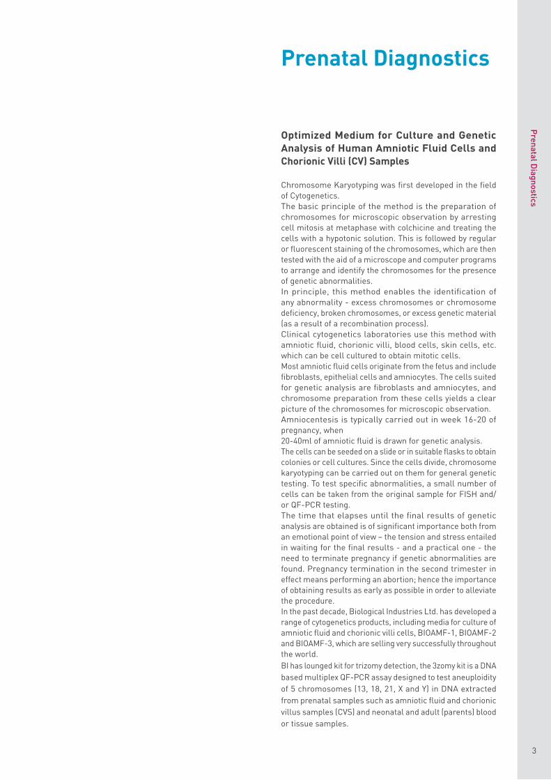
Optimized Medium for Culture and Genetic Analysis of Human Amniotic Fluid Cells and Chorionic Villi ( CV ) Samples
Chromosome Karyotyping was first developed in the field of Cytogenetics.The basic principle of the method is the preparation of chromosomes for microscopic observation by arresting cell mitosis at metaphase with colchicine and treating the cells with a hypotonic solution. This is followed by regular or fluorescent staining of the chromosomes, which are then tested with the aid of a microscope and computer programs to arrange and identify the chromosomes for the presence of genetic abnormalities.In principle, this method enables the identification of any abnormality - excess chromosomes or chromosome deficiency, broken chromosomes, or excess genetic material (as a result of a recombination process).Clinical cytogenetics laboratories use this method with amniotic fluid, chorionic villi, blood cells, skin cells, etc. which can be cell cultured to obtain mitotic cells.Most amniotic fluid cells originate from the fetus and include fibroblasts, epithelial cells and amniocytes. The cells suited for genetic analysis are fibroblasts and amniocytes, and chromosome preparation from these cells yields a clear picture of the chromosomes for microscopic observation.Amniocentesis is typically carried out in week 16-20 of pregnancy, when20-40ml of amniotic fluid is drawn for genetic analysis.The cells can be seeded on a slide or in suitable flasks to obtain colonies or cell cultures. Since the cells divide, chromosome karyotyping can be carried out on them for general genetic testing. To test specific abnormalities, a small number of cells can be taken from the original sample for FISH and/or QF-PCR testing.The time that elapses until the final results of genetic analysis are obtained is of significant importance both from an emotional point of view – the tension and stress entailed in waiting for the final results - and a practical one - the need to terminate pregnancy if genetic abnormalities are found. Pregnancy termination in the second trimester in effect means performing an abortion; hence the importance of obtaining results as early as possible in order to alleviate the procedure.In the past decade, Biological Industries Ltd. has developed a range of cytogenetics products, including media for culture of amniotic fluid and chorionic villi cells, BIOAMF-1, BIOAMF-2 and BIOAMF-3, which are selling very successfully throughout the world. BI has lounged kit for trizomy detection , the 3zomy kit is a DNA based multiplex QF-PCR assay designed to test aneuploidity of 5 chromosomes ( 13 , 18 , 21 , X and Y ) in DNA extracted from prenatal samples such as amniotic fluid and chorionic villus samples ( CVS ) and neonatal and adult ( parents ) blood or tissue samples .
3
Prenatal Diagnostics

Prenatal D
iagnostics
4
The 3ZOMY kit includes a multiplex STR markers set used to perform initial aneuploidy diagnosis of the 21 , 18 , 13 , X & Y chromosomes and two additional chromosomes specific marker sets for 21 & XY chromosomes that may be used to confirm the diagnosis .
BIOAMF-1 Basal Medium and Supplement
BIOAMF-1 is designed for the primary culture of human amniotic fluid cells and chorionic villi (CV) samples in both open (5% CO2) and closed systems.The medium allows rapid growth of amniocytes or chorionic villi for use in karyotyping.No supplementation with serum or serum-substitutes is necessary.The medium consists of two components: basal medium and frozen supplements.
Instructions for Use For the preparation of 500ml complete medium, use 01-190-1A with 01-192-1E. For the preparation of 100ml complete medium, use 01-190-1B with 01-192-1D. Thaw the BIOAMF-1 Supplement by swirling in a 37ºC water bath, and transfer the contents to the bottle of BIOAMF-1 Basal Medium. Mix the complete medium by swirling the bottle, and add 2mM L-Glutamine (L-Glutamine Solution 200mM, cat. no. 03-020-1). Antibiotics may be added if desired (Gentamicin, Cat. No. 03-035-1).
Storage and Stability BIOAMF-1 Basal Medium is stable for 15 months from production date when stored at 2-8ºC.BIOAMF-1 Supplement is stable for 24 months from production date when stored at -20ºC.The complete medium is stable for 14 days when stored at 2-8ºC.Do not freeze the complete medium. Protect both the basal medium and the complete medium from light.
BIOAMF-2 Complete MediumFor faster results
BIOAMF-2 is a complete medium specifically optimized for the primary culture of human amniotic fluid cells and chorionic villi (CV) samples in both open (5% CO2) and closed systems.No addition of serum is required, and chromosome karyotyping time is greatly reduced compared with the conventional medium.Note: this is a one-bottle formulation, which also contains L-Glutamine and antibiotics. Simply thaw and use!
Storage and Stability BIOAMF-2 Medium should be kept frozen at -20°C. After thawing, the medium should be stored at 2-8°C. The medium should be used within 7 days after thawing. If required , after thawing at R.T or 4°C ( recommended ), dispense into aliquots to avoid repeated freezing and thawing cycles . Protect the medium from light.
Figure: Comparison of the Percentage of Harvested Plates According To Harvest Day Between Biological Industries BIOAMF™-2 and a Leading Competitor
40
45
50 BIOAMF™-2 Leading Competitor
5 6 7 8 9 10 11 12 >12
20
15
10
5
0
25
30
35
Harvest Day
% H
arve
sted
Pla
tes
Product Name Cat. No. Unit Size
BIOAMF-1 01-190-1A 450mlBasal Medium 01-190-1B 90ml
BIOAMF-1 01-192-1D 10mlSupplement 01-192-1E 50ml
Product Name Cat. No. Unit Size
BIOAMF-2 01-194-1A 500mlComplete Medium 01-194-1B 100ml
Instructions for use of BIOAMF-1,2,3
BIOAMF Medium may be used for:• Primary culture of amniotic fluid cells• Culture of passaged amniotic fluid cells• Propagation of chorionic villus cells
In Situ Culture of Amniotic Fluid Cells1. Centrifuge 20ml of amniotic fluid at 750 rpm for 10
minutes.2. Carefully decant the amniotic fluid from the cell pellet
into a sterile test tube.3. Re-suspend the cell pellet with 2ml of amniotic fluid.4. Add 2ml of BIOAMF Medium and swirl gently.5. Culture 0.5ml of the cell suspension on each coverslip
in a tissue culture dish.6. Incubate cultures at 37°C in 5% CO2 atmosphere.7. Flood cultures on day 2 with 1.5ml of BIOAMF Medium.8. After 5 days, check the cultures for the presence of
colonies.9. After the colonies first appear (5-7 days), replace the
medium with fresh BIOAMF Medium.10. When the cultures have colonies of sufficient size, proceed
with harvesting.Note: It is recommended to replace the medium with fresh BIOAMF Medium the day before harvesting.
Flask Method Culture of Amniotic Fluid Cells – Open and Closed SystemsUse the same procedure as for the in situ culture, with the following adaptations:1. Re-suspend the cell pellet with 4ml of amniotic fluid. Add
16ml of BIOAMF Medium and swirl gently.2. Culture 5ml per each T25 flask. Place the cap loosely on
the flask and incubate undisturbed at 37°C in 5% CO2
atmosphere.For Closed Systems: Flush each culture flask with 5%CO2 – 95% air through 0.2μ sterile filter for 20 seconds.Tighten the caps and incubate the flasks at 37°C.
3. Check all flasks for growth after 5 days.
BIOAMF-3 Complete MediumFor Increased metaphase yield
An improved version of complete medium specifically optimized for the primary culture of human amniotic fluid cells and chorionic villi samples used in prenatal diagnostic testing. This medium accelerates the growth of the non-epithelial cells used for chromosome karyotyping.The medium is supplied frozen and contains L-Glutamine and antibiotics.
Storage and StabilityBIOAMF-3 Medium should be kept frozen at -20ºC. After thawing, the medium should be stored at 2-8ºC. The medium should be used within 14 days after thawing. Protect the medium from light.If required , after thawing at R.T or 4°C ( recommended ), dispense into aliquots to avoid repeated freezing and thawing cycles .
Product Name Cat. No. Unit Size
BIOAMF-3 01-196-1A 500mlComplete Medium 01-196-1B 100ml
Prenatal D
iagnostics
5
Prenatal D
iagnostics
6
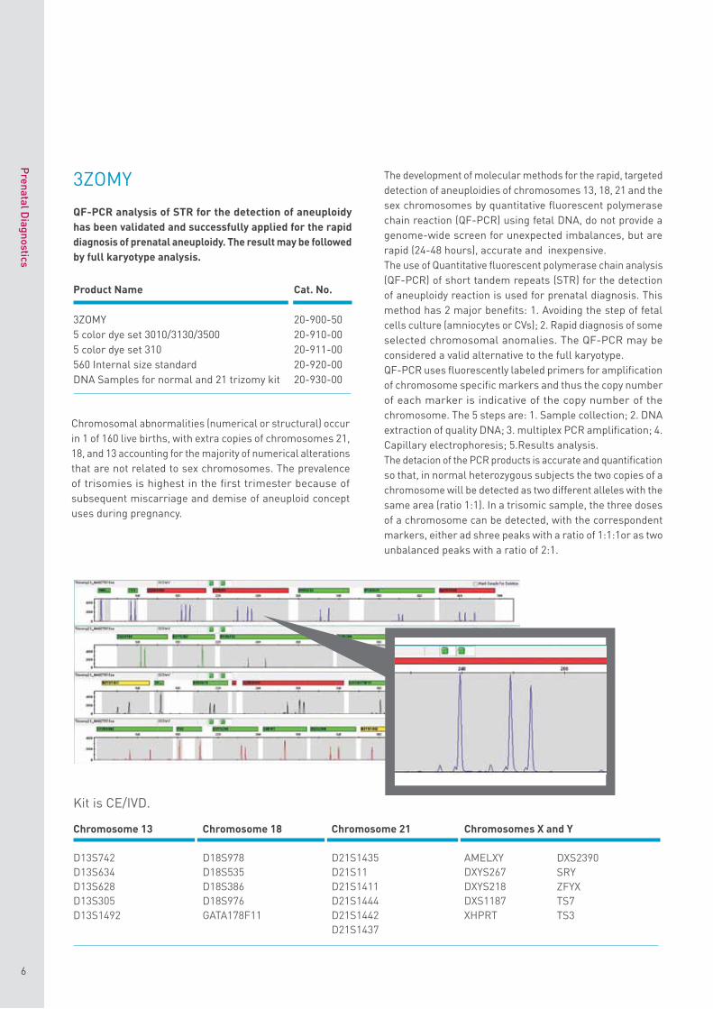
The development of molecular methods for the rapid, targeted detection of aneuploidies of chromosomes 13, 18, 21 and the sex chromosomes by quantitative fluorescent polymerase chain reaction (QF-PCR) using fetal DNA, do not provide a genome-wide screen for unexpected imbalances, but are rapid (24-48 hours), accurate and inexpensive. The use of Quantitative fluorescent polymerase chain analysis (QF-PCR) of short tandem repeats (STR) for the detection of aneuploidy reaction is used for prenatal diagnosis. This method has 2 major benefits: 1. Avoiding the step of fetal cells culture (amniocytes or CVs); 2. Rapid diagnosis of some selected chromosomal anomalies. The QF-PCR may be considered a valid alternative to the full karyotype. QF-PCR uses fluorescently labeled primers for amplification of chromosome specific markers and thus the copy number of each marker is indicative of the copy number of the chromosome. The 5 steps are: 1. Sample collection; 2. DNA extraction of quality DNA; 3. multiplex PCR amplification; 4. Capillary electrophoresis; 5.Results analysis.The detacion of the PCR products is accurate and quantification so that, in normal heterozygous subjects the two copies of a chromosome will be detected as two different alleles with the same area (ratio 1:1). In a trisomic sample, the three doses of a chromosome can be detected, with the correspondent markers, either ad shree peaks with a ratio of 1:1:1or as two unbalanced peaks with a ratio of 2:1.
Chromosome 13
D13S742D13S634D13S628D13S305D13S1492
Kit is CE/IVD.
Chromosome 18
D18S978D18S535D18S386D18S976GATA178F11
Chromosome 21
D21S1435D21S11D21S1411D21S1444D21S1442D21S1437
Chromosomes X and Y
AMELXYDXYS267DXYS218DXS1187XHPRT
DXS2390SRYZFYXTS7TS3
3ZOMY
QF-PCR analysis of STR for the detection of aneuploidy has been validated and successfully applied for the rapid diagnosis of prenatal aneuploidy. The result may be followed by full karyotype analysis.
Chromosomal abnormalities (numerical or structural) occur in 1 of 160 live births, with extra copies of chromosomes 21, 18, and 13 accounting for the majority of numerical alterations that are not related to sex chromosomes. The prevalence of trisomies is highest in the first trimester because of subsequent miscarriage and demise of aneuploid concept uses during pregnancy.
Product Name Cat. No.
3ZOMY 20-900-505 color dye set 3010/3130/3500 20-910-005 color dye set 310 20-911-00560 Internal size standard 20-920-00DNA Samples for normal and 21 trizomy kit 20-930-00
Postnatal D
iagnostics - Bone M
arrow C
ulture
7
Cytogenetic analysis of human hematopoietic cells using bone marrow aspirates is a standard practice in hematology. Cell culture improvements and processing techniques have enabled the identification of a number of recurring abnormalities in solid tumors and hematologic malignant diseases. But even more data are available for leukemias and lymphomas than for solid tumors because of the relative ease of obtaining bone marrow or peripheral blood specimens from leukemia patients.The study of chromosomal abnormalities in leukemia serves two functions:The first is to assist in more accurate diagnosis, thereby providing prognostic information and allowing the more rational selection of therapy for a particular patient. The second is to identify the sites of consistent rearrangements, providing the precise localization required for the isolation and cloning of DNA from these regions. Using molecular techniques the function of the genes can be identified and the mechanisms whereby their altered function is involved in tumorigenesis can be determined.In the past, it was assumed that cytogenetic analysis of hematologic malignant disorders was best performed directly on uncultured bone marrow samples. However, later studies indicate that analysis of cultured samples disclosed a clonal abnormality that would not have been detected if the direct method alone had been used. Thus, for many samples, chromosomal rearrangements were often characterized only after analysis of cultured preparations.
Bone Marrow Karyotyping Medium
Bone Marrow Karyotyping Medium is intended for use in short-term cultivation of primary bone marrow cells for chromosome evaluation.Bone Marrow Karyotyping Medium is based on RPMI-1640 basal medium supplemented with L-Glutamine, foetal bovine serum, and antibiotics (Gentamicin). The medium does not contain any mitogens or conditioned medium.Bone Marrow Karyotyping Medium is supplied as frozen medium, which is ready for use after thawing.
Instructions for useThe bone marrow karyotyping method was developed to provide information about chromosomal abnormalities. The ready-to-use medium is intended for the culture of bone marrow cells without any mitogens or conditioned medium. After 48-72 hours, a mitotic inhibitor is added to the culture to stop mitosis in the metaphase stage. After treatment by hypotonic solution, fixation and staining, chromosomes can be microscopically observed and evaluated for abnormalities.
1. Inoculate approximately 0.5ml of bone marrowsuspension into a plastic tube or tissue culture platewith 10ml of medium. Invert tubes gently to mixspecimen.
2. Incubate the culture for up to 72 hours.3. Add 0.1-0.2ml of Colcemid Solution (Cat.No. 12-004-
1) to each culture tube. Incubate the culture for anadditional 15-30 minutes.
4. Transfer the culture to a centrifuge tube and spin at500g for 5 minutes.
5. Remove the supernatant and re-suspend the cells in5-10ml of hypotonic 0.075M KCl (Cat. No. 12-005-1).Incubate at 37ºC for 10-12 minutes.
6. Spin at 500g for 5 minutes.7. Remove the supernatant, agitate the cellular sediment
and add drop-by-drop 5-10ml of fresh, ice-cold fixativemade up of 1 part acetic acid to 3 parts methanol.Leave in 4ºC for 10 minutes.
8. Repeat steps 6 and 7.9. Re-suspend the cell pellet in a small volume 0.5-1ml
of fresh fixative, drop onto a clean slide and allow to airdry.
10. At this stage, the preparation can be stained withOrecin or Giemsa. Giemsa banding has become themost widely used technique. The most commonmethod to obtain this staining is to treat slides withTrypsin-EDTA 10X (Cat.No. 03-051-5).
Storage and StabilityBone Marrow Karyotyping Medium should be kept frozen at -20ºC. After thawing, the medium should be stored at 2-8ºC. If required , after thawing at R.T or 4°C ( recommended ), dispense into aliquots to avoid repeated freezing and thawing cycles . The medium should be used within 10 days after thawing. Protect the medium from light.
01-199-1A 500ml
01-199-1B 100ml
Postnatal DiagnosticsBone Marrow Culture
Product Name Cat. No. Unit Size
Bone Marrow Karyotyping Medium Without conditioned medium
Postnatal D
iagnostics - Bone M
arrow C
ulture
8
to each culture tube. Incubate the culture for an additional 15-30 minutes.
4. Transfer the culture to a centrifuge tube and spin at500g for 5 minutes.
5. Remove the supernatant and re-suspend the cells in5-10ml of hypotonic 0.075M KCl (Cat.No. 12-005-1).Incubate at 37ºC for 10-12 minutes.
6. Spin at 500g for 5 minutes.7. Remove the supernatant, agitate the cellular sediment
and add drop-by-drop 5-10ml of fresh, ice-cold fixativemade up of 1 part acetic acid to 3 parts methanol.Leave in 4ºC for 10 minutes.
8. Repeat steps 6 and 7.9. Spin at 500g for 5 minutes.10. Re-suspend the cell pellet in a small volume 0.5-1ml
of fresh fixative, drop onto a clean slide and allow toair dry.
11. At this stage, the preparation can be stained withOrecin or Giemsa. Giemsa banding has become themost widely used technique. The most commonmethod to obtain this staining is to treat slides withTrypsin-EDTA 10X (Cat. No. 03-051-5).
Storage and StabilityHematopoietic Cell Karyotyping Medium should be kept frozen at -20ºC.After thawing, the medium should be stored at 2-8ºC. If required , after thawing at R.T or 4°C ( recommended ), dispense into aliquots to avoid repeated freezing and thawing cycles . The medium should be used within 10 days after thawing. Protect the medium from light.
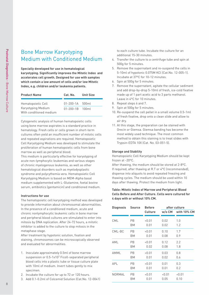
Table: Mitotic Index of Marrow and Peripheral Blood Cells Before and After Culture. Cells were cultured for 4 days with or without 10% CM.
Bone Marrow Karyotyping Medium with Conditioned Medium
Specially developed for use in hematological karyotyping. Significantly improves the Mitotic Index and accelerates cell growth. Designed for use with samples which contain a low amount of cells and/or low Mitotic Index, e.g. children and/or leukemia patients.
Cytogenetic analysis of human hematopoietic cells using bone marrow aspirates is a standard practice in hematology. Fresh cells or cells grown in short-term cultures often yield an insufficient number of mitotic cells and repeated aspirations are required. Hematopoietic Cell Karyotyping Medium was developed to stimulate the proliferation of human hematopoietic cells from bone marrow as well as peripheral blood.This medium is particularly effective for karyotyping of acute non-lymphocytic leukemias and various stages of chronic myelogenous leukemia, as well as other hematological disorders such as myelodysplastic syndrome and polycythemia vera. Hematopoietic Cell Karyotyping Medium is based on MEM-Alpha basal medium supplemented with L-Glutamine, foetal bovine serum, antibiotics (gentamicin) and conditioned medium.
Instructions for useThe hematopoietic cell karyotyping method was developed to provide information about chromosomal abnormalities. In the presence of a conditioned medium, acute and chronic nonlymphocytic leukemic cells in bone marrow and peripheral blood cultures are stimulated to enter into mitosis by DNA replication. After 24-72 hours, a mitotic inhibitor is added to the culture to stop mitosis in the metaphase stage.After treatment by hypotonic solution, fixation and staining, chromosomes can be microscopically observed and evaluated for abnormalities.
1. Inoculate approximately 0.5ml of bone marrowsuspension or 0.5-1x10⁷ Ficoll-separated peripheralblood cells into a plastic tube or tissue culture platewith 10ml of medium. Invert tubes gently to mixspecimen.
2. Incubate the culture for up to 72 or 120 hours.3. Add 0.1-0.2ml of Colcemid Solution (Cat.No. 12-0041)
Product Name Cat. No. Unit Size
Hematopoietic Cell Karyotyping Medium With conditioned medium
01-200-1A 500ml
01-200-1B 100ml
Diagnosis Source Before after culture Culture w/o CM with 10% CM
CML PB <0.01 0.02 1.0BM 0.01 0.02 1.2
CML-BC PB <0.01 0.10 1.7BM 0.01 0.08 0.9
AML PB <0.01 0.12 2.2BM 0.02 0.08 1.8
AMML PB <0.01 0.03 0.8BM 0.01 0.02 0.6
APL PB <0.01 0.01 0.3BM 0.01 0.01 0.2
NORMAL PB <0.01 <0.01 <0.01BM 0.01 0.05 0.10
Postnatal D
iagnostics - Blood Lym
phocyte Culture
9
Blood cell karyotyping is an important tool in modern human cytogenetics, providing information about chromosomal abnormalities, their frequency in the population, and the relationship between specific chromosomal abnormalities and phenotypic effects.Human cytogenetic studies involve the examination of a stimulated lymphocyte after blocking cell division at metaphase with an inhibitor of spindle formation. The nuclear membrane breaks down and chromosome condensation takes place as usual, but the chromosomes fail to organize themselves into a metaphase plate.This gives an appearance quite unlike a natural metaphase, in that the chromosomes are free within the cytoplasm. Subsequent processing and staining allows clear visualization of the chromosomes.The chromosomes can be stained either by a technique that gives a fairly uniform intensity, or by a technique that gives differential staining along the length of the chromosome.
Advantages• Saves time• Excellent growth promotion• No other supplements required
Peripheral Blood Karyotyping Medium
Peripheral Blood (PB) Karyotyping Medium is specifically optimized for short-term culture of peripheral blood lymphocytes for chromosome analysis. No addition of serum, glutamine or antibiotics is required. The medium is supplied frozen.
Instructions for useFor use of Peripheral Blood Karyotypung Media without PHA-M (Cat.No. 01-198-1) only: Add 2-4ml of PHA-M (Cat.No. 12-009-1H) per 100ml PB Karyotyping Medium.
1. Inoculate approximately 0.5ml of Peripheral Bloodinto a plastic tube or tissue culture plate with 10ml ofmedium. Invert tubes gently to mix specimen.
2. Incubate the culture for total of 72 hours.3. Add 0.1-0.2ml of Colcemid Solution (Cat. No. 12-004-
1) to each culture tube. Incubate the culture for anadditional 15-30 minutes.
4. Transfer the culture to a centrifuge tube and spin at500g for 5 minutes.
5. Remove the supernatant and re-suspend the cells in5-10ml of hypotonic 0.075M KCl (Cat. No. 12-005-1).Incubate at 37ºC for 10-12 minutes.
6. Spin at 500g for 5 minutes.7. Remove the supernatant, agitate the cellular sediment
and add drop-by-drop 5-10ml of fresh, ice-cold fixativemade up of 1 part acetic acid to 3 parts methanol.Leave in 4ºC for 10 minutes.
8. Repeat steps 6 and 7.9. Re-suspend the cell pellet in a small volume 0.5-1ml
of fresh fixative, drop onto a clean slide and allow to airdry.
10. At this stage, the preparation can be stained withOrecin or Giemsa. Giemsa banding has become themost widely used technique. The most commonmethod to obtain this staining is to treat slides withTrypsin-EDTA 10X (Cat. No. 03-051-5).
Storage and StabilityPB Karyotyping Medium should be kept frozen at -20ºC. After thawing, the medium should be stored at 2-8ºC. If required , after thawing at R.T or 4°C ( recommended ), dispense into aliquots to avoid repeated freezing and thawing cycles . The medium should be used within 10 days after thawing. Protect the medium from light.
Peripheral Blood Karyotyping Medium with Conditioned Medium
Specially developed for use in hematological karyotyping. Significantly improves the Mitotic Index and accelerates cell growth. Designed for use with samples which contain a low amount of cells and/or low Mitotic Index, e.g. children and/or leukemia patients.
Product Name Cat. No. Unit Size
Hematopoietic Cell Karyotyping Medium With conditioned medium
01-200-1A 500ml
01-200-1B 100ml
Blood Lymphocyte Culture
Product Name Cat. No. Unit Size
Peripheral Blood Karyotyping Medium Without Phytohemagglutinin-M
Peripheral Blood Karyotyping Medium With Phytohemagglutinin-M
01-198-1A 500ml
01-198-1B 100ml
01-201-1A 500ml
01-201-1B 100ml
10
Postnatal D
iagnostics - Blood Lym
phocyte Culture
Cytogenetic analysis of human hematopoietic cells using bone marrow aspirates is a standard practice in hematology. Fresh cells or cells grown in short-term cultures often yield an insufficient number of mitotic cells and repeated aspirations are required. Hematopoietic Cell Karyotyping Medium was developed to stimulate the proliferation of human hematopoietic cells from bone marrow as well as peripheral blood.This medium is particularly effective for karyotyping of acute non-lymphocytic leukemias and various stages of chronic myelogenous leukemia, as well as other hematological disorders such as myelodysplastic syndrome and polycythemia vera. Hematopoietic Cell Karyotyping Medium is based on MEM-Alpha basal medium supplemented with L-Glutamine, foetal bovine serum, antibiotics (gentamicin) and conditioned medium.
Instructions for useThe hematopoietic cell karyotyping method was developed to provide information about chromosomal abnormalities. In the presence of a conditioned medium, acute and chronic nonlymphocytic leukemic cells in bone marrow and peripheral blood cultures are stimulated to enter into mitosis by DNA replication. After 48-72 hours, a mitotic inhibitor is added to the culture to stop mitosis in the metaphase stage.After treatment by hypotonic solution, fixation and staining, chromosomes can be microscopically observed and evaluated for abnormalities.
1. Inoculate approximately 0.5ml of peripheral bloodor 0.5-1x10⁷ Ficoll-separated peripheral blood cellsinto a plastic tube or tissue culture plate with 10ml ofmedium. Invert tubes gently to mix specimen.
2. Incubate the culture for up to 72 or 120 hours.3. Add 0.1-0.2ml of Colcemid Solution (Cat.No. 12-0041)
to each culture tube. Incubate the culture for anadditional 15-30 minutes.
4. Transfer the culture to a centrifuge tube and spin at500g for 5 minutes.
5. Remove the supernatant and re-suspend the cells in5-10ml of hypotonic 0.075M KCl (Cat.No. 12-005-1).Incubate at 37ºC for 10-12 minutes.
6. Spin at 500g for 5 minutes.7. Remove the supernatant, agitate the cellular sediment
and add drop-by-drop 5-10ml of fresh, ice-cold fixativemade up of 1 part acetic acid to 3 parts methanol.Leave in 4ºC for 10 minutes.
8. Repeat steps 6 and 7.9. Spin at 500g for 5 minutes.10. Re-suspend the cell pellet in a small volume 0.5-1ml
of fresh fixative, drop onto a clean slide and allow toair dry.
11. At this stage, the preparation can be stained withOrecin or Giemsa. Giemsa banding has become themost widely used technique. The most commonmethod to obtain this staining is to treat slides withTrypsin-EDTA 10X (Cat. No. 03-051-5).
Storage and StabilityHematopoietic Cell Karyotyping Medium should be kept frozen at -20ºC.After thawing, the medium should be stored at 2-8ºC. The medium should be used within 10 days after thawing. Protect the medium from light.
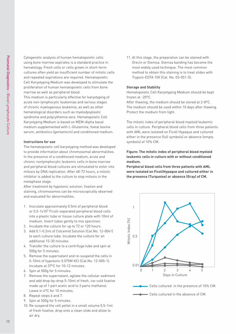
The mitotic index of peripheral blood myeloid leukemic cells in culture. Peripheral blood cells from three patients with AML were isolated on Ficoll Hypaque and cultured either in the presence (full symbols) or absence (empty symbols) of 10% CM.
Figure: The mitotic index of peripheral blood myeloid leukemic cells in culture with or without conditioned medium . Peripheral blood cells from three patients with AML were isolated on FicollHypaque and cultured either in the presence (Turquoise) or absence (Gray) of CM.
Mito
tic In
dex
(%)
Days in Culture
Cells cultured in the presence of 10% CM.
Cells cultured in the absence of CM.
1
0.5
0.010 31 42 5
11
EZ Lympho-Sep™
Preparation of separation tubes is time consuming and their application is technically difficult. The EZ Lympho-Sep™ provide, a competitively priced ready-for-use alternative to the “home made” blood separation tube.
Principal areas requiring lymphocyte assessment
• The determination of malignant proliferation• Suspected deficiency of the immune system• Wide variety of acute and chronic diseases associated
with evidence of some alteration in the immune system
Isolation of lymphocytes from peripheral blood
The most commonly used procedure is for separation of the mononuclear cells by density gradient centrifugation of whole blood. This procedure is performed by carefully layering diluted whole blood over a polysucrose - sodium metrizoate medium (Ficoll-Paque, Lymphoprep, Histopaque, etc). The diluted blood is added to the gradient by gently pipetting with the tubes held at an angle or by pouring the blood onto the separation medium. This latter method requires considerable practice and is not recommended for beginners. To obtain good separations, it is critical that a clear separation be kept between the dense polysucrose - metrizoate and the blood layer before centrifugation. Due to the extreme care that must be exercised when pipetting blood on to the separation medium, alternative methods have been devised. In one such procedure a sample of the diluted blood is placed in a centrifuge tube and Ficoll-Paque (or equivalent separation medium with a density of 1.077) is added by being underlayered under the blood. This method creates cleaner interfaces than those obtained when blood is layered over the Ficoll, since there is less disturbance of the surface of the Ficoll and less mixing.
Unique density gradient separation of lymphocytes from whole blood
Density gradient centrifugation of whole blood on a polysucrose - sodium metrizoate medium is the method of choice for isolation of lymphocytes. The success of the procedure, i.e. the recovery of viable lymphocytes with the lowest proportion of contaminating granulocytes and erythrocytes, depends to a large extent on the careful layering of the blood sample onto the separation medium and the maintenance of a sharp interface between the two solutions prior to centrifugation. The EZ Lympho-Sep™ system allows the blood sample to be poured directly into the centrifuge tube with no special
precautions required to prevent disruption of the polysucrose - sodium metrizoate layer. Thus, a large number of samples may be handled at the same time. The mechanism also reduces the length of centrifugation time required for separation of the lymphocytes.
Features:• Ready-to-use, sterile.• Safe method, minimum contact with biological fluids.• Time saver, quick and easy sample filling.• Maximum yield of viable mononuclear cells.• A large number of samples may be handled at the same
time
EZ Lympho-Sep™ tubes are manufactured with either 2 or 3 ml separation medium in 15 ml centrifuge tubes or with either 10 or 15 ml separation medium in 50 ml centrifuge tubes. For laboratories that have stocks of Ficoll or Lymphoprep, the tubes can be ordered empty for charging with separation medium immediately before use.
Ready-for-use cell separation tubes
The heart of the EZ Lympho-Sep™ is a plastic insert that allows the blood sample to be poured directly into the tube alleviating the need for slow and careful addition of the blood. Secondly, a one-way feature of the insert allows passage of materials during the centrifugation step but prevents the flow of the separation medium during shipping. After centrifugation, if desired, the upper lymphocyte-containing fraction may be poured off without risk of contamination from the erythrocytes, which are trapped under the insert.
Postnatal D
iagnostics - Blood Lym
phocyte Culture
Lymphocyte Separation Tubes
12
Postnatal D
iagnostics - Blood Lym
phocyte Culture
Buffy Booster for EZ Lympho-Sep™
Catalog No. 01-899-U15
DENSITY GRADIENT LYMPHOCYTE SEPARATION FROM BIOLOGICAL FLUIDS HAVING LOW RED CELL CONTENT
Critically important to the function of ready-to-use lymphocyte separation tubes (such as EZ Lymph-Sep™) is the hematocrit, i.e. the volume of the blood fluid occupied by the red blood cells. It is this mass of cells that during the centrifugation procedure displaces the sodium metrizoate – polysucrose so that it rises above the centrifugation device to form a density layer at which the white cells collect. In cases where sufficient red cells are not present, the sodium metrizoate - polysucrose interface forms at or even below the level of the separation device. In this situation recovery of the white cell fraction may not be possible. Diluted whole blood, buffy coat (enriched white cell fraction), bone marrow, lymph and spinal fluid are all examples of this type of biological fluids. When there is not sufficient red cell mass in the sample itself to displace the sodium metrizoate - polysucrose to the required level, extra mass must be added. Buffy Booster, a dense inert liquid, immiscible in water, is added prior to centrifugation. Centrifugal force causes Buffy Booster to sink to the bottom of the tube. Any red blood cells present form a sedimentation layer on top of the Buffy Booster (the red cell layer and Buffy Booster do not mix). The volume of sodium metrizoate - polysucrose displaced upwards is equal to the combined volumes of the Buffy Booster and the sedimented red cells. No contamination with red cells or Buffy Booster is possible even when the whole upper plasma layer is poured off. The red cells do not pass the separation device, while the Buffy Booster is blocked in by the red cell layer.
13
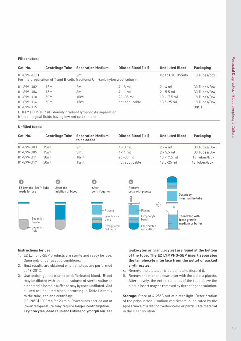
Filled tubes:
Cat. No. Centrifuge Tube Separation Medium Diluted Blood (1:1) Undiluted Blood Packaging
01-899- - U0 1 2ml Up to 8 X 10⁷cells 10 Tubes/boxFor the preparation of T and B cells fractions : Uni-sorb nylon wool column .
01-899-U02 15ml 2ml 4 - 8 ml 2 - 4 ml 30 Tubes/Box01-899-U04 15ml 3ml 4-11 ml 2 - 5.5 ml 30 Tubes/Box01-899-U10 50ml 10ml 20 -35 ml 10 -17.5 ml 18 Tubes/Box01-899-U16 50ml 15ml not applicable 18.5-25 ml 18 Tubes/Box01-899-U15 3 / KIT BUFFY BOOSTER KIT density gradient lymphocyte separation from biological fluids having low red cell content
Unfilled tubes:
Cat. No. Centrifuge Tube Separation Medium Diluted Blood (1:1) Undiluted Blood Packagingto be added
01-899-U03 15ml 2ml 4 - 8 ml 2 - 4 ml 30 Tubes/Box01-899-U05 15ml 3ml 4-11 ml 2 - 5.5 ml 30 Tubes/Box01-899-U11 50ml 10ml 20 -35 ml 10 -17.5 ml 18 Tubes/Box01-899-U17 50ml 15ml not applicable 18.5-25 ml 18 Tubes/Box
Instructions for use:1. EZ Lympho-SEP products are sterile and ready for use.
Open only under aseptic conditions.2. Best results are obtained when all steps are performed
at 18-20°C.3. Use anticoagulant treated or defibrinated blood. Blood
may be diluted with an equal volume of sterile saline orother sterile isotonic buffer or may by used undiluted. Adddiluted or undiluted blood, according to Table I directlyto the tube, cap and centrifuge(18-20°C) 1000 x g for 20 min. Procedures carried out atlower temperature may require longer centrifugation.Erythrocytes, dead cells and PMNs (polymorph nuclear
leukocytes or granulocytes) are found at the bottom of the tube. The EZ LYMPHO-SEP insert separates the lymphocyte interface from the pellet of packed erythrocytes.
4. Remove the platelet-rich plasma and discard it.5. Remove the mononuclear layer with the aid of a pipette.
Alternatively, the entire contents of the tube above theplastic insert may be removed by decanting the solution.
Storage: Store at 4-25°C out of direct light. Deterioration of the polysucrose - sodium metrizoate is indicated by the appearance of a distinct yellow color or particulate material in the clear solution.
EZ Lympho-Sep™ Tubeready for use
1 2 3 4
After theaddition of blood
After centrifugation
Plasma
LymphocytebandSepartion
devicePrecipitatedred cellsSepartion
fluid
Plasma
Lymphocyteband
Precipitatedred cells
Decant by inverting the tube
Removecells with pipette
Then wash with fresh growth medium or buffer
or
Postnatal D
iagnostics - Blood Lym
phocyte Culture
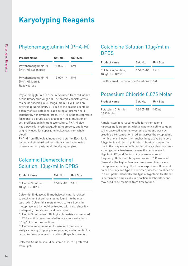
Phytohemagglutinin M (PHA-M)
Phytohemagglutinin is a lectin extracted from red kidney beans (Phaseolus vulgaris). The protein consists of two molecular species, a leucoagglutinin (PHA-L) and an erythroagglutinin (PHA-E). Each of the proteins contains a family of five isolectins, each being a tetramer held together by noncovalent forces. PHA-M is the mucoprotein form and is a crude extract used for the stimulation of cell proliferation in lymphocyte culture. PHA-M also has a powerful erythroagglutinating property and it was originally used for separating leukocytes from whole blood.PHA-M from Biological Industries is sterile. Each lot is tested and standardized for mitotic stimulation using primary human peripheral blood lymphocytes.
Colcemid (Demecolcine) Solution, 10μg/ml in DPBS
Colcemid, N-deacetyl-N-methylcolchicine, is related to colchicine, but animal studies found it to be much less toxic. Colcemid arrests mitotic cultured cells in metaphase and it should be treated with care, since it is mutagenic, tumorigenic, and teratogenic.Colcemid Solution from Biological Industries is prepared in PBS and it is recommended to use a concentration of 0.1μg/ml in culture medium.Colcemid is recommended for use in chromosome analysis during lymphocyte karyotyping and amniotic fluid cell chromosome analysis, and in cell synchronization.
Colcemid Solution should be stored at 2-8ºC, protected from light.
Colchicine Solution 10μg/ml in DPBS
Potassium Chloride 0.075 Molar
A major step in harvesting cells for chromosome karyotyping is treatment with a hypotonic saline solution to increase cell volume. Hypotonic solutions work by creating a concentration gradient across the cytoplasmic membrane and water then rushes in by active transport.A hypotonic solution of potassium chloride in water for use in the preparation of blood lymphocyte chromosomes - the hypotonic treatment causes the cells to swell . Hypotonic KCl and Sodium citrate are used most frequently . Both room temperature and 37°C are used . Generally , the higher temperature is used to increase metaphase spreading . The time of exposure will depend on cell density and type of specimen , whether on slides or in a cell pellet . Generally , the type of hypotonic treatment is determined empirically in a particular laboratory and may need to be modified from time to time .
See Colcemid (Demecolcine) Solutions (p.14)
Product Name Cat. No. Unit Size
Phytohemagglutinin-M 12-006-1H 5ml(PHA-M), Lyophilized
Phytohemagglutinin-M 12-009-1H 5ml(PHA-M), Liquid, Ready-to-use
Product Name Cat. No. Unit Size
Potassium Chloride, 12-005-1B 100ml0.075 Molar
Product Name Cat. No. Unit Size
Colchicine Solution, 12-003-1C 25ml10μg/ml in DPBS
Product Name Cat. No. Unit Size
Colcemid Solution, 12-004-1D 10ml10μg/ml in DPBS
Karyotyping Reagents
Karyotyping R
eagents
14
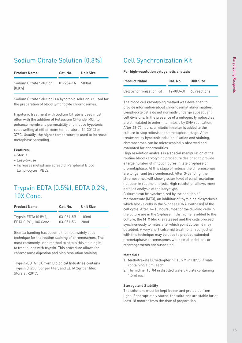
Sodium Citrate Solution (0.8%)
Sodium Citrate Solution is a hypotonic solution, utilized for the preparation of blood lymphocyte chromosomes.
Hypotonic treatment with Sodium Citrate is used most often with the addition of Potassium Chloride (KCl) to enhance membrane permeability and induce hypotonic cell swelling at either room temperature (15-30°C) or 37°C. Usually, the higher temperature is used to increase metaphase spreading.
Features:• Sterile• Easy-to-use• Increases metaphase spread of Peripheral Blood
Lymphocytes (PBL’s)
Trypsin EDTA (0.5%), EDTA 0.2%, 10X Conc.
Giemsa banding has become the most widely used technique for the routine staining of chromosomes. The most commonly used method to obtain this staining is to treat slides with trypsin. This procedure allows for chromosome digestion and high resolution staining.
Trypsin-EDTA 10X from Biological Industries contains Trypsin (1:250) 5gr per liter, and EDTA 2gr per liter.Store at -20ºC.
Cell Synchronization Kit
For high-resolution cytogenetic analysis
The blood cell karyotyping method was developed to provide information about chromosomal abnormalities. Lymphocyte cells do not normally undergo subsequent cell divisions. In the presence of a mitogen, lymphocytes are stimulated to enter into mitosis by DNA replication. After 48-72 hours, a mitotic inhibitor is added to the culture to stop mitosis in the metaphase stage. After treatment by hypotonic solution, fixation and staining, chromosomes can be microscopically observed and evaluated for abnormalities.High resolution analysis is a special manipulation of the routine blood karyotyping procedure designed to provide a large number of mitotic figures in late prophase or prometaphase. At this stage of mitosis the chromosomes are longer and less condensed. After G-banding, the chromosomes will show greater level of band resolution not seen in routine analysis. High resolution allows more detailed analysis of the karyotype.Cultures can be synchronized by the addition of methotrexate (MTX), an inhibitor of thymidine biosynthesis which blocks cells in the S-phase (DNA synthesis) of the cell cycle. After 16-18 hours, most of the dividing cells in the cuture are in the S-phase. If thymidine is added to the culture, the MTX block is released and the cells proceed synchronously to mitosis, at which point colcemid may be added. A very short colcemid treatment in conjuction with this technique may be used to produce extended prometaphase chromosomes when small deletions or rearrangements are suspected.
Materials 1. Methotrexate (Amethopterin), 10-5M in HBSS: 4 vials
containing 1.5ml each2. Thymidine, 10-3M in distilled water: 4 vials containing
1.5ml each
Storage and Stability The solutions must be kept frozen and protected from light. If appropriately stored, the solutions are stable for at least 18 months from the date of preparation.
Product Name Cat. No. Unit Size
Trypsin EDTA (0.5%), 03-051-5B 100mlEDTA 0.2% , 10X Conc. 03-051-5C 20ml
Product Name Cat. No. Unit Size
Sodium Citrate Solution 01-934-1A 500ml(0.8%)
Product Name Cat. No. Unit Size
Cell Synchronization Kit 12-008-60 60 reactions
Karyotyping R
eagents
15
Product Name Cat. No. Size
EZ-PCR Mycoplasma 20-700-20 20 reactionsTest Kit
Product Name Cat. No. Size
BIOMYC-1 Antibiotic Solution 03-036-1D 10 ml100X (Tiamutin) 03-036-1C 20 ml
03-036-1B 100 ml
BIOMYC-2 Antibiotic Solution 03-037-1 D 10 ml100X (Minocycline) 03-037-1 C 20 ml
03-037-1 B 100 ml
BIOMYC-3 Antibiotic Solution 03-038-1D 10 ml100X (Ciprofloxacin) 03-038-1 C 20 ml
03-038-1 B 100 ml
Keep your lab clean
16
Keep your lab cleanMycoplasma Detection and Treatement
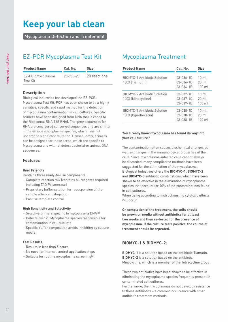
EZ-PCR Mycoplasma Test Kit
DescriptionBiological Industries has developed the EZ-PCR Mycoplasma Test Kit. PCR has been shown to be a highly sensitive, specific and rapid method for the detection of mycoplasma contamination in cell cultures. Specific primers have been designed from DNA that is coded to the Ribosomal RNA(16S RNA). The gene sequences for RNA are considered conserved sequences and are similar in the various mycoplasma species, which have not undergone significant mutation. Consequently, primers can be designed for these areas, which are specific to Mycoplasma and will not detect bacterial or animal DNA sequences.
Features
User Friendly Contains three ready-to-use components:- Complete reaction mix (contains all reagents required
including TAQ Polymerase)- Proprietary buffer solution for resuspension of the
sample after centrifugation- Positive template control
High Sensitivity and Selectivity - Selective primers specific to mycoplasma DNA(1) - Detects over 20 Mycoplasma species responsible for
contamination in cell cultures - Specific buffer composition avoids inhibition by culture
media
Fast Results- Results in less than 5 hours- No need for internal control application steps- Suitable for routine mycoplasma screening(2)
Mycoplasma Treatment
You already know mycoplasma has found its way into your cell culture?
The contamination often causes biochemical changes as well as changes in the immunological properties of the cells. Since mycoplasma-infected cells cannot always be discarded, many complicated methods have been suggested for the elimination of the mycoplasma. Biological Industries offers the BIOMYC-1, BIOMYC-2 and BIOMYC-3 antibiotic combinations, which have been shown to be effective in the elimination of mycoplasma species that account for 90% of the contaminations found in cell cultures. When using according to instructions, no cytotoxic effects will occur.
On completion of the treatment, the cells should be grown on media without antibiotics for at least two weeks and then re-tested for the presence of mycoplasma. If the culture tests positive, the course of treatment should be repeated.
BIOMYC-1 & BIOMYC-2:
BIOMYC-1 is a solution based on the antibiotic Tiamutin. BIOMYC-2 is a solution based on the antibiotic Minocycline, which is a member of the Tetracycline group.
These two antibiotics have been shown to be effective in eliminating the mycoplasma species frequently present in contaminated cell cultures. Furthermore, the mycoplasmas do not develop resistance to these antibiotics – a common occurrence with other antibiotic treatment methods.
Keep your lab clean
17
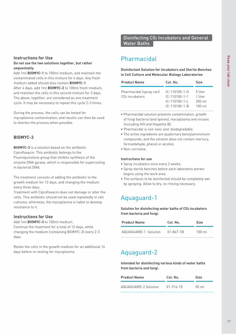
Pharmacidal
Disinfectant Solution for Incubators and Sterile Benches in Cell Culture and Molecular Biology Laboratories
• Pharmacidal solution prevents contamination , growthof fungi bacteria ( and spores ), mycoplasma and viruses( including HIV and Hepatitis B ) .
• Pharmacidal is non-toxic and biodegradable .• The active ingredients are quaternary benzylammonium
compounds , and the solution does not contain mercury , formaldehyde , phenol or alcohol.
• Non-corrosive .
Instructions for use• Spray incubators once every 2 weeks.• Spray sterile benches before each laboratory worker
begins using the work area.• The surfaces to be disinfected should be completely wet
by spraying . Allow to dry , no rinsing necessary.
Aquaguard-1
Solution for disinfecting water baths of CO2 incubators from bacteria and fungi.
Aquaguard-2
Intended for disinfecting various kinds of water baths from bacteria and fungi.
Product Name Cat. No. Size
Pharmacidal ( spray can ) IC-110100-1-G 5 literCO2 incubators IC-110100-1-1 1 liter
IC-110100-1-L 250 mlIC-110100-1-B 100 ml
Product Name Cat. No. Size
AQUAGUARD-1 Solution 01-867-1B 100 ml
Disinfecting C02 Incubators and General Water Baths
Instructions for Use Do not use the two solutions together, but rather sequentially. Add 1ml BIOMYC-1 to 100ml medium, and maintain the contaminated cells in this mixture for 4 days. Any fresh medium added should also contain BIOMYC-1. After 4 days, add 1ml BIOMYC-2 to 100ml fresh medium, and maintain the cells in this second mixture for 3 days. The above, together, are considered as one treatment cycle. It may be necessary to repeat this cycle 2-3 times.
During the process, the cells can be tested for mycoplasma contamination, and results can then be used to shorten the process when possible.
BIOMYC-3
BIOMYC-3 is a solution based on the antibiotic Ciprofloxacin. This antibiotic belongs to the Fluoroquinolone group that inhibits synthesis of the enzyme DNA gyrase, which is responsible for supercoiling in bacterial DNA.
The treatment consists of adding the antibiotic to the growth medium for 12 days, and changing the medium every three days. Treatment with Ciprofloxacin does not damage or alter the cells. This antibiotic should not be used repeatedly in cell cultures; otherwise, the mycoplasma is liable to develop resistance to it.
Instructions for Use Add 1ml BIOMYC-3 to 100ml medium. Continue the treatment for a total of 12 days, while changing the medium (containing BIOMYC-3) every 2-3 days.
Retain the cells in the growth medium for an additional 14 days before re-testing for mycoplasma.
Product Name Cat. No. Size
AQUAGUARD-2 Solution 01-916-1E 50 ml
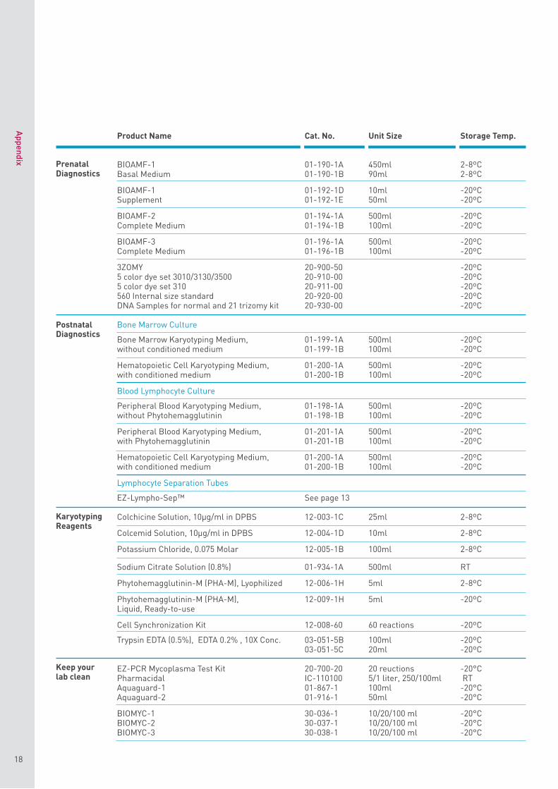
Product Name Cat. No. Unit Size Storage Temp.
BIOAMF-1 01-190-1A 450ml 2-8ºCBasal Medium 01-190-1B 90ml 2-8ºC
BIOAMF-1 01-192-1D 10ml -20ºCSupplement 01-192-1E 50ml -20ºC
BIOAMF-2 01-194-1A 500ml -20ºCComplete Medium 01-194-1B 100ml -20ºC
BIOAMF-3 01-196-1A 500ml -20ºCComplete Medium 01-196-1B 100ml -20ºC
3ZOMY 20-900-50 -20ºC5 color dye set 3010/3130/3500 20-910-00 -20ºC5 color dye set 310 20-911-00 -20ºC560 Internal size standard 20-920-00 -20ºCDNA Samples for normal and 21 trizomy kit 20-930-00 -20ºC
Bone Marrow Culture
Bone Marrow Karyotyping Medium, 01-199-1A 500ml -20ºCwithout conditioned medium 01-199-1B 100ml -20ºC
Hematopoietic Cell Karyotyping Medium, 01-200-1A 500ml -20ºCwith conditioned medium 01-200-1B 100ml -20ºC
Blood Lymphocyte Culture
Peripheral Blood Karyotyping Medium, 01-198-1A 500ml -20ºCwithout Phytohemagglutinin 01-198-1B 100ml -20ºC
Peripheral Blood Karyotyping Medium, 01-201-1A 500ml -20ºCwith Phytohemagglutinin 01-201-1B 100ml -20ºC
Hematopoietic Cell Karyotyping Medium, 01-200-1A 500ml -20ºCwith conditioned medium 01-200-1B 100ml -20ºC
Lymphocyte Separation Tubes
EZ-Lympho-Sep™ See page 13
Colchicine Solution, 10μg/ml in DPBS 12-003-1C 25ml 2-8ºC
Colcemid Solution, 10μg/ml in DPBS 12-004-1D 10ml 2-8ºC
Potassium Chloride, 0.075 Molar 12-005-1B 100ml 2-8ºC
Sodium Citrate Solution (0.8%) 01-934-1A 500ml RT
Phytohemagglutinin-M (PHA-M), Lyophilized 12-006-1H 5ml 2-8ºC
Phytohemagglutinin-M (PHA-M), 12-009-1H 5ml -20ºCLiquid, Ready-to-use
Cell Synchronization Kit 12-008-60 60 reactions -20ºC
Trypsin EDTA (0.5%), EDTA 0.2% , 10X Conc. 03-051-5B 100ml -20ºC03-051-5C 20ml -20ºC
EZ-PCR Mycoplasma Test Kit 20-700-20 20 reuctions -20°CPharmacidal IC-110100 5/1 liter, 250/100ml RTAquaguard-1 01-867-1 100ml -20°CAquaguard-2 01-916-1 50ml -20°C
BIOMYC-1 30-036-1 10/20/100 ml -20°C BIOMYC-2 30-037-1 10/20/100 ml -20°C BIOMYC-3 30-038-1 10/20/100 ml -20°C
Appendix
18
Prenatal Diagnostics
Karyotyping Reagents
Keep your lab clean
Postnatal Diagnostics

NettaD
avids E29/3 8/13
India Contact:
Life Technologies (India) Pvt. Ltd. 306, Aggarwal City Mall, Opposite M2K Pitampura Delhi – 110034. Ph: +91-11-42208000, 42208111, 42208222 Fax: +91-11-42208444
Email: [email protected] Website: www.lifetechindia.com