Embed Size (px)
Citation preview
A Senior Scholars Thesis
by
KIMBERLY ANN KRENEK
HPLC AND MASS SPECTROSCOPIC CHARACTERIZATION OF
MANGO (Mangifera Indica L.) GALLOTANNINS FOLLOWING
ENZYMATIC HYDROLYSIS
Submitted to the Office of Undergraduate Research
Texas A&M University
in partial fulfillment of the requirements for the designation as
UNDERGRADUATE RESEARCH SCHOLAR
April 2009
Major: Food Science
HPLC AND MASS SPECTROSCOPIC CHARACTERIZATION OF
MANGO (Mangifera Indica L.) GALLOTANNINS FOLLOWING
ENZYMATIC HYDROLYSIS
Approved by:
Research Advisor: Steve Talcott
Associate Dean for Undergraduate Research: Robert C. Webb
A Senior Scholars Thesis
by
KIMBERLY ANN KRENEK
Submitted to the Office of Undergraduate Research
Texas A&M University
in partial fulfillment of the requirements for the designation as
UNDERGRADUATE RESEARCH SCHOLAR
April 2009
Major: Food Science
iii
ABSTRACT
HPLC and Mass Spectroscopic Characterization of Mango (Mangifera indica L.)
Gallotannins Following Enzymatic Hydrolysis. (April 2009)
Kimberly Ann Krenek
Department of Nutrition and Food Science
Texas A&M University
Research Advisor: Dr. Steve Talcott
Department of Nutrition and Food Science
Mangos contain numerous compounds that have been shown to exhibit antioxidant
properties. These compounds, most of which are polyphenolics, are linked to anti-cancer
and anti-inflammatory activities in the body. Mangos more specifically boast a large
number of high molecular weight compounds called gallotannins, composed of gallic
acid units attached to glucose via a glycosidic linkage. It is unknown if these compounds
are broken down into smaller molecules through the normal course of human digestion,
or if food processing operations, such as the addition of a gallotannin-active hydrolyases,
could be more effective in lowering the size of these molecules to increase the
absorption and potential bioactivity. This research focused on understanding the
chemical changes that occur to gallotannins derived from mangos following enzymatic
hydrolysis and attempted to draw inferences relating to overall human health.
Polyphenolics in mangos, cv. Ataulfo were extracted using a 1:1:1
acetone:ethanol:methanol mixture and further concentrated and clarified using a reverse-
iv
phase C18 Sep-Pak cartridge. Mango extracts were treated with 20,000 U/ml and
13,000U/ml β-glucosidase with a time course of 2, 4, 6, and 8 hours in an optimal pH 5.0
citric acid buffer, and at a constant temperature of 35◦ C. Changes in mango
polyphenolics following enzyme hydrolysis were monitored using a Thermo Finnigan
LCQ Deca XP Max MSn ion trap mass spectrometer equipped with an ESI ion source.
Β-glucosidase proved to be effective in the hydrolysis of some gallotannins but was
incapable of hydrolyzing all gallotannins into free gallic acid. This was illustrated by the
observance of an increase in penta, hexa, hepta-O- and a subsequent decrease in higher
molecular weight compounds. The limitations for complete hydrolysis explains by the
inability of β-glucosidase to cleave the glycosidic linkage due to steric hindrance created
from having up to five gallic acid moieties attached to glucose, or from the inability of
the enzyme to break m-dipside linkages between two or more galloyl groups. Incubating
mango extract with both 20,000 U/mL and 13,000 U/mL resulted in an equivalent eight-
fold increase in free gallic acid. Enzyme concentration was not the limiting factor in the
hydrolytic reaction. Additionally, reaction time did not have a significant role in the
hydrolytic rate, as the amount of free gallic remained relatively constant from 2 to 8
hours. These findings indicated that it was possible to increase low molecular weight
gallotannin species following enzyme hydrolysis and will aid in future studies to
understand the digestion and bioavailability mango phenolics.
v
ACKNOWLEDGMENTS
I would like to first thank my research advisor Dr. Steve Talcott for his constant support
and guidance. He was instrumental in molding me into the scientist and researcher that I
am becoming, and I am thankful for all the opportunities he gave me to become a great
food scientist. I have learned more in the time I spent in his lab then I have in many
other classes combined.
I would also like to thank my labmates, Lisbeth, Kim, Jorge, Chris, and Michelle. They
each helped me understand what it means to research and the patience it requires. I am
thankful for the support they provided and even more thankful for the friendship they
gave.
I would like to thank my friends, especially, Stephanie, Angie, Jamie, Kristin, Chris,
Stephen, and Liz for their encouragement and uplifting words when things seemed a
little rough. They were a constant reminder of the bigger picture and how to get through
it all.
Lastly, but more importantly, I want to thank my family for being my biggest fans. I
could not have asked for more love and encouragement from anyone. Thank you for
supporting my dreams and being so interested in my work. Amanda, Hannah, and
Colleen, thank you for being amazing sisters and serving as inspiration to me.
vi
NOMENCLATURE
CV Cultivar
L.M.W Low Molecular Weight
H.M.W High Molecular Weight
amu Atomic Mass Unit
vii
TABLE OF CONTENTS
Page
ABSTRACT .................................................................................................................. iii
ACKNOWLEDGMENTS ............................................................................................... v
NOMENCLATURE ...................................................................................................... vi
TABLE OF CONTENTS .............................................................................................. vii
LIST OF FIGURES ..................................................................................................... viii
LIST OF TABLES ......................................................................................................... ix
CHAPTER
I INTRODUCTION: INCREASING MANGO BIOAVAILABILITY ....... 1
Mango fruit overview ................................................................... 1
Tannins ........................................................................................ 2
II METHODOLOGY .................................................................................. 5
Mango extraction ......................................................................... 5
Enzyme hydrolysis ....................................................................... 6
Chemical analysis......................................................................... 6
III RESULTS AND DISCUSSIONS ............................................................ 8
Polyphenolic characterization ....................................................... 8
Enzymatic hydrolysis ................................................................. 10
IV SUMMARY AND CONCLUSIONS ..................................................... 14
REFERENCES ............................................................................................................. 15
CONTACT INFORMATION ....................................................................................... 17
viii
LIST OF FIGURES
FIGURE Page
1 Chromatograph of mango polyphenolics cv. Ataulfo before enzymatic
hydrolysis. .......................................................................................................... 9
2 Chromatograph of mango polyphenolics cv. Ataulfo after enzymatic
hydrolysis with 13,000 U/mL β-glucosidase ..................................................... 10
3 Chromatograph of mango polyphenolics cv. Ataulfo after enzymatic
hydrolysis with 20,000 U/mL β-glucosidase ..................................................... 11
4 The effect of β-glucosidase concentration on the hydrolysis of mango
polyphenolics ................................................................................................... 13
5 The effects incubation time had on mango polyphenolic hydrolysis .................. 13
ix
LIST OF TABLES
TABLE Page
1 Change in identified mango polyphenolic compounds after enzyme hydrolysis ............ 9
1
CHAPTER I
INTRODUCTION: INCREASING MANGO BIOAVAILABILITY
Mango fruit overview
Mango (Mangifera indica L.) is a popular tropical fruit because of its unique taste,
affordability and nutritional qualities. Mangoes are members of the family
Anacardiaceae and are predominately grown in tropical and warm sub-tropical climates
in places such as Asia, Africa, and the Americas. There are over one thousand different
cultivars of mangos growing world-wide and mangos are considered to be one of the
most widely eaten fruits. The mango is known to be an excellent source of many
vitamins such as ascorbic acid, thiamine, riboflavin, and niacin, and ß-carotene. Not only
are mangos rich in these nutrients, mangos are also high in non-nutrient phytochemical
compounds. Recently much attention has been given to phytochemicals and the
distinctive roles they play in anti-inflammatory and anti-cancer properties related to the
consumption of fruits and vegetables. A need exists to chemically identify hydrolytic by-
products from mango polyphenolics in an effort to eventually relate these compounds to
the absorption of mango polyphenolics following human consumption.
_______________
This thesis follows the style of Journal of Food Science.
2
Mango phytochemicals
Extensive research has been conducted to quantify and characterize compounds from
kernels, peels, and leaves of the mango (Barreto and others 2008; Berardini and others
2004). Mango pulp has proven to be more difficult however, and studies have been on-
going to identify unambiguously compounds located in the pulp. Reports suggest that the
phytochemical content of mango pulp consists of gallic acid, mangiferin, quercetin
glycosides and many identified and uncharacterized hydrolyzable tannins (Schieber and
others 2000). Other compounds in smaller concentrations include p-OH-benzoic acid, m-
coumaric acid, p-coumaric acid, and ferulic acid (Kim and others 2007). In most mango
varieties, free gallic acid, 3,4,5-trihydroxybenzoic acid, is the predominant compound
present and has been shown to possess a high antioxidant capacity with numerous
implications to overall human health (Shanrzad and Bitsch 1998). Gallic acid units
possess three hydroxyl groups and an acid group which allow the compound to link with
another gallic acid to form an ester, digallic acid (Masibo and He 2008). Gallic acid is an
essential component to a group of compounds present in mango pulp called gallotannins.
Tannins
Tannins, in general, are water soluble phenolic secondary metabolites that range in size
from 300 to 3000 Daltons (D) and are classified into four different groups based on
similar structural characteristics. Such groups include gallotannins, ellagitannins,
complex tannins and condensed tannins (Mingshu and others 2006). Tannins are known
for their ability to bind proteins to form either soluble or insoluble complexes
3
(Hangerman and others 1992). Gallotannins are the predominant class of tannins
identified from mango pulp and range in size from 787 – 1243 Daltons (Berardini and
others 2004). Gallotannins consist of a sugar, primarily glucose, surrounded by several
gallic acid units which can further be attached to other gallic acid units via a m-depside
bond (Mueller-Harvey 2001). Research has suggested that the larger a polyphenolic
compound is in size, the less the likelihood for intestinal absorption and subsequent
bioavailability. Therefore, an important area of research is to understand the hydrolytic
products of gallotannins as a means of understanding their absorption and bioactivity of
the mango as a whole.
Enzyme treatment
Only by first evaluating enzyme hydrolysis techniques and understanding how these
processes relate to the size and chemical composition of the resultant molecule can a
later assessment of reduced molecular weight be made on potential health benefits of
mangos. It is known that certain bacteria, yeasts, and molds produce enzymes that are
capable of breaking down these gallotannins (Mingshu and others 2006). β-glucosidases
can also be found in the epithelial cells of the small intestine where it is thought that
sugar-linked molecules are broken into smaller units that are subsequently absorbed by
these cells. (Nemeth and others 2003). Both β-glucosidase and tannase have been used
for hydrolysis of tannins, but further studies need to be completed in this area to
determine the most effective enzyme to use, or if a combination of the two enzymes
could be more effective (Kikuzaki and others 2000). It is therefore hypothesized that
4
hydrolysis of gallotannins via the addition of β-glucosidase will increase the
concentration of free gallic acid, as well as lower molecular weight polyphenolics, and
will potentially enhance the bioavailability of mango polyphenolics. Quantifying and
characterizing the gallotannins and their hydrolytic by-products will aid in future
research with a goal of understanding industrial food processing techniques that will aid
in better or more efficient uses of nutritionally dense foods such as mango.
5
CHAPTER II
METHODOLOGY
Mango extraction
Whole mango fruits, Mangifera indica L., cv. Ataulfo, were received in May and
allowed to fully ripen. Upon ripening, peels and kernels were removed. Pulp was
blended and stored at -20◦C until needed.
Polyphenolic extraction
To extract polyphenolic compounds from mango pulp, 200 g of mango pulp was placed
in a beaker with 600 mL of 1:1:1 acetone, methanol, and ethanol (v/v/v). The extract
was initially filtered through cheesecloth to remove large particles and subsequently
filtered through diatomaceous earth. This process was repeated twice, using the pulp
remaining on the cheesecloth in the first filtrations and extracted in 400mL, 200mL,
respectively. The extraction solvents were evaporated under reduced pressure at < 45◦C
using a rotary evaporator. Methanol was added to the extract and the extract was
centrifuged to precipitate pectin. The supernant was collected, precipitate re-extracted,
and methanol was again evaporated under reduced pressure at <45◦C. The mango extract
was washed through a Waters Sep-Pak® Vac 35cc 10g C18 cartridge to remove sugars,
residual pectin, and to concentrate phenolic compounds. Bound phenolic compounds
were fractioned using 25% methanol and 100% methanol to separate (LMW) low
molecular weight compounds from high molecular weight compounds based on their
6
affinity to C18. Each extract was brought to a total volume of 100 mL in a pH 5.0 buffer
solution.
Enzyme hydrolysis
The enzyme β-glucosidase was purchased from MP Biomedicals and stored at -20◦C. An
enzyme solution was prepared immediately prior to incubation in a pH 5.0 buffer with a
concentration of 13,000 U/mL and 20,000 U/ml. One unit (U) of β-glucosidase will
liberate 1.0µmol of glucose per minute at 35◦C. Ataulfo mango fractions (stock, L.M.W.,
H.M.W) at a volume of 2.0 mL were treated with 200 µL of the prepared beta-
glucosidase enzyme solution. The samples were incubated following a time course at
35◦C, with samples pulled at 30 minutes, 1, 2, and 4 hrs. Following incubation, each
sample was acidified to pH 3.0 to inactivate β-glucosidase.
Chemical analysis
Changes in mango polyphenolics following enzyme hydrolysis were monitored
using a Thermo Finnigan LCQ Deca XP Max MSn ion trap mass spectrometer equipped
with an ESI ion source (ThermoFisher, San Jose, CA). Separation of compounds was
completed with a Dionex (Sunnydale, CA) Acclaim® 120 A, (4.6 X 250 mm; 5µM).
Mobile phases used included 0.5% formic acid in water (solvent A), and 0.5% formic
acid in acetonitrile (solvent B) at 0.4 mL/min. The mobile phases were run in a gradient
program and began with 100% A for 5 minutes. Solvent B was changed from 0-30% in
7
25 minutes, to 50% in 15 minutes, to 100% in 25 minutes, back to 0% in 2 minutes. The
gradient run was finished with 100% A for 8 minutes.
8
CHAPTER III
RESULTS AND DISCUSSIONS
Polyphenolic characterization
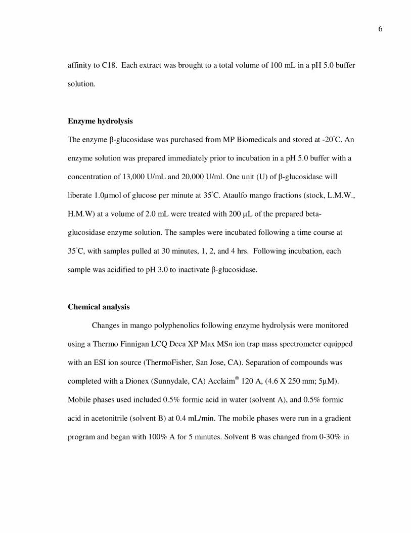
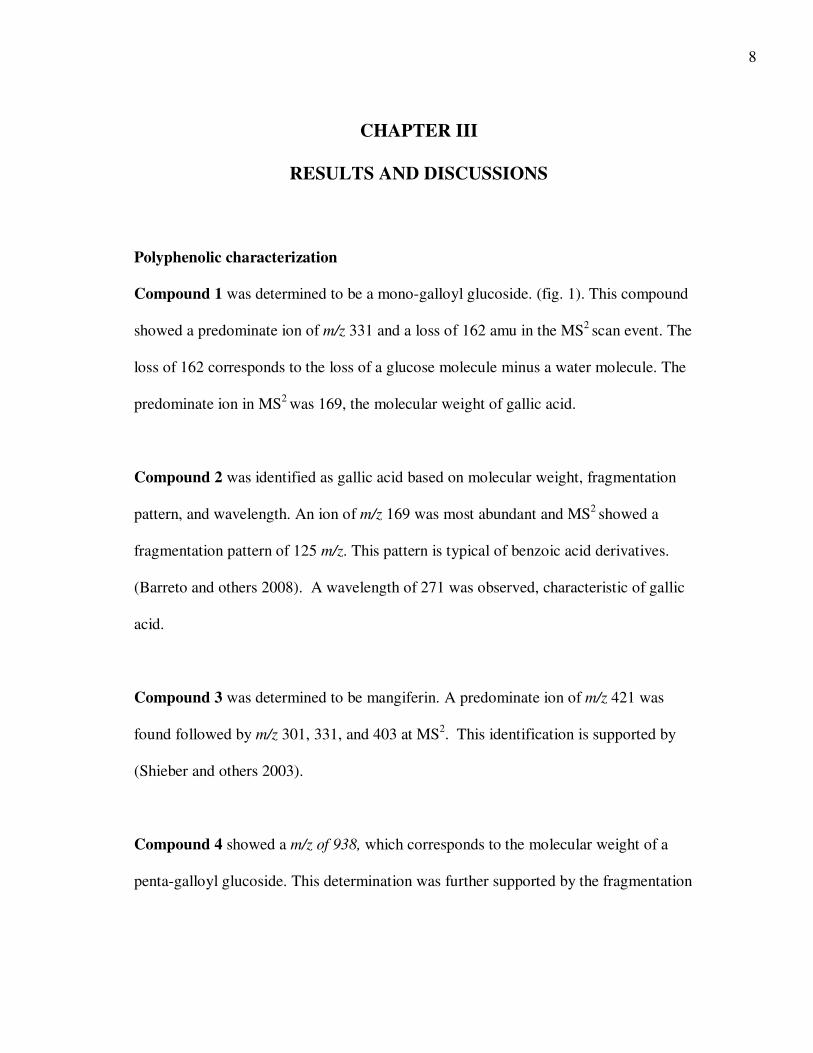
Compound 1 was determined to be a mono-galloyl glucoside. (fig. 1). This compound
showed a predominate ion of m/z 331 and a loss of 162 amu in the MS2 scan event. The
loss of 162 corresponds to the loss of a glucose molecule minus a water molecule. The
predominate ion in MS2 was 169, the molecular weight of gallic acid.
Compound 2 was identified as gallic acid based on molecular weight, fragmentation
pattern, and wavelength. An ion of m/z 169 was most abundant and MS2 showed a
fragmentation pattern of 125 m/z. This pattern is typical of benzoic acid derivatives.
(Barreto and others 2008). A wavelength of 271 was observed, characteristic of gallic
acid.
Compound 3 was determined to be mangiferin. A predominate ion of m/z 421 was
found followed by m/z 301, 331, and 403 at MS2. This identification is supported by
(Shieber and others 2003).
Compound 4 showed a m/z of 938, which corresponds to the molecular weight of a
penta-galloyl glucoside. This determination was further supported by the fragmentation
9
pattern observed at MS2. The predominate ion was 768 followed by 787 m/z. A
molecular weight of 787 is the known molecular weight of a tetra-galloyl glucoside.
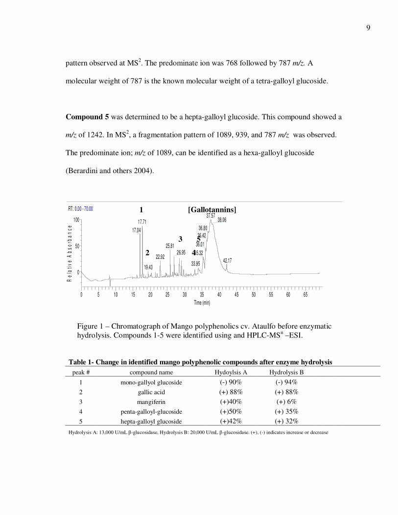
Compound 5 was determined to be a hepta-galloyl glucoside. This compound showed a
m/z of 1242. In MS2, a fragmentation pattern of 1089, 939, and 787 m/z was observed.
The predominate ion; m/z of 1089, can be identified as a hexa-galloyl glucoside
(Berardini and others 2004).
Table 1- Change in identified mango polyphenolic compounds after enzyme hydrolysis
peak # compound name Hydoylsis A Hydrolysis B
1 mono-gallyol glucoside (-) 90% (-) 94%
2 gallic acid (+) 88% (+) 88% 3 mangiferin (+)40% (+) 6%
4 penta-galloyl-glucoside (+)50% (+) 35% 5 hepta-galloyl glucoside (+)42% (+) 32%
Hydrolysis A: 13,000 U/mL β-glucosidase, Hydrolysis B: 20,000 U/mL β-glucosidase. (+), (-) indicates increase or decrease
RT: 0.00 - 70.00
0 5 10 15 20 25 30 35 40 45 50 55 60 65
Time (min)
0
50
100
Re
lati
ve
Ab
so
rba
nc
e
37.5738.0617.71
36.8017.0436.42
36.0125.8126.95 35.32
22.9242.17
33.9519.43
1
2
3
4
5
[Gallotannins]
Figure 1 – Chromatograph of Mango polyphenolics cv. Ataulfo before enzymatic
hydrolysis. Compounds 1-5 were identified using and HPLC-MSn –ESI.
10
Enzymatic hydrolysis
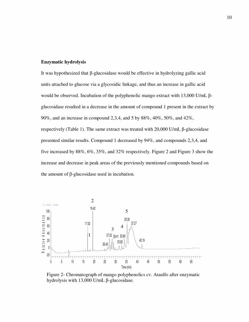
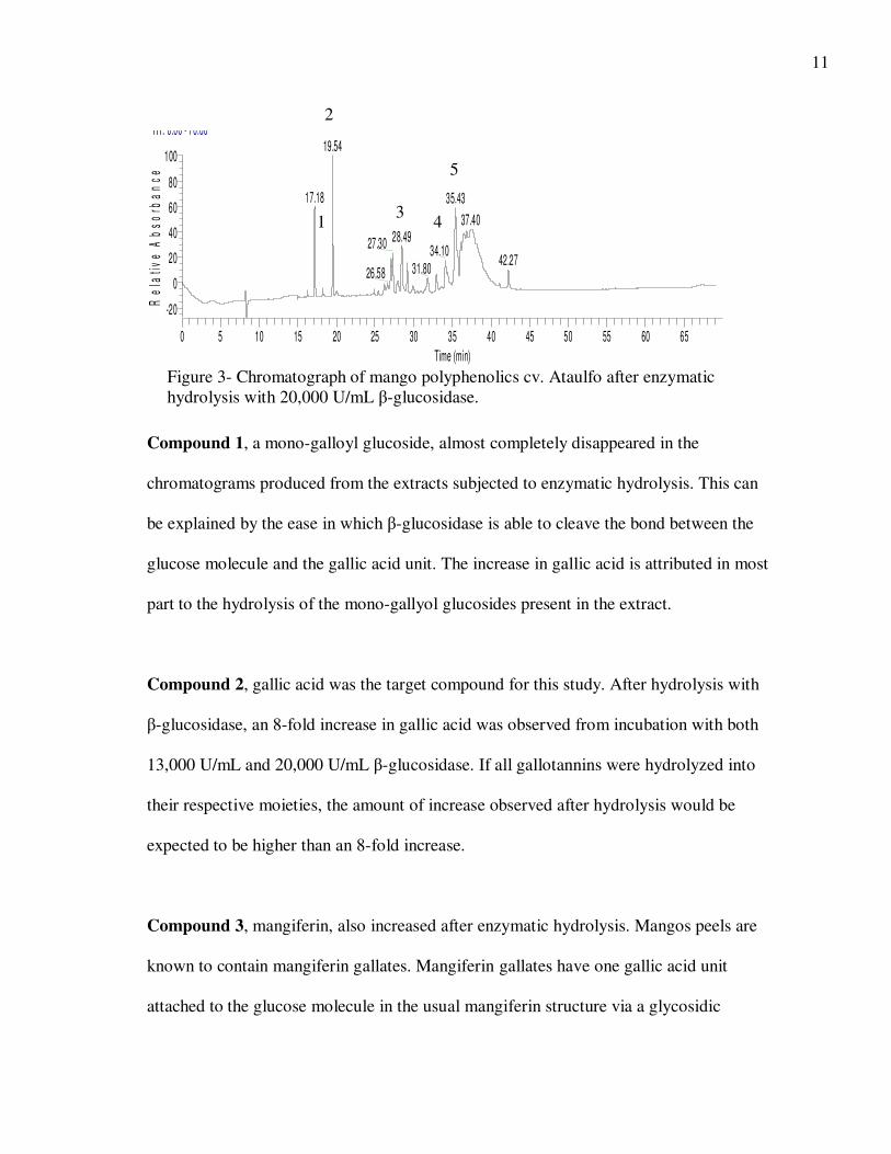
It was hypothesized that β-glucosidase would be effective in hydrolyzing gallic acid
units attached to glucose via a glycosidic linkage, and thus an increase in gallic acid
would be observed. Incubation of the polyphenolic mango extract with 13,000 U/mL β-
glucosidase resulted in a decrease in the amount of compound 1 present in the extract by
90%, and an increase in compound 2,3,4, and 5 by 88%, 40%, 50%, and 42%,
respectively (Table 1). The same extract was treated with 20,000 U/mL β-glucosidase
presented similar results. Compound 1 decreased by 94%, and compounds 2,3,4, and
five increased by 88%, 6%, 35%, and 32% respectively. Figure 2 and Figure 3 show the
increase and decrease in peak areas of the previously mentioned compounds based on
the amount of β-glucosidase used in incubation.
RT: 0.00 - 70.00
0 5 10 15 20 25 30 35 40 45 50 55 60 65
Time (min)
-20
0
20
40
60
80
100
Re
lati
ve
Ab
so
rba
nc
e
19.42
35.3037.2717.02
27.02 33.9928.4142.1932.83
26.45
Figure 2- Chromatograph of mango polyphenolics cv. Ataulfo after enzymatic
hydrolysis with 13,000 U/mL β-glucosidase.
1
2
3 4
5
11
Compound 1, a mono-galloyl glucoside, almost completely disappeared in the
chromatograms produced from the extracts subjected to enzymatic hydrolysis. This can
be explained by the ease in which β-glucosidase is able to cleave the bond between the
glucose molecule and the gallic acid unit. The increase in gallic acid is attributed in most
part to the hydrolysis of the mono-gallyol glucosides present in the extract.
Compound 2, gallic acid was the target compound for this study. After hydrolysis with
β-glucosidase, an 8-fold increase in gallic acid was observed from incubation with both
13,000 U/mL and 20,000 U/mL β-glucosidase. If all gallotannins were hydrolyzed into
their respective moieties, the amount of increase observed after hydrolysis would be
expected to be higher than an 8-fold increase.
Compound 3, mangiferin, also increased after enzymatic hydrolysis. Mangos peels are
known to contain mangiferin gallates. Mangiferin gallates have one gallic acid unit
attached to the glucose molecule in the usual mangiferin structure via a glycosidic
RT: 0.00 - 70.00
0 5 10 15 20 25 30 35 40 45 50 55 60 65
Time (min)
-20
0
20
40
60
80
100R
ela
tiv
e A
bs
orb
an
ce
19.54
17.18 35.43
37.4028.4927.30
34.1042.27
31.8026.58
Figure 3- Chromatograph of mango polyphenolics cv. Ataulfo after enzymatic
hydrolysis with 20,000 U/mL β-glucosidase.
1
2
3 4
5
12
linkage (Barreto and others 2008). This increase in mangiferin can be credited to the
hydrolysis of the previously mentioned mangiferin gallates that are present in lower
concentrations in the pulp of mangos.
Compounds 4 and 5 are both gallotannins that were identifiable. Both the hepta-galloyl
glucosides and the penta-galloyl glucosides increased by 30-50%. This increase in the
middle weight gallotannins can be explained by the ability for β-glucosidase to cleave
only certain strategic bonds on the gallotannin structure, but cannot cleave every bond. A
noticeable decrease in the gallotannin “hump” can be observed. This may be a false
positive for HMW gallotannins hydrolysis because gallotannins have the ability to bind
protein. ( Feldman and others 1998). This binding can be seen as a precipitate formed
during the incubation period and may have resulted in an inaccurate determination of
gallotannin hydrolysis.
Figure 4 illustrates the effect that enzyme concentration had on the amount of hydrolytic
products identified. Incubating the mango extract with 13,000 U/mL β-glucosidase
versus 20,000 U/mL β-glucosidase did not have an effect of hydrolytic products. Figure
5 shows how reaction time affected enzyme hydrolysis. Maximum hydrolysis was
reached between two to four hours of incubation time at 35◦C in the pH 5.0 buffer.
13
-
500,000
1,000,000
1,500,000
2,000,000
2,500,000
3,000,000
identified mango compounds
Pe
ak
Are
amono-galloyl glucoside, ctrl
mono-galloyl glucoside 13,000 U/ml
mono-galloyl glucoside 20,000 U/ml
gallic acid, ctrl
gallic acid, 13,000 U/ml
gallic acid, 20,000 U/ml
penta-galloyl glucoside, ctrl
penta-galloyl glucoside, 13,000 U/ml
penta-galloyl glucoside, 20,000 U/ml
hepta-galloyl glucoside, ctrl
hepta-galloyl glucoside, 13,000 U/ml
hepta-galloyl glucoside, 20,000 U/ml
-
500,000
1,000,000
1,500,000
2,000,000
2,500,000
3,000,000
0 2 4 6 8 10
peak area
incubation time (hrs)
gallic acid 13,000 U/ml
mangiferin 13,000 U/ml
hepta galloyl 13,000 U/ml
penta galloyl 13,000 U/ml
Figure 4 - The effect of β-glucosidase concentration on the hydrolysis of mango
polyphenolics.
Fig. 5 - The effects incubation time had on mango polyphenolic hydrolysis.
14
CHAPTER IV
SUMMARY AND CONCLUSIONS
The hydrolyase enzyme β-glucosidase proved to be effective in the hydrolysis of some
gallotannins, but the hydrolytic reaction was never successfully run to completion. This
was illustrated by the observance of an increase in middle-weight gallotannins, (penta,
hexa, hepta-gallotannins) and a decrease in high molecular weight compounds. The
limitations for complete hydrolysis could be explained by the inability of β-glucosidase
to cleave the beta-linkage due to steric hindrance created from having five gallic acid
moieties attached to glucose. Incubating mango extract with both 20,000 U/mL and
13,000 U/mL resulted in an eight-fold increase in free gallic acid. Therefore, enzyme
concentration did not seem to affect gallotannin hydrolysis when the difference was not
greater than around 10,000 U/mL. Time did not play a significant role in the hydrolysis
of gallotannins, as the amount of gallic cleaved remained relatively stable from 2 to 8
hours. These findings showed an increase in lower molecular weight species following
enzyme hydrolysis and will aid in future studies to understand the digestion and
bioavailability mango phenolics.
15
REFERENCES
Barreto JC, Trevisa MTS, Hull WE, Erben G, deBrito ES, Pfundstein B, Wurtele G,
Spiedgelhalder B, Owen RW. 2008. Characterization and quantitation of
polyphenolic compounds in bark, kernel, leaves, and peel of mango
(Mangifera indica L.) J. Agric. Food Chem. 56(14):5599-5610.
Berardini N, Carle R, Schieber A. 2004. Characterization of gallotannins and
benzophenone derivatives from Mango (Mangifera indica L. cv. “Tommy
Atkins”) peels, pulp and kernels by high-performance liquid
chromatography/electrospray ionization mass spectrometry. Rapid Commun.
Mass Spectrom. 18:2208-2216.
Feldman KS, Sambandam A, Lemon ST, Nicewonger RB, Long GS, Battaglia DF,
Ensel SM, Laci MA. 1998. Binding affinities of gallotannin analogs with
bovine serum albumin: ramifications for polyphenol-protein molecular
recognition. Phythochemistry 51:867-872.
Hangerman AE, Robbins CT, Weerasuriya Y, Wilson TC, McArthur C. 1992. Tannin
chemistry in relation to digestion. J. Range Manage 45:57-62.
Kikuzaki H, Sato A, Mayahara Y, Nakatani N. 2000. Galloylglucosides from berries
of Pimenta dioica. J. Nat. Prod. 63:749-752.
Kim Y, Brecht JK, Talcott ST. 2007. Antioxidant phytochemical and fruit
quality changes in mango (Mangifera indica L.) following hot water
immersion and controlled atmosphere storage. Food Chem 105:1327-1334.
Masibo M, He Q. 2008. Major mango polyphenols and their potential significance to
human health. Comprehensive Reviews in Food Science and Food Safety
7:309-319.
Mingshu L, Kai Y, Qiang H, Dongying J. 2006. Biodegradation of gallotannins
and ellagitannins. J. Basic Microbiol. 46:68-84.
Mueller-Harvey I. 2001. Analysis of hydrolysable tannins. Animal Feed Science
and Technology 91:3-20.
Nemeth K, Plumb GW, Berrin JG, Juge N, Jacob R, Naim HY, Williamson G,
Swallow DM, Kroon PA. 2003. Deglycosylation by small intestinal epithelial
16
cell β-glucosidases is a critical step in the absorption and metabolism of
dietary flavonoid glycosides in humans. Eur. J. Nutr 42:29-42.
Schieber A, Berardini N, Carle R. 2003. Identification of flavonol and xanthone
glycosides from mango (Mangifera indica L. Cv. “Tommy Atkins”) peels by
high-performance liquid chromatography-electrospray ionization mass
spectrometry. J. Agric. Food Chem 51(17):5006-5011.
Schieber A, Ullrich W, Carle R. 2000. Characterization of polyphenols in mango
puree concentrate by HPLC with diode array and mass spectrometric
detection. Innovative Food Science and Emerging Technologies 1:161-166.
Shanrzad S, Bitsch I. 1998. Determination of gallic acid and its metabolites in
human plasma and urine by high-performance liquid chromatography. J.
Chromatogr. Biomed. Sci. Appl. 705:87-95.
17
CONTACT INFORMATION
Name: Kimberly Ann Krenek
Professional Address: c/o Dr. Steve Talcott
Department of Nutrition and Food Science
MS 4227
Texas A&M University
College Station, TX 77843
Email Address: [email protected]
Education: B.S., Food Science, Texas A&M University, May 2009
Magna Cum Laude
Undergraduate Research Scholar


![Spectroscopic Analysis of Poly (Methacrylic Acid-co … · absence of any template molecule [22]. A stock E2 solution was prepared in HPLC grade methanol at a concentration of 1000](https://img.dokumen.tips/doc/110x75/5b933d0009d3f2a22a8ce8b4/spectroscopic-analysis-of-poly-methacrylic-acid-co-absence-of-any-template.jpg)