Embed Size (px)
Citation preview
Zakhary et al. Crit Care (2020) 24:671 https://doi.org/10.1186/s13054-020-03388-2
EDITORIAL
How I approach membrane lung dysfunction in patients receiving ECMOBishoy Zakhary1* , Leen Vercaemst2, Phillip Mason3, Marta V. Antonini4,5, Roberto Lorusso6 and Daniel Brodie7,8
© The Author(s) 2020. Open Access This article is licensed under a Creative Commons Attribution 4.0 International License, which permits use, sharing, adaptation, distribution and reproduction in any medium or format, as long as you give appropriate credit to the original author(s) and the source, provide a link to the Creative Commons licence, and indicate if changes were made. The images or other third party material in this article are included in the article’s Creative Commons licence, unless indicated otherwise in a credit line to the material. If material is not included in the article’s Creative Commons licence and your intended use is not permitted by statutory regulation or exceeds the permitted use, you will need to obtain permission directly from the copyright holder. To view a copy of this licence, visit http://creat iveco mmons .org/licen ses/by/4.0/. The Creative Commons Public Domain Dedication waiver (http://creat iveco mmons .org/publi cdoma in/zero/1.0/) applies to the data made available in this article, unless otherwise stated in a credit line to the data.
IntroductionWith improvements in circuit technology and expanding supportive evidence, extracorporeal membrane oxygena-tion (ECMO) use has grown rapidly over the past decade [1]. Advances in pump and membrane lung (ML) design have led to simpler and more efficient circuits. Circuit-related complications, however, remain frequent and associated with considerable morbidity [2].
Mechanisms of membrane lung dysfunctionThe ML is responsible for oxygen uptake and carbon dioxide removal. The non-biologic surface of the ML activates inflammatory and coagulation pathways with thrombus formation, fibrinolysis, and leukocyte acti-vation [3–5] leading to ML dysfunction. Activation of coagulation and fibrinolysis can precipitate systemic coagulopathy or hemolysis, while clot deposition can obstruct blood flow [6, 7]. Additionally, moisture buildup in the gas phase and protein and cellular debris accu-mulation in the blood phase may contribute to shunt and dead-space physiology, respectively, impairing gas exchange [8, 9]. These three categories—hematologic abnormalities, mechanical obstruction, and inadequate gas exchange—prompt the majority of ML exchanges.
Membrane lung monitoringHematologic profileMonitoring of hematologic variables, including coagula-tion and hemolysis labs, can help identify the develop-ment of an ECMO coagulopathy or hemolysis.
Pressure monitoringThe pressure drop across the ML (ΔP) is measured as (Additional file 1: Supplemental Figure):
where PPre = pre-ML pressure, PPost = post-ML pressure.As clot forms in the ML, increases in resistance (RML)
are reflected as increases in ΔP. To correct for changes in blood flow rate (BFR), monitoring of ΔP normalized for BF rate (ΔP/BFR) more directly reflects RML.
Membrane lung gas transferApplying the Fick principle across the ML, oxygen (O2) transfer may be calculated as:
where V′O2 = O2 transfer across the ML (mL/min), BFR = blood flow rate (L/min), CxO2 = O2 content of (pre-/post-ML) blood (mL/L) for
where Hb = hemoglobin (g/dL), SxO2 = O2 saturation of (pre-/post-ML) blood, PxO2 = O2 partial pressure of (pre-/post-ML) blood (mmHg).
Measurement of V′O2 provides an objective measure of oxygen transfer and can confirm ML dysfunction, when clinically indicated.
Membrane lung dysfunctionPrompt recognition of ML dysfunction is vital for safety, allowing for elective replacement in a controlled manner. On the other hand, replacement of an adequately func-tioning device—requiring temporary cessation of ECMO
�P = PPre − PPost
V′O2 = BFR(CPostO2−CPreO2)
CxO2 = 13.4 ·Hb · SxO2 + 0.03 · PxO2
Open Access
*Correspondence: [email protected] Division of Pulmonary and Critical Care Medicine, Oregon Health and Science University, Portland, OR, USAFull list of author information is available at the end of the article
Page 2 of 4Zakhary et al. Crit Care (2020) 24:671
support—places the patient at unnecessary risk while consuming a limited and expensive resource.
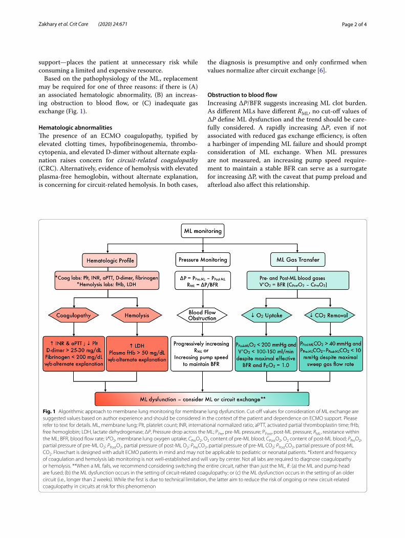
Based on the pathophysiology of the ML, replacement may be required for one of three reasons: if there is (A) an associated hematologic abnormality, (B) an increas-ing obstruction to blood flow, or (C) inadequate gas exchange (Fig. 1).
Hematologic abnormalitiesThe presence of an ECMO coagulopathy, typified by elevated clotting times, hypofibrinogenemia, thrombo-cytopenia, and elevated D-dimer without alternate expla-nation raises concern for circuit-related coagulopathy (CRC). Alternatively, evidence of hemolysis with elevated plasma-free hemoglobin, without alternate explanation, is concerning for circuit-related hemolysis. In both cases,
the diagnosis is presumptive and only confirmed when values normalize after circuit exchange [6].
Obstruction to blood flowIncreasing ΔP/BFR suggests increasing ML clot burden. As different MLs have different RML, no cut-off values of ΔP define ML dysfunction and the trend should be care-fully considered. A rapidly increasing ΔP, even if not associated with reduced gas exchange efficiency, is often a harbinger of impending ML failure and should prompt consideration of ML exchange. When ML pressures are not measured, an increasing pump speed require-ment to maintain a stable BFR can serve as a surrogate for increasing ΔP, with the caveat that pump preload and afterload also affect this relationship.
Fig. 1 Algorithmic approach to membrane lung monitoring for membrane lung dysfunction. Cut-off values for consideration of ML exchange are suggested values based on author experience and should be considered in the context of the patient and dependence on ECMO support. Please refer to text for details. ML, membrane lung; Plt, platelet count; INR, international normalized ratio; aPTT, activated partial thromboplastin time; fHb, free hemoglobin; LDH, lactate dehydrogenase; ΔP, Pressure drop across the ML; PPre, pre-ML pressure; PPost, post-ML pressure; RML, resistance within the ML; BFR, blood flow rate; V′O2, membrane lung oxygen uptake; CPreO2, O2 content of pre-ML blood; CPostO2, O2 content of post-ML blood; PPreO2, partial pressure of pre-ML O2; PPostO2, partial pressure of post-ML O2; PPreCO2, partial pressure of pre-ML CO2; PPostCO2, partial pressure of post-ML CO2. Flowchart is designed with adult ECMO patients in mind and may not be applicable to pediatric or neonatal patients. *Extent and frequency of coagulation and hemolysis lab monitoring is not well-established and will vary by center. Not all labs are required to diagnose coagulopathy or hemolysis. **When a ML fails, we recommend considering switching the entire circuit, rather than just the ML, if: (a) the ML and pump head are fused; (b) the ML dysfunction occurs in the setting of circuit-related coagulopathy; or (c) the ML dysfunction occurs in the setting of an older circuit (i.e., longer than 2 weeks). While the first is due to technical limitation, the latter aim to reduce the risk of ongoing or new circuit-related coagulopathy in circuits at risk for this phenomenon
Page 3 of 4Zakhary et al. Crit Care (2020) 24:671
Inadequate oxygen uptakeWorsening oxygenation during ECMO should prompt quantification of oxygen transfer. When the ML is no longer able to meet patient oxygen demand, ML exchange is indicated. There are three important con-siderations in making this decision.
First, it is necessary that measured V′O2 is truly a maximal value. If circuit BFR is low, for example, the blood will be fully saturated early in the ML path and reserve will exist for additional oxygen transfer as BFR is increased. Similarly, if CPreO2 is artificially elevated, due to high recirculation fraction or impaired tissue extraction, or if the fraction of delivered oxygen in the sweep gas (FDO2) is below 100%, the gradient driving oxygen transfer is reduced, and measured V′O2 may not represent maximal capacity. As such, BFR should be sufficiently high that further increases do not increase arterial saturation, recirculation fraction should be minimized, and ML FDO2 set to 100% to ensure an accurate assessment of maximal V′O2.
Second, though PPost-MLO2 less than 200 mmHg can suggest a failing ML [6], it is vital to calculate V′O2 for confirmation. In the setting of low CPreO2 or high cir-cuit BFR, blood exiting the ML may not be fully satu-rated, with low PPostO2, despite normal V′O2. In this case, if the ML is exchanged, the patient is placed at risk without subsequent improvement in oxygen delivery.
Finally, no absolute values diagnose inadequate oxy-gen transfer and clinical context is important. In gen-eral, however, in a patient with hypoxemia and a ML with maximal V′O2 < 100–150 mL/min, ML exchange is typically indicated.
Inadequate carbon dioxide clearanceML dysfunction can also manifest as inadequate CO2 clearance. Calculation of V′CO2 is not typically per-formed as it varies in a nonlinear fashion with sweep gas flow rate and requires sampling ML exhaust CO2 [10]. However, persistent PPost-MLCO2 greater than 40 mmHg [6] and clearance of less than 10 mmHg PCO2 between pre- and post-ML blood gases despite sweep gas flow rates of 10 L/min or greater is sugges-tive of ML dysfunction and ML exchange should be considered.
Sudden membrane lung failureWhile serial monitoring of the ML may identify markers of dysfunction and allow for elective exchange, acute ML failure is a potentially life-threatening event with unique considerations. Mechanisms to ensure optimal manage-ment are provided in the Additional file 2.
ConclusionThe decision to exchange a ML is complex and without clear guidelines. In this manuscript, we outline a physi-ologic approach to troubleshooting this common yet high risk event.
Supplementary informationSupplementary information accompanies this paper at https ://doi.org/10.1186/s1305 4-020-03388 -2.
Additional file 1. Membrane lung monitoring of pressure drop and oxygen transfer.
Additional file 2. Sudden Membrane Lung Failure.
AbbreviationsECMO: Extracorporeal membrane oxygenation; ML: Membrane lung; COVID-19: Coronavirus disease 2019; ΔP: Pressure drop across the ML; PPre: Pre-ML pressure; PPost: Post-ML pressure; RML: Resistance within the ML; BFR: Blood flow rate; V′O2: Membrane lung oxygen uptake; CPreO2: O2 content of pre-ML blood; CPostO2: O2 content of post-ML blood; Hb: Hemoglobin; SPreO2: Fractional O2 saturation of pre-ML blood; SPostO2: Fractional O2 saturation of post-ML blood; PPreO2: Partial pressure of pre-ML O2; PPostO2: Partial pressure of post-ML O2; V′CO2: CO2 clearance across the ML; ExCO2: ML exhaust CO2; CRC : Circuit-related coagulopathy; FDO2: Membrane lung inlet oxygen fraction; PPreCO2: Partial pressure of pre-ML CO2; PPostCO2: Partial pressure of post-ML CO2; Plt: Platelet count; INR: International normalized ratio; aPTT: Activated partial thromboplastin time; fHb: Free hemoglobin; LDH: Lactate dehydrogenase.
AcknowledgementsNot applicable.
Authors’ contributionsBZ and DB conceived of the presented idea. All authors read and approved the final manuscript.
FundingNone.
Availability of data and materialsNot applicable.
Ethics approval and consent to participateNot applicable.
Consent for publicationNot applicable.
Competing interestsLeen Vercaemst is consultant for Medtronic for conducting/coordinating EMEA region ECMO trainings. Dr. Lorusso is consultant for Medtronic and LivaNova and is on the medical advisory board for Eurosets (all honoraria are paid at the university). Dr. Brodie receives research support from ALung Tech-nologies. He has been on the medical advisory boards for Baxter, Abiomed, Xenios and Hemovent. No other authors report conflicts of interest.
Author details1 Division of Pulmonary and Critical Care Medicine, Oregon Health and Sci-ence University, Portland, OR, USA. 2 Department of Perfusion, University Hospital Gasthuisberg, Leuven, Belgium. 3 Department of Surgery, Brooke Army Medical Center, San Antonio, TX, USA. 4 General ICU, I° Department of Anesthesia and Intensive Care, University Hospital of Parma, Parma, Italy. 5 Department of Biomedical, Metabolic, and Neural Sciences, University of Modena and Reggio Emilia, Modena, Italy. 6 Cardio-Thoracic Surgery Department, Heart and Vascular Centre, Maastricht University Medical Centre (MUMC), Cardiovascular Research Institute Maastricht (CARIM), Maastricht, The Netherlands. 7 Columbia University College of Physicians and Surgeons,
Page 4 of 4Zakhary et al. Crit Care (2020) 24:671
• fast, convenient online submission
•
thorough peer review by experienced researchers in your field
• rapid publication on acceptance
• support for research data, including large and complex data types
•
gold Open Access which fosters wider collaboration and increased citations
maximum visibility for your research: over 100M website views per year •
At BMC, research is always in progress.
Learn more biomedcentral.com/submissions
Ready to submit your researchReady to submit your research ? Choose BMC and benefit from: ? Choose BMC and benefit from:
New York-Presbyterian Hospital, New York, USA. 8 Center for Acute Respiratory Failure, New York-Presbyterian Hospital, New York, NY, USA.
Received: 14 September 2020 Accepted: 13 November 2020
References 1. Brodie D, Slutsky AS, Combes A. Extracorporeal life support for adults
with respiratory failure and related indications: a review. JAMA. 2019;322(6):557–68.
2. Extracorporeal Life Support Organization. ECLS registry report: interna-tional summary. https ://www.elso.org/Regis try/Stati stics /Inter natio nalSu mmary .aspx. Accessed 4 August 2020.
3. Dornia C, Philipp A, Bauer S, Lubnow M, Muller T, Lehle K, et al. Analysis of thrombotic deposits in extracorporeal membrane oxygenators by multidetector computed tomography. ASAIO J. 2014;60(6):652–6.
4. Doyle AJ, Hunt BJ. Current understanding of how extracorporeal mem-brane oxygenators activate haemostasis and other blood components. Front Med (Lausanne). 2018;5:352.
5. Maul TM, Massicotte MP, Wearden PD. ECMO biocompatibility: surface coatings, anticoagulation, and coagulation monitoring. In: Firstenberg
MS, editor. Extracorporeal Membrane Oxygenation: Advances in Therapy. Croatia: IntechOpen; 2016. p. 30–5.
6. Lubnow M, Philipp A, Foltan M, Bull Enger T, Lunz D, Bein T, et al. Technical complications during veno-venous extracorporeal membrane oxygena-tion and their relevance predicting a system-exchange–retrospective analysis of 265 cases. PLoS ONE. 2014;9(12):e112316.
7. Hastings SM, Deshpande SR, Wagoner S, Maher K, Ku DN. Thrombosis in centrifugal pumps: location and composition in clinical and in vitro circuits. Int J Artif Organs. 2016;39(4):200–4.
8. Lehle K, Philipp A, Gleich O, Holzamer A, Muller T, Bein T, et al. Efficiency in extracorporeal membrane oxygenation-cellular deposits on polymeth-ylpentene membranes increase resistance to blood flow and reduce gas exchange capacity. ASAIO J. 2008;54(6):612–7.
9. Epis F, Belliato M. Oxygenator performance and artificial-native lung interaction. J Thorac Dis. 2018;10(Suppl 5):S596–605.
10. Zakhary B, Sheldrake J, Pellegrino V. Extracorporeal membrane oxygena-tion and V/Q ratios: an ex vivo analysis of CO2 clearance within the Maquet Quadrox-iD oxygenator. Perfusion. 2020;35(1):29–33.
Publisher’s NoteSpringer Nature remains neutral with regard to jurisdictional claims in pub-lished maps and institutional affiliations.


![Oridonin protects LPS-induced acute lung injury by ......and acute lung injury (ALI) [1, 2]. Lipopolysaccharide (LPS), from the outer membrane of gram-negative bacteria, has been widely](https://img.dokumen.tips/doc/110x75/608e9a4b0654131b49646243/oridonin-protects-lps-induced-acute-lung-injury-by-and-acute-lung-injury.jpg)


