Embed Size (px)
Citation preview

1521-0103/360/1/206–214$25.00 http://dx.doi.org/10.1124/jpet.116.237040THE JOURNAL OF PHARMACOLOGY AND EXPERIMENTAL THERAPEUTICS J Pharmacol Exp Ther 360:206–214, January 2017Copyright ª 2016 by The American Society for Pharmacology and Experimental Therapeutics
Hormonal and Chemical Regulation of the Glut9 Transporterin Mice
Pengli Bu, Yuan Le, Yue Zhang, and Xingguo ChengDepartment of Pharmaceutical Sciences, College of Pharmacy and Health Sciences (P.B., Y.L., Y.Z., X.C.), and Department ofBiological Sciences, College of Liberal Arts and Sciences (P.B.), St. John’s University, Queens, New York
Received August 8, 2016; accepted October 27, 2016
ABSTRACTGlucose transporter (Glut) 9 plays an important role in maintain-ing the homeostasis of uric acid in the body. Although thephysiologic functions of Glut9 have been well established, theregulation of Glut9 expression is less well understood. In thisstudy, we showed that the mRNA and protein expression ofGlut9 in mouse liver and kidney are female predominant.Ontogeny studies further revealed that the female-predominantGlut9 expression in mouse liver only occurs in adult mice, whichis primarily attributable to the fact that Glut9 expression sustainsin females but gradually decreases in males after it reachesthe peak level at day 22. Hormone replacement studies ingonadectomized mice, lit/lit mice, and hypophysectomizedmice demonstrated that female-predominant Glut9 expressionin mouse liver and kidney are primarily due to the inhibitoryeffects of male-pattern growth hormone secretion, but not sex
hormones. In silico analysis of DNA sequences revealed thatconserved response elements of signal transducer and activa-tor of transcription 5b, which is an established relay molecule inthe growth hormone signaling pathway, are present in mouseand human Glut9/GLUT9 gene promoters, suggesting thatGlut9/GLUT9 is a potential target gene of growth hormone.Analysis of mice treated with a panel of chemicals revealed thatagonists of the aryl hydrocarbon receptor and the peroxisomeproliferator–activated receptor a induced Glut9 mRNA expressionin the liver, which is further supported by the presence of conservedxenobiotic response elements and direct repeat 1 DNAmotifs in themouse Glut9 gene promoter. In summary, Glut9 expression isdownregulated by male-pattern growth hormone secretion but isupregulated by activation of aryl hydrocarbon receptor andperoxisome proliferator–activated receptor a signaling in mice.
IntroductionGlucose transporter (Glut) 9, also named solute carrier (Slc)
2a9, was initially identified as a fructose transporter with lowbinding affinity (Phay et al., 2000). Later, Glut9 was redis-covered as a urate transporter with high binding affinity,which transports urate in an electrogenic and voltage-dependent manner, although it is independent of Na1 or Cl2
transmembrane gradients (Le et al., 2008; Bibert et al.,2009; Anzai et al., 2012). Glut9 is highly expressed in mouseliver and kidney (Keembiyehetty et al., 2006). In mouse liver,Glut9 is localized at the basolateral membrane of hepatocytesand is responsible for transporting urate into hepatocytes,where the enzyme uricase further metabolizes urate intoallantoin for excretion (Hediger et al., 2005; So and Thorens,2010). In mouse kidney, Glut9 is abundantly localized on boththe apical and basolateral membrane of distal convolutedtubules and transports urate from filtrate into blood (Anzaiet al., 2008; Doblado and Moley, 2009; So and Thorens, 2010).The role of Glut9 as amajor regulator of urate homeostasis hasbeen clearly demonstrated by studies using systemic and
liver-specific Glut9 knockout (KO) mice (Preitner et al., 2009).Both mouse models exhibited massive hyperuricosuria, in-dicating the crucial function of Glut9 in urate reabsorption.Mice with liver-specific inactivation of Glut9 also manifestedsevere hyperuricemia, revealing that the Glut9-mediatedurate uptake into hepatocytes is also a critical excretion routein mice.Human GLUT9 was found to be expressed in the liver,
kidney, and placenta (Augustin et al., 2004). A recent studyreported that individuals carrying the homozygous mutantGLUT9 gene exhibited severe hypouricemia and abnormallyhigh renal clearance of urate (Dinour et al., 2010), suggestingan important role of GLUT9 in urate handling in humans.Several genome-wide association studies established a linkbetween single nucleotide polymorphism in the GLUT9/SLC2A9 gene and serum urate levels and risk for gout(Dehghan et al., 2008; Döring et al., 2008; Wallace et al.,2008; Tu et al., 2010). Despite the fact that physiologicfunctions of Glut9 have been well established, there arelimited studies exploring the regulation of Glut9 expression,other than the aforementioned genetic studies. In particular,marked differences in serum urate concentrations betweenmen and women have been noted (Fang and Alderman,2000), and an independent genome-wide association studyreported that the GLUT9/SLC2A9 genotype influenced
This research was supported in part by seed grant and faculty summerresearch support from St. John’s University.
dx.doi.org/10.1124/jpet.116.237040.
ABBREVIATIONS: AhR, aryl hydrocarbon receptor; CAR, constitutive androstane receptor; DHT, dihydrotestosterone; E2, 17b-estradiol; GH,growth hormone; GLUT, glucose transporter; KO, knockout; PCB126, 3,39,4,49,5-Pentachlorobiphenyl; PPAR, peroxisome proliferator–activatedreceptor; PXR, pregnane X receptor.
206
at ASPE
T Journals on February 20, 2022
jpet.aspetjournals.orgD
ownloaded from

urate concentrations with pronounced sex-specific effects(Döring et al., 2008). However, the underlying mechanismsremain largely unknown. In this report, we show that male-pattern growth hormone (GH) secretion downregulates Glut9expression and thus leads to its female-predominant expressionpattern in mouse liver and kidney, and activation of nuclearreceptors aryl hydrocarbon receptor (AhR) and peroxisomeproliferator–activated receptor (PPAR)a upregulates Glut9expression in mouse liver. Because the regulation of trans-porter expression generally occurs at the transcriptional level,we focused our investigation of Glut9 regulation mainly atmRNA levels in this study.
Materials and MethodsAnimals. Animal experiments were performed in accordance with
the guidelines of the Institutional Animal Care and Use Committee atthe University of Kansas Medical Center (Kansas City, KS) or at St.John’s University (Queens, NY). Eight-week-old adult male andfemale C57BL/6 mice were purchased from the Jackson Laboratory(Bar Harbor, ME). In the tissue distribution study, 12 tissues (liver,kidney, lung, stomach, duodenum, jejunum, ileum, colon, heart, brain,testis, and ovary) were collected from 58- to 64-day-old male andfemale C57BL/6 mice. Placentas were removed from pregnant damson gestation day 19. The small intestine was longitudinally dissected,rinsed in saline, and divided into three equal-length sections (duode-num, jejunum, and ileum) before being snap-frozen in liquid nitrogen.In the ontogeny study, livers frommale and femaleC57BL/6micewerecollected at 22 (gestation day 19), 0, 5, 10, 15, 22, 30, 35, 40, and45 days of age (n 5 5 to 6/sex per age), snap-frozen in liquid nitrogen,and stored at 280°C. All animals were allowed free access to waterand standard rodent chow. Tissue samples for immunoblotting werecollected from 8- to 10-week-old adult male and female C57BL/6 mice(n5 4/sex). All tissue collectionswere performed between 8:00AMand9:30 AM.
Uric Acid Measurement in Mouse Serum and Urine. Bloodand urine were collected from 8- to 10-week-old male and femaleC57BL/6mice. Labsand, the urine sample collection hydrophobic sand(Coastline Global Inc., Palo Alto, CA), was used for urine collection.Each mouse was individually housed in a cage with Labsand beddingand urine samples were collected every 2 hours throughout a 12-hourperiod. Uric acid levels were quantified using the Uric Acid Assay Kit(catalog number 700320; CaymanChemical, AnnArbor,MI) accordingto the manufacturer’s instructions.
Sex Hormone Replacement in Gonadectomized Mice.C57BL/6 mice were castrated or ovariectomized at 37 days of age byCharles River Laboratories (Wilmington, MA). At 54 days of age,dihydrotestosterone (DHT; 5 mg), 17b-estradiol (E2; 0.5 mg), orvehicle in 21-day-release pellets (Innovative Research of America,Sarasota, FL) was implanted subcutaneously in the gonadectomizedmale and female mice. The mice were separated into four groups (n56 to 7/sex per treatment): 1) gonadectomized mice (castration in malesand ovariectomy in females) plus placebo, 2) gonadectomized miceplus DHT, 3) gonadectomized mice plus E2, and 4) age-matched naïvemice. Livers and kidneys were collected at 64 days of age for all mice.
Sex Hormone and GH Replacement in HypophysectomizedMice. Mice were hypophysectomized at 38 days of age by CharlesRiver Laboratories. Hypophysectomized mice received water with 5%glucose (w/v) ad libitum. Hypophysectomized mice that gained weightbefore the start of the study were excluded under the assumption thattheir pituitaries were incompletely removed. At 54 days of age, themice (n5 4–6/sex per treatment) were treated for 10 dayswith placeboor 21-day-release pellets (containing 5 mg DHT or 0.5 mg E2). Tomimic the pulsatile secretion pattern of GH in male, GH wasadministered to appropriate mice twice daily via intraperitonealinjection at a dose of 2.5 mg/kg body weight (hence, the male-pattern
GH replacement). To achieve a female secretion pattern of GH inmice,GHwas administered to appropriatemice via continuous release fromsubcutaneously implanted 21-day-release 1-mg recombinant GHpellets (Cheng et al., 2006). Intact, untreated, age-matched mice wereincluded as naïve controls. After 10 days of treatment, livers andkidneys were collected for total RNA isolation.
GH Replacement in Ghrhr-Deficient lit/lit Mice. GH-releasinghormone receptor mutant heterozygous mice (C57BL/6J-Ghrhrlit)were purchased from the Jackson Laboratory and used as breedingpairs to generate Ghrhr homozygous mutant mice (lit/lit). Lit/litmice aged 8–16 weeks were used in the experiment. Lit/lit mice(n 5 6/group) were treated for 10 days with either placebo or GHmimicking amale pattern (twice daily, intraperitoneal injection, dose of2.5 mg GH/kg per day), or a female pattern (continuous releasefrom a subcutaneously implanted 21-day-release 1-mgGHpellet). Uponcompletion of treatment, livers and kidneyswere collected for total RNAisolation.
Chemical Treatment in Mice. Groups of five mice were ad-ministered one of the following chemicals once daily for 4 days: 1)AhR ligands 2,3,7,8-tetrachlorodibenzo-p-dioxin (40 mg/kg, i.p. in cornoil), b-naphthoflavone (200 mg/kg, i.p. in corn oil), and 3,39,4,49,5-Pentachlorobiphenyl (PCB126) (300 mg/kg, p.o. in corn oil); 2)constitutive androstane receptor (CAR) activators phenobarbital(100 mg/kg, i.p. in saline), 1,4-bis[2-(3,5-dichloropyridyloxy)]benzene(3mg/kg, i.p. in corn oil), and diallyl sulfide (200mg/kg, i.p. in corn oil);3) pregnane X receptor (PXR) ligands pregnenolone 16a-carbonitrile(200 mg/kg, i.p. in corn oil), spironolactone (200 mg/kg, i.p. in corn oil),and dexamethasone (75 mg/kg, i.p. in corn oil); 4) PPARa ligandsclofibric acid (500 mg/kg, i.p. in saline), ciprofibrate (40 mg/kg, i.p. insaline), diethylhexylphthalate (1000 mg/kg, p.o. in corn oil), andperfluorodecanoic acid (50 mg/kg, i.p. in corn oil); or 5) nuclear factor,erythroid 2 like 2 (Nrf2) activators butylated hydroxyanisole(350 mg/kg, i.p. in corn oil), ethoxyquin (250 mg/kg, p.o. in corn oil),and oltipraz (150 mg/kg, p.o. in corn oil). Four different vehicle controlgroups (corn oil by i.p., corn oil by p.o., saline by p.o., and saline by i.p.)were chosen. All injections were administered in a volume of 10 ml/kg.Livers were collected on day 5, snap-frozen in liquid nitrogen, andstored at 280°C.
RNA Isolation. Total RNA was isolated using RNA-Bee re-agents (Tel-Test Inc., Friendswood, TX) according to the manufac-turer’s instructions. RNA pellets were resuspended in diethylpyrocarbonate–treated sterile ultrapure water. Total RNA concentra-tions were quantified at 260 nm with a spectrophotometer. RNAsampleswith anA260/A280 ratio higher than 1.8were used for furtheranalysis.
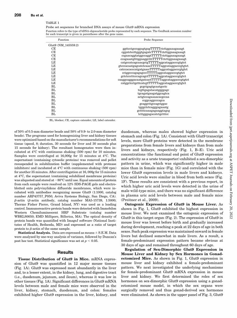
Development of Specific Oligonucleotide Probe Sets for theBranched DNA Assay. Gene sequences of mouse Glut9 wereobtained from GenBank. The strategy used for multiple oligonucleo-tide probe set design was described previously (Cheng et al., 2005).Oligonucleotide probe sets of mouse Glut9 (including captureextenders, label extenders, and blockers) are shown in Table 1.Probe sets were synthesized by Integrated DNA Technologies, Inc.(Coralville, IA).
Branched DNA Assay. Reagents required for the branched DNAassay (i.e., lysis buffer, amplifier/label probe dilution buffer, andsubstrate solution) were supplied in the QuantiGene branched DNAsignal amplification kit (Panomics Inc., Fremont, CA). mRNA expres-sion was analyzed according to the method reported previously(Hartley and Klaassen, 2000). Data are presented as relative lightunits per indicated amount of total RNA.
Membrane Protein Extraction and Immunoblotting. Mem-brane proteins were extracted using the Mem-PER Plus MembraneProtein Extraction Kit (catalog number 89842; Thermo FisherScientific, Grand Island, NY) following the manufacturer’s instruc-tions. Briefly, freshly collected livers and kidneys (40 mg protein/sample) were homogenized in permeabilization buffer (supplementedwith protease inhibitors) using a precooled Bullet Blender (modelBBY24M;Next Advance, Averill Park,NY)with sterile beads (mixture
Regulation of Mouse Glut9 Expression 207
at ASPE
T Journals on February 20, 2022
jpet.aspetjournals.orgD
ownloaded from
of 50% of 0.5-mm diameter beads and 50% of 0.9- to 2.0-mm diameterbeads). The programs used for homogenizing liver and kidney tissueswere optimized based on themanufacturer’s recommendations for softtissue (speed, 8; duration, 30 seconds for liver and 30 seconds plus15 seconds for kidney). The resultant homogenates were then in-cubated at 4°C with continuous shaking (500 rpm) for 10 minutes.Samples were centrifuged at 16,000g for 15 minutes at 4°C. Thesupernatant (containing cytosolic proteins) was removed and pelletresuspended in solubilization buffer (supplemented with proteaseinhibitors) and incubated at 4°C with continuous shaking (500 rpm)for another 35minutes. After centrifugation at 16, 000g for 15minutesat 4°C, the supernatant (containing solubilized membrane proteins)was aliquoted and stored at280°C until use. Equal amounts of proteinfrom each sample were resolved on 12% SDS-PAGE gels and electro-blotted onto polyvinylidene difluoride membranes, which were in-cubated with antibodies recognizing mouse Glut9 (1:1000, catalognumber ARP43757_P050; Aviva Systems Biology, San Diego, CA).b-actin (b-actin antibody, catalog number MA5-15739, 1:5000;Thermo Fisher Pierce, Grand Island, NY) was used as a loadingcontrol. Immunoreactive protein bands were detected with ImmobilonWestern Chemiluminescent HRP Substrate (catalog numberWBKLS0050; EMD Millipore, Billerica, MA). The optical density ofprotein bands was quantified with ImageJ software (National Insti-tutes of Health, Bethesda, MD) and expressed as a ratio of targetprotein to b-actin of the same sample.
Statistical Analysis. Data are expressed asmeans6 S.E.M. Datawere analyzed by one-way analysis of variance, followed by Duncan’spost hoc test. Statistical significance was set at p , 0.05.
ResultsTissue Distribution of Glut9 in Mice. mRNA expres-
sion of Glut9 was quantified in 12 major mouse tissues(Fig. 1A). Glut9 was expressed most abundantly in the liverand, to a lesser extent, in the kidney, lung, and digestive tract(i.e., duodenum, jejunum, and ileum), whereas it was low inother tissues (Fig. 1A). Significant differences in Glut9 mRNAlevels between male and female mice were observed in theliver, kidney, stomach, duodenum, and colon: femalesexhibited higher Glut9 expression in the liver, kidney, and
duodenum, whereas males showed higher expression instomach and colon (Fig. 1A). Consistent with Glut9 transcriptlevels, more Glut9 proteins were detected in the membranepreparations from female livers and kidneys than from malelivers and kidneys, respectively (Fig. 1, B–E). Uric acidconcentrations (the functional end point of Glut9 expressionand activity as a urate transporter) exhibited a sex-dimorphicpattern in urine, which was significantly higher in malemice than in female mice (Fig. 1G) and correlated with thelower Glut9 expression levels in male livers and kidneys.Uric acid levels were similar in blood from both sexes (Fig.1F). These results are consistent with a previous report, inwhich higher uric acid levels were detected in the urine ofmale wild-type mice, and there was no significant differencein plasma uric acid levels between male and female mice(Preitner et al., 2009).Ontogenic Expression of Glut9 in Mouse Liver. As
shown in Fig. 1, Glut9 exhibited the highest expression inmouse liver. We next examined the ontogenic expression ofGlut9 in this target organ (Fig. 2). The expression of Glut9 inmouse liver was lowest before birth and gradually increasedduring development, reaching a peak at 22 days of age in bothsexes. Such peak expressionwasmaintained onward in femalelivers but declined somewhat in male livers. As a result, afemale-predominant expression pattern became obvious at30 days of age and remained throughout 60 days of age.Regulation of Sex-Dimorphic Glut9 Expression in
Mouse Liver and Kidney by Sex Hormones in Gonad-ectomized Mice. As shown in Fig. 1, Glut9 expression inmouse liver and kidney exhibited a female-predominantpattern. We next investigated the underlying mechanismsfor female-predominant Glut9 mRNA expression in mouseliver and kidney. We first determined the roles of sexhormones on sex-dimorphic Glut9 expression using a gonad-ectomized mouse model, in which the sex organs weresurgically removed and thus gonad-derived sex hormoneswere eliminated. As shown in the upper panel of Fig. 3, Glut9
TABLE 1Probe set sequences for branched DNA assays of mouse Glut9 mRNA expressionFunction refers to the type of bDNA oligonucleotide probe represented by each sequence. The GenBank accession numberfor each transcript is given in parentheses after the gene name.
Function Probe Sequence
Glut9 (NM_145559.2)CE ggttcctgcccggagtgagTTTTTctcttggaaagaaagtCE cggatctccttgggtgagatcTTTTTctcttggaaagaaagtCE ccgaacaggtatggccaggtTTTTTctcttggaaagaaagtCE ccagaaatgtttggaaggctttTTTTTctcttggaaagaaagtLE catgccatcagcaacgctgTTTTTaggcataggacccgtgtctLE gtcccacaatgagcatctcaaaTTTTTaggcataggacccgtgtctLE catccacacccatgatgaaccTTTTTaggcataggacccgtgtctLE cctggcccagagagcccTTTTTaggcataggacccgtgtctLE gctctcccttcccagcagctTTTTTaggcataggacccgtgtctLE caaggcagggacaatgatcactTTTTTaggcataggacccgtgtctLE catggctcctgcttcatcgtTTTTTaggcataggacccgtgtctBL gcgctgagtgcaatgcctcBL tcgttgagatacatggggagcBL tgcagatgaagatggcagtcaBL actgtccagaaaacacgccaaBL ccggcaggcccagcaBL gcaggcttgccagctggacBL tgggctctcagggagaaatgBL gcttttcaaagagaaggtagcgBL tcttgggagacatctgctttcc
BL, blocker; CE, capture extender; LE, label extender.
208 Bu et al.
at ASPE
T Journals on February 20, 2022
jpet.aspetjournals.orgD
ownloaded from

mRNA levels increased in male livers upon gonadectomybut remained unchanged in female livers (gonadectomizedgroup plus placebo); as a result, the sex-dimorphic expres-sion pattern of Glut9 in the liver was abolished. DHTadministration to gonadectomized mice did not affect Glut9mRNA levels inmale livers. Administration of estrogen (E2)to gonadectomized mice reduced Glut9 mRNA levels infemale livers compared with placebo treatment. In thekidney, as shown in the lower panel of Fig. 3, surgicalremoval of gonads caused opposite outcomes in Glut9mRNA levels between the two sexes (i.e., an increase inmale kidneys but a decrease in female kidneys). The absenceof gonads not only abolished the female-predominant Glut9expression pattern in the kidney but also caused it to bereversed. Administration of androgen (DHT) or estrogen (E2)to gonadectomized male mice reduced Glut9 mRNA levelsto naïve levels. In contrast, both DHT and E2 had no effecton Glut9 expression in gonadectomized female mice. There-fore, sex hormones appear to inhibit Glut9 expression inmale kidneys. Overall, the regulation of sex hormones inGlut9 gene expression seems to be in tissue- and sex-specificmanners.
Regulation of Sex-Dimorphic Glut9 Expression inMouse Liver and Kidney by GH in lit/lit Mice. Inaddition to sex hormones, the secretion pattern of GH hasbeen shown to regulate sex-dimorphic expression of certaindrug-processing genes. In rats and mice, males secrete GH inhigh-amplitude pulses with a regular frequency, which resultsin pulsatile serum GH levels (Tannenbaum and Martin, 1976;MacLeod et al., 1991). In contrast, female rats and micesecrete GH in low-amplitude pulses with greater frequencyand higher trough levels thanmales; as a result, the serumGHlevel is continuously detectable in females (Saunders et al.,1976; MacLeod et al., 1991). The sex-specific GH secretionpattern was demonstrated to be responsible for sex-specificexpression of Cyp2c11 and Cyp2c12 in rats and Cyp2d9 andCyp2a4 in mice (Noshiro and Negishi, 1986; Waxman et al.,1991; Aida and Negishi, 1993). To investigate whether a sex-specific GH secretion patternmodulates Glut9 expression, thelit/lit mouse model was used. In the lit/lit mouse model, aspontaneous mutation occurred in the GH-releasing hormonereceptor, which leads to impaired secretion of GH (Lin et al.,1993). As shown in the upper panel of Fig. 4, when the male-pattern GH release was abolished, Glut9 expression was
Fig. 1. Tissue distribution of Glut9 in mice. (A) RNA isolated from tissues of adult male and female C57BL/6 mice (n = 6/sex) was analyzed by branchedDNA assay for mRNA expression of Glut9. (B–E) Immunoblotting results of Glut9 protein levels in livers (B and C) and kidneys (D and E) of adult maleand female mice (n = 4/sex). b-actin was included as a loading control. Representative results from three different immunoblotting experiments. Ratios ofthe optical density of the immunoreactive bands (Glut9 versus b-actin of the same sample) in livers (C) and kidneys (E) of both sexes. (F and G) Uric acidlevels in serum (F) and urine (G) of adult male and female C57BL/6 mice (n=5/sex). Data are presented as means 6 S.E.M. *p , 0.05 (statisticallysignificant differences between male and female mice). RLU, relative light unit.
Regulation of Mouse Glut9 Expression 209
at ASPE
T Journals on February 20, 2022
jpet.aspetjournals.orgD
ownloaded from

elevated in the livers of male lit/lit mice; consequently, thefemale-predominant expression pattern seen in naïve liv-ers was lost. Administration of GH via a male patterndecreased Glut9 expression in livers of both male andfemale lit/lit mice, further confirming the inhibitory effectof male-pattern GH. In contrast, administration of GH via afemale pattern to lit/lit mice did not have much effect onGlut9 expression in either sex. Glut9 expression in femalekidneys is much lower in lit/lit mice than in native femalemice. This results in a reversed male-predominant patternin the kidneys of lit/lit mice (Fig. 4, lower panel). Admin-istration of GH via the male pattern reduced Glut9expression in the kidneys of male lit/lit mice but not inthe kidneys of females. Administration of GH via a femalepattern did not have much effect on Glut9 expression.Together, data obtained from the lit/lit mouse modeldemonstrate that male-pattern GH suppresses Glut9 ex-pression in the liver and kidney.Regulation of Sex-Dimorphic Glut9 Expression in
Mouse Liver and Kidney by Sex Hormones and GH inHypophysectomized Mice. Sex hormones can influencesex-specific GH secretion via multiple pathways and thusmay secondarily modulate the regulation of gene expression,which is seemingly governed by sex-specific GH (Legraverendet al., 1992; Painson et al., 1992). The hypophysectomizedmouse model is commonly used to dissect the potentialinterplay of sex hormones and GH. In this model, surgicalremoval of the pituitary obliterates both GH and the pituitaryhormones that are required for biosynthesis of gonad-derivedsex hormones, including luteinizing hormone and follicle-stimulating hormone. As a result, the effects of both sexhormones and GH are eliminated in hypophysectomizedmice, and the GH replacement is considered free of sexhormone influences. As shown in the upper panel of Fig. 5(placebo versus naïve), hypophysectomy caused Glut9 mRNA
levels to increase in male livers. Consequently, the female-predominant Glut9 expression pattern was abolished. Admin-istration of male-pattern, but not female-pattern, GH tohypophysectomized mice suppressed Glut9 expression in thelivers of both sexes. Kidney Glut9 mRNA levels were elevatedin both sexes upon hypophysectomy (Fig. 5, lower panel,placebo versus naïve). In addition, female-predominant Glut9expression in the kidneys was further sustained in hypophy-sectomized mice. Similarly, only administration of male-pattern GH to hypophysectomized mice suppressed kidneyGlut9 expression in both sexes. This evidence corroborates theresults obtained from lit/lit mice, clearly indicating an inhib-itory effect ofmale-patternGH onGlut9 expression in both theliver and kidney of both sexes in mice.Chemical Regulation of Glut9 in Mouse Liver. Male
C57BL/6 mice were administered five classes of prototypicaldrug-metabolizing enzyme inducers. Each class contains threechemicals that are known to transcriptionally activate aspecific nuclear receptor. Because one chemical may influencegene expression through multiple signaling pathways, weconsidered Glut9 to be a potential target gene of thatparticular nuclear receptor only when all three activators of
Fig. 2. Ontogenic expression of Glut9 in mouse liver. RNA isolated frommouse livers at each age during development (n = 5/sex per age) wasanalyzed by the branched DNA assay for mRNA expression of Glut9. Dataare presented as means 6 S.E.M. *p , 0.05 (statistically significantdifferences between male and female mice). RLU, relative light unit.
Fig. 3. Effects of gonadectomy and sex hormone replacement on Glut9mRNA expression in mouse liver and kidney. Total RNA from liversand kidneys of naïve and gonadectomized mice was analyzed by thebranched DNA assay for Glut9 mRNA levels. Data are presented asmeans 6 S.E.M. (n = 5 mice/group). *p , 0.05 (statistically significantdifferences between males and females in naïve mice or the gonadecto-mized group receiving the same treatment); †p , 0.05 (statisticallysignificant differences between the same-sex naïve mice and placebo-treated gonadectomizedmice); ‡p, 0.05 (statistically significant differencesbetween the same-sex placebo-treated and sex hormone–treated gonadec-tomizedmice). +DHT, gonadectomizedmice receiving 5a-dihydroxytestosteronetreatment; +E2, gonadectomized mice receiving 17b-estradiol treat-ment; +Plac., gonadectomizedmice receiving placebo treatment; RLU, relativelight unit.
210 Bu et al.
at ASPE
T Journals on February 20, 2022
jpet.aspetjournals.orgD
ownloaded from

that particular nuclear receptor caused the same alteration inGlut9 gene expression. The doses of chemicals were selectedaccording to the literature, with such doses the activation ofspecific nuclear receptors was confirmed by induction of theirtarget genes (which are also drug-metabolizing enzymes) asfollows: cytochrome P450 enzymes (Cyp1a1 induction by AhRligands, Cyp2b10 induction by CAR ligands, Cyp3a11 in-duction by PXR ligands, and Cyp4a14 induction by PPARaligands) and NAD(P)H/quinone oxidoreductase 1 induction byNrf2 activators, as reported previously (Maher et al., 2005).The induction of Glut9 mRNA by drug-metabolizing enzymeinducers is shown in Fig. 6. All three activators of AhR andPPARa inducedGlut9mRNAexpression. In contrast, only twoof three CAR, PXR, or Nrf2 activators induced Glut9 mRNAexpression. Therefore, these results suggest that Glut9may bea potential target gene of nuclear receptors AhR and/orPPARa.
DiscussionIn this study, we identified a female-predominant expres-
sion pattern of Glut9 inmouse liver and kidney and discoveredthe underlying mechanism to be male-pattern GH secretion,which repressed Glut9 transcription in liver and kidney ofmale mice. Furthermore, we demonstrated that activation ofAhR and PPARa nuclear receptors by their prototypicalagonists induced Glut9 mRNA levels in mouse liver.Upon binding of GH to its receptor, GH signaling is
mediated by the Janus kinase (Jak) 2–signal transducer andactivator of transcription (Stat) 5b signaling pathway. Byscreening a 6-kb promoter sequence of the mouse Glut9 gene,we identified a highly conserved Stat5b response element(Table 2). Similarly, we identified a perfect STAT5b responseelement in the promoter of the human GLUT9 gene (Table 2).The presence of Stat5b response elements in the promoters ofboth the human and mouse GLUT9/Glut9 gene suggests thatthe GH signaling is linked to transcriptional regulation ofGlut9, although further studies are needed to confirmwhetherGlut9 is a bona fide GH target gene. Secretion of GH is
Fig. 5. Effects of hypophysectomy and multiple hormone replacements onGlut9 mRNA expression in mouse liver and kidney RLU. Total RNA fromlivers and kidneys of naïve and hypophysectomized mice was analyzed bythe branched DNA assay for Glut9 mRNA levels. Data are presented asmeans 6 S.E.M. (n = 5 mice/group). *p , 0.05 (statistically significantdifferences between males and females in naïve mice or the hypophysec-tomized mice group receiving the same treatment); †p, 0.05 (statisticallysignificant differences between the same-sex naïve mice and placebo-treated hypophysectomized mice); ‡p , 0.05 (statistically significantdifferences between the same-sex placebo-treated and hormone-treatedhypophysectomized mice). +DHT, hypophysectomized mice receiving5a-dihydroxytestosterone treatment; +E2, hypophysectomized micereceiving 17b-estradiol treatment; +FPGH, hypophysectomized micereceiving continuous infusion of rat GH via subcutaneously implanted21-day-release pellet mimicking female-pattern GH secretion; +MPGH,hypophysectomized mice receiving rat GH twice daily administered byintraperitoneal injection to mimic male-pattern GH secretion; +Plac.,hypophysectomized mice receiving placebo treatment; RLU, relativelight unit.
Fig. 4. Effects of GH on Glut9 mRNA expression in the livers and kidneysof lit/lit mice. Total RNA from livers and kidneys of naïve and lit/lit micewas analyzed by the branched DNA assay for Glut9 mRNA levels. Dataare presented as means 6 S.E.M. (n = 5 mice/group). *p , 0.05(statistically significant differences between males and females in naïvemice or the lit/lit mice group receiving the same treatment); †p , 0.05(statistically significant differences between the same-sex naïve mice andplacebo-treated lit/lit mice); ‡p , 0.05 (statistically significant differencesbetween the same-sex placebo-treated and GH-treated gonadectomizedmice). +FPGH, lit/lit mice receiving continuous infusion of rat GH via asubcutaneously implanted 21-day-release pellet mimicking female-pattern GH secretion; +MPGH, lit/lit mice receiving rat GH twice dailyadministered by intraperitoneal injection to mimic male-pattern GHsecretion; +Plac., lit/lit mice receiving placebo treatment; RLU, relativelight unit.
Regulation of Mouse Glut9 Expression 211
at ASPE
T Journals on February 20, 2022
jpet.aspetjournals.orgD
ownloaded from

pulsatile inmen but continuous inwomen, similarly as inmice(Vance et al., 1985; Veldhuis et al., 2001). Therefore, we expectthat expression of human GLUT9 may also be influencedby sex-specific GH secretion and may depict a female-predominant pattern. This may provide an explanation for aprevious study, which reported that humanGLUT9 influencesserum urate concentrations with significant sex-specific ef-fects (Döring et al., 2008). Circulating urate levels in humansare known to be associated with hyperuricemia and gout. Forinstance, men have higher serum urate levels than women(Mikkelsen et al., 1965; Fang and Alderman, 2000) and areclinically at a significantly higher risk for gout (Hediger et al.,2005; Döring et al., 2008). Therefore, it is of great interest tounderstand the difference in urate handling betweenmen andwomen. Because GLUT9 is a key urate transporter andcontributes significantly to urate homeostasis, discoveries
made on Glut9 sex-dimorphic expression in mouse modelsmay provide useful information on the regulation of humanGLUT9.In addition to Glut9, many transporters that are involved in
urate homeostasis (e.g., organic anion transporter 1, breastcancer resistance protein, and urate anion exchanger 1) showsex-dimorphic expression. For instance, the expression oforganic anion transporter 1 is male predominant in mousekidney (Buist and Klaassen, 2004; Breljak et al., 2016). Theexpression of breast cancer resistance protein is male pre-dominant in both human andmouse liver (Merino et al., 2005;Tanaka et al., 2005). Similarly, urate anion exchanger 1 ex-pression is also male predominant in mouse kidney (Chengand Klaassen, 2009). Therefore, a trend of male-predominantexpression patterns is commonly seen in the transporters thatare involved in urate homeostasis. In this study, we reportedthat Glut9 exhibits a female-predominant expression patternin mouse liver and kidney. Multiple transporters are involvedin urate handling in the body, and it is logical to speculate thatthe regulation of each individual transporter is specific andindependent of one another.Our previous reports showed that male-pattern GH primar-
ily contributes to sex-dimorphic expression of drug-processinggenes including transporters in the liver, whereas sex hor-mones primarily contribute to sex-dimorphic expression ofdrug-processing genes in the kidney (Cheng et al., 2005, 2006;Cheng and Klaassen, 2009). Uniquely, in our study, weshowed that female-predominant Glut9 expression in bothmouse liver and kidney ismainly attributable to the inhibitoryeffects of male-pattern GH secretion but not sex hormones.Male sex hormone (DHT) appears to have a negative effect onGut9 expression only in the kidney (Fig. 3, lower panel, malenaïve versus gonadectomy plus placebo versus gonadectomyplus DHT), but not in the liver (Fig. 3, upper panel, male naïveversus gonadectomy plus placebo versus gonadectomy plusDHT). Female sex hormone, on the other hand, did not seem tohave a consistent influence on Glut9 expression in eitherorgan (Fig. 3). The suppressive effect of male-pattern GH isclearly evident in the lit/lit mouse model (Fig. 4, upper panel),but the effect of female-pattern GH is not so clear, especially inthe kidney (Fig. 4, lower panel). It seems plausible thatfemale-pattern GH is required for maintaining Glut9 expres-sion in female kidneys, because Glut9 expression was mark-edly decreased in the kidney of lit/lit mouse (Fig. 4, lowerpanel, female naïve versus lit/lit plus placebo). However,female-pattern GH replacement failed to restore Glut9 ex-pression (Fig. 4, lower panel, female lit/lit plus placebo versuslit/lit plus female-pattern GH), which argues against such
TABLE 2Putative response elements in the mouse and human Glut9/GLUT9 gene promoter
Response Element Sequencea Location (Relative toTranscription Start Site)
bp
Mouse Glut9 gene promoterXRE CTTGGGCGTGACTCCGGGGAG 210Stat5b response element GAGGCTGCTTCAGGGAACAGCC 23570DR1 CTGAAGGTCATGGTCCATTC 25790
Human GLUT9 gene promoterSTAT5b response element TCCAGGTTCCTGGAATGTCTGC 21260
DR1, direct repeat 1 (also a binding site for PPARa); XRE, xenobiotic response element (also a binding site for AhR).aThe consensus sequence of the response element is underlined.
Fig. 6. Chemical regulation of Glut9 mRNA expression in mouse liver.Adult male C57BL/6 mice were administrated the following prototypicalphase I and phase II drug-metabolizing enzyme inducers (see theMaterials andMethods for details): TCDD, BNF, PCB126, PB, TCPOBOP,DAS, PCN, SPR, DEX, CLFB, CPFB, DEHP, BHA, ETHOXYQ, and OPZ.Total RNA extracted from livers of each chemical treatment group (n = 5/group) was analyzed by the branched DNA assay. Control groupsconsisted of the four individual control groups, in which the Glut9mRNA levels were confirmed not statistically different. All data areexpressed as means 6 S.E.M. *p , 0.05 (statistically significant differ-ence between treated and control mice). BHA, butylated hydroxyanisole;BNF,b-naphthoflavone; CLFB, clofibrate; CONT, control; CPFB, ciprofibrate;DAS, diallyl sulfide; DEHP, diethylhexylphthalate; DEX, dexamethasone;ETHOXYQ, ethoxyquin; OPZ, oltipraz; PB, phenobarbital; PCB126, poly-chlorinated biphenyl 126; PCN, pregnenolone 16a-carbonitrile; SPR,spironolactone; TCDD, 2,3,7,8-tetrachlorodibenzo-p-dioxin; TCPOBOP, 1,4-bis[2-(3,5-dichloropyridyloxy)]benzene.
212 Bu et al.
at ASPE
T Journals on February 20, 2022
jpet.aspetjournals.orgD
ownloaded from

effects of female-pattern GH. Further studies are needed toelucidate this issue. In addition, surgical removal of thepituitary markedly increased Glut9 transcription in femalekidneys, presumably owing to the withdrawal of some pitui-tary inhibitory factor(s) other than gonadotropin (the up-stream hormone for sex hormone) or GH, because abolishmentof either gonad-derived sex hormone by gonadectomy (Fig. 3,lower panel) or GH signaling in lit/lit mice (Fig. 4, lower panel)did not trigger a similar stimulation in female kidneys (Fig. 5,lower panel). We speculate that the relief of pituitary in-hibition on female kidney Glut9 expression is quite drastic,such that any exogenous disturbances (e.g., replacement ofDHT, E2, male-pattern GH, or female-pattern GH) negativelyaffected this stimulation, resulting in a decrease in Glut9mRNA levels. The underlyingmechanism bywhich removal ofthe pituitary stimulates female kidney Glut9 expression isunknown and warrants further investigation.To our knowledge, no studies have yet been conducted to
investigate chemical regulation of Glut9 or signaling path-ways regulating Glut9 gene expression. Our results suggestthat via binding to the response elements (e.g., xenobioticresponse element and direct repeat 1, DR1) present in theGlut9 promoter, nuclear receptors AhR and PPARa are able toregulate the transcription of Glut9 as a target gene (Table 2).Our finding is likely to provide a mechanistic explanation forthe observations made in AhR KO mice that these miceexhibited 10-times higher urate levels in urine and conse-quently developed kidney stones composed of uric acid crystals(Atala, 2012; Butler et al., 2012). Because we have shown thatagonists of AhR markedly stimulated Glut9 transcript levelsin mouse liver (Fig. 6), it is possible that loss of functional AhRresulted in reduced Glut9 expression in the livers and kidneysof these mice. In mice, Glut9 is required for transporting uratefrom blood to hepatocytes for uricase-mediated degradationand for urate reabsorption in proximal and distal convolutedtubules of the kidney. It is therefore logical to postulate thatreduced Glut9 expression contributed to hyperuricosuria andsubsequent uric acid precipitation as kidney stones in AhRKOmice. Our unpublished data showed that two PPARa ligands,perfluorononanoic acid and perfluorodecanoic acid, decreaseduric acid levels in mouse serum after a single intraperitonealadministration. Activation of PPARa by these two ligandscould lead to an increase in Glut9 expression in its targetorgans (i.e., the liver and the kidney). Stimulated expression ofthe Glut9 transporter on the basolateral side of hepatocytes islikely to increase the uptake of uric acid from blood into theliver for uricase-mediated degradation and thus contributes todecreased uric acid levels in serum.In humans, many endogenous and exogenous chemicals,
including environmental chemicals (e.g., dioxins and per-fluorinated compounds) and drugs (e.g., fibrates), are able toactivate AhR and/or PPARa as agonists. Our findings call forconsiderations regarding the effects of these drugs andchemicals on urate homeostasis.Glut9/GLUT9 is abundantly expressed in mouse and hu-
man liver (Fig. 1) (Augustin et al., 2004). However, thefunctions of Glut9/GLUT9 may be species dependent. Forinstance, in mice, Glut9 is expressed on the basolateral side ofhepatocytes, where it mediates influx of serum urate into theliver for breakdown by uricase (Keembiyehetty et al., 2006).The mice use two major routes for eliminating serum urate:one through secretion by the kidney, and the other through
uricase-dependent degradation in the liver; both routes re-quire functional Glut9 (Preitner et al., 2009). However,because of the lack of uricase in humans, it is unlikely thatthe GLUT9 protein located on the basolateral side of humanhepatocytes performs a similar function as it does in mice.Furthermore, considerable amounts of urate are produced inthe liver from metabolizing purine-containing compounds(Hediger et al., 2005). Therefore, in humans, it seems morelogical that GLUT9 functions as an efflux transporter thatexports urate from the liver to blood. Species differences seemto exist regarding the function of hepatic Glut9/GLUT9between mice and humans, which must be taken into consid-eration when modeling regulation of hepatic Glut9 in mice. Inthis regard, primary human hepatocytes may prove to be abetter model.In summary, this study demonstrates that essential phys-
iologic factors, including sex, GH secretion patterns, andactivation of nuclear receptors, contribute to the regulationof Glut9 expression in mice.
Acknowledgments
The authors thankDr. Curtis D. Klaassen for helpful suggestions instudy design and manuscript preparation.
Authorship Contributions
Participated in research design: Bu, Le, Cheng.Conducted experiments: Bu, Le, Zhang, Cheng.Contributed new reagents or analytic tools: Bu, Le, Cheng.Performed data analysis: Bu, Le, Zhang, Cheng.Wrote or contributed to the writing of the manuscript: Bu, Le,
Zhang, Cheng.
References
Aida K and Negishi M (1993) A trans-acting locus regulates transcriptional re-pression of the female-specific steroid 15 alpha-hydroxylase gene in male mice. JMol Endocrinol 11:213–222.
Anzai N, Ichida K, Jutabha P, Kimura T, Babu E, Jin CJ, Srivastava S, Kitamura K,Hisatome I, Endou H, et al. (2008) Plasma urate level is directly regulated by avoltage-driven urate efflux transporter URATv1 (SLC2A9) in humans. J Biol Chem283:26834–26838.
Anzai N, Jutabha P, Amonpatumrat-Takahashi S, and Sakurai H (2012) Recentadvances in renal urate transport: characterization of candidate transporters in-dicated by genome-wide association studies. Clin Exp Nephrol 16:89–95.
Atala A (2012) Re: Uric acid stones in the urinary bladder of aryl hydrocarbon re-ceptor (AhR) knockout mice. J Urol 188:1042.
Augustin R, Carayannopoulos MO, Dowd LO, Phay JE, Moley JF, and Moley KH(2004) Identification and characterization of human glucose transporter-likeprotein-9 (GLUT9): alternative splicing alters trafficking. J Biol Chem 279:16229–16236.
Bibert S, Hess SK, Firsov D, Thorens B, Geering K, Horisberger JD, and Bonny O(2009) Mouse GLUT9: evidences for a urate uniporter. Am J Physiol Renal Physiol297:F612–F619.
Breljak D, Ljubojevi�c M, Hagos Y, Micek V, Balen Eror D, Vrhovac Maduni�c I, BrzicaH, Karaica D, Radovi�c N, Kraus O, et al. (2016) Distribution of organic aniontransporters NaDC3 and OAT1-3 along the human nephron. Am J Physiol RenalPhysiol 311:F227–F238.
Buist SC and Klaassen CD (2004) Rat and mouse differences in gender-predominantexpression of organic anion transporter (Oat1-3; Slc22a6-8) mRNA levels. DrugMetab Dispos 32:620–625.
Butler R, Inzunza J, Suzuki H, Fujii-Kuriyama Y, Warner M, and Gustafsson JA(2012) Uric acid stones in the urinary bladder of aryl hydrocarbon receptor (AhR)knockout mice. Proc Natl Acad Sci USA 109:1122–1126.
Cheng X and Klaassen CD (2009) Tissue distribution, ontogeny, and hormonal reg-ulation of xenobiotic transporters in mouse kidneys. Drug Metab Dispos 37:2178–2185.
Cheng X, Maher J, Chen C, and Klaassen CD (2005) Tissue distribution and ontogenyof mouse organic anion transporting polypeptides (Oatps). Drug Metab Dispos 33:1062–1073.
Cheng X, Maher J, Lu H, and Klaassen CD (2006) Endocrine regulation of gender-divergent mouse organic anion-transporting polypeptide (Oatp) expression. MolPharmacol 70:1291–1297.
Dehghan A, Köttgen A, Yang Q, Hwang SJ, Kao WL, Rivadeneira F, Boerwinkle E,Levy D, Hofman A, Astor BC, et al. (2008) Association of three genetic loci with uricacid concentration and risk of gout: a genome-wide association study. Lancet 372:1953–1961.
Regulation of Mouse Glut9 Expression 213
at ASPE
T Journals on February 20, 2022
jpet.aspetjournals.orgD
ownloaded from

Dinour D, Gray NK, Campbell S, Shu X, Sawyer L, Richardson W, Rechavi G,Amariglio N, Ganon L, Sela BA, et al. (2010) Homozygous SLC2A9 mutationscause severe renal hypouricemia. J Am Soc Nephrol 21:64–72.
Doblado M and Moley KH (2009) Facilitative glucose transporter 9, a unique hexoseand urate transporter. Am J Physiol Endocrinol Metab 297:E831–E835.
Döring A, Gieger C, Mehta D, Gohlke H, Prokisch H, Coassin S, Fischer G, Henke K,Klopp N, Kronenberg F, et al. (2008) SLC2A9 influences uric acid concentrationswith pronounced sex-specific effects. Nat Genet 40:430–436.
Fang J and Alderman MH (2000) Serum uric acid and cardiovascular mortality theNHANES I epidemiologic follow-up study, 1971-1992. National Health and Nu-trition Examination Survey. JAMA 283:2404–2410.
Hartley DP and Klaassen CD (2000) Detection of chemical-induced differential ex-pression of rat hepatic cytochrome P450 mRNA transcripts using branched DNAsignal amplification technology. Drug Metab Dispos 28:608–616.
Hediger MA, Johnson RJ, Miyazaki H, and Endou H (2005) Molecular physiology ofurate transport. Physiology (Bethesda) 20:125–133.
Keembiyehetty C, Augustin R, Carayannopoulos MO, Steer S, Manolescu A,Cheeseman CI, and Moley KH (2006) Mouse glucose transporter 9 splice variantsare expressed in adult liver and kidney and are up-regulated in diabetes. MolEndocrinol 20:686–697.
Le MT, Shafiu M, Mu W, and Johnson RJ (2008) SLC2A9–a fructose transporteridentified as a novel uric acid transporter. Nephrol Dial Transplant 23:2746–2749.
Legraverend C, Mode A, Wells T, Robinson I, and Gustafsson JA (1992) Hepaticsteroid hydroxylating enzymes are controlled by the sexually dimorphic pattern ofgrowth hormone secretion in normal and dwarf rats. FASEB J 6:711–718.
Lin SC, Lin CR, Gukovsky I, Lusis AJ, Sawchenko PE, and Rosenfeld MG (1993)Molecular basis of the little mouse phenotype and implications for cell type-specificgrowth. Nature 364:208–213.
MacLeod JN, Pampori NA, and Shapiro BH (1991) Sex differences in the ultradianpattern of plasma growth hormone concentrations in mice. J Endocrinol 131:395–399.
Maher JM, Cheng X, Slitt AL, Dieter MZ, and Klaassen CD (2005) Induction of themultidrug resistance-associated protein family of transporters by chemical acti-vators of receptor-mediated pathways in mouse liver. Drug Metab Dispos 33:956–962.
Merino G, van Herwaarden AE, Wagenaar E, Jonker JW, and Schinkel AH (2005)Sex-dependent expression and activity of the ATP-binding cassette transporterbreast cancer resistance protein (BCRP/ABCG2) in liver. Mol Pharmacol 67:1765–1771.
Mikkelsen WM, Dodge HJ, and Valkenburg H (1965) The distribution of serum uricacid values in a population unselected as to gout or hyperuricemia: Tecumseh,Michigan 1959-1960. Am J Med 39:242–251.
Noshiro M and Negishi M (1986) Pretranslational regulation of sex-dependenttestosterone hydroxylases by growth hormone in mouse liver. J Biol Chem 261:15923–15927.
Painson JC, Thorner MO, Krieg RJ, and Tannenbaum GS (1992) Short-term adultexposure to estradiol feminizes the male pattern of spontaneous and growthhormone-releasing factor-stimulated growth hormone secretion in the rat. Endo-crinology 130:511–519.
Phay JE, Hussain HB, and Moley JF (2000) Cloning and expression analysis of anovel member of the facilitative glucose transporter family, SLC2A9 (GLUT9).Genomics 66:217–220.
Preitner F, Bonny O, Laverrière A, Rotman S, Firsov D, Da Costa A, Metref S,and Thorens B (2009) Glut9 is a major regulator of urate homeostasis and itsgenetic inactivation induces hyperuricosuria and urate nephropathy. Proc NatlAcad Sci USA 106:15501–15506.
Saunders A, Terry LC, Audet J, Brazeau P, andMartin JB (1976) Dynamic studies of growthhormone and prolactin secretion in the female rat. Neuroendocrinology 21:193–203.
So A and Thorens B (2010) Uric acid transport and disease. J Clin Invest 120:1791–1799.
Tanaka Y, Slitt AL, Leazer TM, Maher JM, and Klaassen CD (2005) Tissue distri-bution and hormonal regulation of the breast cancer resistance protein(Bcrp/Abcg2) in rats and mice. Biochem Biophys Res Commun 326:181–187.
Tannenbaum GS and Martin JB (1976) Evidence for an endogenous ultradianrhythm governing growth hormone secretion in the rat. Endocrinology 98:562–570.
Tu HP, Chen CJ, Lee CH, Tovosia S, Ko AM, Wang SJ, Ou TT, Lin GT, Chiang SL,Chiang HC, et al. (2010) The SLC22A12 gene is associated with gout in HanChinese and Solomon Islanders. Ann Rheum Dis 69:1252–1254.
Vance ML, Kaiser DL, Evans WS, Furlanetto R, Vale W, Rivier J, and Thorner MO(1985) Pulsatile growth hormone secretion in normal man during a continuous24-hour infusion of human growth hormone releasing factor (1-40). Evidence forintermittent somatostatin secretion. J Clin Invest 75:1584–1590.
Veldhuis JD, Anderson SM, Shah N, Bray M, Vick T, Gentili A, Mulligan T, JohnsonML, Weltman A, Evans WS, et al. (2001) Neurophysiological regulation and target-tissue impact of the pulsatile mode of growth hormone secretion in the human.Growth Horm IGF Res 11 (Suppl A):S25–S37.
Wallace C, Newhouse SJ, Braund P, Zhang F, Tobin M, Falchi M, Ahmadi K, DobsonRJ, Marçano AC, Hajat C, et al. (2008) Genome-wide association study identifiesgenes for biomarkers of cardiovascular disease: serum urate and dyslipidemia. AmJ Hum Genet 82:139–149.
Waxman DJ, Pampori NA, Ram PA, Agrawal AK, and Shapiro BH (1991) Interpulseinterval in circulating growth hormone patterns regulates sexually dimorphic ex-pression of hepatic cytochrome P450. Proc Natl Acad Sci USA 88:6868–6872.
Address correspondence to: Dr. Xingguo Cheng, Department of Pharma-ceutical Sciences, College of Pharmacy and Health Sciences, St. John’sUniversity, 8000 Utopia Parkway, Queens, NY 11439. E-mail: [email protected]
214 Bu et al.
at ASPE
T Journals on February 20, 2022
jpet.aspetjournals.orgD
ownloaded from