Embed Size (px)
Citation preview
1
Histopathology of S. tanaceti infection in pyrethrum leaf lamina
M. A. H. B. Bhuiyan1 · T Groom2 · M. E. Nicolas1 · P. W. J. Taylor1*
1Faculty of Veterinary and Agricultural Sciences, The University of Melbourne, Parkville,
Victoria-3010
2Botanical Resources Australia (BRA)-Agricultural Services Pty. Ltd.
*Email: [email protected]
Abstract:
The infection process and life cycle of S. tanaceti in leaf lamina of pyrethrum plants was
investigated using histopathology. Conidia attached firmly to the leaf surface before the
infection hyphae penetrated directly into the epidermal cells of the leaf without forming
appressoria. The maximum germination of conidia on leaf surface was 85% at 54 HAI.
Infection hyphae infected the epidermal and palisade parenchyma cells through the
middle lamella. Brown lesions on the leaf were a result of infected necrotic epidermal
cells. Extensive colonization through both intra- and intercellular hyphae along with
pycnidia formation caused enormous damage to the infected cells at 12 DAI. Unlike the
quadruple stain, both single and dual stains had very limited ability to visualise infection
structures. These results have provided a better understanding of the physical interaction
between the pathogen and the pyrethrum leaf tissues and will help to elucidate the
complete disease cycle of S. tanaceti on pyrethrum plant.
Key words: histopathology, microtome section, pyrethrum, ray blight, staining, S. tanaceti

2
Introduction:
Pyrethrum, Tanacetum cinerariifolium (Trevir.) Schultz-Bip., is a perennial plant which
belongs to the family Asteraceae and is widely used for the production of natural
insecticide pyrethrins (Hitmi et al. 2000). In Australia, two thirds of world production of
pyrethrum is grown commercially in northern Tasmania and around Ballarat in Victoria
(Pethybridge et al. 2013). Ray blight (Stagonosporopsis tanaceti; Vaghefi et al. 2012) is
one of the most important diseases of pyrethrum which produces typical necrotic
symptoms on leaf margins, shoots and developing buds in spring (Pethybridge et al.
2008). Moreover, shepherd crook symptoms are produced on the flower stems with clear
constriction and delineation between the necrotic peduncle and the remainder of the
healthy stem. The common means of reproduction of S. tanaceti is through pycnidia
while the sexual stage is unknown in Australia (Vaghefi et al. 2012). Although necrotic
lesions were reported in pyrethrum leaves by S. tanaceti (Pethybridge and Wilson 1998;
Pethybridge et al. 2008), a detail histopathological study of tissue infection in relation to
the phenotypic symptoms on leaf surface and life cycle of the pathogen have not been
documented. Considering the significance of this disease, a better understanding of the
host-pathogen interaction of S. tanaceti in the tissues of pyrethrum is important for the
implementation of integrated disease control methods.
Various techniques have been used to study the infection events of pathogenic fungi in
plants. Light microscopy and single staining have been used to study the infection events
on the leaf surface of hosts (Ranathunge et al. 2012; Roustaee et al. 2000). Although dual
stain combinations like safranin and aniline blue/fast green (Patton and Spear 1989) or
safranin and fast green (Ribichich et al. 2000) have been used to study the colonization of
host tissue by various fungal pathogens, the structural and chemical changes of infected

3
plant cells were not clear. However, Johansen quadruple stain (Johansen 1940)
combinations have been used to study the events of infection in paraffin embedded plant
tissues. This could differentiate invading fungal mycelia from the cutin, lignin, suberin
and cellulose in cell walls and cell organelles, like plastids and nuclei, with contrasting
colour and was used to study the infection and colonisation of wheat by the crown rot
pathogen Fusarium pseudograminearum (Taylor and Burgess 1983). Besides light
microscopy, both scanning electron microscopy (SEM) and green fluorescent protein
(GFP) transformed fungi have been used to investigate the interaction of fungi in plant
tissue. However, more advanced microscopy and molecular techniques are not practical
to use on a routine basis (Liberato et al. 2005) which is time consuming and limited to a
certain number of fungal strains (Knight and Sutherland 2011).
Therefore, the aim of this study was to use light microscopy and histopathology to
determine the infection process and life cycle of S. tanaceti infection in pyrethrum leaves.
Materials and methods
Fungal isolate and preparation of inoculum
S. tanaceti isolate (Tas-1) was provided by Botanical Resources Australia (BRA) which
was cultured on Potato dextrose agar (PDA; Difco, USA). For sporulation, specialized V8
medium was used followed by incubation for 2-3-weeks at 25 °C with a 12 h photoperiod
until formation of brown coloured pycnidia. Pycniospores generated from these pycnidia
were hyaline ellipsoid to oblong with individual spore size between 7-9 × 3-3.5 µm
(Vaghefi 2012). Conidia were then harvested by gently scrapping the surface of cultures
4
using a sterile bent glass rod with 5 mL sterile distilled water (SDW). The suspension was
then filtered through three layers of muslin cloth. Spore concentration was measured
using a haemocytometer and adjusted to 106 spores/mL with SDW. Tween 20 (0.05%)
was added as a surfactant to the spore suspension.
Germination of conidia and penetration by germinated hyphae in pyrethrum leaves
For the study of the mechanism of penetration on leaf surfaces, detached leaf laminas
were collected from healthy 4-week-old pyrethrum cultivar BR1. Sixty leaf laminas were
surface sterilized with 1% sodium hypochlorite for 2 min and then twice washed in sterile
distilled water. Thirty leaf laminas were inoculated by spraying with 106 spores/ mL spore
suspension on both abaxial and adaxial surface. The other 30 leaf laminas were sprayed
with sterile distilled water as controls. Each leaf lamina was considered as a replicate.
Both inoculated and control replicates were placed separately into petri-dish with
moistened sterile filter paper and incubated at 25 °C at 12 h photoperiod. Leaf laminas
were sampled at 6, 12, 24, 30, 48 and 54 h after inoculation (HAI) for microscopic studies
and 5-replicates from each of inoculated and control were sampled at each time interval.
Germination data were subjected to analysis of variance (ANOVA) using Minitab and
means of each time after inoculation were compared using the Least Significance
Differences (LSDs) at P<0.05 using Microsoft Excel. A modified fixing and clearing
technique was used to prepare the sample (Johansen 1940). Briefly, 4, 1-cm2 inoculated
and control leaf tissue from each replicate at each time interval was cut using a sterile
scalpel. These tissues were then processed for fixation, clearing and staining by using the
steps described in Table 1.
5
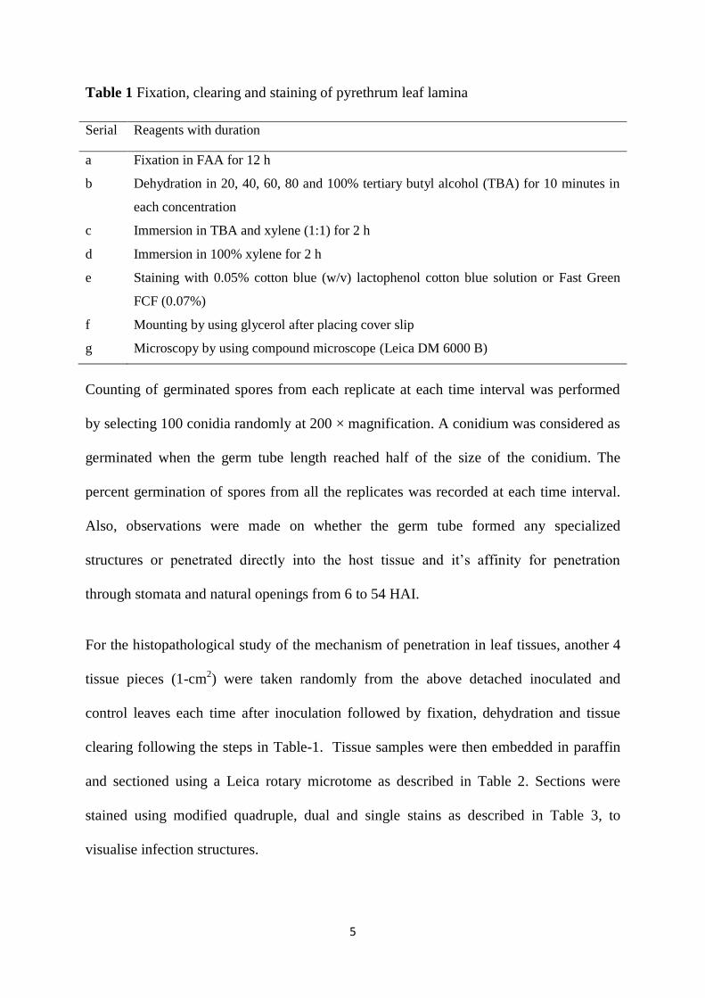
Table 1 Fixation, clearing and staining of pyrethrum leaf lamina
Serial Reagents with duration
a Fixation in FAA for 12 h
b Dehydration in 20, 40, 60, 80 and 100% tertiary butyl alcohol (TBA) for 10 minutes in
each concentration
c Immersion in TBA and xylene (1:1) for 2 h
d Immersion in 100% xylene for 2 h
e Staining with 0.05% cotton blue (w/v) lactophenol cotton blue solution or Fast Green
FCF (0.07%)
f Mounting by using glycerol after placing cover slip
g Microscopy by using compound microscope (Leica DM 6000 B)
Counting of germinated spores from each replicate at each time interval was performed
by selecting 100 conidia randomly at 200 × magnification. A conidium was considered as
germinated when the germ tube length reached half of the size of the conidium. The
percent germination of spores from all the replicates was recorded at each time interval.
Also, observations were made on whether the germ tube formed any specialized
structures or penetrated directly into the host tissue and it’s affinity for penetration
through stomata and natural openings from 6 to 54 HAI.
For the histopathological study of the mechanism of penetration in leaf tissues, another 4
tissue pieces (1-cm2) were taken randomly from the above detached inoculated and
control leaves each time after inoculation followed by fixation, dehydration and tissue
clearing following the steps in Table-1. Tissue samples were then embedded in paraffin
and sectioned using a Leica rotary microtome as described in Table 2. Sections were
stained using modified quadruple, dual and single stains as described in Table 3, to
visualise infection structures.
6
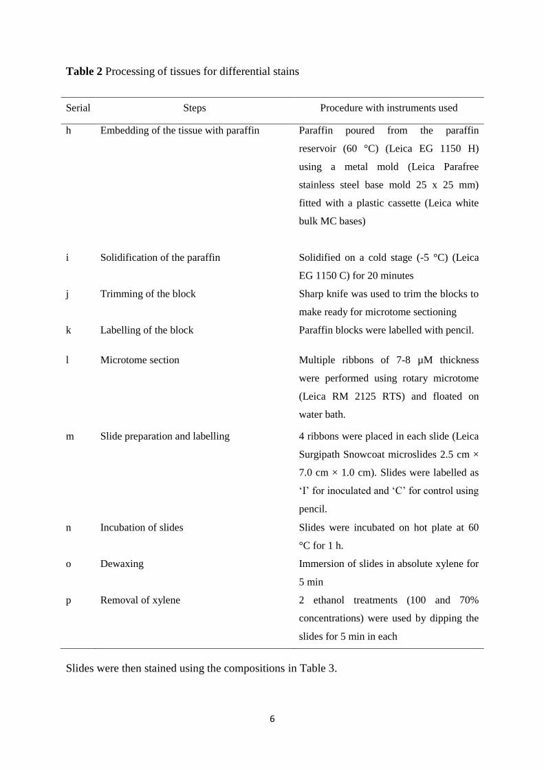
Table 2 Processing of tissues for differential stains
Serial Steps Procedure with instruments used
h Embedding of the tissue with paraffin
Paraffin poured from the paraffin
reservoir (60 °C) (Leica EG 1150 H)
using a metal mold (Leica Parafree
stainless steel base mold 25 x 25 mm)
fitted with a plastic cassette (Leica white
bulk MC bases)
i Solidification of the paraffin Solidified on a cold stage (-5 °C) (Leica
EG 1150 C) for 20 minutes
j Trimming of the block Sharp knife was used to trim the blocks to
make ready for microtome sectioning
k Labelling of the block Paraffin blocks were labelled with pencil.
l Microtome section Multiple ribbons of 7-8 µM thickness
were performed using rotary microtome
(Leica RM 2125 RTS) and floated on
water bath.
m Slide preparation and labelling 4 ribbons were placed in each slide (Leica
Surgipath Snowcoat microslides 2.5 cm ×
7.0 cm × 1.0 cm). Slides were labelled as
‘I’ for inoculated and ‘C’ for control using
pencil.
n Incubation of slides Slides were incubated on hot plate at 60
°C for 1 h.
o Dewaxing Immersion of slides in absolute xylene for
5 min
p Removal of xylene 2 ethanol treatments (100 and 70%
concentrations) were used by dipping the
slides for 5 min in each
Slides were then stained using the compositions in Table 3.
7
Table 3 Composition of differential stains
Quadruple stain (modified from Johansen
1940)
Dual stain (modified from
Ribichich et al. 2000)
Single stain
(Ranathunge et al.
2012)
Immersed in Safranin O for 2 h then rinsed
for 2-3 times using distilled water followed
by 5 min immersed in crystal violet then
washed with distilled water for twice. Then
extra stains were removed by using EMT
for 15 min. Then staining for 5 min in Fast
Green FCF solution followed by removal
of extra stain by using EMT for 15 sec.
Final staining was done by using Orange G
for 3 min. Afterwards; extra stains were
removed by using two changes of CME
and CMX for 15 sec in each. Final step
before mounting with immersion of the
slides in xylene for 5 min.
Safranin O was used as a
primary stain for 2 h
followed by rinses with
sterile distilled water.
Then extra stains were
removed by using EMT
for 15 min. Then Fast
Green FCF solution was
used for 10 min as a
counter stain. Extra stain
was removed by using
EMT and CME
respectively for 15 sec.
Lactophenol cotton
blue solution was
used for 10 min
followed by 2-3
rinses in sterile water.
EMT: Ethanol (95%): methyl cello solve: tert-butanol (1:1:1), CME: Clove oil: methyl cell
solve: ethanol (95%) (1:1:1), CMX: Clove oil: methyl cello solve: xylene (1:1:1)
After staining, all the slides were immersed in xylene for 5 min followed by mounting
with Leica Surgipath Micromount and placement of glass cover slips over the sample.
Infection, colonization and formation of pycnidia in pyrethrum leaves
For the study of phenotypical symptom expression between 1 and 12 DAI, a further 30
leaf laminas were selected from healthy 4-week-old pyrethrum plants (cultivar BR1).
Twenty leaf laminas were inoculated with S. tanaceti at a concentration of 106 spores/mL
following the same process of surface sterilization, inoculation and incubation as
described above. Another 10 leaf laminas were used as a control and were sprayed with
8
SDW. 5 replicates of each of inoculated and control leaflets were kept in a sterile
moistened filter paper placed in a 90 mm petri plates and incubated at 25 °C temperature
in 12 h photoperiod. Each leaf lamina was treated as a replicate. Progressive lesion
development and pycnidia formation was assessed on both sides of leaves using the
dissecting microscope (Leica M 205 FA). Lesion size was measured using the Leica
software.
For histopathological study of infection and colonization, 4 tissue pieces (1 cm2) were
taken randomly from the detached inoculated and control leaves by cutting at the junction
of the necrotic lesion of infected leaf and any point of the control leaf lamina respectively
at 3 and 12 DAI using a sterile scalpel. Then 0.5 cm2 piece was used for culturing in
water agar (1%) for 3 days followed by subculturing in V8 media to confirm the presence
of S. tanaceti using the taxonomy of conidiomata and pycnidia (Vaghefi 2012). The other
0.5 cm2 tissue piece was processed and labelled for histopathology. According to the
results on V8 medium, the corresponding tissue in the paraffin blocks were subsequently
labelled as Infected (IL) Control leaf (Cl) were processed for histopathology. Microtome
sections, slide preparation and use of differential stains were followed as mentioned in
Tables 2 and 3.
All plant tissues on prepared slides were observed under the light microscope (Leica DM
6000 B) and the images were taken using a Leica DFC 450 C camera.

9
Results
Germination of conidia and penetration by germinated hyphae in pyrethrum leaves
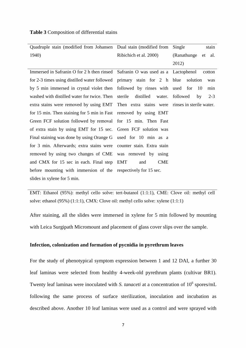
Germination of conidia started at 12 HAI and significantly increased over time. The
germination percentage of conidia was 11, 29, 41, 60 and 85% at 12, 24, 30, 48 and 54
HAI respectively on adaxial leaf surface (Fig. 1). No specialized penetration structures
occurred and the infection hyphae had no affinity towards the natural openings like
stomata (Fig. 2 a) in each observation up to 54 HAI. Both lactophenol cotton blue and
Fast Green FCF (0.07%) were effective to visualize the germinated spores on cleared leaf
surface.
Fig. 1 Percent germination of S. tanaceti conidia over time on adaxial leaf surface of pyrethrum
(Each bar denotes LSD value at significance level P<0.05, there were 5 replications for each time
observation)
-20
0
20
40
60
80
100
6 12 24 30 48 54
% g
erm
inat
ion
Hours after inoculation
10
Fig. 2 a) infection hypha (hy) or germ tube (gt) from the conidia
(co) had no affinity towards stomata (st) at 12 HAI.
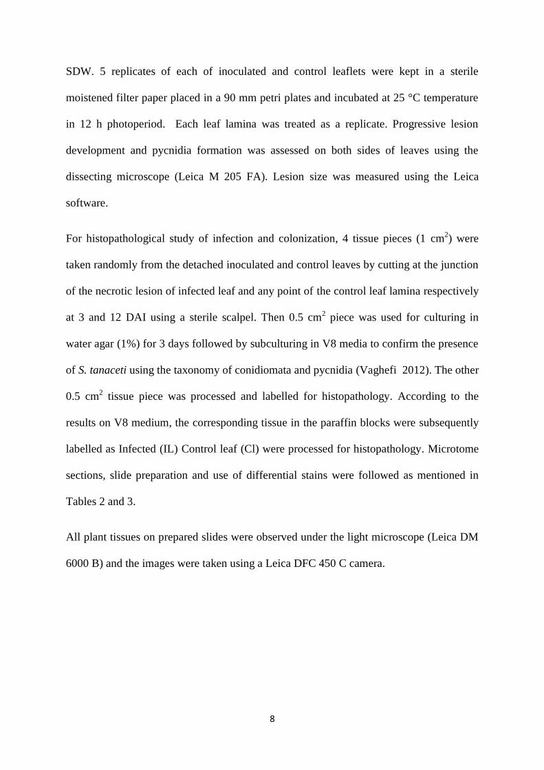
Using quadruple stain it was observed that the germ tubes initiated from the firmly
attached conidia penetrated directly through the upper layer of adaxial epidermis at 12
HAI. Firm attachment was represented as a slight depression at the point of attachment of
conidium on host surface (Fig. 3 a). Over time, infection hyphae from the adaxial
epidermis moved horizontally through the epidermis by invading the middle lamella of
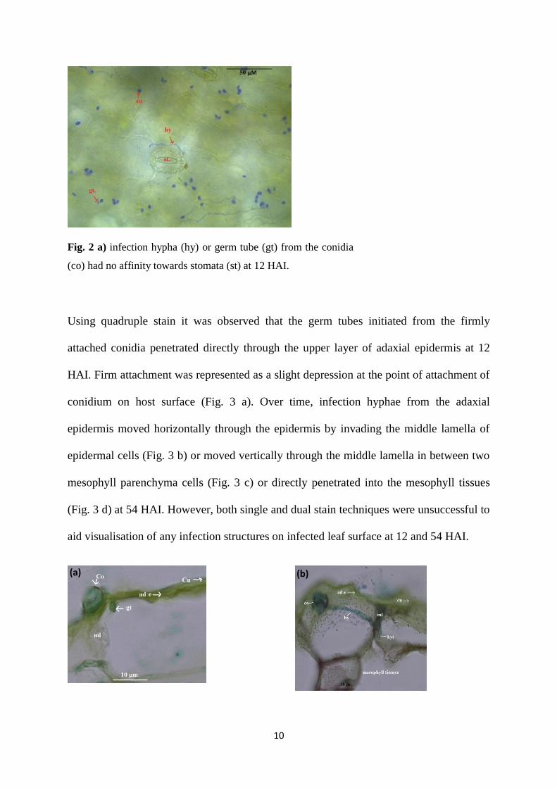
epidermal cells (Fig. 3 b) or moved vertically through the middle lamella in between two
mesophyll parenchyma cells (Fig. 3 c) or directly penetrated into the mesophyll tissues
(Fig. 3 d) at 54 HAI. However, both single and dual stain techniques were unsuccessful to
aid visualisation of any infection structures on infected leaf surface at 12 and 54 HAI.
(a) (b)
11
Fig. 3 Penetration of infection hyphae in pyrethrum leaves by S. tanaceti using quadruple stain. a)
firm attachment of conidium (co) at the point of attachment on host surface, germ tube (gt)
penetrated directly through the cuticle (cu) and adaxial epidermis (ad e); b) infection hypha (hy)
extended horizontally through the middle lamella (ml) of two epidermal cells; c) infection hypha
(hy) from the germinated conidium (co) infected horizontally through the middle lamella (ml) of
two adjacent mesophyll parenchyma cells; d) infection hypha (hy) penetrated directly from the
adaxial epidermis (ad e) to the mesophyll tissues.
Infection, colonization and formation of pycnidia on leaf tissues
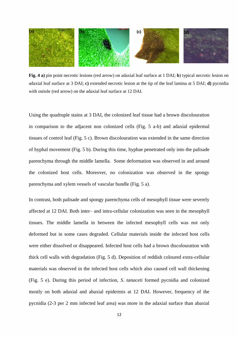
Pin point necrotic lesions developed sparsely on the adaxial surface of inoculated leaves
at 1 DAI (Fig. 4 a) and developed into distinct brown discoloured lesions (300 × 550
µM2) on adaxial leaf surface 3 DAI (Fig. 4 b). Over time, some of the lesions coalesced to
form black to deep brown lesions (3.0 × 3.5 mm2) particularly at the tip of the leaves at 5-
6 DAI which were more obvious on the adaxial surface than abaxial surface (Fig. 4 c).
Pycnidia formed at 9 DAI and the ostiole of the pycnidia was visible at 12 DAI. Almost
the entire leaf lamina was covered by the pycnidia which changed the leaf lamina colour
to brown. Distribution of the pycnidia was more in the adaxial leaf surface than abaxial
leaf surface (Fig. 4 d). Control leaf samples showed no symptoms.
(c) (d)
12
Fig. 4 a) pin point necrotic lesions (red arrow) on adaxial leaf surface at 1 DAI; b) typical necrotic lesion on
adaxial leaf surface at 3 DAI; c) extended necrotic lesion at the tip of the leaf lamina at 5 DAI; d) pycnidia
with ostiole (red arrow) on the adaxial leaf surface at 12 DAI.
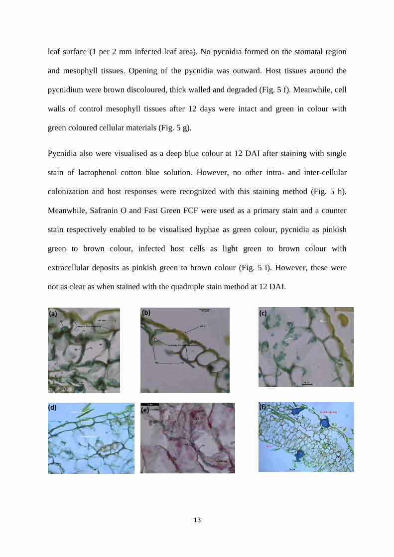
Using the quadruple stains at 3 DAI, the colonized leaf tissue had a brown discolouration
in comparison to the adjacent non colonized cells (Fig. 5 a-b) and adaxial epidermal
tissues of control leaf (Fig. 5 c). Brown discolouration was extended in the same direction
of hyphal movement (Fig. 5 b). During this time, hyphae penetrated only into the palisade
parenchyma through the middle lamella. Some deformation was observed in and around
the colonized host cells. Moreover, no colonization was observed in the spongy
parenchyma and xylem vessels of vascular bundle (Fig. 5 a).
In contrast, both palisade and spongy parenchyma cells of mesophyll tissue were severely
affected at 12 DAI. Both inter– and intra-cellular colonization was seen in the mesophyll
tissues. The middle lamella in between the infected mesophyll cells was not only
deformed but in some cases degraded. Cellular materials inside the infected host cells
were either dissolved or disappeared. Infected host cells had a brown discolouration with
thick cell walls with degradation (Fig. 5 d). Deposition of reddish coloured extra-cellular
materials was observed in the infected host cells which also caused cell wall thickening
(Fig. 5 e). During this period of infection, S. tanaceti formed pycnidia and colonized
mostly on both adaxial and abaxial epidermis at 12 DAI. However, frequency of the
pycnidia (2-3 per 2 mm infected leaf area) was more in the adaxial surface than abaxial
(a) (b) (c) (d)
13
leaf surface (1 per 2 mm infected leaf area). No pycnidia formed on the stomatal region
and mesophyll tissues. Opening of the pycnidia was outward. Host tissues around the
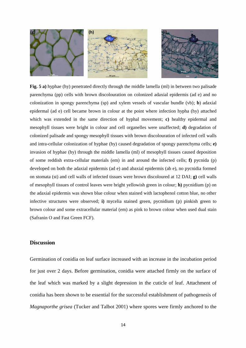
pycnidium were brown discoloured, thick walled and degraded (Fig. 5 f). Meanwhile, cell
walls of control mesophyll tissues after 12 days were intact and green in colour with
green coloured cellular materials (Fig. 5 g).
Pycnidia also were visualised as a deep blue colour at 12 DAI after staining with single
stain of lactophenol cotton blue solution. However, no other intra- and inter-cellular
colonization and host responses were recognized with this staining method (Fig. 5 h).
Meanwhile, Safranin O and Fast Green FCF were used as a primary stain and a counter
stain respectively enabled to be visualised hyphae as green colour, pycnidia as pinkish
green to brown colour, infected host cells as light green to brown colour with
extracellular deposits as pinkish green to brown colour (Fig. 5 i). However, these were
not as clear as when stained with the quadruple stain method at 12 DAI.
(a) (b) (c)
(d) (e) (f)
14
Fig. 5 a) hyphae (hy) penetrated directly through the middle lamella (ml) in between two palisade
parenchyma (pp) cells with brown discolouration on colonized adaxial epidermis (ad e) and no
colonization in spongy parenchyma (sp) and xylem vessels of vascular bundle (vb); b) adaxial
epidermal (ad e) cell became brown in colour at the point where infection hypha (hy) attached
which was extended in the same direction of hyphal movement; c) healthy epidermal and
mesophyll tissues were bright in colour and cell organelles were unaffected; d) degradation of
colonized palisade and spongy mesophyll tissues with brown discolouration of infected cell walls
and intra-cellular colonization of hyphae (hy) caused degradation of spongy parenchyma cells; e)
invasion of hyphae (hy) through the middle lamella (ml) of mesophyll tissues caused deposition
of some reddish extra-cellular materials (em) in and around the infected cells; f) pycnida (p)
developed on both the adaxial epidermis (ad e) and abaxial epidermis (ab e), no pycnidia formed
on stomata (st) and cell walls of infected tissues were brown discoloured at 12 DAI; g) cell walls
of mesophyll tissues of control leaves were bright yellowish green in colour; h) pycnidium (p) on
the adaxial epidermis was shown blue colour when stained with lactophenol cotton blue, no other
infective structures were observed; i) mycelia stained green, pycnidium (p) pinkish green to
brown colour and some extracellular material (em) as pink to brown colour when used dual stain
(Safranin O and Fast Green FCF).
Discussion
Germination of conidia on leaf surface increased with an increase in the incubation period
for just over 2 days. Before germination, conidia were attached firmly on the surface of
the leaf which was marked by a slight depression in the cuticle of leaf. Attachment of
conidia has been shown to be essential for the successful establishment of pathogenesis of
Magnaporthe grisea (Tucker and Talbot 2001) where spores were firmly anchored to the
(g) (h) (i)

15
host surface (Nicole and Gianinazzi-Pearson 1996). Depression on the host surface at the
point of conidia attachment was reported for P. macdonaldii on sunflower cotyledon
surface (Roustaee et al. 2000). The germ tube had no affinity towards natural openings
such as stomata nor formed an appressoria during the incubation period. This was also
found for spores of similar kinds of fungal pathogens such as Leptophaeria maculans in
which hyphae directly penetrated the surface of canola leaf lamina (Brassica napus)
without forming appressoria. In contrast, hyphae of L. maculans on canola leaf lamina
(Hammond et al. 1985; Idnurm and Howlett 2002) and Phomopsis phaseoli on soybean
seedlings (Kulik 1988) also entered through the stomata as well as direct penetration.
Histopathology using quadruple stain showed that the infection hyphae penetrated
directly into the epidermal cells and then further moved toward the mesophyll tissues
inter-cellularly. Direct penetration has commonly been reported in related fungi like
Phoma medicaginis in alfalfa leaf (Castell-Miller et al. 2007), P. clematidina on clematis
(Clematis spp.) leaf surface (Van de Graaf et al. 2002) and S. nodorum in wheat leaf
(Solomon et al. 2006). Direct penetration was mentioned by Roustaee et al. (2000) that at
the point of conidia attachment of P. macdonaldii on sunflower cotyledon surface, germ
tubes formed infection pegs which perforated the host cuticle through exerting
mechanical pressure.
Phenotypic expression of initial pin point brown necrotic lesions appeared on leaf surface
at 1 DAI and the lesions extended alongside the margin which completely covered the
entire leaf lamina within 9-12 DAI. Fully matured pycnidia were almost evenly
distributed on the infected necrotic leaves at 12 DAI. Likewise in histopathology at 3 DAI
brown pigmented infected epidermal cells at the colonized area were distinct in
comparison to the healthy cells away from the colonized or uninfected control epidermal
16
leaf tissues. The brown discolouration of the infected cells by S. tanaceti might have been
caused by the release of some toxins at the infection site, because some related fungi like
Phoma and Ascochyta species (Pedras and Biesenthal 2000; Fogliano et al. 1998; Idnurm
and Howlett 2002) have been shown to release toxins at the site of infection and cause
brown discolouration. Similar brown pigmentation was also reported in the lesion area of
L. maculans infected canola leaves (Sexton and Howlett 2001).
Intercellular colonization by hyphae of S. tanaceti occurred within the palisade
parenchyma without any obvious damage at 3 DAI which was in full agreement with the
infection nature of L. maculans in the infected leaf tissues of oilseed rape at initial stage
of infection (Hammond et al.1985). But, infection hyphae ramified intra- and inter-
cellularly within the spongy and palisade parenchyma tissues at 12 DAI. Middle lamella
of parenchyma cells may have been either degraded or dissolved due to activation of cell
decomposing enzymes like cyanide hydratase as reported by Sexton and Howlett (2001).
Some of the infected cell walls were thickened around the middle lamella of parenchyma
cells which might have indicated the deposition of lignin (Hammond and Lewis 1987;
Sexton and Howlett 2001). Likewise, heavy lignification and deposition of tannin were
observed in the heavily infected tissues of lodgepole pine (Pinus contorta Dougl.)
seedlings after infection by either Anthracobia maurilabra, A. tristis, Geopyxis
carbonaria or Gyromitra infula (Egger and Paden 1986).
The frequency of necrotic lesions and pycnidia after inoculation by S. tanaceti were more
on the adaxial epidermis than abaxial epidermis of leaf lamina. A survey conducted in
1909 by Heald and Wolf (1911) in Texas reported similar nature of abundant brown spots
and pycnidia formation on upper leaf surfaces of cultivated crops infected by Phyllosticta
congesta and P. bumeliifolia. However, further studies are needed to explore the reason

17
for higher frequency of lesions and pycnidia caused by S. tanaceti on the adaxial surface
compared to the abaxial surface of pyrethrum leaves.
The single stain technique was found to be useful to identify the infection events on
cleared leaf surface but almost ineffective in histopathological studies. Whereas, the dual
stain technique, although useless in histopathology at initial stage of infection and was
limited to the latter stage of infection when host cellular materials were almost degraded
and pycnidia formed. In contrast, the quadruple stain was informative for
histopathological analysis of infected tissues at any stages of infection caused by S.
tanaceti in pyrethrum leaves. This technique could be used as an alternate to some costly
tissue staining processes such as GFP and advanced microscopy such as SEM or
Transmission electron microscopy (TEM). Application of the quadruple stain to study
varietal resistance, infection process of S. tanaceti from seed to seedling and to explore
the complete disease cycle of ray blight in pyrethrum is currently underway.
Acknowledgements:
This project was supported by Botanical Resources Australia (BRA)-Agricultural Services Pty.
Ltd.
References
Egger KN, Paden JW (1986) Biotrophic associations between lodgepole pine seedlings and post
fire ascomycetes (Pezizales) in monoxenic culture. Can J Bot 64(11):2719-2725
Fogliano V, Marchese A, Scaloni A, Ritieni A, Visconti A, Randazzo G, Graniti A (1998)
Characterization of a 60 kDa phytotoxic glycoprotein produced by Phoma
tracheiphilaand its relation to malseccin. Physiol Mol Plant Pathol 53(3): 149-161
18
Hammond KE, Lewis B, Musa T (1985) A systemic pathway in the infection of oilseed rape
plants by Leptosphaeria maculans. Plant Pathol 34(4): 557-565
Hammond KE, Lewis BG (1987) Variation in stem infections caused by aggressive and non-
aggressive isolates of Leptosphaeria maculans on Brassica napus var. oleifera. Plant
Pathol 36(1):53-65
Heald FD, Wolf FA (1911) New species of Texas fungi. Mycologia 3(1): 5-22
Hitmi A, Coudret A, Barthomeuf C (2000) The production of pyrethrins by plant cell and
tissue cultures of Chrysanthemum cinerariaefolium and Tagetes species. Critical
reviews in biochemistry and molecular biology 35(5):317-337
Idnurm A, Howlett BJ (2002) Isocitrate lyase is essential for pathogenicity of the fungus
Leptosphaeria maculans to canola (Brassica napus). Eukaryotic Cell 1(5): 719-724
Johansen DA (1940) Plant microtechnique. New York , McGraw-Hill.
Knight NL, Sutherland MW (2011) A rapid differential staining technique for Fusarium
pseudograminearum in cereal tissues during crown rot infections. Plant Pathol 60(6):
1140-1143
Kulik M (1988) Observations by scanning electron and brightfield microscopy on the mode of
penetration of soybean seedlings by Phomopsis phaseoli. Plant Dis 72:115-118
Liberato J, Barreto R, Shivas R (2005) Leaf-clearing and staining techniques for the observation
of conidiophores in the Phyllactinioideae (Erysiphaceae). Australas Plant Pathol
34(3):401-404
Nicole M, Gianinazzi-Pearson V (1996) Histology, ultrastructure and molecular cytology of
plant-microorganism interactions. 1st edition, Kluwer Academic Publishers. The
Netherlands.
Patton RF, Spear RN (1989) Histopathology of colonization in leaf tissue of Castilleja,
Pedicularis, Phaseolus, and Ribes species by Cronartium ribicola. Phytopathology
9(5):539-547
19
Pedras MSC, Biesenthal CJ (2000) Vital staining of plant cell suspension cultures: evaluation of
the phytotoxic activity of the phytotoxins phomalide and destruxin B. Plant Cell Reports
19(11): 1135-1138
Pethybridge SJ, Gent DH, Groom T, Hay FS (2013) Minimizing crop damage through
understanding relationships between pyrethrum phenology and ray blight disease
severity. Plant Dis 97(11):1431-1437
Pethybridge SJ, Hay FS, Esker PD, Gent DH, Wilson CR, Groom T, Nutter Jr FW (2008)
Diseases of pyrethrum in Tasmania: challenges and prospects for management. Plant Dis
92(9):1260-1272
Pethybridge SJ, Wilson C (1998) Confirmation of ray blight disease of pyrethrum in Australia.
Australas Plant Pathol 27(1): 45-48
Ranathunge N, Mongkolporn O, Ford R, Taylor P (2012) Colletotrichum truncatum Pathosystem
on Capsicum spp: infection, colonization and defence mechanisms. Australas Plant Pathol
41(5):463-473
Ribichich KF, Lopez SE, Vegetti AC (2000) Histopathological spikelet changes produced by
Fusarium graminearum in susceptible and resistant wheat cultivars. Plant Dis 84(7): 794-
802
Roustaee A, Dechamp-Guillaume G, Gelie B, Savy C, Dargent R, Barrault G (2000)
Ultrastructural studies of the mode of penetration by Phoma macdonaldii in sunflower
seedlings. Phytopathology 90(8): 915-920
Sexton A, Howlett B (2001) Green fluorescent protein as a reporter in the Brassica–
Leptosphaeria maculans interaction. Physiol Mol Plant Pathol 58(1):13-21
Taylor PWJ, Burgess LW (1983) Histopathology of infection and colonisation of wheat by
Fusarium graminearum Group 1. Proceedings of the 4th International Congress of Plant
Pathology, Melbourne.
20
Tucker SL, Talbot NJ (2001) Surface attachment and pre-penetration stage development by plant
pathogenic fungi. Annu Rev Phytopathol 39(1):385-417
Vaghefi N, Pethybridge S, Ford R, Nicolas M, Crous P, Taylor P (2012) Stagonosporopsis spp.
associated with ray blight disease of Asteraceae. Australas Plant Pathol 41(6): 675-686
Van De Graaf P, Joseph ME, Chartier-Hollis JM, O’Neill TM (2002) Prepenetration stages in
infection of clematis by Phoma clematidina. Plant Pathol 51(3):331-337
21
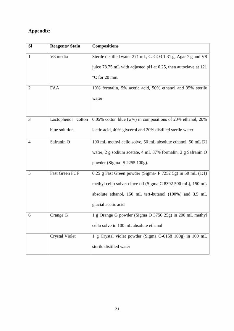
Appendix:
Sl Reagents/ Stain Compositions
1 V8 media Sterile distilled water 271 mL, CaCO3 1.31 g, Agar 7 g and V8
juice 78.75 mL with adjusted pH at 6.25, then autoclave at 121
°C for 20 min.
2 FAA 10% formalin, 5% acetic acid, 50% ethanol and 35% sterile
water
3 Lactophenol cotton
blue solution
0.05% cotton blue (w/v) in compositions of 20% ethanol, 20%
lactic acid, 40% glycerol and 20% distilled sterile water
4 Safranin O 100 mL methyl cello solve, 50 mL absolute ethanol, 50 mL DI
water, 2 g sodium acetate, 4 mL 37% formalin, 2 g Safranin O
powder (Sigma- S 2255 100g).
5 Fast Green FCF 0.25 g Fast Green powder (Sigma- F 7252 5g) in 50 mL (1:1)
methyl cello solve: clove oil (Sigma C 8392 500 mL), 150 mL
absolute ethanol, 150 mL tert-butanol (100%) and 3.5 mL
glacial acetic acid
6 Orange G 1 g Orange G powder (Sigma O 3756 25g) in 200 mL methyl
cello solve in 100 mL absolute ethanol
Crystal Violet 1 g Crystal violet powder (Sigma C-6158 100g) in 100 mL
sterile distilled water
Minerva Access is the Institutional Repository of The University of Melbourne
Author/s:Bhuiyan, MAHB;Groom, T;Nicolas, ME;Taylor, PWJ
Title:Histopathology of S-tanaceti infection in pyrethrum leaf lamina
Date:2015-11-01
Citation:Bhuiyan, M. A. H. B., Groom, T., Nicolas, M. E. & Taylor, P. W. J. (2015). Histopathology ofS-tanaceti infection in pyrethrum leaf lamina. AUSTRALASIAN PLANT PATHOLOGY, 44 (6),pp.629-636. https://doi.org/10.1007/s13313-015-0377-0.
Persistent Link:http://hdl.handle.net/11343/283004