Embed Size (px)
Citation preview
the diffuse neuroendocrine system. This interpretation of the problem was first adopted by Leong and Matthews [9]. In our opinion, the basis for this hypothesis is the fact that PG maintains central levels of regulation and realizes its effects through the central nervous, autonomic nervous, and endocrine systems [6, 13], and that cells of the diffuse APUD system act on homeostasis at organ and system levels, and are under the control of central mechanisms of regulation, including PG.
LITERATURE CITED
i. G. G. Avtandilov, V. P. Nevzorov, and O. F. Nevzorova, Systemic Stereometric Analysis of Cellular Ultrastructures [in Russian], Kishinev (1984).
2. B. K. Aleshin, Current Problems in Endocrinology: Neurobiological Aspects [in Russian], Moscow (1981), pp. 6-24.
3. I. M. Kvetnoi and N. T. Raikhlin, Klin. Med., No. ii, 15 (1978). 4. N. T. Raikhlin, I. M. Kvetnoi, E. A. Smirnova, and V. N. Anders, Current Problems in
Endocrinology: Neurobiological Aspects [in Russian], Moscow (1981), pp. 124-140. 5. V. M. Rozum, Functional Histo-Ultrastructure of Calcification in the Pineal Gland
(Clinical-Experimental Investigation). Author's Abstract of Dissertation for the De- gree of Candidate of Medical Sciences, Moscow (1989).
6. V. D. Slepushkin and V. G. Pashinskii, The Pineal Gland and Adaptation [in Russian], Tomsk (1982).
7. E. I. Chazov and V. A. Isachenkov, The Pineal Gland, Its Place and Role in the System of Endocrine Regulation [in Russian], Moscow (1974).
8. R. Krstid, J. Pineal Res., 2, 21 (1985). 9. A. S. Y. Leong and S. D. Matthews, Med. Hypoth., 5, 265 (1979).
i0. S. D. Matthews and A. S. Y. Leong, Melatonin: Current Status and Perspectives, N. Birau and W. Schloot (eds.), Oxford (1981), pp. 77-82.
ii. M. M~ller, Cell Tissue Res., 152, 13 (1974). 12. M. M~ller, Cell Tissue Res., 169, 7 (1976). 13. W. B. Quay, Pineal Chemistry in Cellular and Physiological Mechanisms, Springfield
(1974). 14. L. Vollrath, The Pineal Gland: Comprehensive Endocrinology, R. J. Reiter (ed.), New
York (1984), pp. 285-321.
HISTOCHEMICAL ANALYSIS OF OXIDOREDUCTASES IN CARDIOMYOCYTES OF
MATURE AND OLD RATS AFTER LOW-DOSE EXTERNAL GAMMA-IRRADIATION
A. P. Amvros'ev, G. G. Vereshchako, UDC 612.173.1.015.11.014.482.4/.06:612.67 and N. V. Banetskaya
KEY WORDS: cardiomyocytes; dehydrogenases; gamma-irradiation
Investigations of myocardial morphology and functions under the influence of ionizing radiation have been conducted mainly with the use of lethal or sublethal doses [3, 6, ii, 12]. Yet the increase in the external environmental radiation factor at the present time has stimulated interest in the effect of small doses of irradiation and its possible after- effects on heart muscle function. The heart is an organ with intensive energy metabolism. In this connection the study of the state of oxidoreduction processes in the cardiomyocytes after irradiation with small doses is very urgent. It is also necessary to study age-related sensitivity of the myocardium to the action of radiation. These problems have been in- adequately dealt with in the literature.
The aim of this investigation was to study changes in the level of activity of the principal oxidoreductases in cardiomyocytes of mature and old rats after total gamma-irradia- tion in a relatively small dose.
Laboratory of Morphology and Cytogenetics, Institute of Radiobiology, Academy of Scien- ces of the Belorussian SSR, Minsk. (Presented by Academician of the Academy of Medical Sci- ences of the USSR V. V. Kupriyanov.) Translated from Byulleten' Eksperimental'noi Biologii i Meditsiny, Vol. ii0, No. ii, pp. 548-550, November, 1990. Original article submitted De- cember 20, 1989.
1580 0007-4888/90/0011-1580512.50 �9 1991 Plenum Publishing Corporation
qo/~ L
7r " :
,9 I 0 3 0
1201 - , 4
. . . . . 2 ~
70'" ' I I i 3 70 30
Fig. 1 Fig. 2
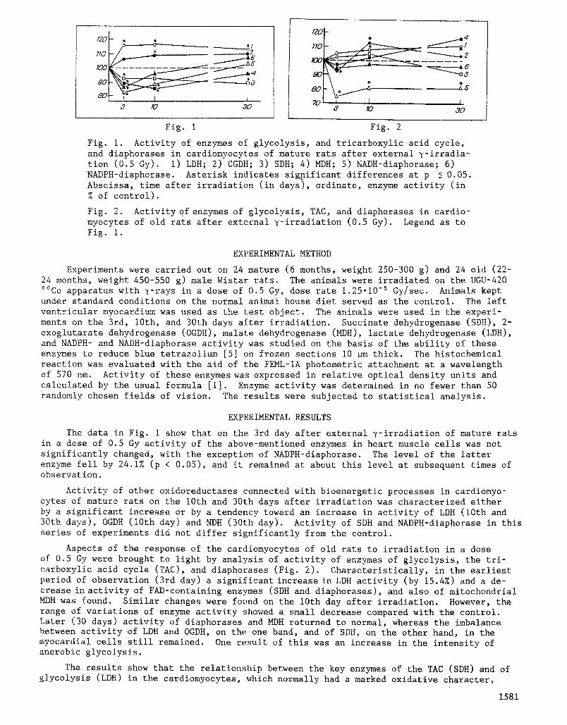
Fig. i. Activity of enzymes of glycolysis, and tricarboxylic acid cycle, and diaphorases in cardiomyocytes of mature rats after external 7-irradia- tion (0.5 Gy). i) LDH; 2) CGDH; 3) SDH; 4) MDH; 5) NADH-diaphorase; 6) NADPH-diaphorase. Asterisk indicates significant differences at p 5 0.05. Abscissa, time after irradiation (in days), ordinate, enzyme activity (in % of control).
Fig. 2. Activity O f enzymes of glycolysis, TAC, and diaphorases in cardio- myocytes of old rats after external 7-irradiation (0.5 Gy). Legend as to Fig. i.
EXPERIMENTAL METHOD
Experiments were carried out on 24 mature (6 months, weight 250-300 g) and 24 old (22- 24 months, weight 450-550 g) male Wistar rats. The animals were irradiated on the UGU-420 ~~ apparatus with ~-rays in a dose of 0.5 Gy, dose rate 1.25.10 -5 Gy/sec. Animals kept under standard conditions on the normal animal house diet served as the control. The left ventricular myocardium was used as the test object. The animals were used in the experi- ments on the 3rd, 10th, and 30th days after irradiation. Succinate dehydrogenase (SDH), 2- oxoglutarate dehydrogenase (OGDH), malate dehydrogenase (MDH), lactate dehydrogenase (LDH), and NADPH- and NADH-diaphorase activity was studied on the basis of the ability of these enzymes to reduce blue tetrazolium [5] on frozen sections I0 ~m thick. The histochemica! reaction was evaluated with the aid of the FEML-IA photometric attachment at a wavelength of 570 nm. Activity of these enzymes was expressed in relative optical density units and calculated by the usual formula [i]. Enzyme activity was determined in no fewer than 50 randomly chosen fields of vision. The results were subjected to statistical analysis.
EXPERIMENTAL RESULTS
The data in Fig. 1 show that on the 3rd day after external 7-irradiation of mature rats in a dose of 0.5 Gy activity of the above-mentioned enzymes in heart muscle cells was not significantly changed, with the exception of NADPH-diaphorase. The level of the latter enzyme fell by 24.1% (p < 0.05), and it remained at about this level at subsequent times of observation.
Activity of other oxidoreductases connected with bioenergetic processes in cardiomyo- cytes of mature rats on the 10th and 30th days after irradiation was characterized either by a significant increase or by a tendency toward an increase in activity of LDH (10th and 30th days), OGDH (10th day) and NDH (30th day). Activity of SDH and NADPH-diaphorase in this series of experiments did not differ significantly from the control.
Aspects of the response of the cardiomyocytes of old rats to irradiation in a dose of 0.5 Gy were brought to light by analysis of activity of enzymes of glycolysis, the tri- carboxylic acid cycle (TAC), and diaphorases (Fig. 2). Characteristically, in the earliest period of observation (3rd day) a significant increase in LDH activity (by 15.4%) and a de- crease in activity of FAD-containing enzymes (SDH and diaphorases), and also of mitochondrial MDH was found. Similar changes were found on the 10th day after irradiation. However, the range of variations of enzyme activity showed a small decrease compared with the control. Later (30 days) activity of diaphorases and MDH returned to normal, whereas the imbalance between activity of LDH and OGDH, on the one band, and of SDH, on the other hand, in the myocardial cells still remained. One result of this was an increase in the intensity of anerobic glycolysis.
The results show that the relationship between the key enzymes of the TAC (SDH) and of glycolysis (LDH) in the cardiomyocytes, which normally had a marked oxidative character,
1581
shifted toward potentiation of anerobic processes after y-irradiation in a dose of 0.5 Gy. In mature animals this was clearly apparent on the 30th day, whereas in the old rats it was found throughout the period of observation. This state of affairs is connected with dif- ferences in the energy metabolism of mature and old animals. With aging, the ability of the myocardium to utilize anerobic glycolysis is known to increase [2].
Intensification of anerobic metabolism in the cardiomyocytes of mature animals, even by the end of the ist month after irradiation, may to a certain degree serve as an indicator of acceleration of age-related changes in the heart. This state of affairs is in agreement with the general idea of ionizating radiation as a factor accelerating aging processes in the body [4].
It can be tentatively suggested that intensification of anerobic processes under the influence of ionizing radiation reflects the state of relative hypoxia in the cardiomyocytes and is compensatory in character.
It must also be pointed out that stimulation of anerobic processes in the heart muscle cells has an inhibitory effect on activity of enzymes of TAC. This is the case more espe- cially with SDH and MDH in old rats. Activity of mitochondrial enzymes also depends on changes in structure of the mitochondria, membrane permeability, and other parameters, aris- ing under the influence of small doses of ionizing radiation [8, 9]. Under these same con- ditions OGDH activity had a tendency to increase. This phenomenon may be based on the adequate supply of oxidation substrate not only in the conversion chain in TAC, but also on account of activation of transamination reactions, as confirmed by data in [7].
Activity of NADH- and NADPH-diaphorases in the cardiomyocytes showed the most significant changes in most series of experiments involving irradiation in a dose of 0.5 Gy. Since these enzymes in mitochondria and the endoplasmic reticulum are relatively firmly bound with mem- branous structures, the changes discovered in the activity of these enzymes are evidence of a disturbance of intracellular oxidative processes, by means of which coenzymes undergo re- generation. Reduction of diaphorase activity may have a considerable influence on the work of the various energy-yielding cycles, for diaphorases play a direct part in the realization of activity of coenzyme-dependent dehydrogenases. A few biochemical investigations have de- monstrated a decrease in the NAD and NADP content in the heart muscle 1 month after irradia- tion in a dose of 0.4 Gy [10], which also confirms the view that intracellular oxidative processes are disturbed.
Thus the changes we found in the activity of certain oxidoreductases after whole-body y-irradiation in a dose of 0.5 Gy lead to the conclusion that a state of hypoxia develops in the animals' cardiomyocytes. This is more characteristic of the myocardium of old rats. Changes in intracellular oxidative processes in the cardiomyocytes of mature rats, just as of old rats, are the result of discoordination of the various stages of energy metabolism, most likely due to the indirect action of ionizing radiation through neurohumoral mechanisms of regulation.
LITERATURE CITED
i. L. S. Agroskin and G. V. Papayan, Cytophotometry: Apparatus and Methods of Analysis of Cells Based on Light Absorption [in Russian], Leningrad (1977).
2. L. N. Bogatskaya and A. Ya. Litoshenko, The Biology of Aging [in Russian], Leningrad (1982), pp. 267-279.
3. V. I. Efimov, Med. Radiol., 28, No. 2, 74 (1983). 4. G. A. Zedgenidze, The Biology of Aging [in Russian], Leningrad (1982), pp. 130-143. 5. Z. Lojda, R. Gossrau, and T. Schiebler, Enzyme Histochemistry: Laboratory Methods [in
Russian], N. T. Raikhlin (ed.), Moscow (1982). 6. E. I. Vorob'ev (ed.), Outlines of Radiation Cardiology [in Russian], Moscow (1978). 7. L. S. Cherkasova (ed.), Role of the Adrenals in Biochemical Changes under the Influence
of Small Doses of Ionizing Radiation [in Russian], Minsk (1969). 8. V. S. Romanov and L. A. Bespalova, Radiobiologiya, 21, No. 2, 238 (1981). 9. V. V. Sirotkin, G. G. Avtandilov, and N. D. Volodin, Byull. Eksp. Biol. Med., No. 12,
751 (1988). i0. L. P. Shalushkova, Izv. Akad. Nauk BSSR, Ser. Biol. Nauk, No. 4, 87 (1968). Ii. M. M. Applefield, Primary Cardiol., 12, No. 5, 159 (1986). 12. K. Jonek and S. Kosmider, Strahlentherapie, 122, No. 2, 198 (1963).
1582