Embed Size (px)
Citation preview
Energies 2012, 5, 1532-1553; doi:10.3390/en5051532
energies ISSN 1996-1073
www.mdpi.com/journal/energies Article
High Lipid Induction in Microalgae for Biodiesel Production
Kalpesh K. Sharma, Holger Schuhmann and Peer M. Schenk *
Algae Biotechnology Laboratory, School of Agriculture and Food Sciences, The University of Queensland, Brisbane, Queensland 4072, Australia; E-Mails: [email protected] (K.K.S.); [email protected] (H.S.)
* Author to whom correspondence should be addressed; E-Mail: [email protected]; Tel.: +61-7-3365-8817; Fax: +61-7-3365-1699.
Received: 30 March 2012; in revised form: 3 May 2012 / Accepted: 8 May 2012 / Published: 18 May 2012
Abstract: Oil-accumulating microalgae have the potential to enable large-scale biodiesel production without competing for arable land or biodiverse natural landscapes. High lipid productivity of dominant, fast-growing algae is a major prerequisite for commercial production of microalgal oil-derived biodiesel. However, under optimal growth conditions, large amounts of algal biomass are produced, but with relatively low lipid contents, while species with high lipid contents are typically slow growing. Major advances in this area can be made through the induction of lipid biosynthesis, e.g., by environmental stresses. Lipids, in the form of triacylglycerides typically provide a storage function in the cell that enables microalgae to endure adverse environmental conditions. Essentially algal biomass and triacylglycerides compete for photosynthetic assimilate and a reprogramming of physiological pathways is required to stimulate lipid biosynthesis. There has been a wide range of studies carried out to identify and develop efficient lipid induction techniques in microalgae such as nutrients stress (e.g., nitrogen and/or phosphorus starvation), osmotic stress, radiation, pH, temperature, heavy metals and other chemicals. In addition, several genetic strategies for increased triacylglycerides production and inducibility are currently being developed. In this review, we discuss the potential of lipid induction techniques in microalgae and also their application at commercial scale for the production of biodiesel.
Keywords: algaculture; biofuels; biodiesel; induction; lipids; microalgae; oil production; triacylglycerides
OPEN ACCESS
Energies 2012, 5
1533
1. Introduction
Sustainable production of renewable energy is being debated globally since it is increasingly understood that first generation biofuels, primarily produced from food crops and mostly oil seeds, compete for arable land, freshwater or biodiverse natural landscapes and are limited in their ability to achieve targets for biofuel production. These concerns have increased the interest in developing second and third generation biofuels such as lignocellulosics and microalgae, respectively, which potentially offer great opportunities in the longer term and do not need to compete for arable land and precious freshwater [1,2]. Due to continuous and increasing combustion of fossil carbon, the amount of greenhouse gas CO2 has increased. As a result global warming and climate change are threatening ecological stability, food security and social welfare [3,4]. The transportation and energy sector are the two major sources, responsible for the generation of 20% and 60% of greenhouse gases (GHG) emissions, respectively, and it is expected that with the development of emerging economies the global consumption of energy will rise considerably and this will lead to more environmental damage [5].
Photosynthesis is the only process that can convert CO2 into organic compounds with high energy content, and thus can provide a source for sustainable transport fuel production. There is an urgent need to develop technologies which are able to produce an additional five to six billion tons of organic carbon apart from the current harvest from agricultural crops [3]. Large-scale cultivation of microalgae may be 10–20 times more productive on a per hectare basis than other biofuel crops, are able to use a wide variety of water sources, and have a strong potential to produce biofuels without the competition for food production [2]. Algae can be produced either as macrophytes via marine aquaculture [6] or in large-scale microalgae cultivation systems in open ponds or in photobioreactors [1]. Microalgae are currently considered the most promising types of algae for biofuel production, based on their high lipid contents. Recent progress in the production of microalgae has been intensively reviewed [7], and future perspectives have been presented by Stephens et al. [5]. Microalgae can also be cultivated as an integrated concept with wastewater treatment to optimize the energetic and financial input for the production process [8].
Triacylglycerides (TAGs) generally serve as energy storage in microalgae that, once extracted, can be easily converted into biodiesel through transesterification reactions [3,9]. These neutral lipids bear a common structure of triple esters where usually three long-chain fatty acids (FAs) are coupled to a glycerol molecule. Transesterification displaces glycerol with small alcohols (e.g., methanol). Recently, the rise in petroleum prices and the need to reduce greenhouse gas emission has seen a renewed interest in large-scale biodiesel production [10].
Within the last few decades the concept of lipid induction in microalgae has been intensively studied to increase TAG production in microalgae, but at present different lipid induction techniques have not been compared to each other. Here we provide a review of different lipid inducing techniques in microalgae and their potential to be used for biodiesel production.
2. Lipids in Microalgae
Lipids produced by microalgae generally include neutral lipids, polar lipids, wax esters, sterols and hydrocarbons, as well as prenyl derivatives such as tocopherols, carotenoids, terpenes, quinines and pyrrole derivatives such as the chlorophylls. Lipids produced by microalgae can be grouped into two

Energies 2012, 5
1534
categories, storage lipids (non-polar lipids) and structural lipids (polar lipids). Storage lipids are mainly in the form of TAG made of predominately saturated FAs and some unsaturated FAs which can be transesterified to produce biodiesel. Structural lipids typically have a high content of polyunsaturated fatty acids (PUFAs), which are also essential nutrients for aquatic animals and humans. Polar lipids (phospholipids) and sterols are important structural components of cell membranes which act as a selective permeable barrier for cells and organelles. These lipids maintain specific membrane functions, providing the matrix for a wide variety of metabolic processes and participate directly in membrane fusion events. In addition to a structural function, some polar lipids may act as key intermediates (or precursors of intermediates) in cell signaling pathways (e.g., inositol lipids, sphingolipids, oxidative products) and play a role in responding to changes in the environment.
Of the non-polar lipids, TAGs are abundant storage products, which can be easily catabolized to provide metabolic energy [11]. In general, TAGs are mostly synthesized in the light, stored in cytosolic lipid bodies, and then reutilized for polar lipid synthesis in the dark [12]. Microalgal TAGs are generally characterized by both, saturated and monounsaturated FAs. However, some oil-rich species have demonstrated a capacity to accumulate high levels of long-chain polyunsaturated fatty acids (PUFA) as TAG [13,14]. A detailed study on both accumulation of TAG in the green microalga Parietochloris incisa and storage into chloroplastic lipids (following recovery from nitrogen starvation) led to the conclusion that TAGs may play an additional role beyond being an energy storage product in this alga [13,15]. Hence, PUFA-rich TAGs are metabolically active and are suggested to act as a reservoir for specific fatty acids. In response to a sudden change in the environmental condition, when the de novo synthesis of PUFA may be slower, PUFA-rich TAG may donate specific acyl groups to monogalactosyldiacylglycerol (MGDG) and other polar lipids to enable rapid adaptive membrane reorganization [15,16].
3. Methods of Lipid Induction
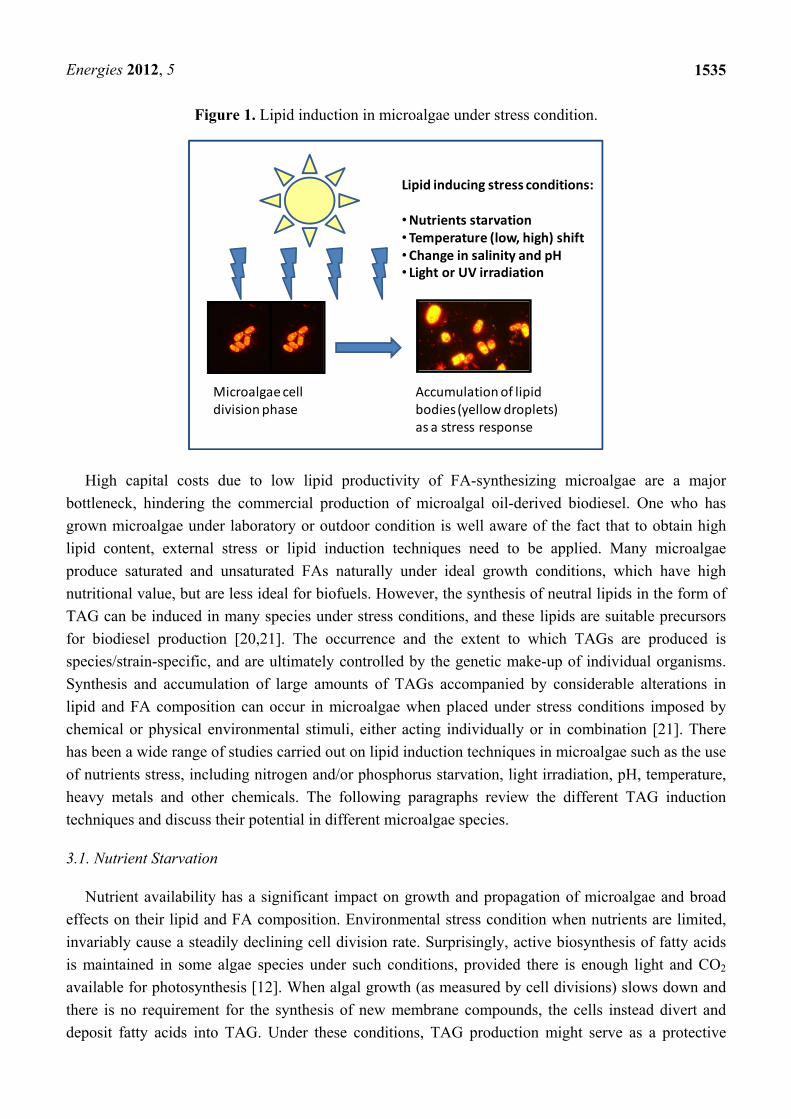
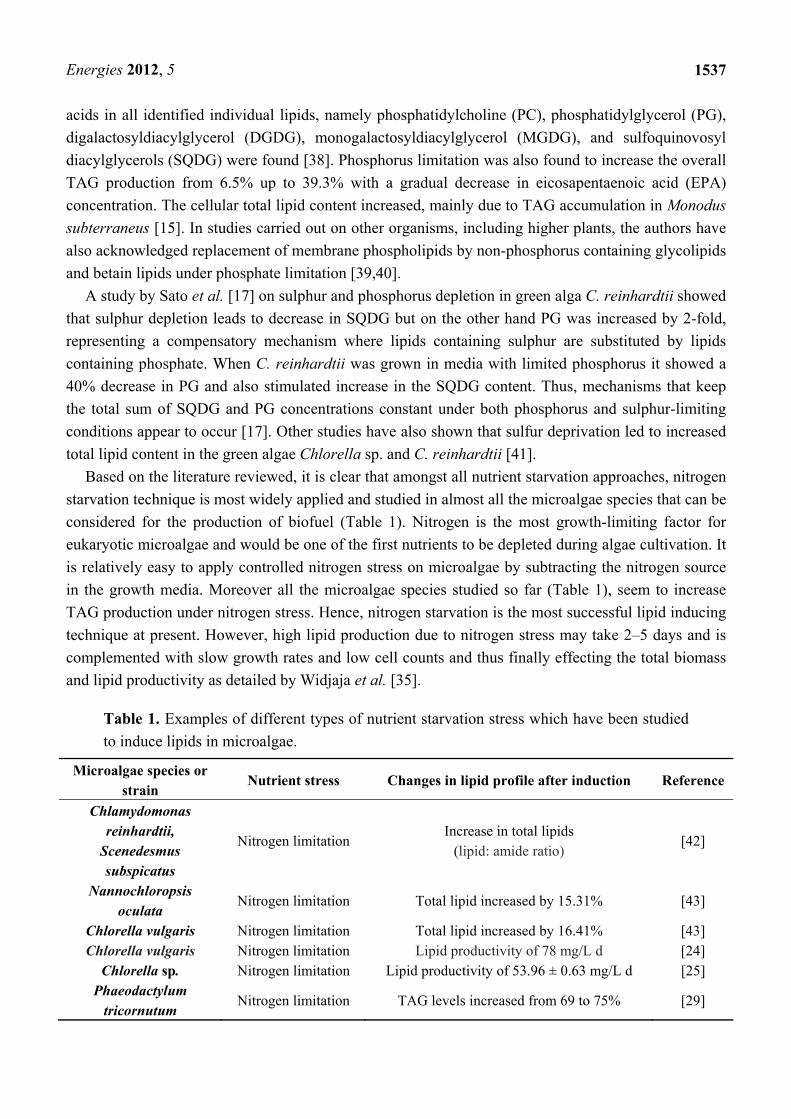
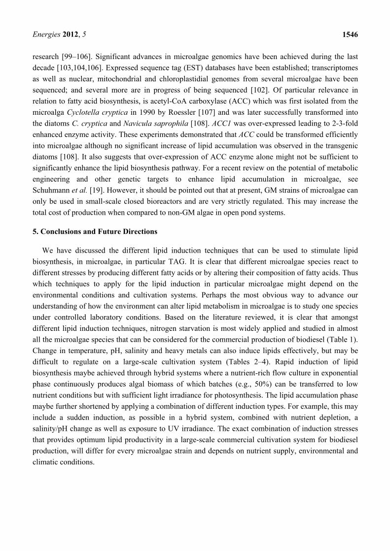
The ability of microalgae to survive in diverse and extreme conditions is reflected in the tremendous diversity and sometimes unusual pattern of cellular lipids obtained from these microalgae [17]. Moreover, some of these microalgae can also modify lipid metabolism efficiently in response to changes in environmental conditions [12,18]. A review of microalgal lipid metabolism has recently been published [19]. Under optimal growth conditions, large amounts of algal biomass are produced but with relatively low lipid contents (Figure 1), which constitute about 5–20% of their dry cell weight (DCW), including glycerol-based membrane lipids. Essentially, microalgae biomass and TAGs compete for photosynthetic assimilate and a reprogramming of physiological pathways is required to stimulate lipid biosynthesis. Under unfavorable environmental or stress conditions many microalgae alter their lipid biosynthetic pathways towards the formation and accumulation of neutral lipids (20–50% DCW), mainly in the form of TAG, enabling microalgae to endure these adverse conditions (Figure 1).
Energies 2012, 5
1535
Figure 1. Lipid induction in microalgae under stress condition.
Lipid inducing stress conditions:
•Nutrients starvation•Temperature (low, high) shift•Change in salinity and pH • Light or UV irradiation
Microalgae cell division phase
Accumulation of lipid bodies (yellow droplets) as a stress response
High capital costs due to low lipid productivity of FA-synthesizing microalgae are a major bottleneck, hindering the commercial production of microalgal oil-derived biodiesel. One who has grown microalgae under laboratory or outdoor condition is well aware of the fact that to obtain high lipid content, external stress or lipid induction techniques need to be applied. Many microalgae produce saturated and unsaturated FAs naturally under ideal growth conditions, which have high nutritional value, but are less ideal for biofuels. However, the synthesis of neutral lipids in the form of TAG can be induced in many species under stress conditions, and these lipids are suitable precursors for biodiesel production [20,21]. The occurrence and the extent to which TAGs are produced is species/strain-specific, and are ultimately controlled by the genetic make-up of individual organisms. Synthesis and accumulation of large amounts of TAGs accompanied by considerable alterations in lipid and FA composition can occur in microalgae when placed under stress conditions imposed by chemical or physical environmental stimuli, either acting individually or in combination [21]. There has been a wide range of studies carried out on lipid induction techniques in microalgae such as the use of nutrients stress, including nitrogen and/or phosphorus starvation, light irradiation, pH, temperature, heavy metals and other chemicals. The following paragraphs review the different TAG induction techniques and discuss their potential in different microalgae species.
3.1. Nutrient Starvation
Nutrient availability has a significant impact on growth and propagation of microalgae and broad effects on their lipid and FA composition. Environmental stress condition when nutrients are limited, invariably cause a steadily declining cell division rate. Surprisingly, active biosynthesis of fatty acids is maintained in some algae species under such conditions, provided there is enough light and CO2 available for photosynthesis [12]. When algal growth (as measured by cell divisions) slows down and there is no requirement for the synthesis of new membrane compounds, the cells instead divert and deposit fatty acids into TAG. Under these conditions, TAG production might serve as a protective
Energies 2012, 5
1536
mechanism. Under normal growth conditions, ATP and NADPH produced by photosynthesis are consumed by generating biomass, with ADP and NADP+ eventually being available again as acceptor molecules in photosynthesis. When cell growth and proliferation is impaired due to the lack of nutrients, the pool of the major electron acceptor for photosynthesis, NADP+, can become depleted. Since photosynthesis is mainly controlled by the abundance of light, and cannot be shut down completely, this can lead to a potentially dangerous situation for the cell, damaging cell components. NADPH is consumed in FA biosynthesis, therefore, increased FAs production (which in turn are stored in TAGs) replenishes the pool of NADP+ under growth-limiting conditions [12,21].
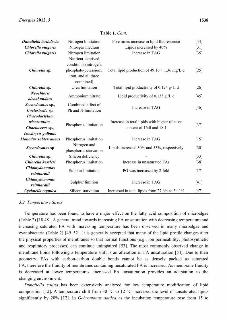
Nutrient starvation is one of the most widely used and applied lipid induction techniques in microalgal TAG production and has been reported for many species (Table 1). For example, when the diatom Stephanodiscus minutulus was grown under silicon, nitrogen or phosphorus limitation, an increase in TAG accumulation and a decrease of polar lipids (as percentage of total lipids) was noticed in all of the nutrient-limited cultures [22]. In the green alga Chlamydomonas moewusii, nutrient limitation resulted in decreased PUFA C16:3, C16:4, and C18:3 contents whereas overall levels of C16:1 and C18:1 FA were increased [23].
Nitrogen is the single most critical nutrient affecting lipid metabolism in algae. A general trend towards accumulation of lipids, particularly TAG, in response to nitrogen deficiency has been observed in numerous species or strains of various microalgae [24–26]. Hu et al. [27] conducted a study on nitrogen stress responses of several green microalgae, diatoms and cyanobacteria and all tested species showed a significant rise in lipid production. A detailed and large-scale model of lipid induction by nutrient starvation (nitrogen, phosphorus) on several diatoms, green algae, red algae, prymnesiophytes and eustimatophytes is presented in a study carried out by Rodolfi et al. [28]. In the diatom Cyclotella cryptica, higher levels of neutral lipids (primarily TAG) and higher proportions of saturated and mono-unsaturated FAs were produced due to silicon deficiency [20]. However, only a small increase in TAG levels (from 69 to 75% from total lipids) together with phospholipids (from 6 to 8%) was reported for the microalga Phaeodactylum tricornutum as a result of reduced nitrogen concentrations [29]. Scenedesmus sp. subjected to nitrogen or phosphorus limitation showed an increase in lipids as high as 30% and 53%, respectively [30]. Lipid content of freshwater green alga Chlorella vulgaris could be significantly increased by 40% in low nitrogen-containing medium [31]. With manipulated culture conditions of 1 mM KNO3, 1.0% CO2, 60 µmol photon m−2 s−1 and 25 °C, lipid production of C. vulgaris was increased by 2.5-fold [32]. In addition, lipid stimulation in Chlorella was also achieved via silicon deficiency [33] and iron supplementation [34]. Moreover, it was found for C. vulgaris that changing from normal nutrient to nitrogen depletion media gradually changed the lipid composition from free FA-rich lipids to lipid mostly contained as TAG [35]. Nitrogen starvation in microalgae not only affects the fatty acid metabolism, but also affects pigment composition. For Parietochloris incise grown in nitrogen-replete medium a considerable increase in the ratio of carotenoid and chlorophyll contents was recorded [36].
Phosphorus limitation resulted in increased lipid content, mainly as TAG, in P. tricornutum, Chaetoceros sp., Isochrysis galbana and Pavlova lutheri, but decreased lipid content in Nannochloris atomus and Tetraselmis sp. [37]. Due to phosphorus deprivation, production of C16:0 and C18:1 was increased and production of C18:4ω3, C20:5ω3 and C22:6ω3 was decreased [37]. In contrast, for phosphorus-starved cells of the green alga Chlorella kessleri, an elevated level of unsaturated fatty
Energies 2012, 5
1537
acids in all identified individual lipids, namely phosphatidylcholine (PC), phosphatidylglycerol (PG), digalactosyldiacylglycerol (DGDG), monogalactosyldiacylglycerol (MGDG), and sulfoquinovosyl diacylglycerols (SQDG) were found [38]. Phosphorus limitation was also found to increase the overall TAG production from 6.5% up to 39.3% with a gradual decrease in eicosapentaenoic acid (EPA) concentration. The cellular total lipid content increased, mainly due to TAG accumulation in Monodus subterraneus [15]. In studies carried out on other organisms, including higher plants, the authors have also acknowledged replacement of membrane phospholipids by non-phosphorus containing glycolipids and betain lipids under phosphate limitation [39,40].
A study by Sato et al. [17] on sulphur and phosphorus depletion in green alga C. reinhardtii showed that sulphur depletion leads to decrease in SQDG but on the other hand PG was increased by 2-fold, representing a compensatory mechanism where lipids containing sulphur are substituted by lipids containing phosphate. When C. reinhardtii was grown in media with limited phosphorus it showed a 40% decrease in PG and also stimulated increase in the SQDG content. Thus, mechanisms that keep the total sum of SQDG and PG concentrations constant under both phosphorus and sulphur-limiting conditions appear to occur [17]. Other studies have also shown that sulfur deprivation led to increased total lipid content in the green algae Chlorella sp. and C. reinhardtii [41].
Based on the literature reviewed, it is clear that amongst all nutrient starvation approaches, nitrogen starvation technique is most widely applied and studied in almost all the microalgae species that can be considered for the production of biofuel (Table 1). Nitrogen is the most growth-limiting factor for eukaryotic microalgae and would be one of the first nutrients to be depleted during algae cultivation. It is relatively easy to apply controlled nitrogen stress on microalgae by subtracting the nitrogen source in the growth media. Moreover all the microalgae species studied so far (Table 1), seem to increase TAG production under nitrogen stress. Hence, nitrogen starvation is the most successful lipid inducing technique at present. However, high lipid production due to nitrogen stress may take 2–5 days and is complemented with slow growth rates and low cell counts and thus finally effecting the total biomass and lipid productivity as detailed by Widjaja et al. [35].
Table 1. Examples of different types of nutrient starvation stress which have been studied to induce lipids in microalgae.
Microalgae species or strain
Nutrient stress Changes in lipid profile after induction Reference
Chlamydomonas reinhardtii,
Scenedesmus subspicatus
Nitrogen limitation Increase in total lipids
(lipid: amide ratio) [42]
Nannochloropsis oculata
Nitrogen limitation Total lipid increased by 15.31% [43]
Chlorella vulgaris Nitrogen limitation Total lipid increased by 16.41% [43] Chlorella vulgaris Nitrogen limitation Lipid productivity of 78 mg/L d [24]
Chlorella sp. Nitrogen limitation Lipid productivity of 53.96 ± 0.63 mg/L d [25] Phaeodactylum
tricornutum Nitrogen limitation TAG levels increased from 69 to 75% [29]
Energies 2012, 5
1538
Table 1. Cont.
Dunaliella tertiolecta Nitrogen limitation Five times increase in lipid fluorescence [44] Chlorella vulgaris Nitrogen medium Lipids increased by 40% [31] Chlorella vulgaris Nitrogen limitation Increase in TAG [35]
Chlorella sp.
Nutrient-deprived conditions (nitrogen, phosphate-potassium,
iron, and all three combined)
Total lipid production of 49.16 ± 1.36 mg/L d [25]
Chlorella sp. Urea limitation Total lipid productivity of 0.124 g/ L d [26] Neochloris
oleoabundans Ammonium nitrate Lipid productivity of 0.133 g /L d [45]
Scenedesmus sp., Coelastrella sp.
Combined effect of Ph and N-limitation
Increase in TAG [46]
Phaeodactylum tricornutum ,
Chaetoceros sp., Isochrysis galbana
Phosphorus limitation Increase in total lipids with higher relative
content of 16:0 and 18:1 [37]
Monodus subterraneus Phosphorus limitation Increase in TAG [15]
Scenedesmus sp Nitrogen and
phosphorus starvation Lipids increased 30% and 53%, respectively [30]
Chlorella sp. Silicon deficiency - [33] Chlorella kessleri Phosphorus limitation Increase in unsaturated FAs [38] Chlamydomonas
reinhardtii Sulphur limitation PG was increased by 2-fold [17]
Chlamydomonas reinhardtii
Sulphur limition Increase in TAG [41]
Cyclotella cryptica Silicon starvation Increased in total lipids from 27.6% to 54.1% [47]
3.2. Temperature Stress
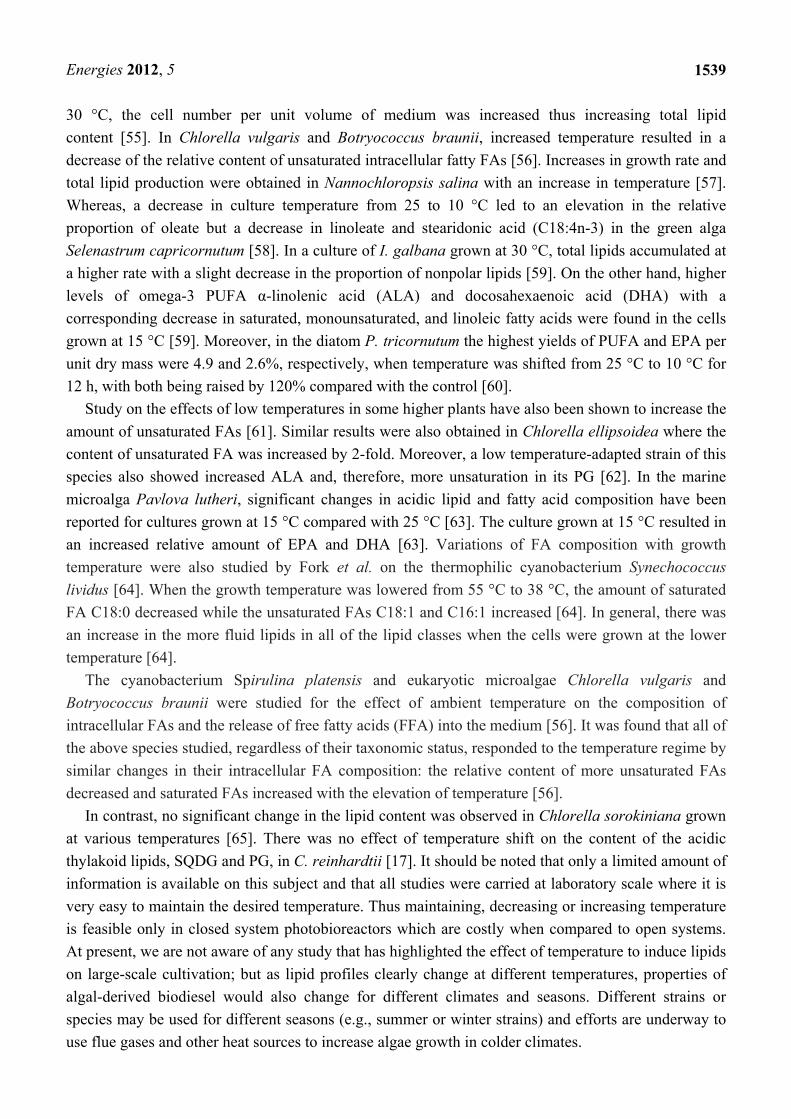
Temperature has been found to have a major effect on the fatty acid composition of microalgae (Table 2) [18,48]. A general trend towards increasing FA unsaturation with decreasing temperature and increasing saturated FA with increasing temperature has been observed in many microalgae and cyanobacteria (Table 2) [49–52]. It is generally accepted that many of the lipid profile changes alter the physical properties of membranes so that normal functions (e.g., ion permeability, photosynthetic and respiratory processes) can continue unimpaired [53]. The most commonly observed change in membrane lipids following a temperature shift is an alteration in FA unsaturation [54]. Due to their geometry, FAs with carbon-carbon double bonds cannot be as densely packed as saturated FA, therefore the fluidity of membranes containing unsaturated FA is increased. As membrane fluidity is decreased at lower temperatures, increased FA unsaturation provides an adaptation to the changing environment.
Dunaliella salina has been extensively analyzed for low temperature modification of lipid composition [12]. A temperature shift from 30 °C to 12 °C increased the level of unsaturated lipids significantly by 20% [12]. In Ochromonas danica, as the incubation temperature rose from 15 to
Energies 2012, 5
1539
30 °C, the cell number per unit volume of medium was increased thus increasing total lipid content [55]. In Chlorella vulgaris and Botryococcus braunii, increased temperature resulted in a decrease of the relative content of unsaturated intracellular fatty FAs [56]. Increases in growth rate and total lipid production were obtained in Nannochloropsis salina with an increase in temperature [57]. Whereas, a decrease in culture temperature from 25 to 10 °C led to an elevation in the relative proportion of oleate but a decrease in linoleate and stearidonic acid (C18:4n-3) in the green alga Selenastrum capricornutum [58]. In a culture of I. galbana grown at 30 °C, total lipids accumulated at a higher rate with a slight decrease in the proportion of nonpolar lipids [59]. On the other hand, higher levels of omega-3 PUFA α-linolenic acid (ALA) and docosahexaenoic acid (DHA) with a corresponding decrease in saturated, monounsaturated, and linoleic fatty acids were found in the cells grown at 15 °C [59]. Moreover, in the diatom P. tricornutum the highest yields of PUFA and EPA per unit dry mass were 4.9 and 2.6%, respectively, when temperature was shifted from 25 °C to 10 °C for 12 h, with both being raised by 120% compared with the control [60].
Study on the effects of low temperatures in some higher plants have also been shown to increase the amount of unsaturated FAs [61]. Similar results were also obtained in Chlorella ellipsoidea where the content of unsaturated FA was increased by 2-fold. Moreover, a low temperature-adapted strain of this species also showed increased ALA and, therefore, more unsaturation in its PG [62]. In the marine microalga Pavlova lutheri, significant changes in acidic lipid and fatty acid composition have been reported for cultures grown at 15 °C compared with 25 °C [63]. The culture grown at 15 °C resulted in an increased relative amount of EPA and DHA [63]. Variations of FA composition with growth temperature were also studied by Fork et al. on the thermophilic cyanobacterium Synechococcus lividus [64]. When the growth temperature was lowered from 55 °C to 38 °C, the amount of saturated FA C18:0 decreased while the unsaturated FAs C18:1 and C16:1 increased [64]. In general, there was an increase in the more fluid lipids in all of the lipid classes when the cells were grown at the lower temperature [64].
The cyanobacterium Spirulina platensis and eukaryotic microalgae Chlorella vulgaris and Botryococcus braunii were studied for the effect of ambient temperature on the composition of intracellular FAs and the release of free fatty acids (FFA) into the medium [56]. It was found that all of the above species studied, regardless of their taxonomic status, responded to the temperature regime by similar changes in their intracellular FA composition: the relative content of more unsaturated FAs decreased and saturated FAs increased with the elevation of temperature [56].
In contrast, no significant change in the lipid content was observed in Chlorella sorokiniana grown at various temperatures [65]. There was no effect of temperature shift on the content of the acidic thylakoid lipids, SQDG and PG, in C. reinhardtii [17]. It should be noted that only a limited amount of information is available on this subject and that all studies were carried at laboratory scale where it is very easy to maintain the desired temperature. Thus maintaining, decreasing or increasing temperature is feasible only in closed system photobioreactors which are costly when compared to open systems. At present, we are not aware of any study that has highlighted the effect of temperature to induce lipids on large-scale cultivation; but as lipid profiles clearly change at different temperatures, properties of algal-derived biodiesel would also change for different climates and seasons. Different strains or species may be used for different seasons (e.g., summer or winter strains) and efforts are underway to use flue gases and other heat sources to increase algae growth in colder climates.
Energies 2012, 5
1540
Table 2. Lipid induction in microalgae with different temperatures.
Microalgae species or strain Stressing agent Lipid profile change after
induction Reference
Chaetoceros sp. Grown at 25 °C Total lipid increased by 16.8% [49] Rhodomonas sp., Cryptomonas sp.,
Isochrysis sp. Range of 27 °C to 30 °C
Lipid production increased by 15.5, 12.7, and 21.7%
respectively [49]
Nannochloropsis oculata Increase from 20 °C to 25 °C Lipid production increased by
14.92% [43]
Isochrysis galbana Increase from 15 °C to 30 °C Increase in neutral lipids [59]
Chlorella ellipsoidea Lowering temperature Unsaturated FA was increased by
2-fold [62]
Nannochloropsis salina Increase in temperature Increase in total lipids [57] Dunaliella salina Shift from 30 °C to 12 °C Increase in unsaturated lipids [12]
Ochromonas danica Increase from 15 °C to 30 °C Increase in total lipids [55]
Selenastrum capricornutum Temperature from 25 °C to
10 °C Increase in oleate fatty acid [58]
Isochrysis galbana Grown at 30 °C Increase in total lipids [59]
Phaeodactylum tricornutum Shifted from 25 °C to 10 °C
for 12 h Highest yields of PUFA and EPA [60]
Pavlova lutheri Grown at 15 °C Increased relative amount of EPA [63] Spirulina platensis, Chlorella
vulgaris, Botryococcus braunii
Increase in temperature Saturated FAs increased [56]
3.3. Salinity-Induced Lipid Production
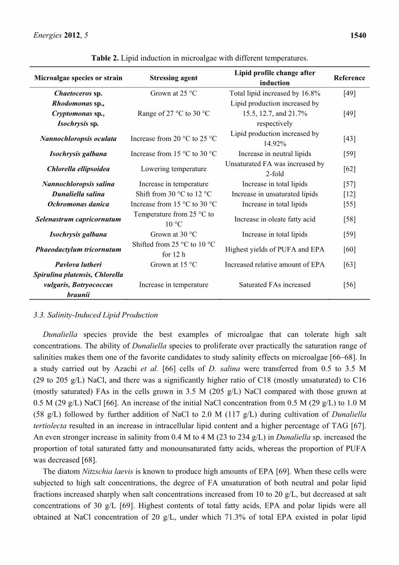
Dunaliella species provide the best examples of microalgae that can tolerate high salt concentrations. The ability of Dunaliella species to proliferate over practically the saturation range of salinities makes them one of the favorite candidates to study salinity effects on microalgae [66–68]. In a study carried out by Azachi et al. [66] cells of D. salina were transferred from 0.5 to 3.5 M (29 to 205 g/L) NaCl, and there was a significantly higher ratio of C18 (mostly unsaturated) to C16 (mostly saturated) FAs in the cells grown in 3.5 M (205 g/L) NaCl compared with those grown at 0.5 M (29 g/L) NaCl [66]. An increase of the initial NaCl concentration from 0.5 M (29 g/L) to 1.0 M (58 g/L) followed by further addition of NaCl to 2.0 M (117 g/L) during cultivation of Dunaliella tertiolecta resulted in an increase in intracellular lipid content and a higher percentage of TAG [67]. An even stronger increase in salinity from 0.4 M to 4 M (23 to 234 g/L) in Dunaliella sp. increased the proportion of total saturated fatty and monounsaturated fatty acids, whereas the proportion of PUFA was decreased [68].
The diatom Nitzschia laevis is known to produce high amounts of EPA [69]. When these cells were subjected to high salt concentrations, the degree of FA unsaturation of both neutral and polar lipid fractions increased sharply when salt concentrations increased from 10 to 20 g/L, but decreased at salt concentrations of 30 g/L [69]. Highest contents of total fatty acids, EPA and polar lipids were all obtained at NaCl concentration of 20 g/L, under which 71.3% of total EPA existed in polar lipid

Energies 2012, 5
1541
fractions [69]. The amount of total free sterols was also increased with an increase in salt concentration. In three marine heterotrophic microalgae strains, Crythecodinium cohnii ATCC 30556, C. cohnii ATCC 50051 and C. cohnii RJH grown at different salinities, the FA composition was also affected [70]. At 9 g/L NaCl, C. cohnii ATCC 30556 had the highest total FA content and DHA (C22:6) proportion. In contrast, C. cohnii ATCC 50051 and C. cohnii RJH had the highest DHA content at 5 g/L NaCl. C. cohnii ATCC 30556 and ATCC 50051 had the highest DHA yield (132 and 68 mg/L respectively) at 9 g/L NaCl while C. cohnii RJH had the highest DHA yield (129 mg/L) at 5 g/L NaCl [70]. Growth, lipid content and FA composition of heterotrophic microalga Schizochytrium limacinum OUC88 at different temperatures (16 °C, 23 °C, 30 °C and 37 °C) and salinities (0, 9, 18, 27 and 36 g/L) were analyzed [71]. Highest lipid content was obtained at salinities of 9–36 g/L at a temperature range of 16–30 °C and the content of saturated fatty acids C15:0 and C17:0 was increased greatly [71]. In addition, the ratio of DHA to DPA changed at different temperatures and salinities [71].
3.4. The Effect of pH and Heavy Metals Stress
Fluctuations of the pH in the medium also have been found to alter the lipid composition of microalgae (Table 3). For example, alkaline pH stress led to TAG accumulation in Chlorella CHLOR1 and was not dependent on nitrogen or carbon limitation levels, and led to a decrease in membrane lipids [72]. Based on morphological observations, alkaline pH inhibited the growth of microalgae, thus diverting the energy to form TAG [72]. The effects of pH on the lipid and FA composition of a Chlamydomonas sp. isolated from a volcanic acidic lake, and C. reinhardtii have been studied and compared [73]. In the unidentified Chlamydomonas sp., FAs of polar lipids were more saturated than those in C. reinhardtii. The relative proportion of TAG (as percentage of total lipids) was higher in Chlamydomonas sp. grown at pH 1 than that in the cells cultivated at higher pH. The increase in saturation of fatty acids in membrane lipids of Chlamydomonas has been suggested to represent an adaptive reaction at low pH to decrease membrane lipid fluidity [73].
Heavy metals like cadmium, iron, copper and zinc have also been found to increase the lipid content in some microalgae [74]. The effect of high levels of cadmium was studied in Euglena gracilis grown as autotrophic, heterotrophic (in the dark) and mixotrophic (in the light with an organic carbon source) cultures [74]. Cadmium caused an increase in the total lipid content per cell in all three culture systems [74]. Among the membrane lipids, sterol content was lower in cadmium-treated cells cultivated under illumination. There were no changes in the total phospholipid content, although there was an increase in PG. E. gracilis has also been shown to display somewhat different sensitivities to copper and zinc [74]. The effect of iron on growth and lipid accumulation in Chlorella vulgaris was investigated by Liu et al. [34]. The culture in the late exponential growth phase when supplemented with Fe3+ at different concentrations, showed increased total lipid content of up to 56.6% biomass by dry weight [34].
Energies 2012, 5
1542
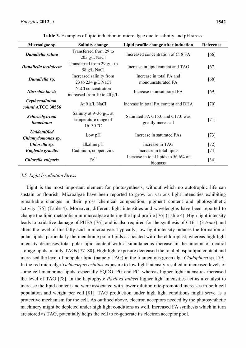
Table 3. Examples of lipid induction in microalgae due to salinity and pH stress.
Microalgae sp Salinity change Lipid profile change after induction Reference
Dunaliella salina Transferred from 29 to
205 g/L NaCl Increased concentration of C18 FA [66]
Dunaliella tertiolecta Transferred from 29 g/L to
58 g/L NaCl Increase in lipid content and TAG [67]
Dunaliella sp. Increased salinity from
23 to 234 g/L NaCl Increase in total FA and
monounsaturated FA [68]
Nitzschia laevis NaCl concentration
increased from 10 to 20 g/L Increase in unsaturated FA [69]
Crythecodinium. cohnii ATCC 30556
At 9 g/L NaCl Increase in total FA content and DHA [70]
Schizochytrium limacinum
Salinity at 9–36 g/L at temperature range of
16–30 °C
Saturated FA C15:0 and C17:0 was greatly increased
[71]
Unidentified Chlamydomonas sp.
Low pH Increase in saturated FAs [73]
Chlorella sp. alkaline pH Increase in TAG [72] Euglenia gracilis Cadmium, copper, zinc Increase in total lipids [74]
Chlorella vulgaris Fe3+ Increase in total lipids to 56.6% of biomass
[34]
3.5. Light Irradiation Stress
Light is the most important element for photosynthesis, without which no autotrophic life can sustain or flourish. Microalgae have been reported to grow on various light intensities exhibiting remarkable changes in their gross chemical composition, pigment content and photosynthetic activity [75] (Table 4). Moreover, different light intensities and wavelengths have been reported to change the lipid metabolism in microalgae altering the lipid profile [76] (Table 4). High light intensity leads to oxidative damage of PUFA [76], and is also required for the synthesis of C16:1 (3 trans) and alters the level of this fatty acid in microalgae. Typically, low light intensity induces the formation of polar lipids, particularly the membrane polar lipids associated with the chloroplast, whereas high light intensity decreases total polar lipid content with a simultaneous increase in the amount of neutral storage lipids, mainly TAGs [77–80]. High light exposure decreased the total phospholipid content and increased the level of nonpolar lipid (namely TAG) in the filamentous green alga Cladophora sp. [79]. In the red microalga Tichocarpus crinitus exposure to low light intensity resulted in increased levels of some cell membrane lipids, especially SQDG, PG and PC, whereas higher light intensities increased the level of TAG [78]. In the haptophyte Pavlova lutheri higher light intensities act as a catalyst to increase the lipid content and were associated with lower dilution rate-promoted increases in both cell population and weight per cell [81]. TAG production under high light conditions might serve as a protective mechanism for the cell. As outlined above, electron acceptors needed by the photosynthetic machinery might be depleted under high light conditions as well. Increased FA synthesis which in turn are stored as TAG, potentially helps the cell to re-generate its electron acceptor pool.
Energies 2012, 5
1543
Light intensity not only affects the fatty acid composition in microalgae, but also the pigment composition. In the green microalga Parietochloris incise under low irradiance photosynthetically active radiation, cultures displayed slow growth and a relatively low carotenoid-to-chlorophyll ratio [36]. At higher irradiances on complete medium, the alga displayed a higher growth rate and an increase in the carotenoid content, especially that of β-carotene and lutein [36].
Light/dark cycles at different growth phases also have a significant effect on algal lipid composition, as was successfully demonstrated in a detailed study on various light regimes on lipids of the diatom Thalassiosira pseudonana [77]. A culture grown to stationary phase under strong continuous light or under 12:12 h strong light/dark conditions had a higher amount of TAG with saturated and monounsaturated fatty acids compared to cultures grown with less light. At the exponential growth phase, however, the proportion of PUFA was highest under high light conditions [77]. This demonstrates the important role of growth phase in the accumulation of certain fatty acids. With the onset of stationary phase, algae typically show increased proportions of saturated and monounsaturated fatty acids and decreased amounts of PUFA [77]. The lipid and fatty acid compositions of three species of sea ice diatoms grown in chemostats have been analyzed and compared when cultivated at light-limiting conditions of 2 and 15 µmol photons m−2 s−1 [82]. Growing cultures at 2 µmol photons m−2 s−1 resulted in 50% more MGDG containing EPA than those grown at 15 µmol photons m−2 s−1. Growing cultures at 2 µmol photons m−2 s−1 resulted in higher amounts of non-polar lipid bilayer-forming MGDG in relation to total bilayer-forming lipids, especially DGDG (the ratio of MGDG:DGDG increased from 3.4 to 5.7) than in cultures grown at 15 µmol photons m−2 s−1. A shorter light period seemed to increase the production of PUFA in Isochrysis galbana [83]. Sitosterol and stigmasterol were the two main sterols detected at 246.3 and 220.0 mg/100 g, respectively. A continuous increase in the level of total sterols was recorded during the life cycle at 24 h lighting [83]. The reduction of the photoperiod led to a decrease in the total sterols produced in the decay phase. A gradual increase in α-tocopherol production during the life cycle was also recorded [83]. Dark treatment caused a decrease in the relative proportion of oleate fatty acid and an increase in linoleate fatty acid in the green alga Selenastrum capricornutum [58]. In the dinoflagellate Prorocentrum minimum dark exposure led to a reduced content of TAG and galactosylglycerides, while the total content of phospholipids changed little [58].
Light irradiation can only be controlled in a closed system bioreactors or in laboratory-scale cultures, as shown by the examples above. Moreover, operational costs for controlled light add up to the production cost of biofuels from microalgae, although there are several commercial approaches of using LEDs or diverted sunlight in large-scale photobioreactors. Light is essential for TAG production, but if high light irradiation is used as a stimulant for increased TAG production, based on the examples above and in Table 4, it can be expected that this will differ for different species. In addition, TAG FA composition is different for different species in response to different light exposures. For example, in Nannochloropsis sp. the degree of unsaturation of FAs was lower with increasing irradiance with a significant decrease in omega-3 fatty acids (29% to 8% of total FA), caused mainly by a decrease of EPA (20:5n-3) [84]. In conclusion, light will normally stimulate fatty acid synthesis, growth and the formation of (particularly chloroplast) membranes. Therefore, the overall lipid content of algae will reflect such morphological changes.
Energies 2012, 5
1544
3.6. UV Irradiance for Lipid Induction
Current research on the effect of UV irradiance in microalgae is mainly focused on the impact of UV-A and UV-B radiation on algal growth, morphology, physiology and oxidative stress [85–89], with a special emphasis on pigments and photosynthesis [90]. Examples of studies on UV radiation on lipid profiles in microalgae are shown in Table 4.
In a study carried out by Srinivas and Ochs [91] on Nannochloropsis oculata the effect of UV-A at different levels of exposure on total lipid accumulation was investigated. UV-A treatments significantly increased the PUFA (chlorophyll-specific lipid concentration) of N. oculata cells, and UV-A and decreased nutrients had a synergistic effect on lipid accumulation. The effects of UV-B radiation on the total lipid, FA and sterol composition and content of three Antarctic marine phytoplankton species Odontella weissflogii, Chaetoceros simplex and the haptophyte Phaeocystis antarctica were examined in a preliminary culture experiment [92]. The cultures were exposed to constant UV-A and low or high UV-B radiation. The sterol, fatty acid and total lipid content for Odontella weissflogii changed little under low UV-B when compared with control conditions. In contrast, when P. antarctica was exposed to low UV-B irradiance, storage lipids were reduced and structural lipids increased [92]. P. antarctica also contained a higher proportion of polyunsaturated fatty acids under low UV-B exposure. Exposure of P. antarctica to high UV-B irradiance increased total lipid, TAG and FFA concentrations. Lipid concentrations per cell also increased when C. simplex was exposed to high UV-B irradiance [92]. This resulted from increases in FFA concentration principally saturated FA and may indicate degradation of complex lipids during high UV-B treatment [92]. Effect of UV-B radiation on lipid productivity was studied in detail in Tetraselmis sp. [93]. A 4 hour-exposure to UV-B radiation resulted in an overall increase in saturated FA and monounsaturated FA, whereas the PUFA content was decreased by 50% [93]. In addition, UV irradiance caused a decline in the overall rate of carbon incorporated into amino acids and a reduction in the pool size of total cellular amino acids [93]. In contrast, intracellular dissolved free amino acid increased [93].
The effect of UV radiation on growth and fatty acid composition of two diatoms, P. tricornutum and Chaetoceros muelleri, were examined in batch cultures [94]. UV radiation induced significant differences in all the major fatty acids of P. tricornutum. The percentages of EPA and PUFA increased while monounsaturated FA decreased in the UV-A treatment in comparison with no UV irradiance or combined UV-A + UV-B treatments [94]. On the other hand, all the major fatty acids of C. muelleri varied with harvest stage and UV irradiance. The percentage of monounsaturated FA in C. muelleri increased, while EPA and PUFA decreased under combined UV-A + UV-B treatment [94]. The study indicated that UV-A exposure may promote EPA and PUFA formation in P. tricornutum, whereas combined UV-A + UV-B exposure enhanced short FA and monounsaturated FA content, but suppressed PUFA formation in C. muelleri [94].
PUFAs, especially EPA and DHA, are abundantly synthesized by some phytoplankton species and play a key role in the marine food chain. However, they are generally considered to be sensitive to oxidation by UV radiation. P. lutheri and Odontella aurita were exposed to a combination of UV-A and UV-B radiation with a total daily dose of 110 kJ/m2 and lipid composition was then determined on days 3, 5, and 8 of UV exposure [95]. In P. lutheri exposure to UV led to a decrease in the proportion
Energies 2012, 5
1545
of PUFAs, especially those in structural lipids (glycolipids and phospholipids) and a reduction of 20% in EPA levels and 16% in DHA levels, after 8 days; whereas for O. aurita, exposure to UV did not change the fatty acid composition of the total lipids and lipid fractions of the cells [95].
Interestingly, UV radiation has been suggested for microalgal lipid induction in large-scale cultivation systems. As UV radiation has genetically and physiologically deleterious effects on many life forms including microalgae [95], the impact is conceivably related to the radiation intensity. A recent study showed that the modulated use of UV-A radiation for seven days could lead to an increased production of fatty acids in Nannocloropsis sp. [96]. However, there is concern that constant use of UV-A light may not be viable for industrial-scale cultivation, while shorter, but stronger UV radiation could also affect microalgal lipid composition and production as a result of inhibiting nutrient uptake, carbon assimilation mechanism and damaging DNA [97].
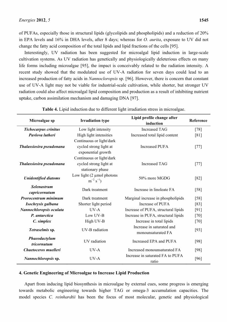
Table 4. Lipid induction due to different light irradiation stress in microalgae.
Microalgae sp Irradiation type Lipid profile change after
induction Reference
Tichocarpus crinitus Low light intensity Increased TAG [78] Pavlova lutheri High light intensities Increased total lipid content [81]
Thalassiosira pseudonana Continuous or light/dark
cycled strong light at exponential growth
Increased PUFA [77]
Thalassiosira pseudonana Continuous or light/dark
cycled strong light at stationary phase
Increased TAG [77]
Unidentified diatoms Low light (2 µmol photons
m−2 s−1) 50% more MGDG [82]
Selenastrum capricornutum
Dark treatment Increase in linoleate FA [58]
Prorocentrum minimum Dark treatment Marginal increase in phospholipids [58] Isochrysis galbana Shorter light period Increase of PUFA [83]
Nannochloropsis oculata UV-A Increase of PUFA, structural lipids [91] P. antarctica Low UV-B Increase in PUFA, structural lipids [70] C. simplex High UV-B Increase in total lipids [70]
Tetraselmis sp. UV-B radiation Increase in saturated and
monounsaturated FA [93]
Phaeodactylum tricornutum
UV radiation Increased EPA and PUFA [98]
Chaetoceros muelleri UV-A Increased monounsaturated FA [98]
Nannochloropsis sp. UV-A Increase in saturated FA to PUFA
ratio [96]
4. Genetic Engineering of Microalgae to Increase Lipid Production
Apart from inducing lipid biosynthesis in microalgae by external cues, some progress is emerging towards metabolic engineering towards higher TAG or omega-3 accumulation capacities. The model species C. reinhardtii has been the focus of most molecular, genetic and physiological
Energies 2012, 5
1546
research [99–106]. Significant advances in microalgae genomics have been achieved during the last decade [103,104,106]. Expressed sequence tag (EST) databases have been established; transcriptomes as well as nuclear, mitochondrial and chloroplastidial genomes from several microalgae have been sequenced; and several more are in progress of being sequenced [102]. Of particular relevance in relation to fatty acid biosynthesis, is acetyl-CoA carboxylase (ACC) which was first isolated from the microalga Cyclotella cryptica in 1990 by Roessler [107] and was later successfully transformed into the diatoms C. cryptica and Navicula saprophila [108]. ACC1 was over-expressed leading to 2-3-fold enhanced enzyme activity. These experiments demonstrated that ACC could be transformed efficiently into microalgae although no significant increase of lipid accumulation was observed in the transgenic diatoms [108]. It also suggests that over-expression of ACC enzyme alone might not be sufficient to significantly enhance the lipid biosynthesis pathway. For a recent review on the potential of metabolic engineering and other genetic targets to enhance lipid accumulation in microalgae, see Schuhmann et al. [19]. However, it should be pointed out that at present, GM strains of microalgae can only be used in small-scale closed bioreactors and are very strictly regulated. This may increase the total cost of production when compared to non-GM algae in open pond systems.
5. Conclusions and Future Directions
We have discussed the different lipid induction techniques that can be used to stimulate lipid biosynthesis, in microalgae, in particular TAG. It is clear that different microalgae species react to different stresses by producing different fatty acids or by altering their composition of fatty acids. Thus which techniques to apply for the lipid induction in particular microalgae might depend on the environmental conditions and cultivation systems. Perhaps the most obvious way to advance our understanding of how the environment can alter lipid metabolism in microalgae is to study one species under controlled laboratory conditions. Based on the literature reviewed, it is clear that amongst different lipid induction techniques, nitrogen starvation is most widely applied and studied in almost all the microalgae species that can be considered for the commercial production of biodiesel (Table 1). Change in temperature, pH, salinity and heavy metals can also induce lipids effectively, but may be difficult to regulate on a large-scale cultivation system (Tables 2–4). Rapid induction of lipid biosynthesis maybe achieved through hybrid systems where a nutrient-rich flow culture in exponential phase continuously produces algal biomass of which batches (e.g., 50%) can be transferred to low nutrient conditions but with sufficient light irradiance for photosynthesis. The lipid accumulation phase maybe further shortened by applying a combination of different induction types. For example, this may include a sudden induction, as possible in a hybrid system, combined with nutrient depletion, a salinity/pH change as well as exposure to UV irradiance. The exact combination of induction stresses that provides optimum lipid productivity in a large-scale commercial cultivation system for biodiesel production, will differ for every microalgae strain and depends on nutrient supply, environmental and climatic conditions.
Energies 2012, 5
1547
References
1. Schenk, P.M.; Thomas-Hall, S.R.; Stephens, E.; Marx, U.; Mussgnug, J.; Posten, C.; Kruse, O.; Hankamer, B. Second generation biofuels: High-efficiency microalgae for biodiesel production. BioEnergy Res. 2008, 1, 20–43.
2. Chisti, Y. Biodiesel from microalgae. Biotechnol. Adv. 2007, 25, 294–306. 3. Chisti, Y. Biodiesel from microalgae beats bioethanol. Trends Biotech. 2008, 26, 126–131. 4. Christenson, L.; Sims, R. Production and harvesting of microalgae for wastewater treatment,
biofuels, and bioproducts. Biotechnol. Adv. 2011, 29, 686–702. 5. Stephens, E.; Ross, I.L.; Mussgnug, J.H.; Wagner, L.D.; Borowitzka, M.A.; Posten, C.;
Kruse, O.; Hankamer, B. Future prospects of microalgal biofuel production systems. Trends Plant Sci. 2010, 15, 554–564.
6. Bruton, T.; Lyons, H.; Lerat, Y.; Stanley, M.; Rasmussen, M.B. A Review of the Potential of Marine Algae as a Source of Biofuel in Ireland; Technical Report; Sustainable Energy Ireland: Dublin, Ireland, 2009.
7. Brennan, L.; Owende, P. Biofuels from microalgae—A review of technologies for production, processing, and extractions of biofuels and co-products. Renew. Sustain. Energy Rev. 2010, 14, 557–577.
8. McGinn, P.; Dickinson, K.; Bhatti, S.; Frigon, J.-C.; Guiot, S.; O’Leary, S. Integration of microalgae cultivation with industrial waste remediation for biofuel and bioenergy production: Opportunities and limitations. Photosynth. Res. 2011, 109, 231–247.
9. Fukuda, H.; Kondo, A.; Noda, H. Biodiesel fuel production by transesterification of oils. J. Biosci. Bioeng. 2001, 92, 405–416.
10. Chen, Y.F. Production of Biodiesel from Algal Biomass: Current Perspectives and Future; Academic Press: Waltham, MA, USA, 2011; p. 399.
11. Gurr , M.I.; Harwood, J.L.; Frayn, K.N. Lipid Biochemistry: An Introduction, 5th ed.; Blackwell: Oxford, UK, 2002; p. 320.
12. Thompson, G.A. Lipids and membrane function in green algae. Biochim. Biophys. Acta 1996, 1302, 17–45.
13. Bigogno, C.; Khozin-Goldberg, I.; Cohen, Z. Accumulation of arachidonic acid-rich triacylglycerols in the microalga Parietochloris incisa (trebuxiophyceae, chlorophyta). Phytochemistry 2002, 60, 135–143.
14. Alonso, D.L.; Belarbi, E.-H.; Rodríguez-Ruiz, J.; Segura, C.I.; Giménez, A. Acyl lipids of three microalgae. Phytochemistry 1998, 47, 1473–1481.
15. Khozin-Goldberg, I.; Cohen, Z. The effect of phosphate starvation on the lipid and fatty acid composition of the fresh water eustigmatophyte Monodus subterraneus. Phytochemistry 2006, 67, 696–701.
16. Makewicz, A.; Gribi, C.; Eichenberger, W. Lipids of Ectocarpus fasciculatus (phaeophyceae). Incorporation of [l-14C]oleate and the role of TAG and MGDG in lipid metabolism. Plant Cell Physiol. 1997, 38, 952–962.
17. Sato, N.; Hagio, M.; Wada, H.; Tsuzuki, A.M. Environmental effects on acidic lipids of thylakoid membranes. Biochem. Soc. Trans. 2000, 28, 912–914.
Energies 2012, 5
1548
18. Guschina, I.A.; Harwood, J.L. Lipids and lipid metabolism in eukaryotic algae. Prog. Lipid Res. 2006, 45, 160–186.
19. Schuhmann, H.; Lim, D.K.Y.; Schenk, P.M. Perspectives on metabolic engineering for increased lipid contents in microalgae. Biofuels 2011, 3, 71–86.
20. Miao, X.; Wu, Q. Biodiesel production from heterotrophic microalgal oil. Bioresour. Technol. 2006, 97, 841–846.
21. Hu, Q.; Sommerfeld, M.; Jarvis, E.; Ghirardi, M.; Posewitz, M.; Seibert, M.; Darzins, A. Microalgal triacylglycerols as feedstocks for biofuel production: Perspectives and advances. Plant J. 2008, 54, 621–639.
22. Lynn, S.G.; Kilham, S.S.; Kreeger, D.A.; Interlandi, S.J. Effect of nutrient availability on the biochemical and elemental stoichiometry in the freshwater diatom Stephanodiscus minutulus (bacillariophyceae). J. Phycol. 2000, 36, 510–522.
23. Arisz, S.A.; van Himbergen, J.A.J.; Musgrave, A.; van den Ende, H.; Munnik, T. Polar glycerolipids of Chlamydomonas moewusii. Phytochemistry 2000, 53, 265–270.
24. Yeh, K.L.; Chang, J.S. Nitrogen starvation strategies and photobioreactor design for enhancing lipid production of a newly isolated microalga Chlorella vulgaris esp-31: Implications for biofuels. Biotechnol. J. 2011, 6, 1358–1366.
25. Praveenkumar, R.; Shameera, K.; Mahalakshmi, G.; Akbarsha, M.A.; Thajuddin, N. Influence of nutrient deprivations on lipid accumulation in a dominant indigenous microalga Chlorella sp., bum11008: Evaluation for biodiesel production. Biomass Bioenerg. 2012, 37, 60–66.
26. Hsieh, C.-H.; Wu, W.-T. Cultivation of microalgae for oil production with a cultivation strategy of urea limitation. Bioresour. Technol. 2009, 100, 3921–3926.
27. Hu, Q. PSA abstracts. J. Phycol. 2006, 42, 1–48. 28. Rodolfi, L.; Chini Zittelli, G.; Bassi, N.; Padovani, G.; Biondi, N.; Bonini, G.; Tredici, M.R.
Microalgae for oil: Strain selection, induction of lipid synthesis and outdoor mass cultivation in a low-cost photobioreactor. Biotechnol. Bioeng. 2009, 102, 100–112.
29. Alonso, D.L.; Belarbi, E.-H.; Fernández-Sevilla, J.M.; Rodríguez-Ruiz, J.; Grima, E.M. Acyl lipid composition variation related to culture age and nitrogen concentration in continuous culture of the microalga Phaeodactylum tricornutum. Phytochemistry 2000, 54, 461–471.
30. Xin, L.; Hong-ying, H.; Ke, G.; Ying-xue, S. Effects of different nitrogen and phosphorus concentrations on the growth, nutrient uptake, and lipid accumulation of a freshwater microalga Scenedesmus sp. Bioresour. Technol. 2010, 101, 5494–5500.
31. Illman, A.M.; Scragg, A.H.; Shales, S.W. Increase in Chlorella strains calorific values when grown in low nitrogen medium. Enzyme Microb. Technol. 2000, 27, 631–635.
32. Lv, J.-M.; Cheng, L.-H.; Xu, X.-H.; Zhang, L.; Chen, H.-L. Enhanced lipid production of Chlorella vulgaris by adjustment of cultivation conditions. Bioresour. Technol. 2010, 101, 6797–6804.
33. Griffiths, M.; Harrison, S. Lipid productivity as a key characteristic for choosing algal species for biodiesel production. J. Appl. Phycol. 2009, 21, 493–507.
34. Liu, Z.-Y.; Wang, G.-C.; Zhou, B.-C. Effect of iron on growth and lipid accumulation in Chlorella vulgaris. Bioresour. Technol. 2008, 99, 4717–4722.
Energies 2012, 5
1549
35. Widjaja, A.; Chien, C.-C.; Ju, Y.-H. Study of increasing lipid production from fresh water microalgae Chlorella vulgaris. J. Taiwan Inst. Chem. Eng. 2009, 40, 13–20.
36. Solovchenko, A.; Khozin-Goldberg, I.; Didi-Cohen, S.; Cohen, Z.; Merzlyak, M. Effects of light and nitrogen starvation on the content and composition of carotenoids of the green microalga Parietochloris incisa. Russ. J. Plant Physiol. 2008, 55, 455–462.
37. Reitan, K.I.; Rainuzzo, J.R.; Olsen, Y. Effect of nutrient limitation on fatty acid and lipid content of marine microalgae. J. Phycol. 1994, 30, 972–979.
38. El-Sheek, M.M.; Rady, A.A. Effect of phosphorus starvation on growth, photosynthesis and some metabolic processes in the unicellular green alga Chlorella kessleri. Phyton 1995, 35, 139–151.
39. Härtel, H.; Dörmann, P.; Benning, C. DGD1-independent biosynthesis of extraplastidic galactolipids after phosphate deprivation in Arabidopsis. Proc. Nat. Acad. Sci. USA 2000, 97, 10649–10654.
40. Andersson, M.X.; Stridh, M.H.; Larsson, K.E.; Liljenberg, C.; Sandelius, A.S. Phosphate-deficient oat replaces a major portion of the plasma membrane phospholipids with the galactolipid digalactosyldiacylglycerol. FEBS Lett. 2003, 537, 128–132.
41. Matthew, T.; Zhou, W.; Rupprecht, J.; Lim, L.; Thomas-Hall, S.R.; Doebbe, A.; Kruse, O.; Hankamer, B.; Marx, U.C.; Smith, S.M.; et al. The metabolome of Chlamydomonas reinhardtii following induction of anaerobic H2 production by sulfur depletion. J. Biol. Chem. 2009, 284, 23415–23425.
42. Dean, A.P.; Sigee, D.C.; Estrada, B.; Pittman, J.K. Using FTIR spectroscopy for rapid determination of lipid accumulation in response to nitrogen limitation in freshwater microalgae. Bioresour. Technol. 2010, 101, 4499–4507.
43. Converti, A.; Casazza, A.A.; Ortiz, E.Y.; Perego, P.; Del Borghi, M. Effect of temperature and nitrogen concentration on the growth and lipid content of Nannochloropsis oculata and Chlorella vulgaris for biodiesel production. Chem. Eng. Process. 2009, 48, 1146–1151.
44. Chen, M.; Tang, H.; Ma, H.; Holland, T.C.; Ng, K.Y.S.; Salley, S.O. Effect of nutrients on growth and lipid accumulation in the green algae Dunaliella tertiolecta. Bioresour. Technol. 2011, 102, 1649–1655.
45. Li, Y.; Horsman, M.; Wang, B.; Wu, N.; Lan, C. Effects of nitrogen sources on cell growth and lipid accumulation of green alga Neochloris oleoabundans. Appl. Microbiol. Biotechnol. 2008, 81, 629–636.
46. Gardner, R.; Peters, P.; Peyton, B.; Cooksey, K.E. Medium ph and nitrate concentration effects on accumulation of triacylglycerol in two members of the chlorophyta. J. Appl. Phycol. 2011, 23, 1005–1016.
47. Roessler, P.G. Effects of silicon deficiency on lipid composition and metabolism in the diatom Cyclotella cryptica. J. Phycol. 1988, 24, 394–400.
48. Morgan-Kiss, R.M.; Priscu, J.C.; Pocock, T.; Gudynaite-Savitch, L.; Huner, N.P.A. Adaptation and acclimation of photosynthetic microorganisms to permanently cold environments. Microbiol. Mol. Biol. Rev. 2006, 70, 222–252.
Energies 2012, 5
1550
49. Renaud, S.M.; Thinh, L.-V.; Lambrinidis, G.; Parry, D.L. Effect of temperature on growth, chemical composition and fatty acid composition of tropical Australian microalgae grown in batch cultures. Aquaculture 2002, 211, 195–214.
50. Lynch, D.V.; Thompson, T.G., Jr. Low temperature-induced alterations in the chloroplast and microsomal membranes of Dunaliella salina. Plant Physiol. 1982, 69, 1369–1375.
51. Murata, N.; Troughton, J.H.; Fork, D.C. Relationships between the transition of the physical phase of membrane lipids and photosynthetic parameters in Anacystis nidulans and lettuce and spinach chloroplasts. Plant Physiol. 1975, 56, 508–517.
52. Sato, N.; Murata, N. Temperature shift-induced responses in lipids in the blue-green alga, Anabaena variabilis: The central role of diacylmonogalactosylglycerol in thermo-adaptation. BBA-Lipid Lipid Metab. 1980, 619, 353–366.
53. Somerville, C. Direct tests of the role of membrane lipid composition in low-temperature-induced photoinhibition and chilling sensitivity in plants and cyanobacteria. Proc. Nat. Acad. Sci. USA 1995, 92, 6215–6218.
54. Harwood, J.L.; Jones, A.L. Lipid Metabolism in Algae. In Advances in Botanical Research; Callow, J.A., Ed.; Academic Press: Waltham, MA, USA, 1989; Volume 16, pp. 1–53.
55. Aaronson, S. Effect of incubation temperature on the macromolecular and lipid content of the phytoflagellate Ochromonas danica. J. Phycol. 1973, 9, 111–113.
56. Sushchik, N.N.; Kalacheva, G.S.; Zhila, N.O.; Gladyshev, M.I.; Volova, T.G. A temperature dependence of the intra- and extracellular fatty-acid composition of green algae and cyanobacterium. Russ. J. Plant Physiol. 2003, 50, 374–380.
57. Boussiba, S.; Vonshak, A.; Cohen, Z.; Avissar, Y.; Richmond, A. Lipid and biomass production by the halotolerant microalga Nannochloropsis salina. Biomass 1987, 12, 37–47.
58. McLarnon-Riches, C.J.; Rolph, C.E.; Greenway, D.L.A.; Robinson, P.K. Effects of environmental factors and metals on Selenastrum capricornutum lipids. Phytochemistry 1998, 49, 1241–1247.
59. Zhu, C.; Lee, Y.; Chao, T. Effects of temperature and growth phase on lipid and biochemical composition of Isochrysis galbana tk1. J. Appl. Phycol. 1997, 9, 451–457.
60. Jiang, H.; Gao, K. Effects of lowering temperature during culture on the production of polyunsaturated fatty acids in the marine diatom Phaeodactylum tricornutum (bacillariophyceae). J. Phycol. 2004, 40, 651–654.
61. Murata, N. Molecular species composition of phosphatidylglycerols from chilling-sensitive and chilling-resistant plants. Plant Cell Physiol. 1983, 24, 81–86.
62. Joh, T.; Yoshida, T.; Yoshimoto, M.; Miyamoto, T.; Hatano, S. Composition and positional distribution of fatty acids in polar lipids from Chlorella ellipsoidea differing in chilling susceptibility and frost hardiness. Physiol. Plant. 1993, 89, 285–290.
63. Tatsuzawa, H.; Takizawa, E. Changes in lipid and fatty acid composition of Pavlova lutheri. Phytochemistry 1995, 40, 397–400.
64. Fork, D.C.; Murata, N.; Sato, N. Effect of growth temperature on the lipid and fatty acid composition, and the dependence on temperature of light-induced redox reactions of cytochrome f and of light energy redistribution in the thermophilic blue-green alga Synechococcus lividus. Plant Physiol. 1979, 63, 524–530.
Energies 2012, 5
1551
65. Patterson, G. Effect of culture temperature on fatty acid composition of Chlorella sorokiniana. Lipids 1970, 5, 597–600.
66. Azachi, M.; Sadka, A.; Fisher, M.; Goldshlag, P.; Gokhman, I.; Zamir, A. Salt induction of fatty acid elongase and membrane lipid modifications in the extreme halotolerant alga Dunaliella salina. Plant Physiol. 2002, 129, 1320–1329.
67. Takagi, M.; Yoshida, T. Effect of salt concentration on intracellular accumulation of lipids and triacylglyceride in marine microalgae Dunaliella cells. J. Biosci. Bioeng. 2006, 101, 223–226.
68. Xu, X.-Q.; Beardall, J. Effect of salinity on fatty acid composition of a green microalga from an antarctic hypersaline lake. Phytochemistry 1997, 45, 655–658.
69. Chen, G.-Q.; Jiang, Y.; Chen, F. Salt-induced alterations in lipid composition of diatom Nitzschia laevis (bacillariophyceae) under heterotrophic culture condition1. J. Phycol. 2008, 44, 1309–1314.
70. Jiang, Y.; Chen, F. Effects of salinity on cell growth and docosahexaenoic acid content of the heterotrophic marine microalga Crypthecodinium cohnii. J. Ind. Microbiol. Biotechnol. 1999, 23, 508–513.
71. Zhu, L.Y.; Zhang, X.C.; Ji, L.; Song, X.J.; Kuang, C.H. Changes of lipid content and fatty acid composition of Schizochytrium limacinum in response to different temperatures and salinities. Process Biochem. 2007, 42, 210–214.
72. Guckert, J.B.; Cooksey, K.E. Triglyceride accumulation and fatty acid profile changes in Chlorella (chlorophyta) during high ph-induced cell cycle inhibition. J. Phycol. 1990, 26, 72–79.
73. Tatsuzawa, H.; Takizawa, E.; Wada, M.; Yamamoto, Y. Fatty acid and lipid composition of the acidophilic green alga Chlamydomonas sp. J. Phycol. 1996, 32, 598–601.
74. Einicker-Lamas, M.; Mezian, G.A.; Fernandes, T.B.; Silva, F.L.S.; Guerra, F.; Miranda, K.; Attias, M.; Oliveira, M.M. Euglena gracilis as a model for the study of Cu2+ and Zn2+ toxicity and accumulation in eukaryotic cells. Environ. Pollut. 2002, 120, 779–786.
75. Richardson, K.; Beardall, J.; Raven, J.A. Adaptation of unicellular algae to irradiance: An analysis of strategies. New Phytol. 1983, 93, 157–191.
76. Harwood, J.L. Membrane Lipids in Algae. In Lipids in Photosynthesis: Structure, Fuction and Genetics; Siegenthaler, P.-A., Murata, N., Eds.; Kluwer Academic Publishers: Kluwer, The Netherlands, 1998; pp. 53–64.
77. Brown, M.R.; Dunstan, G.A.; Norwood, S.J.; Miller, K.A. Effects of harvest stage and light on the biochemical composition of the diatom Thalassiosira pseudonana. J. Phycol. 1996, 32, 64–73.
78. Khotimchenko, S.V.; Yakovleva, I.M. Lipid composition of the red alga Tichocarpus crinitus exposed to different levels of photon irradiance. Phytochemistry 2005, 66, 73–79.
79. Napolitano, G.E. The relationship of lipids with light and chlorophyll measurements in freshwater algae and periphyton. J. Phycol. 1994, 30, 943–950.
80. Orcutt, D.; Patterson, G. Effect of light intensity upon lipid composition Nitzschia closterium (cylindrotheca fusiformis). Lipids 1974, 9, 1000–1003.
81. Carvalho, A.P.; Malcata, F.X. Optimization of ω-3 fatty acid production by microalgae: Crossover effects of CO2 and light intensity under batch and continuous cultivation modes. Mar. Biotech. 2005, 7, 381–388.
Energies 2012, 5
1552
82. Mock, T.; Kroon, B.M.A. Photosynthetic energy conversion under extreme conditions—ii: The significance of lipids under light limited growth in antarctic sea ice diatoms. Phytochemistry 2002, 61, 53–60.
83. Bandarra, N.M.; Pereira, P.A.; Batista, I.; Vilela, M.H. Fatty acids, sterols and α-tocopherol in isochrysis galbana. J. Food Lipids 2003, 10, 25–34.
84. Fábregas, J.; Maseda, A.; Domínguez, A.; Otero, A. The cell composition of Nannochloropsis sp. changes under different irradiances in semicontinuous culture. World J. Microbiol. Biotechnol. 2004, 20, 31–35.
85. Holzinger, A.; Lütz, C. Algae and UV irradiation: Effects on ultrastructure and related metabolic functions. Micron 2006, 37, 190–207.
86. Xue, L.; Zhang, Y.; Zhang, T.; An, L.; Wang, X. Effects of enhanced ultraviolet-B radiation on algae and cyanobacteria. Crit. Rev. Microbiol. 2005, 31, 79–89.
87. He, Y.-Y.; Häder, D.-P. UV-B-induced formation of reactive oxygen species and oxidative damage of the cyanobacterium Anabaena sp.: Protective effects of ascorbic acid and n-acetyl-cysteine. J. Photochem. Photobiol. B-Biol. 2002, 66, 115–124.
88. Bhandari, R.; Sharma, P. Photosynthetic and biochemical characterization of pigments and UV-absorbing compounds in Phormidium tenue due to UV-B radiation. J. Appl. Phycol. 2011, 23, 283–292.
89. Wiley, P.S. Photosynthetic and Oxidative Stress in the Green Alga Dunaliella tertiolecta: The Effects of UV-B and UV-A Radiation. Ph.D. Thesis, University of New Hampshire, Durham, NH, USA, 2009.
90. Fouqueray, M.; Mouget, J.-L.; Morant-Manceau, A.; Tremblin, G. Dynamics of short-term acclimation to UV radiation in marine diatoms. J. Photochem. Photobiol. B-Biol. 2007, 89, 1–8.
91. Srinivas, R.; Ochs, C. Effect of UV-A irradiance on lipid accumulation in Nannochloropsis oculata. Photochem. Photobiol. 2012, 88, 684–689.
92. Skerratt, J.H.; Davidson, A.D.; Nichols, P.D.; McMeekin, T.A. Effect of UV-B on lipid content of three antarctic marine phytoplankton. Phytochemistry 1998, 49, 999–1007.
93. Goes, J.I.; Handa, N.; Taguchi, S.; Hama, T.; Saito, H. Impact of uv radiation on the production patterns and composition of dissolved free and combined amino acids in marine phytoplankton. J. Plankton Res. 1995, 17, 1337–1362.
94. Liang, Y.; Beardall, J.; Heraud, P. Effect of uv radiation on growth, chlorophyll fluorescence and fatty acid composition of Phaeodactylum tricornutum and Chaetoceros muelleri (bacillariophyceae). Phycologia 2006, 45, 605–615.
95. Guihéneuf, F.; Fouqueray, M.; Mimouni, V.; Ulmann, L.; Jacquette, B.; Tremblin, G. Effect of UV stress on the fatty acid and lipid class composition in two marine microalgae Pavlova lutheri (pavlovophyceae) and Odontella aurita (bacillariophyceae). J. Appl. Phycol. 2010, 22, 629–638.
96. Forján, E.; Garbayo, I.; Henriques, M.; Rocha, J.; Vega, J.; Vílchez, C. UV-A mediated modulation of photosynthetic efficiency, xanthophyll cycle and fatty acid production of Nannochloropsis. Mar. Biotechnol. 2011, 13, 366–375.
97. Guihéneuf, F.; Mimouni, V.; Ulmann, L.; Tremblin, G. Combined effects of irradiance level and carbon source on fatty acid and lipid class composition in the microalga Pavlova lutheri commonly used in mariculture. J. Exp. Mar. Biol. Ecol. 2009, 369, 136–143.
Energies 2012, 5
1553
98. Liang, Y.; Beardall, J.; Heraud, P. Effects of nitrogen source and uv radiation on the growth, chlorophyll fluorescence and fatty acid composition of Phaeodactylum tricornutum and Chaetoceros muelleri (bacillariophyceae). J. Photochem. Photobiol. B-Biol. 2006, 82, 161–172.
99. Shrager, J.; Hauser, C.; Chang, C.-W.; Harris, E.H.; Davies, J.; McDermott, J.; Tamse, R.; Zhang, Z.; Grossman, A.R. Chlamydomonas reinhardtii genome project. A guide to the generation and use of the cdna information. Plant Physiol. 2003, 131, 401–408.
100. Jain, M.; Shrager, J.; Harris, E.H.; Halbrook, R.; Grossman, A.R.; Hauser, C.; Vallon, O. Est assembly supported by a draft genome sequence: An analysis of the Chlamydomonas reinhardtii transcriptome. Nucl. Acid. Res. 2007, 35, 2074–2083.
101. Molnar, A.; Schwach, F.; Studholme, D.J.; Thuenemann, E.C.; Baulcombe, D.C. Mirnas control gene expression in the single-cell alga Chlamydomonas reinhardtii. Nature 2007, 447, 1126–1129.
102. Courchesne, N.M.D.; Parisien, A.; Wang, B.; Lan, C.Q. Enhancement of lipid production using biochemical, genetic and transcription factor engineering approaches. J. Biotechnol. 2009, 141, 31–41.
103. Merchant, S.S.; Kropat, J.; Liu, B.; Shaw, J.; Warakanont, J. TAG, You’re it! Chlamydomonas as a reference organism for understanding algal triacylglycerol accumulation. Curr. Opin. Biotechnol. 2011, 23, 1–12.
104. Boyle, N.R.; Page, M.D.; Liu, B.; Blaby, I.K.; Casero, D.; Kropat, J.; Cokus, S.; Hong-Hermesdorf, A.; Shaw, J.; Karpowicz, S.J.; et al. Three acyltransferases and a nitrogen responsive regulator are implicated in nitrogen starvation-induced triacylglycerol accumulation in Chlamydomonas. J. Biol. Chem. 2012, 287, 15811–15825; doi:10.1074/jbc.M111.334052.
105. Yohn, C.; Mendez, M.; Behnke, C.; Brand, A. Stress-Induced Lipid Trigger. Patent No. WO/2011/097261, 11 August 2011.
106. Siaut, M.; Cuine, S.; Cagnon, C.; Fessler, B.; Nguyen, M.; Carrier, P.; Beyly, A.; Beisson, F.; Triantaphylides, C.; Li-Beisson, Y.; et al. Oil accumulation in the model green alga Chlamydomonas reinhardtii: Characterization, variability between common laboratory strains and relationship with starch reserves. BMC Biotechnol. 2011, 11, 7.
107. Roessler, P.G. Purification and characterization of acetyl-coa carboxylase from the diatom Cyclotella cryptica. Plant Physiol. 1990, 92, 73–78.
108. Dunahay, T.G.; Jarvis, E.E.; Roessler, P.G. Genetic transformation of the diatoms Cyclotella cryptica and Navicula saprophila. J. Phycol. 1995, 31, 1004–1012.
© 2012 by the authors; licensee MDPI, Basel, Switzerland. This article is an open access article distributed under the terms and conditions of the Creative Commons Attribution license (http://creativecommons.org/licenses/by/3.0/).