Embed Size (px)
Citation preview

Hibernoma: unusual location in the submental space
Arsa J. Minid
Institute of Pathology (Head." Prof. A. T. Paljm, MD, PhD), Faculty of Stomatology, University of Belgrade, Serbia, Yugoslavia.
S U M M A R Y. A 40-year-old woman with a hibernoma, of unusual location in the submental space is described. The patient presented with a submental space mass, without other associated symptoms. There was no recurrence after a 4 year follow-up.
KEY W O R D S : Hibernoma - Submental space
INTRODUCTION
Hibernoma, the ' l ipoma' of brown fat, is a benign tumour of which about 50 examples have been reported in the literature (Enzinger, 1988). The tumour most commonly occurs in the scapular and inter- scapular region, but there are also a number of cases originating in the thigh, a site normally devoid of brown fat. Other common locations are the chest wall and the back, as well as the axillary and inguinal regions (Seiber and Heller, 1952; Enzinger, 1988). A review of the literature indicated that the hibernoma in the head and neck region is very rare (Brines and Johnson, 1949; Cox, 1954; Mesara and Batsakis, 1967; Enterline et al., 1979; Makino, 1979; Batsakis, 1979; Hall et al., 1988). In this report we add a case of hibernoma of the submental space.
Case report
A 40-year-old woman was admitted for evaluation of a submental space tumour. Two years before admission the patient noted a mass in the submental area. On physical examination the submental mass was less than 2.0 cm in diameter, mobile, and non-tender. A thorough examination of the head and neck failed to reveal any other ab- normalities. The lesion was removed and the patient was well 4 years later.
Fig. 1 Gross appearance of the tumour.
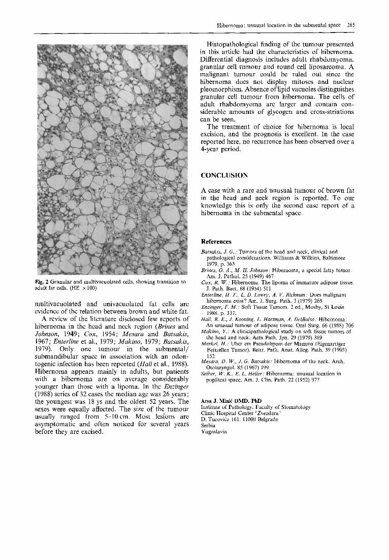
content of lipoproteins. In some multilocular cells coalescence of smaller into larger vacuoles was noted, with eccentric displacement of the nucleus. In ad- dition, there were numerous univacuolated, mature fat cells (Fig. 2). They were scattered throughout the neoplasm. The cells of tumour did not show mitoses or nuclear pleomorphism. Chronic inflammatory cells, aggregated into focal nests, were associated with the presence of dilated capillaries. Myxoid areas were focally distributed within the tumour.
P A T H O L O G I C A L FINDINGS
The tumour was yellow, lobulated and soft (Fig. 1). It measured 2 x 1.5 x 0.6cm.
Microscopically, the neoplasm was thinly encapsu- lated and had a lobular pattern. The lesion was composed predominantly of closely spaced round-to- oval cells having a distinct cellular membrane. The nuclei were usually centrally placed, blanded in appearance, and sharply defined. The cytoplasm of the cells was either granulated or multivacuolated. Periodic acid-Schiff staining demonstrated a diastase- resistant glycoprotein material, indicating a high
264
DISCUSSION
Hibernoma was first mentioned by Merkel (1905). The term hibernoma has been given to these tumours because the multivacuolated cells present in them resemble the cells seen in the brown fat of hibernating animals (Brines and Johnson, 1949; Cox, 1954). In humans, brown fat is largely restricted to the interscapular region, neck, axillae and retroperito- neum. It is seldom found after infancy. Transitions between brown and white fat are frequent; white fat may also revert to brown fat (Enzinger, 1988). In the present case, transitional stages between granular/
Hibernoma: unusual location in the submental space 265
Histopathological finding of the tumour presented in this article had the characteristics of hibernoma. Differential diagnosis includes adult rhabdomyoma, granular cell tumour and round cell liposarcoma. A malignant tumour could be ruled out since the hibernoma does not display mitoses and nuclear pleomorphism. Absence of lipid vacuoles distinguishes granular cell tumour from hibernoma. The cells of adult rhabdomyoma are larger and contain con- siderable amounts of glycogen and cross-striations can be seen.
The treatment of choice for hibernoma is local excision, and the prognosis is excellent. In the case reported here, no recurrence has been observed over a 4-year period.
CONCLUSION
A case with a rare and unusual tumour of brown fat in the head and neck region is reported. To our knowledge this is only the second case report of a hibernoma in the submental space.
Fig. 2 Granular and multivacuolated cells, showing transition to adult fat cells. (HE x 100).
multivacuolated and univacuolated fat cells are evidence of the relation between brown and white fat.
A review of the literature disclosed few reports of hibernoma in the head and neck region (Brines and Johnson, 1949; Cox, 1954; Mesara and Batsakis, 1967; Enterline et al., 1979; Makino, 1979; Batsakis, 1979). Only one tumour in the submental/ submandibular space in association with an odon- togenic infection has been reported (Hall et al., 1988). Hibernoma appears mainly in adults, but patients with a hibernoma are on average considerably younger than those with a lipoma. In the Enzinger (1988) series of 32 cases the median age was 26 years; the youngest was 18 ys and the oldest 52 years. The sexes were equally affected. The size of the tumour usually ranged from 5-10cm. Most lesions are asymptomatic and often noticed for several years before they are excised.
References
Batsakis, J. G. : Tumors of the head and neck, clinical and pathological considerations. Williams & Wilkins, Baltimore 1979, p. 363
Brines, O. A., M. H. Johnson : Hibernoma, a special fatty tumor. Am. J. Pathol. 25 (1949) 467
Cox, R. W.: Hibernoma. The lipoma of immature adipose tissue. J. Path. Bact. 68 (1954) 511
Enterline, H. T., L. D. Lowry, A. V. Richman ." Does malignant hibernoma exist? Am. J. Surg. Path. 3 (1979) 265
Enzinger, F. M. : Soft Tissue Tumors. 2 ed., Mosby, St Louis 1988, p. 337.
Hall, R. E., J. Kooning, L. Hartman, A. DelBalso : Hibernoma: An unusual tumour of adipose tissue. Oral Surg. 66 (1988) 706
Makino, Y. : A clinicipathological study on soft tissue tumors of the head and neck. Acta Path. Jpn. 29 (1979) 389
Merkel, H. : Uber ein Pseudolipom der Mamma (Eigenartiger Fettzellen Tumor). Beitr. Path. Anat. Alleg. Path. 39 (1905) 152
Mesara, D. W., J. G. Batsakis : Hibernoma of the neck. Arch. Otolaryngol. 85 (1967) 199
Seiber, W. K., E. L. Heller." Hibernoma: unusual location in popliteal space. Am. J. Clin. Path. 22 (1952) 977
Arsa J. Minid DMD, PhD Institute of Pathology, Faculty of Stomatology Clinic Hospital Center 'Zvezdara' D. Tucovida 161, 11000 Belgrade Serbia Yugoslavia