Embed Size (px)
Citation preview
180
EPIDEMIOLOGY AND PATHOGENESISHerpes zoster (zoster, shingles) is a neurocutaneous disease caused by the human herpes virus 3, the same virus that causes varicella (chick-enpox). It is a member of the herpes virus family (Herpesviridae) and exclusively infects human or simian cells. Varicella zoster virus (VZV) is the smallest of the alphaherpes viruses and has very stable linear double-stranded DNA. It has an icosohedral capsid and lipid envelope, which contains glycoproteins for cell entry.1
Herpes zoster has the highest incidence of any neurologic disease, affecting approximately 500 000 people in the United States yearly. The lifetime risk is 20–30%, and 50% of those living until 85 years of age will be affected.2–4 The reported incidence varies from 2.2–3.4 per 1000 individuals per year. Herpes zoster develops more frequently in the elderly, with an incidence rate of 10 per 1000 per year in people over 80 years. The incidence of zoster in immunosuppressed patients is substantially higher.5
In temperate climates, primary infection with this virus usually occurs before the age of 10, manifesting clinically as chickenpox (vari-cella). The virus then establishes a latent state in the sensory ganglia. In circumstances of diminished virus-specific and cell-mediated immu-nity, the virus may reactivate and spread to the corresponding der-matome along a spinal or cranial nerve (usually the trigeminal nerve) to generate the characteristic unilateral vesicular exanthem. The accompanying inflammation of the sensory nerve and skin damage are purportedly responsible for the acute pain.6,7
Physical trauma and surgery have been correlated with the develop-ment of zoster.8–11 Other reported triggers include tuberculosis, syphilis, radiation therapy, and steroids.12
The epidemiology of zoster is ultimately dependent on the transmis-sion and spread of VZV in a population. The spread of primary varicella (chickenpox) infection is of primary importance, but latent and reacti-vated infections also play an important role in maintaining VZV infec-tions within a population. Latently infected elderly adults and immunosuppressed patients are important reservoirs of the virus, as these groups are more likely to experience reactivation. When zoster does occur, the virus can be transmitted to a seronegative individual during the vesicular phase of the rash and cause a primary varicella infection. A zoster exposure with a seropositive, latently infected indi-vidual may result in a subclinical re-infection and boost humoral and cellular VZV immunity but is unlikely to cause varicella or herpes zoster.13 Herpes zoster may also develop in immunologically competent patients who harbor the latent virus and who are re-exposed to it by contact with someone who has an active varicella or zoster infection (primary, spontaneous, or infectious zoster).12,14,15
Ophthalmic Herpes ZosterThe frequency of herpes zoster ophthalmicus (HZO) is second only to thoracic dermatomal occurrence, with up to 250 000 cases occurring yearly in the United States. Of these, 50–70% suffer visual morbidity with severity increasing in the 5th–8th decades of life.3,16 The virus most commonly establishes latency in the trigeminal sensory ganglion and reactivates in 10–25% of the population.16 The ophthalmic division of the trigeminal nerve is affected 20 times more frequently than the maxillary or mandibular divisions.17 Ocular involvement occurs in more than 70% of patients with zoster of the first (ophthalmic) division of the trigeminal nerve. VZV more commonly resides in the portions of the ganglion that supply the upper lid and nasociliary branch of the trigeminal nerve. Nasociliary branch involvement with skin lesions located on the inner corner of the eye, tip of the nose, and the root or side of the nose (Hutchinson’s sign) is much more predictive (50–85%) of ocular involvement and is strongly prognostic for ocular inflamma-tion and corneal sensory denervation.12,18 It is important to remember that the eye may be seriously affected in up to 50% of cases in the absence of Hutchinson’s sign.19
HZO usually begins with a prodrome of influenza-like illness char-acterized by fatigue, malaise, nausea and mild fever accompanied by progressive pain and skin hyperesthesia. A diffuse erythematous or maculopapular rash then appears over a single dermatome 3–5 days later. These eruptions progress to form clusters of papules and clear vesicles, which then evolve through stages of pustulation and crusting. Patients with deeper involvement of the dermis may develop perma-nent scars with loss of normal pigmentation. Rarely, herpes zoster may manifest with ophthalmic symptoms in the absence of cutaneous eruptions.7,20
CLINICAL MANIFESTATIONSHZO can affect all ocular and adnexal tissues, and manifests with a diverse array of signs and symptoms. Ocular or extraocular involve-ment may occur at the time of the cutaneous eruptions or years later.
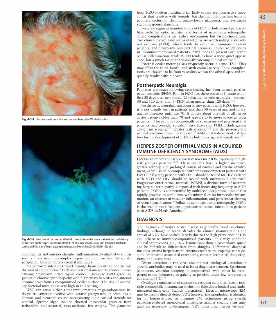
The skin of the forehead and upper eyelid is commonly affected and strictly obeys the midline with involvement of one or more branches of the ophthalmic division of the trigeminal nerve (Fig. 4-5-1). Zoster involves the deep dermis, in contrast to herpes simplex, which is lim-ited to the epidermis. The deep involvement causes numerous lid complications such as scarring, entropion and ectropion. Conjunctival findings include hyperemia, petechial hemorrhages, papillary or follicu-lar reaction and rarely a pseudomembrane. Episcleritis and scleritis are common and tend to progress towards the limbus causing vasculitis and sterile corneal infiltrates.21
Corneal pathology tends to results from three pathophysiologic mechanics: (1) active viral infection; (2) immune-mediated inflamma-tion; and (3) chronic neurotrophic keratopathy. Active viral infections tend to affect the epithelium, leading to punctate epithelial keratitis and pseudodendrites (Fig. 4-5-2). These lesions likely contain live virus. Pseudodendrites are typically smaller than typical dendrites, and lack terminal end-bulb formations. An immune-mediated stromal keratitis can take multiple forms. Nummular keratitis is the earliest finding of corneal stromal involvement and presents during the second week of the disease in 25–30% of patients.21 It is characterized by mul-tiple, fine, granular coin-shaped infiltrates of different diameters in the anterior stroma and can lead to permanent scarring. Chronic intersti-tial keratitis can lead to deep corneal neovascularization and lipid keratopathy. Disciform keratitis is a deep stromal infiltrate that devel-ops 3–4 months after the acute phase. Examination reveals a central disc-shaped area of diffuse corneal edema that results from
Gene Kim, Majid Moshirfar 4.5Herpes Zoster Ophthalmicus (HZO)
SECTION 3 External Diseases
PART 4 CORNEA AND OCULAR SURFACE DISEASES
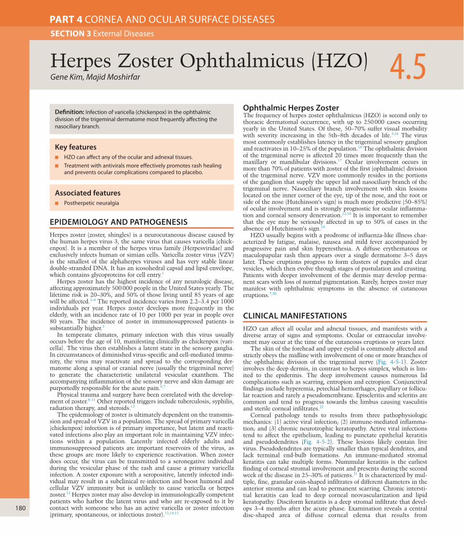
Definition: Infection of varicella (chickenpox) in the ophthalmic division of the trigeminal dermatome most frequently affecting the nasociliary branch.
Key features■ HZO can affect any of the ocular and adnexal tissues.■ Treatment with antivirals more effectively promotes rash healing
and prevents ocular complications compared to placebo.
Associated features■ Postherpetic neuralgia
Herpes Zoster O
phthalmicus (H
ZO)
4.5
181
from HZO is often multifactorial. Early causes are from active trabe-culitis that resolves with steroids, but chronic inflammation leads to papillary seclusion, chronic angle-closure glaucoma, and eventually steroid-response glaucoma.
Posterior segment manifestations of HZO include retinal perivascu-litis, ischemic optic neuritis, and forms of necrotizing retinopathy. These complications are rather uncommon but vision-threatening. Two clinical recognizable forms of retinitis are worth noting: acute reti-nal necrosis (ARN), which tends to occur in immunocompetent patients, and progressive outer retinal necrosis (PORN), which occurs in immunocompromised patients. ARN tends to present with severe ocular inflammation, while PORN tends to have a more quiet appear-ance, but a much faster and vision-threatening clinical course.21
External ocular motor palsies frequently occur in acute HZO. They may affect the third, fourth, and sixth cranial nerves. These complica-tions are thought to be from vasculitis within the orbital apex and fre-quently resolve within a year.
Postherpetic NeuralgiaPain that continues following rash healing has been termed posther-petic neuralgia (PHN). Pain in HZO has three phases: (1) acute pain – first 30 days after rash onset; (2) subacute herpetic neuralgia – between 30 and 120 days; and (3) PHN when greater than 120 days.22,23
Postherpetic neuralgia can occur in any patient with HZO; however, it is not usually seen in patients less than 50 years of age, and its fre-quency increases until age 70. It afflicts about one-half of all herpes zoster patients older than 70 and appears to be more severe in older patients.12 The pain may occasionally be so extreme and persistent that patients may consider suicide.12 Risk factors for PHN include greater acute pain severity,23,24 greater rash severity,25,26 and the presence of a painful prodrome preceding the rash.27 Additional independent risk fac-tors for the development of PHN include older age and female sex.22
HERPES ZOSTER OPHTHALMICUS IN ACQUIRED IMMUNE DEFICIENCY SYNDROME (AIDS)HZO is an important early clinical marker for AIDS, especially in high-risk younger patients.28–30 These patients have a higher incidence, greater severity, and prolonged course of corneal and uveitic involve-ment, as well as PHN compared with immunocompetent patients with HZO.31 All young patients with HZO should be tested for HIV. Patients with HZO and HIV should be treated with intravenous acyclovir.32 Progressive outer retinal necrosis (PORN), a distinct form of necrotiz-ing herpetic retinopathy, is reported with increasing frequency in AIDS patients. PORN is characterized by multifocal, deep retinal lesions that rapidly progress to confluence with minimal or no intraocular inflam-mation, an absence of vascular inflammation, and perivenular clearing of retinal opacification.19 Following cytomegalovirus retinopathy, PORN is the second most frequent opportunistic retinal infection in patients with AIDS in North America.33
DIAGNOSISThe diagnosis of herpes zoster disease is generally based on clinical findings, although in recent decades the clinical manifestations and spread of VZV have shifted, largely due to the high prevalence of HIV and otherwise immunocompromised patients. This may confound clinical impressions, e.g., HSV lesions may show a zosteriform spread and be difficult to differentiate from shingles. Differential diagnosis includes eczema herpeticatum, eczema vaccinatum, impetigo contagio-sum, enterovirus-associated exanthema, contact dermatitis, drug erup-tions, and insect bites.
Direct detection of the virus and indirect serological detection of specific antibodies may be used to boost diagnostic acumen. Specimens (cutaneous vesicular scraping or conjunctival swab) must be trans-ported to the laboratory as quickly as possible under low temperature conditions (4 °C).33
Cytologic examination of cutaneous vesicular scrapings reveals mul-tiple eosinophilic intranuclear inclusions (Lipschutz bodies) and multi-nucleated giant cells (Tzanck preparation). Electron microscopy (EM) may be used to directly detect VZV; however, the morphology is identical for all herpesviridae, so immune EM techniques using specific peroxidase-labeled monoclonal antibodies against specific virus anti-gens are necessary to distinguish VZV from other herpes viruses.34
endotheliitis and anterior chamber inflammation. Perilimbal vasculitis results from immune-complex deposition and can lead to sterile, peripheral, anterior cornea stromal infiltrates.
Active zoster infections travel through branches of the ophthalmic division of cranial nerve.5 Each reactivation damages the corneal nerves causing progressive neurotrophic cornea. Late-stage HZO gives the picture of chronic epitheliopathy with filamentary keratitis and anterior stromal scars from a compromised ocular surface. The risk of second-ary bacterial infection is very high in this setting.19
HZO can cause either a nongranulomatous or granulomatous iri-docyclitis (anterior uveitis) with keratic precipitates. It often has a chronic and recurrent course necessitating topic cortical steroids for control. Specific signs include elevated intraocular pressure from trabeculitis and sectorial, vaso-occlusive iris atrophy. The glaucoma
Fig. 4-5-1 Herpes zoster ophthalmicus involving the V1 distribution.
Fig. 4-5-2 Peripheral corneal epithelial pseudodendrites in a patient with a history of herpes zoster ophthalmicus. (From Hu AY, et al. Late varicella-zoster virus dendriform keratitis in patients with histories of herpes zoster ophthalmicus. Am J Ophthalmol 2010;149:214–220 e3.)
4
182
CORN
EA A
ND
OCU
LAR SU
RFACE DISEA
SES
effective than tricyclic antidepressants against treating allodynia, another subtype of neuralgia.46 Capsaicin cream (0.025%) is effective when applied to the involved skin 3–4 times daily, although 2 weeks of treatment are often required for pain relief.47 PHN may be severe, intractable, and permanent, with some patients requiring psychiatric and pain clinic care, and occasionally trigeminal rhizotomy or stellate ganglion block.48
The use of systemic steroids in the treatment of PHN remains con-troversial. Recent studies have found no benefit of oral prednisolone compared with placebo in preventing PHN.49 Prednisolone-treated patients did have more pain relief initially; however, at 6 months there was no difference between the two groups.
Findings of a meta-analysis reveal that famciclovir and valacyclovir significantly reduce the duration but not incidence of PHN. Steroids have no effect on PHN. Amitriptyline for 90 days reduced the incidence of pain at 6 months. Finally, a single trial of percutaneous electrical nerve stimulation (PENS) in 50 patients suggested a decrease in pain incidence at 3 and 6 months when compared with famciclovir.50
PREVENTIONThe varicella vaccine is available in two formulations which are both given subcutaneously: Varivax (Merck) and Zostavax (Merck). The former prevents primary varicella infections in infants while the latter prevents reactivation of zoster in adults. Both utilize live attenuated virus. The main differences between the two vaccines are their dosing and concentration. Zostavax is only administrated once and has 14 times the concentration of Varivax, which needs to be given twice.51
The Shingles Prevention Study (SPS) showed that the vaccine reduced the incidence of HZO by 49% and reduced the severity of the illness in those that did have a reactivation of the virus.52 The vaccine was gener-ally safe and well tolerated with the rate of adverse events being 1.4% and no difference in the vaccine versus placebo group. The most com-mon adverse effect was pain and erythema at the injection site. The vaccinated group, however, did have a higher rate of serious adverse events. A couple of case reports in the literature report exacerbation of chronic HZO and also of new onset retinitis in immunocompromised patients within the first 6 weeks following vaccination.51,53
Currently, the Centers for Disease Control and Prevention (CDC) and Advisory Committee on Immunization Practices (ACIP) recom-mend that the zoster vaccine be administrated to adults aged 60 and above for the prevention of herpes zoster, including those who have already had a case of zoster.
KEY REFERENCESArvin AM. The varicella-zoster virus. In: Watson CPN, Gershon AA, editors. Herpes zoster and
post herpetic neuralgia. 2nd ed. Pain research and clinical management, vol. 11. New York: Elsevier Science BV; 2001. p. 25–39.
Balfour Jr HH, Bean B, Laskin OL, et al. Acyclovir halts progression of herpes zoster in immunocompromised patients. N Engl J Med 1983;308:1448.
Brinsson M, Edmunds WJ, Law B, et al. Epidemiology of varicella zoster virus infection in Canada and the United Kingdom. Epidemiol Infect 2001;127:305–14.
Choo PW, Galil K, Donahue JG, et al. Risk factors for postherpetic neuralgia. Arch Intern Med 1997;157:1217–24.
Colin J, Pristant O, Beatrice C, et al. Comparison of the efficacy and safety of valaciclovir and acyclovir for the treatment of herpes zoster ophthalmicus. Ophthalmology 2000;107: 1507–11.
Gelb LD. Preventing herpes zoster through vaccination. Ophthalmology 2008; 115(2 Suppl): S35–8.
Liesegang TJ. Herpes zoster ophthalmicus natural history, risk factors, clinical presentation, and morbidity. Ophthalmology 2008;115(2 Suppl):S3–12.
Mahalingam R, Wellish M, Lederer D, et al. Quantitation of latent varicella-zoster virus DNA in human trigeminal ganglia by polymerase chain reaction. J Virol 1993;67:2381–4.
Pavan-Langston D. Herpes zoster antivirals and pain management. Ophthalmology 2008; 115(2 Suppl):S13–20.
Sellitti TP, Huang AJ, Schiffman J, et al. Association of herpes zoster ophthalmicus with acquired immunodeficiency syndrome and acute retinal necrosis. Am J Ophthalmol 1993;116:297.
Wolff MH, Schunemann S, Rahaus M, et al. Diagnosis of varicella-zoster virus associated diseases with special emphasis on infections in the immunocompromised host. In: Wolff MH, Schunemann S, Schimdt A, editors. Varicella-zoster virus: molecular biology, pathogenesis and clinical aspects. Contributions to Microbiology, vol. 3. Basle: Karger; 1999. p. 150–7.
Zaal MJW, Volker-Dieben HJ, D’Amaro J. Prognostic value of Hutchinson’s sign in acute herpes zoster ophthalmicus. Graefes Arch Clin Exp Ophthalmol 2003;241:187–91.
VZV-DNA can be directly obtained via anterior chamber paracentesis or vitreous tap and analyzed using real-time PCR.35 Fluorescent antibody techniques, cytospin direct immunofluorescence staining, and rapid direct immunofluorescence assays (SimulFluor direct fluorescent anti-body) are additional methods for detecting VZV virus.36
Serologic testing to detect herpes zoster antibodies is of limited use as cross-reactivation between VZV and HSV can occur. Following zoster, a booster of IgG is detected for 2 weeks after infection, which then falls to lower levels, where it may remain for years.37
MANAGEMENTHZO is treated with oral antivirals: acyclovir, famciclovir, or valacyclo-vir. Acyclovir (800 mg, five times daily) for 7–10 days, which reduces viral shedding and the chance of systemic dissemination of the virus as well as the incidence and severity of ocular complications, particularly if used within 72 hours of onset of symptoms.38,39 Acyclovir can also shorten the duration of pain if taken within the first 3 days of onset of symptoms.40,41 Intravenous acyclovir is recommended in immunocom-promised patients.38
Famciclovir is a prodrug of penciclovir and has a much higher bioa-vailability (77%) than acyclovir (18%). The recommended dosage is 500 mg three times daily for 7 days, which has been shown to be well tolerated and safe with similar efficacy to acyclovir 800 mg five times daily.42
Valacyclovir is the L-valine ester of acyclovir and has higher bioavail-ability (80%) than acyclovir (18%). The usual dosage is 1000 mg three times daily. It has similar activity to acyclovir in the prevention of the sequelae of herpes zoster. Valacyclovir has been shown to be as effective as acyclovir in preventing ocular complications of HZO, including con-junctivitis, superficial and stromal keratitis, and pain. This compara-tive analysis also showed that tolerability of the two drugs was similar, but the dosing schedule of valacyclovir was simpler.43 Valacyclovir has been shown to significantly accelerate the resolution of pain when com-pared with acyclovir.44 Comparisons between valacyclovir and famciclo-vir treatment in HZO have not shown a statistically significant difference in resolution of pain or rash.45
Management of Ocular ManifestationPalliative therapy, including Burow’s solution, cool compresses, mechanical cleansing of the involved skin, and topical antibiotic oint-ment without steroid, are helpful in treating skin lesions.
Oral acyclovir has been shown to be effective for the punctate, pseu-dodendritic, and delayed corneal mucous plaque forms of herpes zoster epithelial keratitis.40,41 Debridement may also be helpful.
Neurotrophic keratitis or the epithelial defects associated with her-pes zoster keratitis may be treated with nonpreserved artificial tears, eye ointments, punctal occlusion, pressure patching, or therapeutic soft contact lenses. If these measures are unsuccessful then tarsorrhaphy, conjunctival flap, or autologous conjunctival transplantation should be considered.
Topical steroids are useful in the management of sclerokeratitis, keratouveitis, interstitial keratitis, anterior stromal infiltrates, and disciform keratitis.21 Steroids should not be used in cases of exposure or neurotrophic keratitis because of the possibility of keratolysis.21 Topical cycloplegics prevent ciliary spasm associated with herpes zoster inflammatory disease. Aqueous suppressants and topical corticoster-oids should be used to treat HZO glaucoma. Herpes zoster vitritis, vitreous hemorrhage, or vitreous debris is usually secondary to severe inflammation and may respond to topical, periocular, or systemic steroids. Herpes zoster infections affecting the cranial nerves are best treated with a combination of systemic steroids and intravenous acy-clovir. Retinitis (acute retinal necrosis syndrome and progressive outer retinal necrosis) is best treated with a combination of intravitreal injec-tion and oral valacyclovir.
Postherpetic NeuralgiaPostherpetic neuralgia may be treated with analgesics, tricyclic antide-pressants (nortriptyline, amitriptyline, desipramine, clomipramine), and anticonvulsants (carbamazepine and phenytoin), often in combina-tion. Newer medications, e.g., gabapentin and pregabalin, are more
Access the complete reference list online at
Herpes Zoster O
phthalmicus (H
ZO)
4.5
182.e1
REFERENCES1. Arvin AM. The varicella-zoster virus. In: Watson CPN, Gershon AA, editors. Herpes zoster and
post herpetic neuralgia. 2nd ed. Pain research and clinical management, vol. 11. New York: Elsevier Science BV; 2001. p. 25–39.
2. Kurtzke JF. Neuroepidemiology. Ann Neurol 1984;16:265–77.3. Donahue JG, Choo PW, Manson JE, et al. The incidence of herpes zoster. Arch Intern Med
1995;155:1605–9.4. Brinsson M, Edmunds WJ, Law B, et al. Epidemiology of varicella zoster virus infection in
Canada and the United Kingdom. Epidemiol Infect 2001;127:305–14.5. Chapman RS, Cross KW, Fleming DM. The incidence of shingles and its implications for
vaccination policy. Vaccine 2003;21:2541–7.6. Haanpaa M, Dastidar P, Weinberg A, et al. CSF and MRI findings in patients with acute herpes
zoster. Neurology 1998;51:1405–11.7. Opstelten W, Zaal MJW. Managing ophthalmic herpes zoster in primary care. BMJ
2005;331:147–51.8. Evans RW, Lee AG. Herpes zoster ophthalmicus, ophthalmoplegia and trauma.
Headache 2004;44:286–8.9. Netland PA, Zierhut M, Raizman MB. Post traumatic herpes zoster ophthalmicus as a
presenting sign of human immunodeficiency virus infection. Ann Ophthalmol 1993;25:14–5.10. Wachym PA, Gray GF, Avant GR. Herpes zoster of the larynx after intubational trauma.
J Laryngeal Otol 1986;100:839–41.11. Weiss R. Herpes zoster following spinal surgery. Clin Exp Dermatol 1989;14:56–7.12. Ostler HB, Thygeson P. The ocular manifestations of herpes zoster, varicella, infectious
mononucleosis and cytomegalovirus disease. Surv Ophthalmol 1976;21:148.13. Arvin AM. The varicella-zoster virus. In: Fields BN, Knipe DM, Howley PM, editors.
Fields virology. 3rd ed. Philadelphia, PA: Lippincott Raven; 1996. p. 2547–87.14. Ragozzino MW, Melton LJ, Kurland LT, et al. Risk of cancer after herpes zoster: a population-
based study. N Engl J Med 1982;307:393.15. Weller TH. Varicella and herpes zoster: changing concepts of the natural history, control and
importance of a not-so-benign virus. N Engl J Med 1983;309:1362.16. Ragozzino MW, Melton LJ, Kurland LT, et al. Population-based study of herpes zoster and its
sequelae. Medicine 1982;61:310–16.17. Mahalingam R, Wellish M, Lederer D, et al. Quantitation of latent varicella-zoster virus DNA in
human trigeminal ganglia by polymerase chain reaction. J Virol 1993;67:2381–4.18. Zaal MJW, Volker-Dieben HJ, D’Amaro J. Prognostic value of Hutchinson’s sign in acute
herpes zoster ophthalmicus. Graefes Arch Clin Exp Ophthalmol 2003;241:187–91.19. Chang SD, De Luise VP, Tasman W, et al. editors. Duane’s ophthalmology, vol. 4, ch. 20
(CD-Rom). Philadelphia, PA: Lippincott Williams & Wilkins; 2001.20. Schwab IR. Herpes zoster sine erupticum: anterior segment equivalent of acute retinal
necrosis. Poster presented at the 97th Annual Meeting of the American Academy of Ophthalmology, Chicago, IL, 16 November, 1993.
21. Liesegang TJ. Herpes zoster ophthalmicus natural history, risk factors, clinical presentation, and morbidity. Ophthalmology 2008;115(2 Suppl):S3–12.
22. Zaal MJW, Volker-Dieben HJ, D’Amaro J. Risk and prognostic factors of post herpetic neuralgia and focal sensory denervation: a prospective evaluation in acute herpes zoster ophthalmicus. Clin J Pain 2000;16:345–51.
23. Jung BF, Johnson RN, Griffin DRJ, et al. Risk factors for post herpetic neuralgia in patients with herpes zoster. Neurology 2004;62:1545–51.
24. Scott FT, Leedham-Gree ME, Barrett-Muir WY, et al. A study of shingles and the development of post herpetic neuralgia in East London. J Med Virol 2003;70:S24–30.
25. Hinga K, Mori M, Hirata K, et al. Severity of skin lesions of herpes zoster at the worst phase rather than age and involved region most influences the duration of acute herpetic pain. Pain 1997;69:245–53.
26. Herr H. Prognostic factors of post herpetic neuralgia. J Korean Med Sci 2002;17:655–9.27. Choo PW, Galil K, Donahue JG, et al. Risk factors for postherpetic neuralgia. Arch Intern Med
1997;157:1217–24.28. Engstrom Jr RE, Holland GN, Margolis TP, et al. The progressive outer retinal necrosis
syndrome: a variant of necrotizing herpetic retinopathy in patients with AIDS. Ophthalmology 1994;101:1488.
29. Sellitti TP, Huang AJ, Schiffman J, et al. Association of herpes zoster ophthalmicus with acquired immunodeficiency syndrome and acute retinal necrosis. Am J Ophthalmol 1993;116:297.
30. Kestelyn P, Stevens AM, Bakkers E, et al. Severe herpes zoster ophthalmicus in young African adults: a marker for HTLV-III seropositivity. Br J Ophthalmol 1987;71:806.
31. Cole EL, Miesler DM, Calabrese LH, et al. Herpes zoster ophthalmicus and acquired immune deficiency syndrome. Arch Ophthalmol 1984;102:1027.
32. Seiff S, Margolis T, Graham S, et al. Use of intravenous acyclovir for treatment of herpes zoster ophthalmicus in patients at risk for AIDS. Ann Ophthalmol 1988;20:480.
33. Engstrom RE Jr, Holland GN, Margolis TP, et al. The progressive outer retinal necrosis syndrome: a variant of necrotizing herpetic retinopathy in patients with AIDS. Ophthalmology 1994;101:1488.
34. Wolff MH, Schunemann S, Rahaus M, et al. Diagnosis of varicella-zoster virus associated diseases with special emphasis on infections in the immunocompromised host. In: Wolff MH, Schunemann S, Schimdt A, editors. Varicella-zoster virus. Molecular biology, pathogenesis and clinical aspects. (Contributions to microbiology, 3.) Basle: Karger; 1999. p. 150–7.
35. Kaneko H, Iida T, Aoki K, et al. Sensitive and rapid detection of herpes simplex virus and varicella-zoster virus by loop-mediated isothermal amplification (LAMP). J Clin Microbiol 2005;43:3290–6.
36. Chan EL, Brandt K, Horsman GB. Comparison of chemicon SimulFluor direct fluorescent antibody staining with cell culture and shell viral direct immunoperoxidase staining for detection of herpes simplex virus and with cytospin direct immunofluorescence staining for detection of varicella-zoster virus. Clin Diagnost Lab Immunol 2001;8:909–12.
37. Arvin AM, Koropchak CM. Immunoglobulins M and G to varicella-zoster virus measured by solid phase radioimmunoassay. Antibody responses to varicella and herpes zoster infections. J Clin Microbiol 1980;12:367–74.
38. Balfour HH Jr, Bean B, Laskin OL, et al. Acyclovir halts progression of herpes zoster in immunocompromised patients. N Engl J Med 1983;308:1448.
39. Bean B, Braun C, Balfour HH Jr. Acyclovir therapy for acute herpes zoster. Lancet 1982;2:118.40. Cobo LM, Foulks GN, Liesegang T, et al. Oral acyclovir in the treatment of acute herpes zoster
ophthalmicus. Ophthalmology 1986;93:763.41. Cobo LM, Foulks GN, Liesegang T, et al. Oral acyclovir in the therapy of acute herpes zoster
ophthalmicus: an interim report. Ophthalmology 1985;92:1574.42. Tyring S, Engst R, Corriveau CH, et al. Famciclovir for ophthalmic zoster: a randomised
acyclovir controlled study. Br J Ophthalmol 2001;85:576–81.43. Colin J, Pristant O, Beatrice C, et al. Comparison of the efficacy and safety of valaciclovir and
acyclovir for the treatment of herpes zoster ophthalmicus. Ophthalmology 2000;107: 1507–11.
44. Lin W, Lin H, Lee SS, et al. Comparative study of the efficacy and safety of valaciclovir versus acyclovir in the treatment of herpes zoster. J Microbiol Immunol Infect 2001;34:138–42.
45. Tyring S, Beutner K, Tucker B, et al. Antiviral therapy for herpes zoster: randomized, controlled clinical trial of valacyclovir and famciclovir therapy in immunocompetent patients 50 years and older. Arch Fam Med 2000;9:863–96.
46. Pavan-Langston D. Herpes zoster antivirals and pain management. Ophthalmology 2008;115(2 Suppl):S13–20.
47. Watson P, Ross D, Soltani K, et al. Therapeutic advances in the management of post-herpetic neuralgia. Geriatr Med Today 1988;7:20.
48. Olson ER, Ivy HB. Stellate block for trigeminal zoster. J Clin Neuro Ophthalmol 1981;1:53.49. Esmann V, Geil J, Kroon S, et al. Prednisolone does not prevent post-herpetic neuralgia.
Lancet 1987;2:126.50. Alper BS, Lewis PR. Does treatment of acute herpes zoster prevent or shorten postherpetic
neuralgia? A systematic review of the literature. J Family Practice 2000;49:255–64.51. Charkoudian LD, Kaiser GM, Steinmetz RL, et al. Acute retinal necrosis after herpes zoster
vaccination. Arch Ophthalmol 2011;129:1495–7.52. Gelb LD. Preventing herpes zoster through vaccination. Ophthalmology 2008;
115(2 Suppl):S35–38.53. Khalifa YM, Jacoby RM, Margolis TP. Exacerbation of zoster interstitial keratitis after zoster
vaccination in an adult. Arch Ophthalmol 2010;128:1079–80.


![AnAcuteCaseofHerpesZosterOphthalmicuswith Ophthalmoplegiadownloads.hindawi.com/journals/criopm/2012/953910.pdf · 2019-07-31 · [5] A. E. Edgerton, “Herpes Zoster ophthalmicus:](https://img.dokumen.tips/doc/110x75/5e537d95ba71a240a47e403d/anacutecaseofherpeszosterophthalmicuswith-opht-2019-07-31-5-a-e-edgerton.jpg)