Embed Size (px)
Citation preview

2695Research Article
IntroductionATM (ataxia telangiectasia-mutated), ATR (ATM and Rad3-related) and DNA-PKcs (DNA-dependent protein kinasecatalytic subunit) are phosphoinositide-3-kinase-relatedprotein kinases (PIKKs) that are rapidly activated in responseto different forms of genotoxic stress. Through thephosphorylation of downstream targets, these stress kinasesdirectly or indirectly activate proteins that participate in cellcycle checkpoints and DNA repair. If the DNA lesion isirreparable, then cells are eliminated through the induction ofapoptosis (reviewed in Burma and Chen, 2004; Kurz and Lees-Miller, 2004; Shechter et al., 2004). Double-strand DNAbreaks (DSBs) can result in the activation of DNA-PKcsleading to DNA repair via the nonhomologous end-joining(NHEJ) pathway (Burma and Chen, 2004) or in the activationof ATM leading to repair through the homologousrecombination (HR) pathway (Kurz and Lees-Miller, 2004).ATR on the other hand, is activated by ssDNA intermediatesthat are generated by a variety of genotoxic assaults includingthose that cause replication fork perturbations. ATR alsoresponds to DSBs but with slower kinetics than ATM. The ATRpathway also plays a crucial role in the maintenance of active,unperturbed replication forks (Shechter et al., 2004).
PIKK-mediated signal transduction involves the recruitmentof the stress kinase to the site of DNA damage. PIKKrecruitment is mediated through a conserved interaction motif
located within the C-terminus of interacting partners (Falck etal., 2005). ATM recruitment to DSBs is mediated throughNBS1, a component of the MRE11-RAD50-NBS1 (MRN)repair complex (Falck et al., 2005). ATR is recruited to ssDNAthrough its tightly associated binding partner ATRIP, which canefficiently recognize replication protein A (RPA)-boundssDNA (Ball et al., 2005; Falck et al., 2005; Zou and Elledge,2003). DNA-PKcs recruitment to DSBs is facilitated by itsinteraction with the Ku80 subunit of the Ku70/Ku80 DNA end-binding complex (Falck et al., 2005). Nuclear events such asDNA replication and repair are commonly studied bymonitoring the subcellular distribution of key proteinsinvolved. For example, immunofluorescence (IF) microscopyof cells exposed to DNA-damaging agents have demonstratedthat ATM and the MRN complex accumulate at DSBs asdetected by staining for �H2AX (phosphorylated histoneH2AX) which is a sensitive marker for DSBs (Falck et al.,2005). It has been similarly shown that ATR, ATRIP and RPAaccumulate at stretches of ssDNA generated in response toagents that induce DNA damage or replication stress (Ball etal., 2005; Barr et al., 2003).
As obligate intracellular parasites, viruses rely on and oftenmanipulate host cell replication and repair factors presumablyfor their own benefit. Herpes simplex virus type 1 (HSV-1) isa large linear dsDNA virus that replicates in the nucleus ofthe infected cell within globular domains called replication
Like other DNA viruses, herpes simplex virus type 1 (HSV-1) interacts with components of the cellular response toDNA damage. For example, HSV-1 sequesters endogenous,uninduced, hyperphosphorylated RPA (replication proteinA) away from viral replication compartments. RPA is assDNA-binding protein that signals genotoxic stressthrough the ATR (ataxia telangiectasia-mutated and Rad3-related) pathway. The sequestration of endogenoushyperphosphorylated RPA away from replicating viralDNA suggests that HSV-1 prevents the normal ATR-signaling response. In this study we examine the spatialdistribution of endogenous hyperphosphorylated RPA withrespect to ATR, its recruitment factor, ATRIP, and thecellular dsDNA break marker, ��H2AX, during HSV-1infection. The accumulation of these repair factors at DNA
lesions has previously been identified as an early event insignaling genotoxic stress. We show that HSV-1 infectiondisrupts the ATR pathway by a mechanism that preventsthe recruitment of repair factors, spatially uncouplesATRIP from ATR and sequesters ATRIP and endogenoushyperphosphorylated RPA within virus-induced nucleardomains containing molecular chaperones and componentsof the ubiquitin proteasome. The HSV-1 immediate earlyprotein ICP0 is sufficient to induce the redistribution ofATRIP. This is the first report that a virus can disrupt theusually tight colocalization of ATR and ATRIP.
Key words: Herpesvirus, ICP0, DNA-damage response, ATR,ATRIP, Hyperphosphorylated RPA, Phosphorylated H2AX,Chaperones, Proteasome
Summary
Herpes simplex virus type I disrupts the ATR-dependent DNA-damage response during lyticinfectionDianna E. Wilkinson and Sandra K. Weller*Department of Molecular, Microbial and Structural Biology MC3205, University of Connecticut Health Center, 263 Farmington Avenue, Farmington,CT 06030, USA*Author for correspondence (e-mail: [email protected])
Accepted 20 March 2006Journal of Cell Science 119, 2695-2703 Published by The Company of Biologists 2006doi:10.1242/jcs.02981
Jour
nal o
f Cel
l Sci
ence

2696
compartments (Quinlan et al., 1984). The first indication thatHSV-1 activates a component of the cellular DNA damageresponse was the observation that lytic infection induces thephosphorylation of NBS1, a DNA damage signaling event thatcorrelates with the recruitment of NBS1 to viral precursors ofreplication compartments (Wilkinson and Weller, 2004). Theobservation that replication compartments recruit componentsof the MRN complex (Taylor and Knipe, 2004; Wilkinson andWeller, 2004) suggests that the MRN complex is used by HSV-1 to promote viral DNA replication, perhaps for the formationof greater-than-unit-length concatemers (Wilkinson andWeller, 2004). Subsequent studies have demonstrated thatHSV-1 infection induces an ATM-dependent activation ofNBS1 and other downstream targets and that HSV-1 infectionis somewhat compromised in cells deficient for Mre11 orNBS1 (Lilley et al., 2005; Shirata et al., 2005). These resultssupport the suggestion that the MRN complex is important forefficient HSV-1 infection.
Although HSV-1 appears to activate components of theATM-dependent signaling pathway, several lines of evidencesuggest that HSV-1 also inactivates components of the NHEJmachinery. In some cell types the HSV-1-encoded immediateearly protein ICP0 targets DNA-PKcs for degradation by theproteasome (Lees-Miller et al., 1996; Parkinson et al., 1999).In Vero cells, we have shown that the Ku80 subunit of theDNA-PK complex is not recruited to the earliest prereplicativesites for HSV-1 (Wilkinson and Weller, 2004). Furthermore,HSV-1 yields are actually increased in Ku70- or DNA-PKcs-deficient cell lines suggesting that NHEJ may have aninhibitory effect on infection (Parkinson et al., 1999; Taylorand Knipe, 2004). Thus, HSV-1 apparently uses ATM-mediated events while inactivating the NHEJ pathway. In thispaper we extend these studies to the analysis of the subcellularATR-mediated damage response during HSV-1 infection.
One predominant ATR signaling event for replication stressor DNA damage is the hyperphosphorylation of the 32-kDasubunit (RPA32) of the heterotrimeric RPA protein, whichoccurs in response to the accumulation of ssDNA. Thehyperphosphorylation of RPA32, which is catalyzed by PIKKs,is thought to direct the role of RPA from DNA replication tothat of repair (Vassin et al., 2004). Hyperphosphorylated RPA,which accumulates at ssDNA lesions serves as a marker forDNA damage and repair (reviewed by Binz et al., 2004) andhas been shown to colocalize with ATR and Mre11 in nuclearfoci following DNA damage (Robison et al., 2004; Wu et al.,2005). Two populations of RPA have been observed in HSV-1infected cells. One RPA population, which can be detected withgeneral anti-RPA antibodies, is found within viral replicationcompartments (Uprichard and Knipe, 1997; Wilcock and Lane,1991; Wilkinson and Weller, 2004) and may act at viralreplication forks. The other population of RPA is recognizedby a phosphospecific anti-RPA antibody and representsuninduced, endogenous levels of hyperphosphorylated RPAthat is sequestered away from replication compartments(Wilkinson and Weller, 2005). The sequestration of thisuninduced population of hyperphosphorylated RPA (hereafterreferred to as P-RPA) may be part of a global mechanism bywhich HSV-1 prevents triggering stress signals that could bedeleterious to the progression of infection (Wilkinson andWeller, 2005). In this study, IF microscopy was used to showthat HSV-1 infection prevents the ATR-dependent signaling of
Journal of Cell Science 119 (13)
cellular replication stress or DNA damage by a mechanism thatthat spatially uncouples ATRIP from ATR and sequestersATRIP and P-RPA within virus-induced domains enriched withmolecular chaperones and the ubiquitin proteasome (VICEdomains). We also show that the HSV-1 immediate earlyprotein, ICP0, is sufficient to induce the relocalization ofATRIP.
ResultsSubcellular distribution of �H2AX and P-RPA in HSV-1-infected cells�H2AX and hyperphosphorylated RPA are commonly used asmarkers for DNA damage. The presence of �H2AX is asensitive indicator of DSBs within cellular chromatin, whereashyperphosphorylated RPA accumulates at stretches of ssDNAgenerated from the processing of DNA lesions or perturbedreplication forks. �H2AX and hyperphosphorylated RPA arecommonly found colocalized in DNA-damage-induced foci(Vassin et al., 2004; Wu et al., 2005). We first examined thelocalization of �H2AX in relation to viral replicationcompartments as detected by staining for the HSV-1 ssDNA-binding protein, UL29. Double labeling of HSV-1-infectedcells revealed an accumulation of �H2AX in areas surroundingreplication compartments (Fig. 1A-C). The marginalization of�H2AX outside replication compartments is in agreement withprevious reports describing the peripheral displacement ofcellular DNA during HSV-1 infection (Monier et al., 2000;Simpson-Holley et al., 2004). Previous reports havedemonstrated that the frequency and intensity of �H2AX fociincrease linearly with the amount of DNA damage sustained(Rogakou et al., 1999; Rogakou et al., 1998), with each discrete�H2AX focus corresponding to one DSB (Sedelnikova et al.,2002). The accumulation of �H2AX staining seen immediatelysurrounding replication compartments suggests that themarginated cellular chromatin has sustained significant DSBsduring lytic infection. The marginalization of �H2AX wasobserved at early times post infection when cells displayedsmall, developing replication compartments (data not shown).Although we cannot rule out the presence of undetectableamounts of �H2AX at viral DNA, our results suggest that�H2AX does not mark the occurrence of DSBs within thereplicating HSV-1 genome. This is not surprising becausereplicating viral DNA is not found in an ordered nucleosomalform with only a fraction of the HSV-1 genomes associatedwith histones (Leinbach and Summers, 1980).
We next assessed the spatial relationship between P-RPAand �H2AX in HSV-1-infected cells. Since the staining patternof �H2AX was shown to define the periphery of replicationcompartments (Fig. 1A-C), it was possible to characterize thelocalization of �H2AX and P-RPA with respect to replicationcompartments simply by double staining for �H2AX and P-RPA. Mock-infected cells typically displayed low levels of P-RPA distributed either in a diffuse or rough granular patternwithin the nucleus as described previously (Wilkinson andWeller, 2005) (Fig. 1E). A low-level nuclear staining patternfor �H2AX was also observed in mock-infected cells (Fig. 1D)with no significant co-staining of �H2AX and P-RPA (Fig. 1F).Western analysis has demonstrated that productive HSV-1infection does not induce the hyperphosphorylation of RPA(Wilkinson and Weller, 2005). Fig. 1G-I indicates that thisendogenous P-RPA does not colocalize with the marginated
Jour
nal o
f Cel
l Sci
ence

2697HSV-1 disruption of the ATR response
�H2AX in HSV-1-infected cells. In fact, the two markers wereconsistently present at locations in the nucleus which aremutually exclusive of each other (Fig. 1J-L). This result wasunexpected in light of previous demonstrations showing thecolocalization of these two markers in repair foci followingDNA damage (Vassin et al., 2004; Wu et al., 2005). To rule outthe possibility that Vero cells were defective in the formationof this type of repair foci during the DNA damage response,we performed a control experiment in which cells were treatedwith 1 �M camptothecin (CPT), a genotoxic agent known toinduce the nuclear colocalization of hyperphosphorylated RPAand �H2AX (Vassin et al., 2004; Wu et al., 2005). Indeed, CPTtreatment of Vero cells induced the increased staining ofhyperphosphorylated RPA and its accumulation with �H2AX(Fig. 2) indicating that the segregation of P-RPA and �H2AXinto separate nuclear compartments during infection is not dueto an inherent defect of Vero cells in the response to DNAdamage. Thus, although Vero cells are competent to induce thecolocalization of �H2AX and hyperphosphorylated RPA into
CPT-induced DNA repair foci (Fig. 2), endogenous P-RPA isactually excluded from sites staining for �H2AX during HSV-1 infection (Fig. 1J-L). We conclude that HSV-1 sequestersendogenous P-RPA away from both replicating viral DNA andcellular DSBs.
P-RPA is present in HSV-1-induced foci called VICEdomainsWe and others have previously reported that HSV-1 infectioncauses the relocalization of cellular heat-shock proteins (Hsps)into domains that also contain components of the ubiquitinproteasome (Burch and Weller, 2004; Everett, 2000). Weproposed that these virus-induced chaperone-enriched (VICE)domains represent a mechanism by which HSV-1 sequestersmisfolded, modified or unwanted proteins. This sequestrationmay prevent cellular events, such as premature apoptosis,which would be catastrophic to the virus (Burch and Weller,2004). The P-RPA staining pattern observed during infectionexhibited a nuclear organization reminiscent of that observedfor VICE domains (Burch and Weller, 2004; Burch and Weller,2005; Everett, 2000). To confirm this, we examined cells thatwere triple-labeled with antibodies to UL29, Hsc70 (Heatshock cognate 70) and P-RPA to visualize replicationcompartments, VICE domains and endogenous P-RPA,respectively (Fig. 3). As previously described, HSV-1 infectionresults in the redistribution of Hsc70 from the nucleolus intoVICE domains located adjacent to replication compartments(Fig. 3C,G) (Burch and Weller, 2004; Burch and Weller, 2005).A subset of P-RPA foci also colocalized with the Hsc70-staining VICE domains (Fig. 3E-I). Additional triple labelingexperiments using antibodies directed against UL29, Hsc70and �H2AX indicate that VICE domains themselves areexcluded from sites of cellular DSBs (Fig. 4). The cells shownin Figs 3 and 4 were extracted with Triton X-100 to removethe cytoplasm and nucleosolic proteins before fixation. Similarprotein localization patterns were observed in cells that werefixed for detection of total proteins (results not shown).
HSV-1 infection induces the spatial uncoupling of theATR-ATRIP complexThe accumulation of RPA on stretches of ssDNA (RPA-ssDNA) is thought to be the signaling intermediate that triggersthe ATR-dependent response to replication stress or DNAdamage (reviewed by Cortez, 2005). Upon activation, ATR isrecruited to RPA-ssDNA by its binding partner, ATRIP (Zouand Elledge, 2003). To determine whether the ATR and ATRIP
Fig. 1. Endogenous hyperphosphorylated RPA32 (P-RPA) does notmark sites of DSBs in HSV-1-infected cells. Vero cells were infectedwith HSV-1 (A-C,G-I) or mock-infected (D-F) and fixed with 4%PFA as described in the Materials and Methods. (A-C) HSV-1infected cells were double labeled with mouse anti-�H2AX (green)and rabbit anti-UL29 (red) to detect the nuclear localization of DSBswith respect to HSV-1 replication compartments, respectively. Themerged image shown in C indicates that �H2AX clearly surroundreplication compartments. Mock-infected cells (D-F) and HSV-1-infected cells (G-I) were double labeled using mouse anti-�H2AX(green) and rabbit phosphospecific anti-P-RPA (red) to determine thenuclear localization of cellular DSBs and endogenous P-RPA,respectively. (J-L) Digital enlargements of an area of the infected cellnucleus show in G-I. Arrows indicate typical P-RPA foci that areexclusive of �H2AX staining. Images were obtained at 100�magnification with 2� zoom.
Fig. 2. P-RPA and �H2AX accumulate at camptothecin-inducedDNA repair foci in Vero cells. Vero cells were treated with 1 �Mcamptothecin (CPT) for 5 hours and prepared for IF analysis asdescribed in the legend to Fig. 1. Cells were double labeled to detectthe localization of �H2AX (green) and P-RPA (red). The mergedimage shows a significant colocalization of the two signals (yellow).100� magnification with 2� zoom.
Jour
nal o
f Cel
l Sci
ence

2698
proteins are recruited to replication compartments or to P-RPAfoci during HSV-1 infection, we examined their intracellularlocalization in cells fixed with 4% paraformaldehyde (PFA) todetect total protein populations or extracted with 0.5% TritonX-100. In situ detergent extraction allows for the visualizationof the subpopulation of factors that are chromatin associatedand which may otherwise be obscured by the nucleoplasmicpool. It is this chromatin-associated population that is generallyconsidered to be relevant to DNA replication and repair(Andegeko et al., 2001; Dimitrova and Gilbert, 2000; Mirzoevaand Petrini, 2001; Vassin et al., 2004).
In mock-infected cells fixed with PFA, ATR was found tobe predominantly nucleolar (Fig. 5A,C) whereas ATRIP wasdiffusely nuclear with some cytoplasmic staining (Fig. 5B,C).Some ATRIP staining was occasionally observed in thenucleolus of mock-infected cells (results not shown). UponTriton extraction, mock-infected cells displayed strong signalsfor both ATR and ATRIP within the nucleolus (Fig. 5D-F).Despite reports suggesting that these proteins are foundthroughout the nucleus of unstressed cells (Itakura et al.,
2004a; Itakura et al., 2004b; Pichierri et al., 2003; Zou andElledge, 2003), our finding that ATR and ATRIP colocalize inthe nucleolus is consistent with a recent study that identifiedATR and ATRIP as components of the nucleolar proteome(http://www.lamondlab.com/NOPdbl) (Andersen et al., 2005).The nucleolar localization of ATRIP and ATR in Triton-extracted Vero cells was confirmed by double labelinguninfected cells with anti-ATRIP and anti-nucleolin (H-250)antibodies (results not shown). We next examined the spatialorganization of ATR and ATRIP in HSV-1-infected cells (Fig.5G-L). In PFA-fixed infected cells, the staining patterns forATRIP and ATR were similar to those observed in mock-infected cells; although faint focal staining for ATRIP couldoccasionally be discerned within the diffuse nuclearbackground (Fig. 5, compare I with C). When cells were pre-extracted with Triton X-100, however, the nucleolar staining ofATR was retained in infected cells (Fig. 5J) whereas ATRIPwas found to be distributed into nuclear foci (Fig. 5K). Thus,infection results in the redistribution of a detergent-resistantpopulation of ATRIP from the nucleolus into nuclear foci (Fig.
Journal of Cell Science 119 (13)
Fig. 3. Endogenous hyperphosphorylated RPA32 (P-RPA) is found within VICE domains during HSV-1 infection. Vero cells were mock-infected (A-D), or infected with HSV-1 (E-I). Pre-extracted cells were triple labeled with mouse anti-UL29 (green), rabbit anti-P-RPA (red) andrat anti-Hsc70 (blue) to detect the nuclear localization of replication compartments, endogenous P-RPA and VICE domains, respectively.(D,H) Merged images for P-RPA and Hsc70 with purple indicating the colocalization of the two proteins. (I) Merged image of UL29, P-RPAand Hsc70 for the HSV-1-infected cell. 100� magnification with 2� zoom.
Fig. 4. VICE domains are excludedfrom cellular DSBs during HSV-1infection. Vero cells were mockinfected (A-D), or infected withHSV-1, strain KOS (E-I). Pre-extracted cells were triple labeledwith mouse anti-UL29 (red), rabbitanti-�H2AX (green) and rat anti-Hsc70 (blue) to detect thelocalization of replicationcompartments, cellular DSBs andVICE domains, respectively. Imageswere obtained at 100�magnification with 2� zoom.(J-L) Digital enlargements of anarea of the cell shown on the left inpanels F-H. Arrows indicate typicalVICE domains that are exclusive of�H2AX staining.
Jour
nal o
f Cel
l Sci
ence
2699HSV-1 disruption of the ATR response
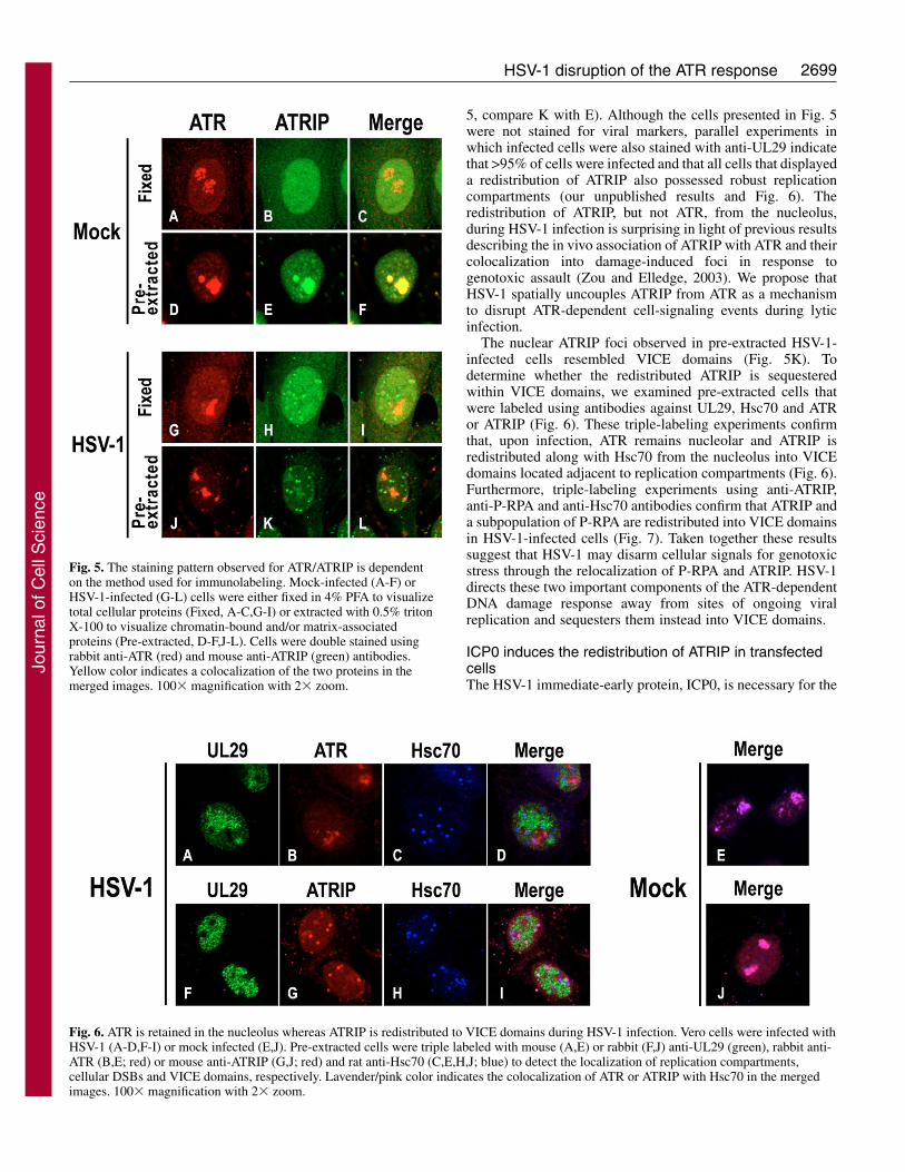
5, compare K with E). Although the cells presented in Fig. 5were not stained for viral markers, parallel experiments inwhich infected cells were also stained with anti-UL29 indicatethat >95% of cells were infected and that all cells that displayeda redistribution of ATRIP also possessed robust replicationcompartments (our unpublished results and Fig. 6). Theredistribution of ATRIP, but not ATR, from the nucleolus,during HSV-1 infection is surprising in light of previous resultsdescribing the in vivo association of ATRIP with ATR and theircolocalization into damage-induced foci in response togenotoxic assault (Zou and Elledge, 2003). We propose thatHSV-1 spatially uncouples ATRIP from ATR as a mechanismto disrupt ATR-dependent cell-signaling events during lyticinfection.
The nuclear ATRIP foci observed in pre-extracted HSV-1-infected cells resembled VICE domains (Fig. 5K). Todetermine whether the redistributed ATRIP is sequesteredwithin VICE domains, we examined pre-extracted cells thatwere labeled using antibodies against UL29, Hsc70 and ATRor ATRIP (Fig. 6). These triple-labeling experiments confirmthat, upon infection, ATR remains nucleolar and ATRIP isredistributed along with Hsc70 from the nucleolus into VICEdomains located adjacent to replication compartments (Fig. 6).Furthermore, triple-labeling experiments using anti-ATRIP,anti-P-RPA and anti-Hsc70 antibodies confirm that ATRIP anda subpopulation of P-RPA are redistributed into VICE domainsin HSV-1-infected cells (Fig. 7). Taken together these resultssuggest that HSV-1 may disarm cellular signals for genotoxicstress through the relocalization of P-RPA and ATRIP. HSV-1directs these two important components of the ATR-dependentDNA damage response away from sites of ongoing viralreplication and sequesters them instead into VICE domains.
ICP0 induces the redistribution of ATRIP in transfectedcellsThe HSV-1 immediate-early protein, ICP0, is necessary for the
Fig. 5. The staining pattern observed for ATR/ATRIP is dependenton the method used for immunolabeling. Mock-infected (A-F) orHSV-1-infected (G-L) cells were either fixed in 4% PFA to visualizetotal cellular proteins (Fixed, A-C,G-I) or extracted with 0.5% tritonX-100 to visualize chromatin-bound and/or matrix-associatedproteins (Pre-extracted, D-F,J-L). Cells were double stained usingrabbit anti-ATR (red) and mouse anti-ATRIP (green) antibodies.Yellow color indicates a colocalization of the two proteins in themerged images. 100� magnification with 2� zoom.
Fig. 6. ATR is retained in the nucleolus whereas ATRIP is redistributed to VICE domains during HSV-1 infection. Vero cells were infected withHSV-1 (A-D,F-I) or mock infected (E,J). Pre-extracted cells were triple labeled with mouse (A,E) or rabbit (F,J) anti-UL29 (green), rabbit anti-ATR (B,E; red) or mouse anti-ATRIP (G,J; red) and rat anti-Hsc70 (C,E,H,J; blue) to detect the localization of replication compartments,cellular DSBs and VICE domains, respectively. Lavender/pink color indicates the colocalization of ATR or ATRIP with Hsc70 in the mergedimages. 100� magnification with 2� zoom.
Jour
nal o
f Cel
l Sci
ence

2700
formation of VICE domains during infection (Burch andWeller, 2004). Furthermore, when cells are transfected with aplasmid expressing the ICP0 gene, nuclear and cytoplasmicinclusions are observed that contain ICP0, Hsc70, conjugatedubiquitin and misfolded proteins (Burch and Weller, 2004;Everett, 2000; Lukonis and Weller, 1996). To determinewhether ICP0 was sufficient for the redistribution of P-RPAand ATRIP, plasmids expressing ICP0 were used to transfectVero cells which were then double labeled with antibodiesagainst ICP0 and either P-RPA or ATR (Fig. 8A,C). Since bothanti-ICP0 and anti-ATRIP are mouse monoclonal antibodies,the anti-ICP0 antibody could not be used in double-labelingexperiments for detection of ATRIP; therefore, Hsc70 was usedas a surrogate marker for the ICP0 inclusions (Burch andWeller, 2004). Transfected cells were double stained withantibodies against ATRIP and Hsc70. (Fig. 8B). Untransfectedcontrols displayed staining patterns similar to that seen inmock-infected controls reported above (data not shown).Examination of cells transfected with the ICP0-expressingplasmid indicate that this viral protein is not sufficient forthe redistribution of P-RPA into ICP0-containing nuclearinclusions (Fig. 8A) suggesting that cellular or viral factorsother than ICP0 are needed for sequestering P-RPA. On theother hand, Fig. 8B shows a transfected cell displaying ATRIPin typical ICP0-induced nuclear inclusions, the interior ofwhich stained for Hsc70 (Burch and Weller, 2004; Everett,2000; Lukonis and Weller, 1996). Nuclear ATRIP inclusionswere never seen in untransfected cells, suggesting that ICP0 issufficient for its redistribution. Finally, the staining pattern ofATR remained nucleolar in ICP0-expressing cells (Fig. 8C).
Taken together, these results suggest that ICP0 is sufficient forthe redistribution of ATRIP but is insufficient for theredistribution of P-RPA.
DiscussionATR is an essential signaling kinase that plays crucial roles inthe regulation of DNA replication during an unperturbed cellcycle as well as in response to genotoxic stress. The ATR-ATRIP complex is thought to sense replication stress or DNAdamage by recognizing persistent RPA-ssDNA intermediatesthat arise from many types of DNA lesions (reviewed byShechter et al., 2004). The hyperphosphorylation of RPA onssDNA is an additional signal for DNA damage (Vassin et al.,2004). We previously showed that productive HSV-1 infectiondoes not result in the induction of RPA hyperphosphorylation;instead, endogenous hyperphosphorylated RPA is sequesteredaway from replication compartments into discrete nuclear foci(Wilkinson and Weller, 2004; Wilkinson and Weller, 2005). Inthis study, IF microscopy was used to examine the subcellularATR response during HSV-1 infection. We found that: (1)although infection induced DSBs in cellular chromatin, asdetected by �H2AX staining, P-RPA did not accumulate atthese breaks; (2) P-RPA was found within VICE domainswhich are enriched for cellular components involved in proteinfolding and degradation; (3) HSV-1 infection induced thespatial uncoupling of ATRIP from ATR and its redistributionfrom the nucleolus into VICE domains; (4) The HSV-1immediate-early protein, ICP0 was sufficient to induce theredistribution of ATRIP. Taken together, these findings suggestthat HSV-1 has evolved a strategy to disarm the ATR pathwayduring lytic infection.
Dismantling of ATR signaling during HSV-1 lytic infectionOne hallmark of the ATR-mediated DNA damage responseis the colocalization of ATR, ATRIP, RPA and/orhyperphosphorylated RPA in nuclear foci where DNA repair isthought to take place (Cortez et al., 2001; Tibbetts et al., 2000;Vassin et al., 2004; Wu et al., 2005; Zou and Elledge, 2003).ATR stress signaling is dependent upon the recruitment of ATRto DNA which is mediated by ATRIP (Falck et al., 2005;Itakura et al., 2004a; Zou and Elledge, 2003). IF microscopicanalysis has allowed us to demonstrate that crucial componentsof the ATR damage-response pathway are mislocalized withinthe infected cell nucleus. Although the general population ofRPA can be found within replication compartments, possiblyacting at unperturbed viral forks or replication intermediates(Uprichard and Knipe, 1997; Wilcock and Lane, 1991;Wilkinson and Weller, 2004), endogenous P-RPA issequestered in VICE domains. Furthermore, in situ extractionof cells revealed that HSV-1 induces the uncoupling of thenucleolar population of ATRIP from ATR and redistributes
Journal of Cell Science 119 (13)
Fig. 7. ATRIP and P-RPA colocalize in VICEdomains during HSV-1 infection. HSV-1-infectedVero cells were pre-extracted with 0.5% Triton X-100 as described in the Materials and Methods. Cellswere triple labeled with mouse anti-ATRIP (green),rabbit anti-P-RPA (red) and rat anti-Hsc70 (blue).White foci shown in the merged image indicate thecolocalization of the three proteins within VICEdomains. 100� magnification with 2� zoom.
Fig. 8. ICP0 is sufficient to redistribute ATRIP in transfected cells.Vero cells were transfected with an expression plasmid encoding theICP0 gene (pICP0) and fixed in 4% PFA as described in theMaterials and Methods. (A) Merged image of a transfected celldouble labeled with mouse anti-ICP0 (green) and rabbit anti-P-RPA(red). No colocalization of the two proteins was observed.(B) Merged image of a transfected cell double labeled with rat anti-Hsc70, a cellular marker that stains the interior of ICP0 inclusions(green), and mouse anti-ATRIP (red). Note the ATRIP-stained ICP0-like nuclear inclusions surrounding Hsc70. (C) Merged image of atransfected cell double labeled with mouse anti-ICP0 (green) andrabbit anti-ATR (red). No colocalization of the two proteins wasobserved. 100� magnification with 2� zoom.
Jour
nal o
f Cel
l Sci
ence

2701HSV-1 disruption of the ATR response
ATRIP into VICE domains. Although it is possible that someATRIP is associated with ATR in the nucleosol of infectedcells, several lines of evidence indicate that neither thenucleosolic nor the detergent-resistant fractions of ATR andATRIP is activated during infection. (1) We have neverobserved either the induction of RPA hyperphosphorylation orthe recruitment of ATR-ATRIP or P-RPA to replicationcompartments indicating that these signaling molecules areexcluded from sites which contain viral DNA. (2) Using�H2AX as a marker for the formation of DSBs, wedemonstrated that ATR-dependent factors are not recruited tocellular DSBs that arise within marginated cellular chromatinof infected cells. (3) Human Chk1, a specific target of theactivated ATR kinase is not phosphorylated during HSVinfection; however Chk2, a target of the ATM kinase isphosphorylated (Lilley et al., 2005; Shirata et al., 2005) (ourunpublished results). (4) Virus yield is unaffected in HSV-1-infected cells expressing a transdominant, kinase-dead mutantATR (Lilley et al., 2005). Thus, we propose that HSV-1 avoidsATR-mediated signaling by preventing the recruitment ofATR-ATRIP and P-RPA to either viral or cellular DNA,uncoupling ATRIP from ATR and sequestering ATRIP andendogenous P-RPA within VICE domains.
VICE domains and sequestration of non-native proteinsDuring heat shock and other forms of stress such as infection,proteins become denatured or misfolded. The Hsc70/Hsp70and Hsp90 multi-chaperone systems play important roles intargeting misfolded proteins to the ubiquitin proteasome forrefolding or elimination (e.g. Connell et al., 2001; Demand etal., 2001; Doong et al., 2003). The redistribution of Hsc70,Hsp70, Hsp40, Hsp90, ubiquitylated proteins and the corecatalytic complex of the proteasome into VICE domains ininfected cells suggests that HSV-1 has evolved a mechanism todeal with misfolded and unwanted proteins (Burch and Weller,2004; Burch and Weller, 2005; Parkinson and Everett, 2001).VICE domains are reminiscent of nuclear aggresomes, whichform in response to misfolded proteins and contain heat-shockproteins and components of the ubiquitin proteasome (Antonet al., 1999; Fu et al., 2005). P-RPA and ATRIP may be targetedto VICE domains because they are recognized as misfolded,perhaps because they have been separated from their normalinteraction partners as described below. Alternatively, thesequestration of P-RPA and ATRIP may reflect a more activetargeting mechanism by which the virus removes particularcellular response signals. Thus, HSV-1 appears to manipulatenot only the DNA damage response but also the unfoldedprotein response to its own advantage.
ICP0 plays a key role in disabling the ATR-dependentpathwayICP0, which is an E3 ubiquitin ligase, plays a central role indismantling the NHEJ pathway in some cell types by inducingthe proteasomal-dependent degradation of DNA-PKcs duringinfection (Lees-Miller et al., 1996; Parkinson et al., 1999). Weprovide evidence here that ICP0 also plays an active role indismantling the ATR pathway. ICP0 has been shown to localizeat the nucleolus early upon infection possibly as part of amechanism that induces the degradation or dispersal ofnucleolar protein(s) (Morency et al., 2005). In this paper, wereport that a significant population of ATR and ATRIP is
present in the nucleolus in uninfected Vero cells. We proposethat ICP0 may dismantle ATR-dependent events by localizingto the nucleolus and inducing the uncoupling of the ATR-ATRIP complex. The uncoupling of ATRIP from ATR mayexpose hydrophobic residues resulting in its recognition byHsc70/Hsp70. Since ICP0 can induce the redistribution ofHsc70/Hsp70 (Burch and Weller, 2004), it is possible thatATRIP is redistributed to VICE domains by virtue of itsinteraction with Hsp70 (Cortez et al., 2001). Although it is notclear how ICP0 could induce the uncoupling of the ATR-ATRIP complex, it is possible that the ubiquitin ligase activitytargets either ATRIP itself or another cellular componentresponsible for maintaining the association of ATR and ATRIPfor degradation.
When expressed in cells by transfection, ICP0 induces theformation of nuclear and cytoplasmic inclusions that containICP0, ubiquitylated proteins and Hsc70 (Burch and Weller,2004; Everett, 2000). Misfolded viral proteins are also foundwithin these ICP0-induced inclusions (Lukonis and Weller,1996). The helicase-primase complex of HSV-1 is aheterotrimer whose full activity depends on the co-expressionof all three subunits (UL5, UL8 and UL52). If one subunit isexpressed without the other two in cells cotransfected withICP0, that subunit is found within the ICP0 inclusions.Moreover, a subpopulation of the UL6 portal protein, whichmay need chaperone assistance during assembly into a portalring, is also found within ICP0-induced inclusions (Lukonisand Weller, 1996). We proposed that proteins expressedwithout their normal interaction partners or that are misfoldedmight be sequestered in an ICP0-dependent fashion (Lukonisand Weller, 1996). In this paper we have demonstrated thatATRIP, which is spatially uncoupled from its binding partnerduring infection, is also found within ICP0-induced inclusionsin transfected cells. Thus, it now appears that the ability ofICP0 to form nuclear inclusions around misfolded proteins intransfected cells and to induce VICE domain formation ininfected cells may reflect an evolutionary strategy to takeadvantage of the host stress response to misfolded proteins. Itis known that the accumulation of non-native proteins cantrigger deleterious events such as apoptosis. Thus, the abilityof ICP0 to sequester and potentially degrade non-nativeproteins may allow the virus to delay the induction of apoptosisat least until viral progeny are made. Experiments using ICP0mutant viruses and proteasomal inhibitors are underway tocharacterize further the role of ICP0 in dismantling the ATRsignaling pathway.
Both HSV-1 and adenovirus disarm components of thecellular DNA damage responseHSV-1 is not the only virus that disarms components of thecellular DNA damage response. Adenovirus type 5 (Ad5)infection inactivates the MRN complex by inducing therelocalization and degradation of one or more of the MRNsubunits. Ad5 sequesters MRN subunits into nuclear focilocated adjacent to viral replication centers as well as intocytoplasmic inclusion bodies (Araujo et al., 2005; Evans andHearing, 2003; Evans and Hearing, 2005; Liu et al., 2005;Stracker et al., 2002). The MRN-containing nuclear foci inAd5-infected cells are reminiscent of the HSV-1-induced VICEdomains, and it will be of interest to determine whether theytoo are enriched for cellular components involved in protein
Jour
nal o
f Cel
l Sci
ence

2702
folding and degradation. The Ad5-induced, MRN-containingcytoplasmic inclusions, however, do appear to be aggresomesenriched with chaperones and the ubiquitin proteasome(Araujo et al., 2005; Evans and Hearing, 2005; Liu et al.,2005), suggesting that both nuclear and cytoplasmic MRN-containing foci are part of a larger cellular response to theaccumulation of misfolded proteins (reviewed by Garcia-Mataet al., 2002). Thus both adenoviruses and herpesviruses appearto disarm cellular responses to DNA damage throughmanipulation of the stress response and sequestration of repairfactors. The fact that HSV-1 targets components of the ATR-response pathway whereas Ad5 targets the MRN pathway mayreflect different DNA replication strategies used by theseviruses (Wilkinson and Weller, 2004).
HSV-1 infection appears to activate the ATM-dependentpathway while dismantling othersIn HSV-1 infected cells, �H2AX foci can be seen surroundingreplication compartments in infected cells indicating thepresence of DSBs; however, instead of being recruited to theseDSBs in host chromatin, the NHEJ and ATR pathways aredisrupted. On the other hand, HSV-1 appears to activatecomponents of the ATM-dependent pathway (Lilley et al.,2005; Shirata et al., 2005; Wilkinson and Weller, 2004). TheMRN repair complex and activated ATM, are apparently notrecruited to cellular DSBs, but instead are found in viralreplication compartments (Lilley et al., 2005; Shirata et al.,2005; Taylor and Knipe, 2004; Wilkinson and Weller, 2004).Thus, components of the ATM-pathway are activated andrecruited to sites of viral DNA synthesis. Although ATR- andATM-mediated pathways are related and both can be activatedby similar genotoxic events, HSV-1 distinguishes betweenthese two pathways, inactivating one and potentially using theother. The MRN complex and other downstream componentsof the ATM-mediated pathway may participate directly in HSVDNA synthesis, which we and others have suggested mayinvolve recombination (reviewed by Wilkinson and Weller,2003).
Materials and MethodsCell lines, virus and infectionsAfrican green monkey kidney fibroblasts (Vero cells) were purchased from theAmerican Type Culture Collection (ATCC) (Manassas, VA) and maintained asmonolayers in Dulbecco’s modified Eagle’s medium (Invitrogen, Carlsbad, CA)supplemented with 5% fetal bovine serum (Gemini Bio-Products, Woodland, CA),penicillin, streptomycin and amphotericin B (Invitrogen). Cell cultures weremaintained at 37°C in a humidified atmosphere containing 5% CO2. Strain KOSwas used as the wild-type strain of HSV-1. For infections, subconfluent cells oncoverslips were adsorbed for 1 hour with 10 particle-forming units of virus per cell,and incubated 5.5-6.0 hours postinfection. Mock infections were carried out inparallel.
TransfectionspW3 (also called pICP0), which expresses ICP0 under its own promoter (Sacks andSchaffer, 1987) was used in transient transfection experiments. For each 35-mmdish, subconfluent cells on coverslips were transfected with 0.25 �g pICP0 and 0.75�g pUC18 carrier DNA using Lipofectamine and PLUS reagents (Invitrogen)according to the manufacturer’s instructions.
Antibodies and reagentsMouse monoclonal anti-UL29 antibody (39S) (Showalter et al., 1981) was obtainedfrom ATCC. Rabbit polyclonal anti-UL29 (367) (Shelton et al., 1994) was suppliedby William Ruyechan (University of SUNY, Buffalo, NY). Rabbit polyclonal anti-ATR and phosphospecific anti-phospho-RPA32 pSer4/pSer8 (BL647) antibodieswere purchased from Bethyl Laboratories (Montgomery, TX). Mouse monoclonalanti-�H2AX was purchased from Upstate (Lake Placid, NY). Mouse monoclonalanti-ATRIP was obtained from Cell Signaling Technology (Beverly, MA). Rat
monoclonal anti-Hsc70 was purchased from Stressgen Biotechnologies (San Diego,CA). Rabbit anti-nucleolin antibody (H-250) was obtained from Santa CruzBiotechnology (Santa Cruz, CA). Mouse monoclonal anti-ICP0 antibody wasobtained from the Goodwin Institute for Cancer Research (Plantation, FL).Secondary antibodies directed against mouse, rabbit or rat IgG and conjugated toAlexa Fluor 488, Alexa Fluor 546, Alexa Fluor 594 or Alexa Fluor 647 werepurchased from Molecular Probes (Eugene, OR). Camptothecin was purchased fromSigma Aldrich (St Louis, MO).
Labeling for indirect immunofluorescence microscopyAt 5.5 to 6.0 hours post infection or 16-18 hours post transfection, cells were brieflywashed in PBS and processed for immunolabeling. For visualization of total cellproteins, cells were first fixed in 4% PFA then permeabilized for 10 minutes in 1%Triton X-100 in PBS. For the visualization of detergent-resistant or chromatin-associated nuclear proteins, an in situ extraction method that removes the cytoplasmand nucleosolic proteins was used. Cells were pre-extracted for 5 minutes on icewith 0.5% Triton X-100 in cytoskeletal buffer as described (Dimitrova and Gilbert,2000) then fixed in 4% PFA for 10 minutes. After washing with PBS and blockingin 3% normal goat serum (NGS) in PBS, cells were incubated with primaryantibodies diluted in 3% NGS for at least 30 minutes. Antibodies 39S, anti-�H2AXanti-ATR and anti-ICP0 were used at a concentration of 1:200 and antibodies 367,BL647, anti-Hsc70 and anti-ATRIP were used at 1:400. After thorough washingwith PBS, cells were incubated with secondary antibodies diluted 1:200. After afinal wash in PBS, coverslips were mounted in glycerol gelatin containing 2.5%DABCO (Sigma Aldrich) to retard photobleaching.
Confocal microscopyCells stained for IF were examined using a Zeiss LSM 410 confocal microscopesystem equipped with an argon-krypton laser, an Axiovert 135 inverted microscopeand a Zeiss �100 Plan Neofluar objective. Alexa Fluor 488 was excited at 488 nm.Alexa Fluor 546 or 594 excited at 568 nm and Alexa Fluor 647 at 647 nm.Appropriate emission filters were used for double- and triple-labeling experiments.Channels were scanned individually using settings established with control slidesand images were merged by computer. To control for crossreactivity, samples werestained with one primary antibody and appropriate secondary antibodies. No overlapbetween the channels was observed for any of the samples at the settings used.Collected images were arranged using Adobe Photoshop 6.0.
We thank the members of our laboratory for helpful comments onthe manuscript. This research was supported by Public Health ServiceGrant AI21747. D.E.W. was supported by NIH training grant F32AI054042.
ReferencesAndegeko, Y., Moyal, L., Mittelman, L., Tsarfaty, I., Shiloh, Y. and Rotman, G.
(2001). Nuclear retention of ATM at sites of DNA double strand breaks. J. Biol. Chem.276, 38224-38230.
Andersen, J. S., Lam, Y. W., Leung, A. K. L., Ong, S.-E., Lyon, C. E., Lamond, A.I. and Mann, M. (2005). Nucleolar proteome dynamics. Nature 433, 77-83.
Anton, L. C., Schubert, U., Bacik, I., Princiotta, M. F., Wearsch, P. A., Gibbs, J., Day,P. M., Realini, C., Rechsteiner, M. C., Bennink, J. R. et al. (1999). Intracellularlocalization of proteasomal degradation of a viral antigen. J. Cell Biol. 146, 113-124.
Araujo, F. D., Stracker, T. H., Carson, C. T., Lee, D. V. and Weitzman, M. D. (2005).Adenovirus type 5 E4orf3 protein targets the Mre11 complex to cytoplasmicaggresomes. J. Virol. 79, 11382-11391.
Ball, H. L., Myers, J. S. and Cortez, D. (2005). ATRIP binding to replication proteinA-single-stranded DNA promotes ATR-ATRIP localization but is dispensable for Chk1phosphorylation. Mol. Biol. Cell 16, 2372-2381.
Barr, S. M., Leung, C. G., Chang, E. E. and Cimprich, K. A. (2003). ATR kinaseactivity regulates the intranuclear translocation of ATR and RPA following ionizingradiation. Curr. Biol. 13, 1047-1051.
Binz, S. K., Sheehan, A. M. and Wold, M. S. (2004). Replication protein Aphosphorylation and the cellular response to DNA damage. DNA Repair Amst. 3, 1015-1024.
Burch, A. D. and Weller, S. K. (2004). Nuclear sequestration of cellular chaperone andproteasomal machinery during herpes simplex virus type 1 infection. J. Virol. 78, 7175-7185.
Burch, A. D. and Weller, S. K. (2005). Herpes simplex virus type 1 DNA polymeraserequires the Mammalian chaperone hsp90 for proper localization to the nucleus. J.Virol. 79, 10740-10749.
Burma, S. and Chen, D. J. (2004). Role of DNA-PK in the cellular response to DNAdouble-strand breaks. DNA Repair Amst. 3, 909-918.
Connell, P., Ballinger, C. A., Jiang, J., Wu, Y., Thompson, L. J., Hohfeld, J. andPatterson, C. (2001). The co-chaperone CHIP regulates protein triage decisionsmediated by heat-shock proteins. Nat. Cell Biol. 3, 93-96.
Cortez, D. (2005). Unwind and slow down: checkpoint activation by helicase andpolymerase uncoupling. Genes Dev. 19, 1007-1012.
Cortez, D., Guntuku, S., Qin, J. and Elledge, S. J. (2001). ATR and ATRIP: partnersin checkpoint signaling. Science 294, 1713-1716.
Journal of Cell Science 119 (13)
Jour
nal o
f Cel
l Sci
ence

2703HSV-1 disruption of the ATR response
Demand, J., Alberti, S., Patterson, C. and Hohfeld, J. (2001). Cooperation of aubiquitin domain protein and an E3 ubiquitin ligase during chaperone/proteasomecoupling. Curr. Biol. 11, 1569-1577.
Dimitrova, D. S. and Gilbert, D. M. (2000). Stability and nuclear distribution ofmammalian replication protein A heterotrimeric complex. Exp. Cell Res. 254, 321-327.
Doong, H., Rizzo, K., Fang, S., Kulpa, V., Weissman, A. M. and Kohn, E. C. (2003).CAIR-1/BAG-3 abrogates heat shock protein-70 chaperone complex-mediated proteindegradation: accumulation of poly-ubiquitinated Hsp90 client proteins. J. Biol. Chem.278, 28490-28500.
Evans, J. D. and Hearing, P. (2003). Distinct roles of the Adenovirus E4 ORF3 proteinin viral DNA replication and inhibition of genome concatenation. J. Virol. 77, 5295-5304.
Evans, J. D. and Hearing, P. (2005). Relocalization of the Mre11-Rad50-Nbs1 complexby the adenovirus E4 ORF3 protein is required for viral replication. J. Virol. 79, 6207-6215.
Everett, R. D. (2000). ICP0 induces the accumulation of colocalizing conjugatedubiquitin. J. Virol. 74, 9994-10005.
Falck, J., Coates, J. and Jackson, S. P. (2005). Conserved modes of recruitment of ATM,ATR and DNA-PKcs to sites of DNA damage. Nature 434, 605-611.
Fu, L., Gao, Y.-S., Tousson, A., Shah, A., Chen, T.-L. L., Vertel, B. M. and Sztul, E.(2005). Nuclear aggresomes form by fusion of PML-associated aggregates. Mol. Biol.Cell 16, 4905-4917.
Garcia-Mata, R., Gao, Y. S. and Sztul, E. (2002). Hassles with taking out the garbage:aggravating aggresomes. Traffic 3, 388-396.
Itakura, E., Takai, K. K., Umeda, K., Kimura, M., Ohsumi, M., Tamai, K. andMatsuura, A. (2004a). Amino-terminal domain of ATRIP contributes to intranuclearrelocation of the ATR-ATRIP complex following DNA damage. FEBS Lett. 577, 289-293.
Itakura, E., Umeda, K., Sekoguchi, E., Takata, H., Ohsumi, M. and Matsuura, A.(2004b). ATR-dependent phosphorylation of ATRIP in response to genotoxic stress.Biochem. Biophys. Res. Commun. 323, 1197-1202.
Kurz, E. U. and Lees-Miller, S. P. (2004). DNA damage-induced activation of ATM andATM-dependent signaling pathways. DNA Repair Amst. 3, 889-900.
Lees-Miller, S. P., Long, M. C., Kilvert, M. A., Lam, V., Rice, S. A. and Spencer, C.A. (1996). Attenuation of DNA-dependent protein kinase activity and its catalyticsubunit by the herpes simplex virus type 1 transactivator ICP0. J. Virol. 70, 7471-7477.
Leinbach, S. S. and Summers, W. C. (1980). The structure of herpes simplex virus type1 DNA as probed by micrococcal nuclease digestion. J. Gen. Virol. 51, 45-59.
Lilley, C. E., Carson, C. T., Muotri, A. R., Gage, F. H. and Weitzman, M. D. (2005).DNA repair proteins affect the lifecycle of herpes simplex virus 1. Proc. Natl. Acad.Sci. USA 102, 5844-5849.
Liu, Y., Shevchenko, A. and Berk, A. J. (2005). Adenovirus exploits the cellularaggresome response to accelerate inactivation of the MRN complex. J. Virol. 79, 14004-14016.
Lukonis, C. J. and Weller, S. K. (1996). The herpes simplex virus type 1 transactivatorICP0 mediates aberrant intracellular localization of the viral helicase/primase complexsubunits. Virololgy 220, 495-501.
Mirzoeva, O. K. and Petrini, J. H. J. (2001). DNA damage-dependent nuclear dynamicsof the Mre11 complex. Mol. Cell. Biol. 21, 281-288.
Monier, K., Armas, J. C., Etteldorf, S., Ghazal, P. and Sullivan, K. F. (2000).Annexation of the interchromosomal space during viral infection. Nat. Cell Biol. 2,661-665.
Morency, E., Coute, Y., Thomas, J., Texier, P. and Lomonte, P. (2005). The proteinICP0 of herpes simplex virus type 1 is targeted to nucleoli of infected cells. Arch. Virol.150, 2387-2395.
Parkinson, J. and Everett, R. D. (2001). Alphaherpesvirus proteins related to herpessimplex virus type 1 ICP0 induce the formation of colocalizing, conjugated ubiquitin.J. Virol. 75, 5357-5362.
Parkinson, J., Lees-Miller, S. P. and Everett, R. D. (1999). Herpes simplex virus type1 immediate-early protein vmw110 induces the proteasome-dependent degradation ofthe catalytic subunit of DNA-dependent protein kinase. J. Virol. 73, 650-657.
Pichierri, P., Rosselli, F. and Franchitto, A. (2003). Werner’s syndrome protein isphosphorylated in an ATR/ATM-dependent manner following replication arrest andDNA damage induced during the S phase of the cell cycle. Oncogene 22, 1491-1500.
Quinlan, M. P., Chen, L. B. and Knipe, D. M. (1984). The intranuclear location of aherpes simplex virus DNA-binding protein is determined by the status of viral DNAreplication. Cell 36, 857-868.
Robison, J. G., Elliott, J., Dixon, K. and Oakley, G. G. (2004). Replication protein Aand the Mre11/Rad50/Nbs1 complex co-localize and interact at sites of stalledreplication forks. J. Biol. Chem. 279, 34802-34810.
Rogakou, E. P., Pilch, D. R., Orr, A. H., Ivanova, V. S. and Bonner, W. M. (1998).DNA double-stranded breaks induce histone H2AX phosphorylation on serine 139. J.Biol. Chem. 273, 5858-5868.
Rogakou, E. P., Boon, C., Redon, C. and Bonner, W. M. (1999). Megabasechromatin domains involved in DNA double-strand breaks in vivo. J. Cell Biol. 146,905-916.
Sacks, W. R. and Schaffer, P. A. (1987). Deletion mutants in the gene encoding theherpes simplex virus type 1 immediate-early protein ICP0 exhibit impaired growth incell culture. J. Virol. 61, 829-839.
Sedelnikova, O. A., Rogakou, E. P., Panyutin, I. G. and Bonner, W. M. (2002).Quantitative detection of (125)IdU-induced DNA double-strand breaks with gamma-H2AX antibody. Radiat. Res. 158, 486-492.
Shechter, D., Costanzo, V. and Gautier, J. (2004). Regulation of DNA replication byATR: signaling in response to DNA intermediates. DNA Repair Amst. 3, 901-908.
Shelton, L. S., Albright, A. G., Ruyechan, W. T. and Jenkins, F. J. (1994). Retentionof the herpes simplex virus type 1 (HSV-1) UL37 protein on single-stranded DNAcolumns requires the HSV-1 ICP8 protein. J. Virol. 68, 521-525.
Shirata, N., Kudoh, A., Daikoku, T., Tatsumi, Y., Fujita, M., Kiyono, T., Sugaya, Y.,Isomura, H., Ishizaki, K. and Tsurumi, T. (2005). Activation of ataxia telangiectasia-mutated DNA damage checkpoint signal transduction elicited by herpes simplex virusinfection. J. Biol. Chem. 280, 30336-30341.
Showalter, S. D., Zweig, M. and Hampar, B. (1981). Monoclonal antibodies to herpessimplex virus type 1 proteins, including the immediate-early protein ICP 4. Infect.Immun. 34, 684-692.
Simpson-Holley, M., Baines, J., Roller, R. and Knipe, D. M. (2004). Herpes simplexvirus 1 UL31 and UL34 gene products promote the late maturation of viral replicationcompartments to the nuclear periphery. J. Virol. 78, 5591-5600.
Stracker, T. H., Carson, C. T. and Weitzman, M. D. (2002). Adenovirus oncoproteinsinactivate the Mre11-Rad50-NBS1 DNA repair complex. Nature 418, 348-352.
Taylor, T. J. and Knipe, D. M. (2004). Proteomics of herpes simplex virus replicationcompartments: association of cellular DNA replication, repair, recombination, andchromatin remodeling proteins with ICP8. J. Virol. 78, 5856-5866.
Tibbetts, R. S., Cortez, D., Brumbaugh, K. M., Scully, R., Livingston, D., Elledge, S.J. and Abraham, R. T. (2000). Functional interactions between BRCA1 and thecheckpoint kinase ATR during genotoxic stress. Genes Dev. 14, 2989-3002.
Uprichard, S. L. and Knipe, D. M. (1997). Assembly of herpes simplex virus replicationproteins at two distinct intranuclear sites. Virology 229, 113-125.
Vassin, V. M., Wold, M. S. and Borowiec, J. A. (2004). Replication protein A (RPA)phosphorylation prevents RPA association with replication centers. Mol. Cell. Biol. 24,1930-1943.
Wilcock, D. and Lane, D. P. (1991). Localization of p53, retinoblastoma and hostreplication porteins at sites of viral replication in herpes-infected cells. Nature 349,429-431.
Wilkinson, D. E. and Weller, S. K. (2003). The role of DNA recombination in herpessimplex virus DNA replication. IUBMB Life 55, 451-458.
Wilkinson, D. E. and Weller, S. K. (2004). Recruitment of cellular recombination andrepair proteins to sites of herpes simplex virus type 1 DNA replication Is dependenton the composition of viral proteins within prereplicative sites and correlates with theinduction of the DNA damage response. J. Virol. 78, 4783-4796.
Wilkinson, D. E. and Weller, S. K. (2005). Inhibition of the herpes simplex virus type1 DNA polymerase induces hyperphosphorylation of replication protein A and itsaccumulation at S-phase-specific sites of DNA damage during infection. J. Virol. 79,7162-7171.
Wu, X., Yang, Z., Liu, Y. and Zou, Y. (2005). Preferential localization ofhyperphosphorylated replication protein A to double-strand break repair andcheckpoint complexes upon DNA damage. Biochem. J. 391, 473-480.
Zou, L. and Elledge, S. J. (2003). Sensing DNA damage through ATRIP recognition ofRPA-ssDNA complexes. Science 300, 1542-1548.
Jour
nal o
f Cel
l Sci
ence