Embed Size (px)
Citation preview
Bone 43 (2008) 689–699
Contents lists available at ScienceDirect
Bone
j ourna l homepage: www.e lsev ie r.com/ locate /bone
Heparanase expression and activity influences chondrogenic and osteogenicprocesses during endochondral bone formation
A.J. Brown a, M. Alicknavitch a, S.S. D’Souza b, T. Daikoku c, C.B. Kirn-Safran a,D. Marchetti d, D.D. Carson a, M.C. Farach-Carson a,e,f,⁎a Department of Biological Sciences, University of Delaware, 326 Wolf, Hall, Newark, DE 19716, USAb Department of Chemistry and Biochemistry, University of Delaware, Newark, DE 19716, USAc Division of Reproductive and Developmental Biology, Vanderbilt Medical Center, Nashville, TN 37232, USAd Department of Pathology and Molecular and Cellular Biology, Baylor College of Medicine, Houston, TX 77030, USAe Department of Material Sciences, University of Delaware, Newark, DE 19716, USAf Center for Translational Cancer Research, University of Delaware, Newark, DE 19716, USA
⁎ Corresponding author. Department of Biological Sci326 Wolf, Hall, Newark, DE 19716, USA. Fax: +1 302 831
E-mail address: [email protected] (M.C. Farach-Cars
8756-3282/$ – see front matter © 2008 Elsevier Inc. Aldoi:10.1016/j.bone.2008.05.022
a b s t r a c t
a r t i c l e i n f oArticle history:
Endochondral bone format Received 21 December 2007Revised 28 April 2008Accepted 20 May 2008Available online 6 June 2008Edited by: M. Noda
Keywords:HeparanaseChondrogenesisOsteogenesisBonePI-88Migration
ion is a highly orchestrated process involving coordination among cell–cell,cell–matrix and growth factor signaling that eventually results in the production of mineralized bone from acartilage template. Chondrogenic and osteogenic differentiation occur in sequence during this process, and thetemporospatial patterning clearly requires the activities of heparin binding growth factors and their receptors.Heparanase (HPSE) plays a role in osteogenesis, but the mechanism by which it does so is incompletelyunderstood. We used a combination of ex vivo and in vitro approaches and a well described HPSE inhibitor,PI-88 to study HPSE in endochondral bone formation. In situ hybridization and immunolocalization with HPSEantibodies revealed that HPSE is expressed in the peri-chondrium, peri-osteum, and at the chondro–osseousjunction, all sites of key signaling events and tissuemorphogenesis. Transcripts encodingHpse alsowere observedin the pre-hypertrophic zone. Addition of PI-88 to metatarsals in organ culture reduced growth and suggestedthat HPSE activity aids the transition from chondrogenic to osteogenic processes in growth of long bones. Tostudy this, we used high density cultures of ATDC5 pre-chondrogenic cells grown under conditions favoringchondrogenesis or osteogenesis. Under chondrogenic conditions, HPSE/Hpsewas expressed at high levels duringthe mid-culture period, at the onset of terminal chondrogenesis. PI-88 addition reduced chondrogenesis andaccelerated osteogenesis, including a dramatic up-regulation of osteocalcin levels. In normal growth medium,addition of PI-88 reduced migration of ATDC-5 cells, suggesting that HPSE facilitates cartilage replacement bybone at the chondro–osseous junction by removing theHS component of proteoglycans, such as perlecan/HSPG2,that otherwise prevent osteogenic cells from remodeling hypertrophic cartilage.
© 2008 Elsevier Inc. All rights reserved.
Introduction
Osteogenesis, the development of bone, is a complex process thatrequires coordination between cell–cell, cell–matrix, and growthfactor mediated signaling events that conclude with the ossificationand mineralization of bone [1]. In the craniofacial skeleton, mesench-ymal cells differentiate into osteoblasts and form bone by intramem-braneous ossification. In contrast, in the axial and limb skeleton,mesenchymal cells differentiate into chondrocytes and are laterreplaced by osteoblasts, a process known as endochondral ossification.Key events in this process include the commitment and differentiationof mesenchymal cells from the mesoderm into chondrocytes, conden-sation and proliferation of these committed mesenchymal cells, and
ences, University of Delaware,2281.on).
l rights reserved.
subsequent invasion of blood vessels along with osteoblasts, boneforming cells, osteoclasts, bone resorbing cells, and hematopoietic cellsto form the bone marrow [2]. Various studies have demonstrated thatbioactive heparin binding factors such as Indian hedgehog (Ihh),Wnts,fibroblast growth factors (FGFs), and bone morphogenetic proteins(BMPs), in addition to components of the pericellular matrix incartilage, are critical regulators of lineage commitment and cellproliferation and differentiation [3-9]. Dynamic paracrine interactionsexist between the developing cartilage core of endochondral bone andthe perichondrium (Pc) and support bone elongation and widening[10,11]. A hallmark of developing endochondral bone is the chondro–osseous junction (COJ) the demarcation between terminally differ-entiated cartilage and mineralizing bone.
Investigations in our laboratory and others, using both human andanimal models, revealed that heparan sulfate proteoglycan (HSPG)core proteins, including perlecan/HSPG2 [12] and their heparansulfate (HS) chains influence the key events that occur during cartilage
690 A.J. Brown et al. / Bone 43 (2008) 689–699
development [12–20] and fracture repair [21]. More specifically, it hasbeen demonstrated that glycosaminoglycan (GAG)-bearing perlecan/HSPG2 domain I, but not perlecan/HSPG2 domain I lacking GAGchains, supports aggregation and expression of mature chondrogenicmarkers in C3H10T1/2 cells and assists growth factor delivery [22–25].Accumulating evidence implicates a critical function for HSPGs infacilitating the interactions among HBGFs and their receptors [26].Cleavage of HS chains facilitates formation of diffusible complexes ofgrowth factors complexed with HS fragments which together form atrimolecular signaling complex [27]; however, the importance of HPSEas a modulator of growth factor bioavailability and HS catabolismduring the transition from chondrogenesis to osteogenesis duringendochondral ossification remains unclear.
HPSE, an endo-β-D-glucuronidase, is synthesized as an inactive65 kDa form (pro-HPSE) and is processed to an active heterodimerconsisting of 50 and 8 kDa subunits (active HPSE). As an activeheterodimer, it cleaves HS chains attached to proteoglycans includingperlecan/HSPG2 [28–30]. HPSE activity is inhibited by a structuralmimic of HS, PI-88 [31]. HPSE also possesses non-enzymatic functionssuch as cell adhesion, proliferation, migration, and invasion [32]. HPSEhas been identified in normal and malignant cells including skinfibroblasts, cytotrophoblasts, hepatocytes, Chinese hamster ovary(CHO) cells, and endothelial cells and tissues including skin, placenta,lung, and kidney [33–38] and is implicated in a number of normal andpathological conditions [31,39–41]. Interestingly, HS chains have beenidentified in the pericellular matrix of both developing and maturecartilage and the Pc of developing growth plate. [12] Identifying therelationship between HPSE and HS expression will allow us to betterunderstand the role of HPSE in growth plate development.
Most studies of HPSE have focused on its regulatory role in cancerprogression. Constitutive overexpression of human HPSE in micealters tissue architecture, vascularization, and metabolism [42]. Inbone, HPSE overexpression creates a complex phenotype that favorsosteogenesis, increases bone mass, but retards bone elongation infemale transgenic mice [43]. Previous studies in our laboratory de-monstrated a dramatic loss of HS at the COJ as endochondral boneformation progresses, suggesting that HS inhibits osteogenesis [12]. Toexpand on these observations, we sought to determine if HPSEinfluences the transition from chondrogenesis to osteogenesis duringendochondral bone formation in mouse models. We designed ourstudies to identify HPSE/Hpse localization within the developinggrowth plate and to provide additional functional insight in the role ofHPSE/Hpse during the process of endochondral bone formation.
Materials and methods
Animals
All animal handling procedureswere approved by the University ofDelaware Institutional Animal Care and Use Committee. Long bonesfrom C57/BL6 and ICR strain mice (Taconic, Germantown, NY) weredissected at various developmental stages and preserved in Tissue-Tek® Optimal Cutting Temperature (O.C.T.) (Sakura Finetechnical,Torrance, CA) at −80 °C until cryosectioning.
Cell culture
ATDC5 cells, a murine carcinoma-derived chondrogenic cell line,were obtained from Dr. Véronique Lefebvre (The Cleveland Clinic,Cleveland, OH) and maintained as monolayer cultures under conditionspreviously described [44]. Briefly, they were cultured at 37 °C in air:CO2
[95:5% (v/v)] in regular growth media, Dulbecco's Modified Eagle'smedium-Ham's F12 (DMEM-F12) supplemented with 5% (v/v) fetalbovine serum (FBS), 100 U/ml pencillin G sodium and 100 μg/mlstreptomycin sulfate in 0.085% (w/v) saline (PS). All cell culture reagentswere purchased from Invitrogen (Carlsbad, CA) unless otherwise stated.
For monolayer differentiation, cells were seeded into six-well tissueculture plates (Becton Dickinson Labware, Franklin Lakes, NJ) until theyreached post-confluency at which culture medium was replaced withregular growth media (DMEM-F12+5% [v/v] FBS) containing 10 μg/mlbovine insulin (I), 10 μg/ml human transferrin (T), and 3 3×10⁻⁸ mol/lsodium selenite (S), (ITS). After 21 days in differentiation media, cellswere transferred to 37 °C in 97% air: 3% (v/v) CO2 and switched to α-MEM containing 5% (v/v) FBS, PS, and 1% (v/v) ITS for further differ-entiation into calcifying chondrocytes as described in [44].
In situ hybridization
In situ hybridization followed a protocol previously described[31,45]. Briefly, E18.5 mouse limbs were cryosectioned at 12 μmthickness andmounted onto poly-L-lysine coated slides. Hybridizationprobes were generated as described in [31]. Sections hybridized withsense probes served as negative controls and indicated specificity ofanti-sense probe. Sections were post-stained with hematoxylin andeosin to confirm orientation.
Immunohistochemistry
Frozen limb sections (10–12 μm thickness) were fixed in methanolfor 10 min at room temperature, rehydrated in PBS containing Ca2+ andMg2+ and blocked with 1% (w/v) bovine serum albumin (BSA) (Sigma-Aldrich) in PBS for 30min at room temperature. Sectionswere incubatedat room temperature for 1 h with mouse monoclonal anti-human HPSE1 antibody (Insight Biopharmaceuticals Limited, Rehevot, Israel) andZenon™ reagent at 1:40 dilution and Draq5™ (Biostatus Limited,Leicestershire, UK) at 1:2000 dilution. 1 μg of this antibody was used toprepare Zenon™ labeling complex using Zenon™ Technology (Invitro-gen/Molecular Probes, Eugene, OR) according to the manufacturer'sprotocol. Samples were rinsed three timeswith PBS containing Ca2+ andMg2+ for 5 min at room temperature each. Next, sections and cells werepost-fixed for 10 min at room temperature in 4% paraformaldehyde (w/v) in PBS. Finally, samples were mounted in Gel/Mount ™ (BioMedaCorp., Foster City, CA) to prevent fading. As a control for specificity,sectionswere incubatedwithmouse IgG2b. Sampleswere imagedwith aZeiss LSM510multi-photon confocalmicroscope at theMicroscopy CoreFacility of the University of Delaware.
Organ culture
The three center metatarsals were microdissected from the feet of 2day old ICR mice as described in [10,46–48]. Metatarsals were placed in24well tissue culture treated plates (Corning Inc) containing500 μl ofα-MEM (Gibco-BRL) supplemented with 0.05 05 mg/ml ascorbic acid,0.3 mg/ml L-glutamine (SIGMA-Aldrich, St Louis, MO),1 mM β-glycerophosphate (SIGMA-Aldrich), 250 μg/ml of Amphotericin B(Invitrogen) (Fungizone Equivalent) and PS (Invitrogen). Explants weregrown at 37 °C in a humidified air:CO2 (95:5, v/v) incubator. Explantswere cultured inmedia containing0,100, or 500 μg/mlof PI-88. For PI-88treated samples, the drug was generously provided by ProgenPharmaceuticals Ltd. (Toowong, Queensland, Australia). Media waschangedevery 2–3days. FreshPI-88wasaddedwitheachmedia change.Cultures were observed for contamination and photographed using adigital camera DXM1200F (Nikon, Japan) attached to phase-contrastmicroscope SMZ1500 (Nikon, Japan). Histomorphometric analyses ofdissected metatarsals were performed as reported by Ueda et al., 2007[49] with the relative contributions of the Rz, Pz, and Hz to thelongitudinal length of the bone indicated. Images then were processedfurther and the length of the bones as well as the lengths of the growthplates and marrow cavities were measured on day 0 and after 5 days ofculture using Adobe Photoshop® 7.0. The percentage of total bonegrowth and relative contribution of growth of each compartment wascalculated from themeasured values. The mean and standard deviation
Table 1PCR primers used in these studies
Gene Forward primer Reverse primer Product size (bp) Annealing temperature (°C) Reference
Aggrecan/Agg 5′-CCAAGTTCCAGGGTCACTGT-3′ 5′-TCTAGCATGCTCCACCACTG-3′ 398 60 [84]Type II Collagen/ColII 5′-TCATCGAGTACCGATCACAGA-3′ 5′-GTTCGGGGGTTTTACAAGAAG-3′ 130 55–60 [56]Type X Collagen/ColX 5′-TAGGAGCTAAAGGAGTGCCT-3′ 5′-TCACCCTTAGATCCAGGGAAT-3′ 124 55–60 [56]Hpse 5′-TTTGCAGCTGGCTTTATGTG-3′ 5′-ACTGGTTTCCTGAACAACGG-3′ 349 57.8 NewOsteocalcin/Ocn 5′-AAGTCCCACACAGCAGCTTG-3′ 5′-AGCCGAGCTGCCAGAGTTTG-3′ 400 65 [85]ActB 5′-GTGGGCCGCTCTAGGCACCAA-3′ 5′-CTCTTTGATGTCACGCACGATTTC-3′ 502 55–60 NewHpse (for QPCR) 5′-TGTCCTGAACCTCCATAATGTC-3′ 5′-TACGTATCCACTGGTTTCCTGA-3′ 79 60 NewActB (for QPCR) 5′-AAATCGTGCGTGACATCAAAGA-3′ 5′-GCCATCTCCTGCTTCGAAGTC-3′ 80 60 [56]
691A.J. Brown et al. / Bone 43 (2008) 689–699
and significance were determined using GraphPad Software©. Groupswith probability value less than 0.05 (Pb.05) were consideredsignificantly different. To observe any morphological differences inthe growth plates that could result from treatment with PI-88, boneswere sectioned on D5 of culture as described above and stained withhematoxylin and eosin. Bones were photographed at 10X.
Real Time PCR. Total RNA of differentiated ATDC5 cell was extractedusing the RNeasy Mini Kit and QIAshredder column (Qiagen) anddigested with DNAse I with the DNA free kit (Ambion) following themanufacturer's instructions. Reverse transcription (RT) was performedwith Omniscript Reverse Transcriptase (RT) kit (Qiagen) for conven-tional and real time quantitative (Q) polymerase chain reaction (PCR).Conventional PCR and Q-PCR were performed using the HotStar TaqDNA Polymerase kit (Qiagen) and the SYBR® green PCR master mix(Applied Biosystems, Foster City, CA). Oligonucleotide sequences weredesigned using Primer Express (Applied Biosystems, Foster City, CA)unless otherwise indicated. Oligonucleotide sequences used in PCRreactions are listed in Table 1. After 10 min of preincubation at 95 °C,samples were incubated for 30 s at 55–65 °C, and 60 s at 72 °C for35 cycles with a final extension for 10 min at 72 °C. For Q-PCR, sampleswere cycled for 15 s at 95 °C and 60 s at 60 °C for 45 cycles. All datacollected from real-time PCR was calculated using the ABI Prism® 7000software (Applied Biosystems). The relative amounts of Hpse mRNAwere identified using the comparative threshold cycle (Ct)method (UserBulletin No.2, ABI Prism 7700 Sequence Detection System). The Ct valueof each target sequence was subtracted from the Ct value of ACTB, toderiveΔCt. The calculation ofΔΔCt involved subtraction of theΔCt valueof D0 differentiated ATDC5 cells. Similarity in amplifying efficiency(slope differenceb0.1) of the targets (Hpse) and reference (ActB) werevalidated. Each sample was assessed in triplicate. To confirm amplifica-tion specificity of the fluorescent detection system, Q-PCR productswere analyzed in a 2% (w/v) agarose gel stainedwith ethidium bromide.
Immunoblotting
For analysis of protein expression, total cell extract was used fromATDC5 cells that were lysed in buffer including 0.05 M Tris pH 7.0, 8 Murea,1% (v/v) sodium dodecyl sulfate (SDS),1% (v/v) β-mercaptoaetha-nol and 0.01% (v/v) phenylmethylsulphonyl fluoride (PMSF). Cellextracts were incubated for 5 min at 100 °C with Laemmli SampleBuffer (BioRad Laboratories, Hercules, CA) containing 1% (v/v) β-mercaptoethanol and separated by SDS-polyacrylamide gel electro-phoresis (PAGE) using a 4–12% (w/v) Bis-Tris gradient polyacrylamidegel with MES buffer (Invitrogen) or a 10% (w/v) polyacrylamide Porzioand Pearson SDS-PAGE gel [50,51]. After electrophoresis, gels weretransferred to Protran® Pure Nitrocellulose Transfer and Immobiliza-tionMembrane (Scheleicher and Schuell Bioscience Inc, Keene, NH) forimmunoblotting with anti-heparanase polyclonal (1453) raisedagainst the entire 65 kDa HPSE precursor which was kindly providedby Dr. Israel Vlodavsky (Cancer and Vascular Biology Research Center,The Bruce Rappaport Faculty of Medicine, Technion, Haifa 31096,Israel). Briefly, the blots were blocked for 3 h-overnight at 4 °C with 5%(w/v) non-fat dry milk in phosphate buffer saline (PBS) plus 0.1% (v/v)
Tween 20 (PBS-T) then incubated overnight at 4 °C with the primaryantibody at a final dilution of 1:2500 in 5% (w/v) non-fat drymilk/PBS-T. Blots were rinsed three times, 5 min each at room temperature withPBS-T to remove unbound antibody. Next, blots were incubated for 2 hat 4 °C with anti-rabbit IgG peroxidase conjugate (Sigma, St Louis, MO)at a final dilution of 1:200,000 in 5% (w/v) non-fat dry milk/PBS-T.Finally, the blots were rinsed three times with PBS for 5 min each atroom temperature and detected using the ECL system (Pierce, Rock-ford, IL) as described by the manufacturer. Blots were immunoblottedwith anti-ACTB to determine equivalent loading. Statistical analyseswere performed using one-way ANOVA and the Tukey–Kramermultiple comparisons test (GraphPad InStat program; GraphPadSoftware Inc., San Diego, CA).
Effects of PI-88 on HPSE Activity
Radiolabeled extracellular matrix-heparan sulfate proteoglycans(ECM-HSPGs) were prepared as described previously [31]. In sum-mary, WiDR cells were cultured in Eagle's Minimum EssentialMedium (ATCC, Manassas, VA) supplemented with 10% (v/v) FBS,2 mM L-glutamine, 100 U/ml penicillin and 100 μg/ml streptomycinsulfate. Following the second passage, the cells were plated in a 24-well cell culture treated plate (Corning Inc) until they reached 80%confluency. The media was removed, cells were rinsed with lowsulfate media containing RPMI-1640 (Invitrogen), 3.3 mMmagnesiumchloride, 1.5 mM HEPES, 1.2 g/l sodium bicarbonate and 0.05% (v/v)penicillin/streptomycin solution. The pH of low sulfate media wasadjusted to 7.3 before use. Cells were incubated in 1 ml of low sulfatemedia containing 100 μCi of Na235SO4 for 48 h. After 48 h, cells wererinsed, briefly, four times with PBS free of Ca2+ and Mg2+ to removeunincorporated Na235SO4. The wells were treated with 0.5% (v/v)Triton X-100/20 20 mM ammonium hydroxide in PBS for 10 min tosolubilize the cell layer followed by four washes with PBS free of Ca2+
and Mg2+. The ECM-H(35S)PGs which remained intact and attachedto the cell culture well were used immediately to test for HPSEactivity as described [31,52]. Total cellular extract from differentiatedATDC5 cells and B16BL6 cell was homogenized in a buffer containing10 mM TBS pH 7.2, 0.5% (v/v) Triton X-100, 0.1 μg/ml (w/v) leupeptin,0.1 μg/ml (w/v) pepstatin and 0.2 mM PMSF and prepared for HPSEactivity assay as described in [31]. Prior to incubating samples withthe ECM-H(35S)PGs, 50 μg protein extracts were incubated withvarious concentrations of PI-88.
High density chondrogenic culture
To prepare micromass cultures, ATDC5 cells were plated at adensity of 1.0×10⁵ cells in a total volume of 10 μl of DMEM/F-12supplemented with 5% (w/v) FBS in the center of four-well tissueculture treated plate (Nunc™, Rosklide, Denmark) as described inDenker et al. (1999) [53]. Cells were allowed to attach for 3 h. Afterattachment, 0.5 ml of regular growth media containing ITS and PSwas added to each well. Media was changed every other day.For micromass cultures treated with PI-88, the procedure was as
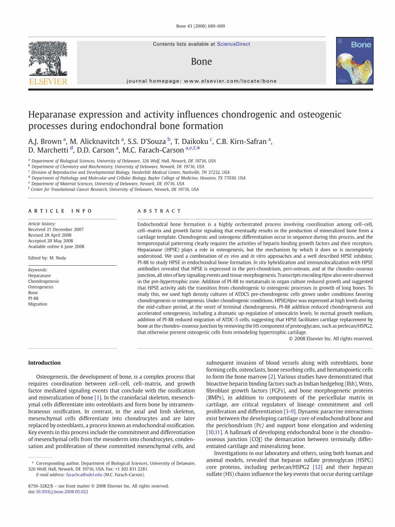
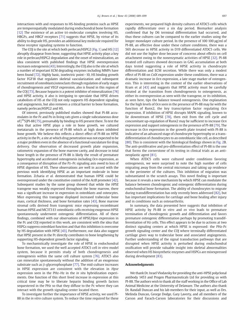
Fig. 1.HPSE is present in the Pc and COJ of developing mouse limb. Panel A shows hematoxylin and eosin counterstaining of the cryosection shown in (B). (B) HpsemRNA (orange andyellow) was detected by in situ hybridization, in the periosteum (Po) and perichondrium (Pc), hypertrophic zone (Hz), and chondro-osseous junction (COJ) and mineralized tissue(MT) of day 18.5 embryos. (C) A cryosection of mouse hindlimbwas analyzed by indirect immunofluorescence using a mouse monoclonal anti-human HPSE antibody as described in“Materials and methods”. Positive HPSE immumostaining is indicated by green fluorescence in the Pc, Po, COJ, and MT. The sectionwas double labeled with anti-perlecan/HSPG2, themajor HSPG in the matrix of developing endochondral bone (red). (D) Close up view of HPSE immunostaining (green) in the Pc at the PHz/Hz boundary shownwith perlecan/HSPG2double stain (red). (E) HPSE staining of the COJ. No specific signal was observed using isoform matched normal mouse IgG under similar conditions used as a negative control (notshown). Arrows highlight areas of high HPSE/Hpse expression.
692 A.J. Brown et al. / Bone 43 (2008) 689–699
follows: a single cell suspension of cells was pretreatedwith 100 μg/mlof PI-88 for 30 min at room temperature with occasional gentleresuspension prior to plating. Following 30min incubation,10 μl of thepretreated cell suspensionwas plated at a density of 1.0 0×105 cells inDMEM/F-12 supplemented with 5% (w/v) FBS in the center of four-well tissue culture treated plate. Fresh PI-88 was added with eachmedia change. For subsequent experiments, total RNA of differen-tiated ATDC5 micromasses were extracted using the RNeasy Mini Kitand QIAshredder column (Qiagen, Valencia, CA) and digested withDNAse I with the DNA free kit (Ambion, Austin, TX) following themanufacturer's instructions. Reverse transcription (RT)was performedwith Omniscript Reverse Transcriptase kit (Qiagen) for conventionaland real time quantitative (Q) RT-polymerase chain reaction (PCR).
High density mineralization culture
ATDC5 cells were cultured in micromass as described above withminimal modifications. Following attachment, 0.5 ml of α-MEMcontaining 5% (v/v) FBS, 0.05 mg/ml ascorbic acid, 10 mM β-glycerophosphate (SIGMA-Aldrich), ITS, and PS. Media was changedevery other day. For PI-88 treated samples, fresh PI-88was addedwitheach media change. Total RNA was extracted as described above.Whole-mount cell cultures were fixed and stained with Alcian Blue8GX (Sigma-Aldrich) as described in [54–57] and Von Kossa stain asdescribed in [46,48] at the indicated time point(s).
Migration assay
The effect of PI-88 on cell migration was determined using woundhealing assay procedures as described [58]. Prior to plating cells,ATDC5 cell suspension was pre-incubated with 100 μg/ml of PI-88 for30 min at room temperature with occasional mixing. As a negativecontrol single cell suspension was incubated with PBS alone.Immediately following incubation, cells were plated in six well tissue
culture treated plates (Becton Dickinson) and grown to confluency inregular growth media. For cells treated with PI-88, fresh PI-88 wasadded to the regular growth media. Next, the cell monolayer waswounded by a sterile plastic micropipette tip and rinsed two timeswith PBS containing Ca2+ and Mg2+ to remove any unbound cells. Thecells were imaged using a digital camera CoolPix 990 (Nikon Japan)attached to a phase contrast microscope (Nikon Japan) immediatelyfollowing the creation of the wound. Cells were incubated at 37 °C in ahumidified incubator of air:CO2 (95:5). After 210 min, cells were re-imaged under phase-contrast microscopy. The percentage of cellsrepopulating the scratch zone was calculated by direct observation ofthe cell density in this area using Image J software (http://rsb.info.nih.gov/ij/).
Statistical analysis
Statistical analyses were performed using one-way analysis ofvariance (ANOVA) and the Tukey–Kramer multiple comparisons testGraphPad Software©. Groups with probability value less than 0.05%(Pb.05) were considered significantly different.
Results
HPSE is present in the developing growth plate
In the first series of experiments, we determined the expression ofHpse mRNA and HPSE protein in developing mouse long bone andthroughout the different regions of the growth plate. To determineHpse mRNA localization in vivo, in situ hybridization was conductedusing radioactively-labeled Hpse antisense probes on limbs obtainedfrom day 18.5 mouse embryos, a time when all regional zones ofendochondral bone are clearly evident. To confirm chondrogenicregions and orientation, cryosections were counterstained withhematoxylin and eosin (Fig.1A). The proliferative zone (Pz) is indicated
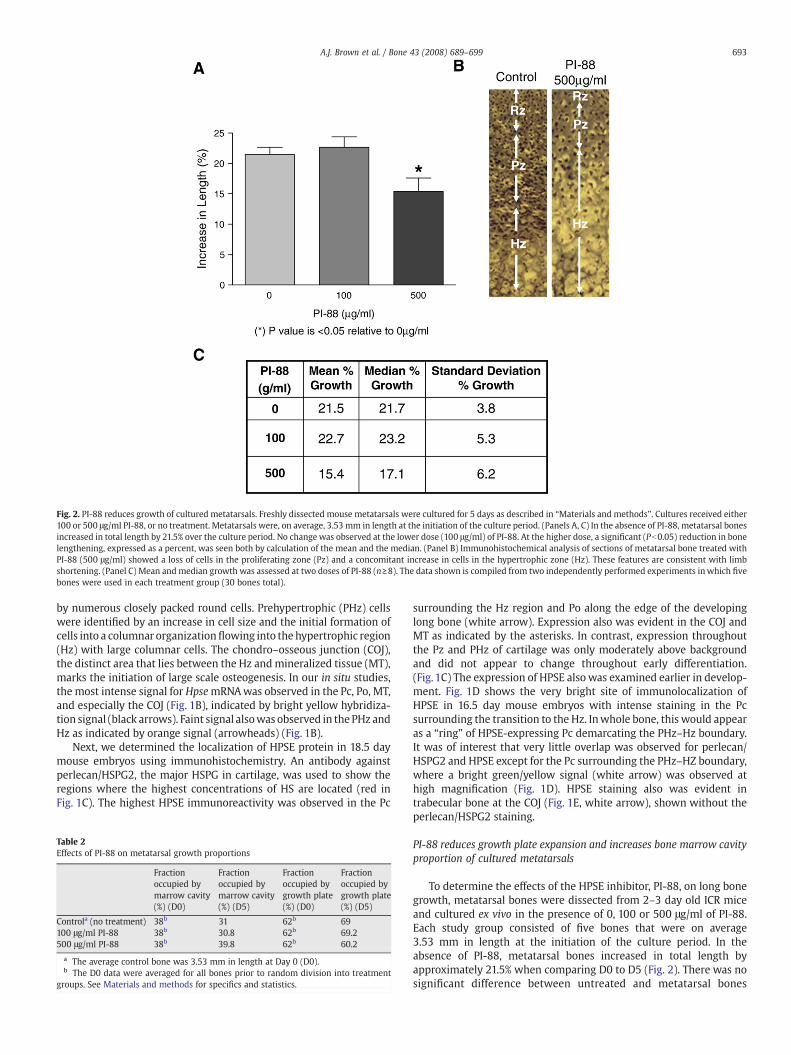
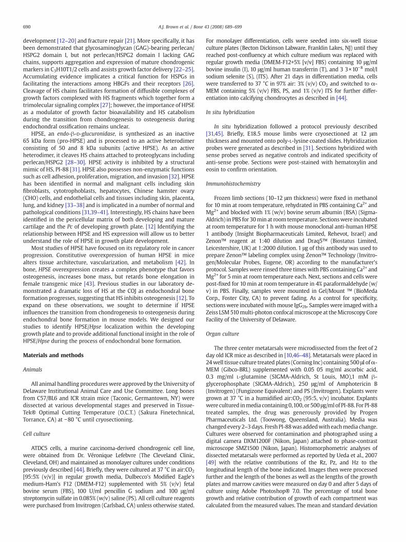
Fig. 2. PI-88 reduces growth of cultured metatarsals. Freshly dissected mouse metatarsals were cultured for 5 days as described in “Materials and methods”. Cultures received either100 or 500 μg/ml PI-88, or no treatment. Metatarsals were, on average, 3.53 mm in length at the initiation of the culture period. (Panels A, C) In the absence of PI-88, metatarsal bonesincreased in total length by 21.5% over the culture period. No change was observed at the lower dose (100 μg/ml) of PI-88. At the higher dose, a significant (Pb0.05) reduction in bonelengthening, expressed as a percent, was seen both by calculation of the mean and the median. (Panel B) Immunohistochemical analysis of sections of metatarsal bone treated withPI-88 (500 μg/ml) showed a loss of cells in the proliferating zone (Pz) and a concomitant increase in cells in the hypertrophic zone (Hz). These features are consistent with limbshortening. (Panel C) Mean andmedian growth was assessed at two doses of PI-88 (n≥8). The data shown is compiled from two independently performed experiments inwhich fivebones were used in each treatment group (30 bones total).
693A.J. Brown et al. / Bone 43 (2008) 689–699
by numerous closely packed round cells. Prehypertrophic (PHz) cellswere identified by an increase in cell size and the initial formation ofcells into a columnar organizationflowing into the hypertrophic region(Hz) with large columnar cells. The chondro–osseous junction (COJ),the distinct area that lies between the Hz andmineralized tissue (MT),marks the initiation of large scale osteogenesis. In our in situ studies,the most intense signal for HpsemRNAwas observed in the Pc, Po, MT,and especially the COJ (Fig. 1B), indicated by bright yellow hybridiza-tion signal (black arrows). Faint signal alsowas observed in the PHz andHz as indicated by orange signal (arrowheads) (Fig. 1B).
Next, we determined the localization of HPSE protein in 18.5 daymouse embryos using immunohistochemistry. An antibody againstperlecan/HSPG2, the major HSPG in cartilage, was used to show theregions where the highest concentrations of HS are located (red inFig. 1C). The highest HPSE immunoreactivity was observed in the Pc
Table 2Effects of PI-88 on metatarsal growth proportions
Fractionoccupied bymarrow cavity(%) (D0)
Fractionoccupied bymarrow cavity(%) (D5)
Fractionoccupied bygrowth plate(%) (D0)
Fractionoccupied bygrowth plate(%) (D5)
Controla (no treatment) 38b 31 62b 69100 μg/ml PI-88 38b 30.8 62b 69.2500 μg/ml PI-88 38b 39.8 62b 60.2
a The average control bone was 3.53 mm in length at Day 0 (D0).b The D0 data were averaged for all bones prior to random division into treatment
groups. See Materials and methods for specifics and statistics.
surrounding the Hz region and Po along the edge of the developinglong bone (white arrow). Expression also was evident in the COJ andMT as indicated by the asterisks. In contrast, expression throughoutthe Pz and PHz of cartilage was only moderately above backgroundand did not appear to change throughout early differentiation.(Fig. 1C) The expression of HPSE also was examined earlier in develop-ment. Fig. 1D shows the very bright site of immunolocalization ofHPSE in 16.5 day mouse embryos with intense staining in the Pcsurrounding the transition to the Hz. Inwhole bone, this would appearas a “ring” of HPSE-expressing Pc demarcating the PHz–Hz boundary.It was of interest that very little overlap was observed for perlecan/HSPG2 and HPSE except for the Pc surrounding the PHz–HZ boundary,where a bright green/yellow signal (white arrow) was observed athigh magnification (Fig. 1D). HPSE staining also was evident intrabecular bone at the COJ (Fig. 1E, white arrow), shown without theperlecan/HSPG2 staining.
PI-88 reduces growth plate expansion and increases bone marrow cavityproportion of cultured metatarsals
To determine the effects of the HPSE inhibitor, PI-88, on long bonegrowth, metatarsal bones were dissected from 2–3 day old ICR miceand cultured ex vivo in the presence of 0, 100 or 500 μg/ml of PI-88.Each study group consisted of five bones that were on average3.53 mm in length at the initiation of the culture period. In theabsence of PI-88, metatarsal bones increased in total length byapproximately 21.5% when comparing D0 to D5 (Fig. 2). There was nosignificant difference between untreated and metatarsal bones
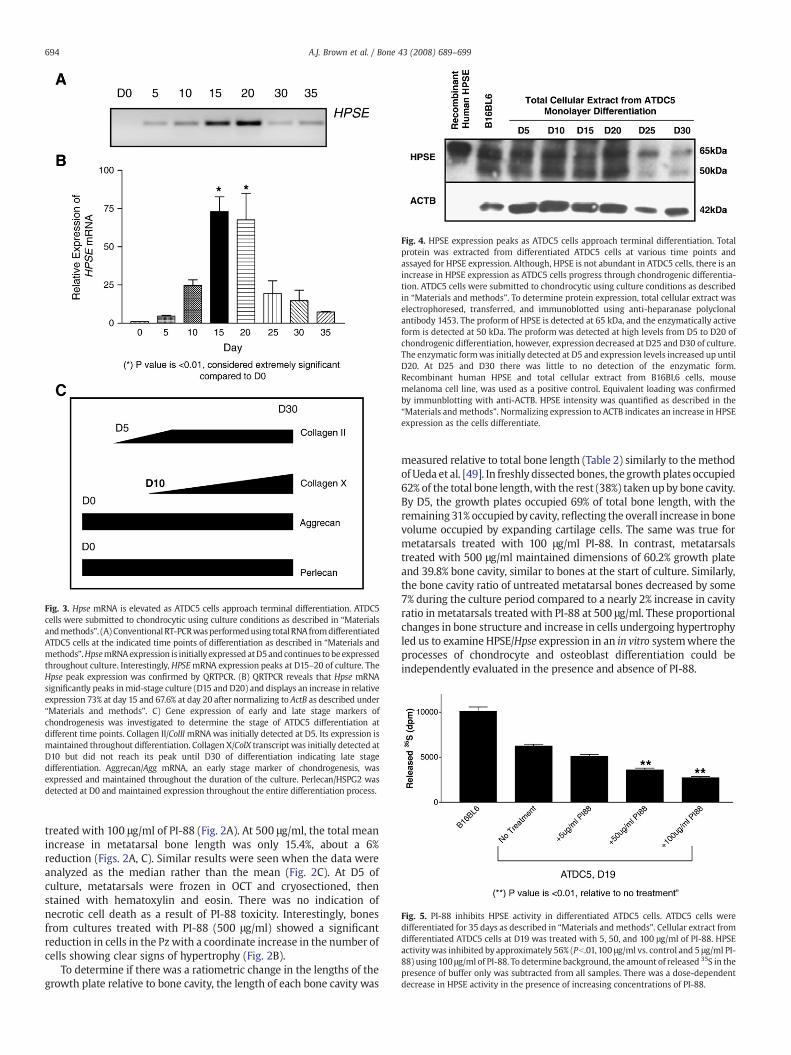
Fig. 4. HPSE expression peaks as ATDC5 cells approach terminal differentiation. Totalprotein was extracted from differentiated ATDC5 cells at various time points andassayed for HPSE expression. Although, HPSE is not abundant in ATDC5 cells, there is anincrease in HPSE expression as ATDC5 cells progress through chondrogenic differentia-tion. ATDC5 cells were submitted to chondrocytic using culture conditions as describedin “Materials and methods”. To determine protein expression, total cellular extract waselectrophoresed, transferred, and immunoblotted using anti-heparanase polyclonalantibody 1453. The proform of HPSE is detected at 65 kDa, and the enzymatically activeform is detected at 50 kDa. The proform was detected at high levels from D5 to D20 ofchondrogenic differentiation, however, expression decreased at D25 and D30 of culture.The enzymatic formwas initially detected at D5 and expression levels increased up untilD20. At D25 and D30 there was little to no detection of the enzymatic form.Recombinant human HPSE and total cellular extract from B16BL6 cells, mousemelanoma cell line, was used as a positive control. Equivalent loading was confirmedby immunblotting with anti-ACTB. HPSE intensity was quantified as described in the“Materials and methods”. Normalizing expression to ACTB indicates an increase in HPSEexpression as the cells differentiate.
Fig. 5. PI-88 inhibits HPSE activity in differentiated ATDC5 cells. ATDC5 cells weredifferentiated for 35 days as described in “Materials and methods”. Cellular extract fromdifferentiated ATDC5 cells at D19 was treated with 5, 50, and 100 μg/ml of PI-88. HPSEactivitywas inhibited by approximately 56% (Pb.01,100 μg/ml vs. control and 5 μg/ml PI-88) using 100 μg/ml of PI-88. To determine background, the amount of released 35S in thepresence of buffer only was subtracted from all samples. There was a dose-dependentdecrease in HPSE activity in the presence of increasing concentrations of PI-88.
Fig. 3. Hpse mRNA is elevated as ATDC5 cells approach terminal differentiation. ATDC5cells were submitted to chondrocytic using culture conditions as described in “Materialsandmethods”. (A) Conventional RT-PCRwasperformedusing total RNA fromdifferentiatedATDC5 cells at the indicated time points of differentiation as described in “Materials andmethods”.HpsemRNAexpression is initially expressed atD5and continues tobeexpressedthroughout culture. Interestingly, HPSEmRNA expression peaks at D15–20 of culture. TheHpse peak expression was confirmed by QRTPCR. (B) QRTPCR reveals that Hpse mRNAsignificantly peaks inmid-stage culture (D15 and D20) and displays an increase in relativeexpression 73% at day 15 and 67.6% at day 20 after normalizing to ActB as described under“Materials and methods”. C) Gene expression of early and late stage markers ofchondrogenesis was investigated to determine the stage of ATDC5 differentiation atdifferent time points. Collagen II/ColII mRNAwas initially detected at D5. Its expression ismaintained throughout differentiation. Collagen X/ColX transcript was initially detected atD10 but did not reach its peak until D30 of differentiation indicating late stagedifferentiation. Aggrecan/Agg mRNA, an early stage marker of chondrogenesis, wasexpressed and maintained throughout the duration of the culture. Perlecan/HSPG2 wasdetected at D0 and maintained expression throughout the entire differentiation process.
694 A.J. Brown et al. / Bone 43 (2008) 689–699
treated with 100 μg/ml of PI-88 (Fig. 2A). At 500 μg/ml, the total meanincrease in metatarsal bone length was only 15.4%, about a 6%reduction (Figs. 2A, C). Similar results were seen when the data wereanalyzed as the median rather than the mean (Fig. 2C). At D5 ofculture, metatarsals were frozen in OCT and cryosectioned, thenstained with hematoxylin and eosin. There was no indication ofnecrotic cell death as a result of PI-88 toxicity. Interestingly, bonesfrom cultures treated with PI-88 (500 μg/ml) showed a significantreduction in cells in the Pz with a coordinate increase in the number ofcells showing clear signs of hypertrophy (Fig. 2B).
To determine if there was a ratiometric change in the lengths of thegrowth plate relative to bone cavity, the length of each bone cavity was
measured relative to total bone length (Table 2) similarly to themethodof Ueda et al. [49]. In freshly dissected bones, the growth plates occupied62% of the total bone length,with the rest (38%) taken upby bone cavity.By D5, the growth plates occupied 69% of total bone length, with theremaining 31% occupied by cavity, reflecting the overall increase in bonevolume occupied by expanding cartilage cells. The same was true formetatarsals treated with 100 μg/ml PI-88. In contrast, metatarsalstreated with 500 μg/ml maintained dimensions of 60.2% growth plateand 39.8% bone cavity, similar to bones at the start of culture. Similarly,the bone cavity ratio of untreated metatarsal bones decreased by some7% during the culture period compared to a nearly 2% increase in cavityratio in metatarsals treated with PI-88 at 500 μg/ml. These proportionalchanges in bone structure and increase in cells undergoing hypertrophyled us to examine HPSE/Hpse expression in an in vitro systemwhere theprocesses of chondrocyte and osteoblast differentiation could beindependently evaluated in the presence and absence of PI-88.
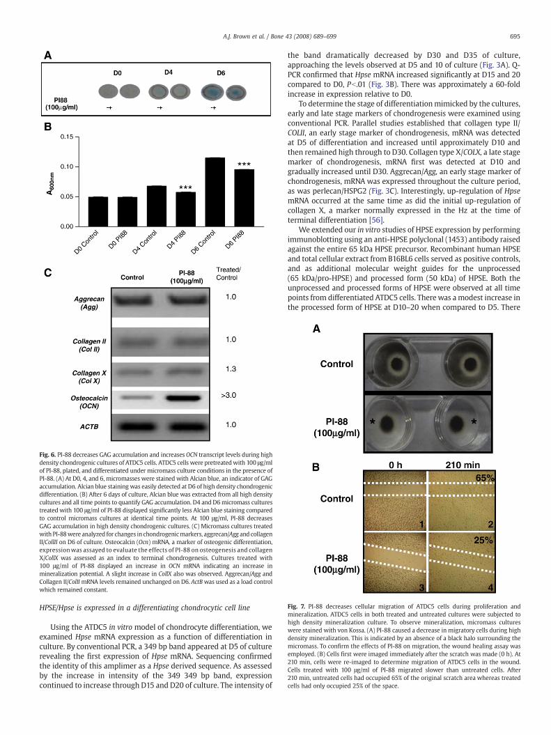
Fig. 6. PI-88 decreases GAG accumulation and increases OCN transcript levels during highdensity chondrogenic cultures of ATDC5 cells. ATDC5 cells were pretreatedwith 100 μg/mlof PI-88, plated, and differentiated under micromass culture conditions in the presence ofPI-88. (A) At D0, 4, and 6, micromasses were stained with Alcian blue, an indicator of GAGaccumulation. Alcian blue staining was easily detected at D6 of high density chondrogenicdifferentiation. (B) After 6 days of culture, Alcian blue was extracted from all high densitycultures and all time points to quantify GAG accumulation. D4 and D6micromass culturestreated with 100 μg/ml of PI-88 displayed significantly less Alcian blue staining comparedto control micromass cultures at identical time points. At 100 μg/ml, PI-88 decreasesGAG accumulation in high density chondrogenic cultures. (C) Micromass cultures treatedwith PI-88were analyzed for changes in chondrogenicmarkers, aggrecan/Agg and collagenII/CollII on D6 of culture. Osteocalcin (Ocn) mRNA, a marker of osteogenic differentiation,expressionwas assayed to evaluate the effects of PI-88 on osteogenesis and collagenX/CollX was assessed as an index to terminal chondrogenesis. Cultures treated with100 μg/ml of PI-88 displayed an increase in OCN mRNA indicating an increase inmineralization potential. A slight increase in CollX also was observed. Aggrecan/Agg andCollagen II/ColIImRNA levels remained unchanged on D6. ActBwas used as a load controlwhich remained constant.
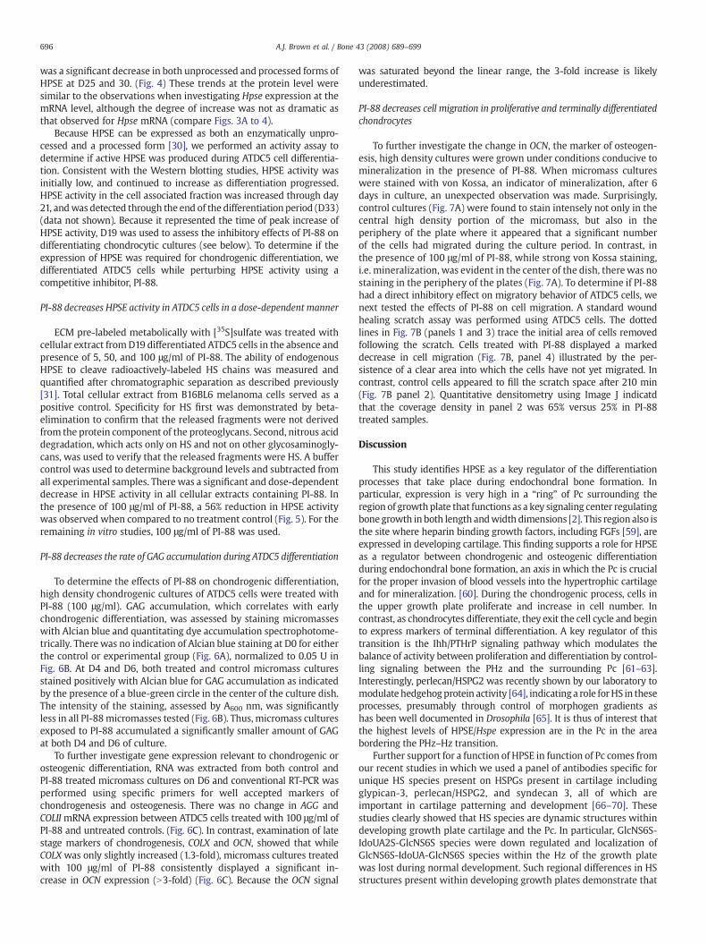
Fig. 7. PI-88 decreases cellular migration of ATDC5 cells during proliferation andmineralization. ATDC5 cells in both treated and untreated cultures were subjected tohigh density mineralization culture. To observe mineralization, micromass cultureswere stained with von Kossa. (A) PI-88 caused a decrease in migratory cells during highdensity mineralization. This is indicated by an absence of a black halo surrounding themicromass. To confirm the effects of PI-88 on migration, the wound healing assay wasemployed. (B) Cells first were imaged immediately after the scratch was made (0 h). At210 min, cells were re-imaged to determine migration of ATDC5 cells in the wound.Cells treated with 100 μg/ml of PI-88 migrated slower than untreated cells. After210 min, untreated cells had occupied 65% of the original scratch area whereas treatedcells had only occupied 25% of the space.
695A.J. Brown et al. / Bone 43 (2008) 689–699
HPSE/Hpse is expressed in a differentiating chondrocytic cell line
Using the ATDC5 in vitro model of chondrocyte differentiation, weexamined Hpse mRNA expression as a function of differentiation inculture. By conventional PCR, a 349 bp band appeared at D5 of culturerevealing the first expression of Hpse mRNA. Sequencing confirmedthe identity of this amplimer as a Hpse derived sequence. As assessedby the increase in intensity of the 349 349 bp band, expressioncontinued to increase through D15 and D20 of culture. The intensity of
the band dramatically decreased by D30 and D35 of culture,approaching the levels observed at D5 and 10 of culture (Fig. 3A). Q-PCR confirmed that Hpse mRNA increased significantly at D15 and 20compared to D0, Pb.01 (Fig. 3B). There was approximately a 60-foldincrease in expression relative to D0.
To determine the stage of differentiationmimicked by the cultures,early and late stage markers of chondrogenesis were examined usingconventional PCR. Parallel studies established that collagen type II/COLII, an early stage marker of chondrogenesis, mRNA was detectedat D5 of differentiation and increased until approximately D10 andthen remained high through to D30. Collagen type X/COLX, a late stagemarker of chondrogenesis, mRNA first was detected at D10 andgradually increased until D30. Aggrecan/Agg, an early stage marker ofchondrogenesis, mRNA was expressed throughout the culture period,as was perlecan/HSPG2 (Fig. 3C). Interestingly, up-regulation of HpsemRNA occurred at the same time as did the initial up-regulation ofcollagen X, a marker normally expressed in the Hz at the time ofterminal differentiation [56].
We extended our in vitro studies of HPSE expression by performingimmunoblotting using an anti-HPSE polyclonal (1453) antibody raisedagainst the entire 65 kDa HPSE precursor. Recombinant human HPSEand total cellular extract from B16BL6 cells served as positive controls,and as additional molecular weight guides for the unprocessed(65 kDa/pro-HPSE) and processed form (50 kDa) of HPSE. Both theunprocessed and processed forms of HPSE were observed at all timepoints from differentiated ATDC5 cells. There was amodest increase inthe processed form of HPSE at D10–20 when compared to D5. There
696 A.J. Brown et al. / Bone 43 (2008) 689–699
was a significant decrease in both unprocessed and processed forms ofHPSE at D25 and 30. (Fig. 4) These trends at the protein level weresimilar to the observations when investigating Hpse expression at themRNA level, although the degree of increase was not as dramatic asthat observed for Hpse mRNA (compare Figs. 3A to 4).
Because HPSE can be expressed as both an enzymatically unpro-cessed and a processed form [30], we performed an activity assay todetermine if active HPSE was produced during ATDC5 cell differentia-tion. Consistent with the Western blotting studies, HPSE activity wasinitially low, and continued to increase as differentiation progressed.HPSE activity in the cell associated fraction was increased through day21, andwas detected through the end of the differentiation period (D33)(data not shown). Because it represented the time of peak increase ofHPSE activity, D19 was used to assess the inhibitory effects of PI-88 ondifferentiating chondrocytic cultures (see below). To determine if theexpression of HPSE was required for chondrogenic differentiation, wedifferentiated ATDC5 cells while perturbing HPSE activity using acompetitive inhibitor, PI-88.
PI-88 decreases HPSE activity in ATDC5 cells in a dose-dependent manner
ECM pre-labeled metabolically with [35S]sulfate was treated withcellular extract fromD19 differentiated ATDC5 cells in the absence andpresence of 5, 50, and 100 μg/ml of PI-88. The ability of endogenousHPSE to cleave radioactively-labeled HS chains was measured andquantified after chromatographic separation as described previously[31]. Total cellular extract from B16BL6 melanoma cells served as apositive control. Specificity for HS first was demonstrated by beta-elimination to confirm that the released fragments were not derivedfrom the protein component of the proteoglycans. Second, nitrous aciddegradation, which acts only on HS and not on other glycosaminogly-cans, was used to verify that the released fragments were HS. A buffercontrol was used to determine background levels and subtracted fromall experimental samples. There was a significant and dose-dependentdecrease in HPSE activity in all cellular extracts containing PI-88. Inthe presence of 100 μg/ml of PI-88, a 56% reduction in HPSE activitywas observed when compared to no treatment control (Fig. 5). For theremaining in vitro studies, 100 μg/ml of PI-88 was used.
PI-88 decreases the rate of GAG accumulation during ATDC5 differentiation
To determine the effects of PI-88 on chondrogenic differentiation,high density chondrogenic cultures of ATDC5 cells were treated withPI-88 (100 μg/ml). GAG accumulation, which correlates with earlychondrogenic differentiation, was assessed by staining micromasseswith Alcian blue and quantitating dye accumulation spectrophotome-trically. There was no indication of Alcian blue staining at D0 for eitherthe control or experimental group (Fig. 6A), normalized to 0.05 U inFig. 6B. At D4 and D6, both treated and control micromass culturesstained positively with Alcian blue for GAG accumulation as indicatedby the presence of a blue-green circle in the center of the culture dish.The intensity of the staining, assessed by A600 nm, was significantlyless in all PI-88micromasses tested (Fig. 6B). Thus, micromass culturesexposed to PI-88 accumulated a significantly smaller amount of GAGat both D4 and D6 of culture.
To further investigate gene expression relevant to chondrogenic orosteogenic differentiation, RNA was extracted from both control andPI-88 treated micromass cultures on D6 and conventional RT-PCR wasperformed using specific primers for well accepted markers ofchondrogenesis and osteogenesis. There was no change in AGG andCOLIImRNA expression between ATDC5 cells treated with 100 μg/ml ofPI-88 and untreated controls. (Fig. 6C). In contrast, examination of latestage markers of chondrogenesis, COLX and OCN, showed that whileCOLX was only slightly increased (1.3-fold), micromass cultures treatedwith 100 μg/ml of PI-88 consistently displayed a significant in-crease in OCN expression (N3-fold) (Fig. 6C). Because the OCN signal
was saturated beyond the linear range, the 3-fold increase is likelyunderestimated.
PI-88 decreases cell migration in proliferative and terminally differentiatedchondrocytes
To further investigate the change in OCN, the marker of osteogen-esis, high density cultures were grown under conditions conducive tomineralization in the presence of PI-88. When micromass cultureswere stained with von Kossa, an indicator of mineralization, after 6days in culture, an unexpected observation was made. Surprisingly,control cultures (Fig. 7A) were found to stain intensely not only in thecentral high density portion of the micromass, but also in theperiphery of the plate where it appeared that a significant numberof the cells had migrated during the culture period. In contrast, inthe presence of 100 μg/ml of PI-88, while strong von Kossa staining,i.e. mineralization, was evident in the center of the dish, therewas nostaining in the periphery of the plates (Fig. 7A). To determine if PI-88had a direct inhibitory effect on migratory behavior of ATDC5 cells, wenext tested the effects of PI-88 on cell migration. A standard woundhealing scratch assay was performed using ATDC5 cells. The dottedlines in Fig. 7B (panels 1 and 3) trace the initial area of cells removedfollowing the scratch. Cells treated with PI-88 displayed a markeddecrease in cell migration (Fig. 7B, panel 4) illustrated by the per-sistence of a clear area into which the cells have not yet migrated. Incontrast, control cells appeared to fill the scratch space after 210 min(Fig. 7B panel 2). Quantitative densitometry using Image J indicatdthat the coverage density in panel 2 was 65% versus 25% in PI-88treated samples.
Discussion
This study identifies HPSE as a key regulator of the differentiationprocesses that take place during endochondral bone formation. Inparticular, expression is very high in a “ring” of Pc surrounding theregion of growthplate that functions as a key signaling center regulatingbonegrowth in both length andwidth dimensions [2]. This region also isthe site where heparin binding growth factors, including FGFs [59], areexpressed in developing cartilage. This finding supports a role for HPSEas a regulator between chondrogenic and osteogenic differentiationduring endochondral bone formation, an axis in which the Pc is crucialfor the proper invasion of blood vessels into the hypertrophic cartilageand for mineralization. [60]. During the chondrogenic process, cells inthe upper growth plate proliferate and increase in cell number. Incontrast, as chondrocytes differentiate, they exit the cell cycle and beginto express markers of terminal differentiation. A key regulator of thistransition is the Ihh/PTHrP signaling pathway which modulates thebalance of activity between proliferation and differentiation by control-ling signaling between the PHz and the surrounding Pc [61–63].Interestingly, perlecan/HSPG2 was recently shown by our laboratory tomodulate hedgehogprotein activity [64], indicatinga role forHS in theseprocesses, presumably through control of morphogen gradients ashas been well documented in Drosophila [65]. It is thus of interest thatthe highest levels of HPSE/Hspe expression are in the Pc in the areabordering the PHz–Hz transition.
Further support for a function of HPSE in function of Pc comes fromour recent studies in which we used a panel of antibodies specific forunique HS species present on HSPGs present in cartilage includingglypican-3, perlecan/HSPG2, and syndecan 3, all of which areimportant in cartilage patterning and development [66–70]. Thesestudies clearly showed that HS species are dynamic structures withindeveloping growth plate cartilage and the Pc. In particular, GlcNS6S-IdoUA2S-GlcNS6S species were down regulated and localization ofGlcNS6S-IdoUA-GlcNS6S species within the Hz of the growth platewas lost during normal development. Such regional differences in HSstructures present within developing growth plates demonstrate that
697A.J. Brown et al. / Bone 43 (2008) 689–699
interactions with and responses to HS-binding proteins such as HPSEare temporospatially modulated during endochondral bone formation[12] The existence of an active tri-molecular complex involving HS,HBGFs, and HBGF receptors [71] suggests that HPSE, by virtue of itsability to degrade HS, provides an additional keymolecule required forthese receptor signaling systems to function.
The COJ is the site at which both perlecan/HSPG2 (Fig.1) and HS [12]abruptly disappear from bone, suggesting that HPSE activity plays a keyrole in perlecan/HSPG2 degradation and the onset of mineralization, anidea consistent with published findings that HPSE overexpressionincreases osteogenesis [43]. Interestingly, theCOJ also is the site atwhicha number of important ECM degrading enzymes including MMPs havebeen found [72]. Highly basic, isoelectric pointN10, HS binding growthfactor FGF18 that regulates skeletal vascularization and subsequentrecruitment of osteoblasts/osteoclasts through regulation of early stagesof chondrogenesis and VEGF expression, also is found in this region ofthe COJ [73]. Because heparin is a potent inhibitor of mineralization [74]and HPSE activity, it also is intriguing to speculate that HPSE drivencatabolism of HS at the COJ not only supports HS dependent signalingand angiogenesis, but also removes a critical barrier to bone formation,namely perlecan/HSPG2 and HS.
Of interest, previously published studies showed that PI-88 accu-mulates in the Pc and Po in living rats given a single subcutaneous doseof [35S]PI-88 [75], presumably by binding toHSpresent there. To test theidea that active HPSE plays a role in bone growth, we culturedmetatarsals in the presence of PI-88 which at high doses inhibitedbone growth. We believe this reflects a direct effect of PI-88 on HPSEactivity in the Pc, a site atwhichdrugpenetrationwasnot expected to beamajor problem even in the absence of a functional vasculature for drugdelivery. Our observation of decreased growth plate expansion,ratiometric expansion of the bone marrow cavity, and decrease in totalbone length in the presence of PI-88 is likely the result of prematurehypertrophy and accelerated osteogenesis including Ocn expression, asa consequence of disruption of the Pz–Pc signaling axis owed to loss ofHPSE digestion of HS. These observations are well in accord with theprevious work identifying HPSE as an important molecule in boneformation. Zcharia et al. demonstrated that human HPSE could beoverexpressed under the constitutive control of the ActB promoter [42].Subsequent studies by the same group showed that while the HPSEtransgene was weakly expressed throughout the bone marrow, therewas a significant increase in expression in osteoblasts and osteocytes.Mice expressing this transgene displayed increased trabecular bonemass, cortical thickness, and bone formation rates [43]. Bone marrowstromal cells derived from transgenic mice expressing recombinanthumanHPSE andMC3T3 E1 osteoblastic cells receiving exogenousHPSEspontaneously underwent osteogenic differentiation. All of thesefindings, combined with our observations of HPSE/Hpse expression inthe Pc and COJ reported in this work, are consistent with the idea thatHSPGs suppress osteoblast function and that this inhibition is overcomeby HS degradation with HPSE [43]. Furthermore, our data also suggestthat HPSE present in the Pc directly contributes to bone lengthening bysupporting HS-dependent growth factor signaling.
To mechanistically investigate the role of HPSE in endochondralbone formation, we used the well accepted ATDC5 cell in vitro modelsystem, because it permits study of both chondrogenesis andosteogenesis within the same cell culture system [76]. ATDC5 alsocan mineralize spontaneously without the addition of an exogenoussubstrate such as β-glycerophosphate [77]. The mid-culture increasesin HPSE expression are consistent with the elevation in Hpseexpression seen in the PHz–Hz in the in situ hybridization experi-ments. One function of this short lived increase in expression at thiscritical time may be to liberate heparin binding growth factorssequestered in the PHz so that they diffuse to the Pc where they caninteract with the growth signaling center located there.
To investigate further the importance of HPSE activity, we used PI-88 in the in vitro culture system. To reduce the time required for these
experiments, we prepared high density cultures of ATDC5 cells whichrapidly differentiate over a six day period. Biomarker analysisconfirmed that by D6 terminal differentiation had occurred, andthus these cultures can be compared to the earlier studies using thelonger monolayer culture period. In the presence of 100 100 μg/ml ofPI-88, an effective dose under these culture conditions, there was a56% decrease in HPSE activity in D19 differentiated ATDC5 cells. Wedid not use the higher dose because of concerns about effects on cellattachment owing to the nonenzymatic activities of HPSE [32]. PI-88treated cell cultures showed decreases in GAG accumulation at bothdays tested suggesting a role of HPSE activity in chondrocytedifferentiation and ECM secretion. While there was only a modesteffect of PI-88 on ColX expression under these conditions, there was adramatic increase in Ocn expression, a late stage marker of osteogen-esis. This is interesting in the context of the earlier observations ofKram et al [43] and suggests that HPSE activity must be carefullytitrated at the transition from chondrogenesis to osteogenesis, aseither its overexpression as seen with the transgene, or its inhibition,as seen here, tips the balance toward osteogenesis. One explanationfor the high levels of Ocn seen in the presence of PI-88may lie with thefunction of Runx2, the key transcription factor regulating Ocnexpression. If inhibition of HPSE disrupts MAPK signaling, known tolie downstream of HPSE [78], then exit from the cell cycle andconcomitant up-regulation of Runx2 may be sufficient to increase Ocnexpression and support osteogenesis in the presence of PI-88 [79]. Theincrease in Ocn expression in the growth plate treated with PI-88 isindicative of an advanced stage of chondrocyte hypertrophy or a trans-differentiation of chondrocytes to an osteoblastic like cell, as described[80]. This is consistent with the histological findings shown in Fig. 2B.The anti-proliferative and pro-differentiation effect of PI-88 is the onethat forms the cornerstone of its testing in clinical trials as an anti-neoplastic agent [81].
When ATDC5 cells were cultured under conditions favoringosteogenesis, we were surprised to note the high number of cellsmigrating away from the central micromass and taking up residencein the perimeter of the cultures. This inhibition of migration wassubstantiated in the scratch assays. This novel finding is importantbecause it reveals a newmechanism by which HPSE can modulate thebalance between chondrogenic and osteogenic differentiation duringendochondral bone formation. The ability of chondrocytes to migrateduring transdifferentiation has only recently been addressed [82], buthas important implications for cartilage and bone healing after injuryand in conditions such as osteoarthritis.
In summary, the data presented here suggests that inhibition ofHPSE activity by PI-88 in vitro and ex vivo leads to prematuretermination of chondrogenic growth and differentiation and favorspremature osteogenic differentiation perhaps by promoting transdif-ferentiation of Hz cells. This effect appears to be due to actions at twodistinct signaling centers at which HPSE is expressed: the PHz-Pcgrowth signaling center and the COJ where terminally differentiatedcartilage gives way to trabecular bone and associated angiogenesis.Further understanding of the signal transduction pathways that aredisrupted when HPSE activity is perturbed during endochondralossification will provide valuable insight into skeletal abnormalitiesobservedwhenHS biosynthetic enzymes and HSPGs aremisexpressedduring development [83].
Acknowledgments
We thankDr. Israel Vlodavsky for providing the anti-HPSEpolyclonalantibody 1453 and Progen Pharmaceuticals Ltd for providing us withPI-88. The authorswish to thank all the staff working in the Office of LabAnimal Medicine at the University of Delaware. The authors also thankDr. Randall Duncan and his lab members for their input, as well as Drs.Melinda Duncan, George Dodge, Gary Laverty, and all members of theCarson and Farach-Carson laboratories for their discussions and
698 A.J. Brown et al. / Bone 43 (2008) 689–699
insightful suggestions. We are grateful to Dr. Rosa Serra, University ofAlabama at Birmingham, for her assistance in setting up the metatarsalculture assays.We especiallywish to thankMs. Sharron Kingston for herassistance in the preparation of this manuscript. The project describedwas partially supported by grants P20RR016458, DE13542 and P01CA098912. The content is solely the responsibility of the authors anddoes not necessarily represent the official views of the National Centerfor Research Resources or the National Institutes of Health.
References
[1] DeLise AM, Fischer L, Tuan RS. Cellular interactions and signaling in cartilagedevelopment. Osteoarthritis Cartilage 2000;8:309–34.
[2] Olsen BR, Reginato AM, Wang W. Bone development. Annu Rev Cell Dev Biol2000;16:191–220.
[3] Ohta S, Muramatsu H, Senda T, Zou K, Iwata H, Muramatsu T. Midkine is expressedduring repair of bone fracture and promotes chondrogenesis. J Bone Miner Res1999;14:1132–44.
[4] Gritli-Linde A, Lewis P, McMahon AP, Linde A. The whereabouts of a morphogen:direct evidence for short- and graded long-range activity of hedgehog signalingpeptides. Dev Biol 2001;236:364–86.
[5] Murakami S, Kan M, McKeehan WL, de Crombrugghe B. Up-regulation of thechondrogenic Sox9 gene by fibroblast growth factors is mediated by the mitogen-activated protein kinase pathway. Proc Natl Acad Sci U S A 2000;97:1113–8.
[6] Pizette S, Niswander L. BMPs are required at two steps of limb chondrogenesis:formation of prechondrogenic condensations and their differentiation intochondrocytes. Dev Biol 2000;219:237–49.
[7] Tapp H, Hernandez DJ, Neame PJ, Koob TJ. Pleiotrophin inhibits chondrocyteproliferation and stimulates proteoglycan synthesis in mature bovine cartilage.Matrix Biol 1999;18:543–56.
[8] Vortkamp A, Lee K, Lanske B, Segre GV, Kronenberg HM, Tabin CJ. Regulation of rateof cartilage differentiation by Indian hedgehog and PTH-related protein. Science1996;273:613–22.
[9] Yano F, Kugimiya F, Ohba S, Ikeda T, Chikuda H, Ogasawara T, Ogata N, Takato T,Nakamura K, Kawaguchi H, Chung UI. The canonical Wnt signaling pathwaypromotes chondrocyte differentiation in a Sox9-dependent manner. BiochemBiophys Res Commun 2005;333:1300–8.
[10] Mukherjee A, Dong SS, Clemens T, Alvarez J, Serra R. Co-ordination of TGF-beta andFGF signaling pathways in bone organ cultures. Mech Dev 2005;122:557–71.
[11] Lee K, Lanske B, Karaplis AC,Deeds JD, KohnoH, Nissenson RA, KronenbergHM, SegreGV. Parathyroid hormone-related peptide delays terminal differentiation of chon-drocytes during endochondral bone development. Endocrinology 1996;137:5109–18.
[12] Gomes Jr RR, Van Kuppevelt TH, Farach-Carson MC, Carson DD. Spatiotemporaldistribution of heparan sulfate epitopes during murine cartilage growth platedevelopment. Histochem Cell Biol 2006;126:713–22.
[13] Atha DH, Stephens AW, Rimon A, Rosenberg RD. Sequence variation in heparinoctasaccharides with high affinity for antithrombin III. Biochemistry 1984;23:5801–12.
[14] Chiao E, Fisher P, Crisponi L, Deiana M, Dragatsis I, Schlessinger D, Pilia G,Efstratiadis A. Overgrowth of a mouse model of the Simpson–Golabi–Behmelsyndrome is independent of IGF signaling. Dev Biol 2002;243:185–206.
[15] Esko JD, Selleck SB. Order out of chaos: assembly of ligand binding sites in heparansulfate. Annu Rev Biochem 2002;71:435–71.
[16] Nicole S, Davoine CS, Topaloglu H, Cattolico L, Barral D, Beighton P, Hamida CB,Hammouda H, Cruaud C, White PS, Samson D, Urtizberea JA, Lehmann-Horn F,Weissenbach J, Hentati F, Fontaine B. Perlecan, themajor proteoglycan of basementmembranes, is altered in patients with Schwartz–Jampel syndrome (chondrody-strophic myotonia). Nat Genet 2000;26:480–3.
[17] Ashikari S, Habuchi H, Kimata K. Characterization of heparan sulfate oligosacchar-ides that bind to hepatocyte growth factor. J Biol Chem 1995;270:29586–93.
[18] Faham S, Hileman RE, Fromm JR, Linhardt RJ, Rees DC. Heparin structure andinteractions with basic fibroblast growth factor. Science 1996;271:1116–20.
[19] Lindahl U, Thunberg L, BackstromG, Riesenfeld J, Nordling K, Bjork I. Extension andstructural variability of the antithrombin-binding sequence in heparin. J Biol Chem1984;259:12368–76.
[20] So CL, Kaluarachchi K, Tam PP, Cheah KS. Impact of mutations of cartilage matrixgenes on matrix structure, gene activity and chondrogenesis. OsteoarthritisCartilage 2001;9(Suppl A):S160–173.
[21] Pacicca DM, Patel N, Lee C, Salisbury K, Lehmann W, Carvalho R, Gerstenfeld LC,Einhorn TA. Expression of angiogenic factors during distraction osteogenesis. Bone2003;33:889–98.
[22] Gomes Jr RR, Joshi SS, Farach-CarsonMC, Carson DD. Ribozyme-mediated perlecanknockdown impairs chondrogenic differentiation of C3H10T1/2 fibroblasts.Differentiation 2006;74:53–63.
[23] Gomes Jr RR, Farach Carson MC, Carson DD. Perlecan-stimulated nodules undergochondrogenic maturation in response to rhBMP-2 treatment in vitro. ConnectTissue Res 2003;44(Suppl 1):196–201.
[24] FrenchMM, Gomes Jr RR, Timpl R, HookM, Czymmek K, Farach-CarsonMC, CarsonDD. Chondrogenic activity of the heparan sulfate proteoglycan perlecan maps tothe N-terminal domain I. J Bone Miner Res 2002;17:48–55.
[25] French MM, Smith SE, Akanbi K, Sanford T, Hecht J, Farach-Carson MC, Carson DD.Expression of the heparan sulfate proteoglycan, perlecan, during mouse
embryogenesis and perlecan chondrogenic activity in vitro. J Cell Biol 1999;145:1103–15.
[26] Kirn-Safran CB, Gomes RR, Brown AJ, Carson DD. Heparan sulfate proteoglycans:coordinators of multiple signaling pathways during chondrogenesis. Birth DefectsRes C Embryo Today 2004;72:69–88.
[27] Zcharia E, Zilka R, Yaar A, Yacoby-Zeevi O, Zetser A,Metzger S, Sarid R, Naggi A, CasuB, Ilan N, Vlodavsky I, Abramovitch R. Heparanase accelerates wound angiogenesisand wound healing in mouse and rat models. Faseb J 2005;19:211–21.
[28] Miao HQ, Navarro E, Patel S, Sargent D, Koo H, Wan H, Plata A, Zhou Q, Ludwig D,Bohlen P, Kussie P. Cloning, expression, and purification of mouse heparanase.Protein Expr Purif 2002;26:425–31.
[29] Podyma-Inoue KA, Yokote H, Sakaguchi K, IkutaM, YanagishitaM. Characterizationof heparanase from a rat parathyroid cell line. J Biol Chem 2002;277:32459–65.
[30] Levy-Adam F, Miao HQ, Heinrikson RL, Vlodavsky I, Ilan N. Heterodimer formationis essential for heparanase enzymatic activity. Biochem Biophys Res Commun2003;308:885–91.
[31] D'Souza SS, Daikoku T, Farach-Carson MC, Carson DD. Heparanase Expression andFunction During Early Pregnancy in Mice. Biol Reprod 2007;77:433–41.
[32] Vlodavsky I, Ilan N, Naggi A, Casu B. Heparanase: structure, biological functions,and inhibition by heparin-derived mimetics of heparan sulfate. Curr Pharm Des2007;13:2057–73.
[33] Bame KJ. Heparanases: endoglycosidases that degrade heparan sulfate proteogly-cans. Glycobiology 2001;11:91R–8R.
[34] Freeman C, Browne AM, Parish CR. Evidence that platelet and tumour heparanasesare similar enzymes. Biochem J 1999;342(Pt 2):361–8.
[35] Sewell RF, Brenchley PE, Mallick NP. Human mononuclear cells contain anendoglycosidase specific for heparan sulphate glycosaminoglycan demonstrablewith the use of a specific solid-phase metabolically radiolabelled substrate.Biochem J 1989;264:777–83.
[36] Laskov R, Michaeli RI, Sharir H, Yefenof E, Vlodavsky I. Production of heparanase bynormal and neoplastic murine B-lymphocytes. Int J Cancer 1991;47:92–8.
[37] Matzner Y, Bar-Ner M, Yahalom J, Ishai-Michaeli R, Fuks Z, Vlodavsky I.Degradation of heparan sulfate in the subendothelial extracellular matrix by areadily released heparanase from human neutrophils. Possible role in invasionthrough basement membranes. J Clin Invest 1985;76:1306–13.
[38] Mollinedo F, Nakajima M, Llorens A, Barbosa E, Callejo S, Gajate C, Fabra A. Majorco-localization of the extracellular-matrix degradative enzymes heparanase andgelatinase in tertiary granules of human neutrophils. Biochem J 1997;327(Pt 3):917–23.
[39] Vlodavsky I, Goldshmidt O, Zcharia E, Metzger S, Chajek-Shaul T, Atzmon R, Guatta-Rangini Z, Friedmann Y. Molecular properties and involvement of heparanase incancer progression and normal development. Biochimie 2001;83:831–9.
[40] Elkin M, Ilan N, Ishai-Michaeli R, Friedmann Y, Papo O, Pecker I, Vlodavsky I.Heparanase as mediator of angiogenesis: mode of action. Faseb J 2001;15:1661–3.
[41] Parish CR, Freeman C, Hulett MD. Heparanase: a key enzyme involved in cellinvasion. Biochim Biophys Acta 2001;1471:M99–108.
[42] Zcharia E, Metzger S, Chajek-Shaul T, Aingorn H, ElkinM, Friedmann Y,Weinstein T,Li JP, Lindahl U, Vlodavsky I. Transgenic expression of mammalian heparanaseuncovers physiological functions of heparan sulfate in tissue morphogenesis,vascularization, and feeding behavior. Faseb J 2004;18:252–63.
[43] Kram V, Zcharia E, Yacoby-Zeevi O, Metzger S, Chajek-Shaul T, Gabet Y, Muller R,Vlodavsky I, Bab I. Heparanase is expressed in osteoblastic cells and stimulatesbone formation and bone mass. J Cell Physiol 2006;207:784–92.
[44] Shukunami C, Ishizeki K, Atsumi T, Ohta Y, Suzuki F, Hiraki Y. Cellular hypertrophyand calcification of embryonal carcinoma-derived chondrogenic cell line ATDC5 invitro. J Bone Miner Res 1997;12:1174–88.
[45] Das SK, Wang XN, Paria BC, Damm D, Abraham JA, Klagsbrun M, Andrews GK, Dey SK.Heparin-bindingEGF-likegrowth factorgene is induced in themouseuterus temporallyby the blastocyst solely at the site of its apposition: a possible ligand for interactionwithblastocyst EGF-receptor in implantation. Development 1994;120: 1071–83.
[46] Addison WN, Azari F, Sorensen ES, Kaartinen MT, McKee MD. Pyrophosphateinhibits mineralization of osteoblast cultures by binding to mineral, up-regulatingosteopontin, and inhibiting alkaline phosphatase activity. J Biol Chem 2007;282:15872–83.
[47] MacRae VE, Farquharson C, Ahmed SF. The restricted potential for recovery ofgrowth plate chondrogenesis and longitudinal bone growth following exposure topro-inflammatory cytokines. J Endocrinol 2006;189:319–28.
[48] McKee MD, Addison WN, Kaartinen MT. Hierarchies of extracellular matrix andmineral organization in bone of the craniofacial complex and skeleton. CellsTissues Organs 2005;181:176–88.
[49] Ueda K, Yamanaka Y, Harada D, Yamagami E, Tanaka H, Seino Y. PTH has thepotential to rescue disturbed bone growth in achondroplasia. Bone 2007;41:13–8.
[50] Laemmli UK. Cleavage of structural proteins during the assembly of the head ofbacteriophage T4. Nature 1970;227:680–5.
[51] Porzio MA, Pearson AM. Improved resolution of myofibrillar proteins with sodiumdodecyl sulfate-polyacrylamide gel electrophoresis. Biochim Biophys Acta 1977;490:27–34.
[52] Reiland J, Sanderson RD, Waguespack M, Barker SA, Long R, Carson DD, MarchettiD. Heparanase degrades syndecan-1 and perlecan heparan sulfate: functionalimplications for tumor cell invasion. J Biol Chem 2004;279:8047–55.
[53] Denker AE, Haas AR, Nicoll SB, Tuan RS. Chondrogenic differentiation of murineC3H10T1/2multipotential mesenchymal cells: I. Stimulation by bonemorphogeneticprotein-2 in high-density micromass cultures. Differentiation 1999;64:67–76.
[54] Atkinson BL, Fantle KS, Benedict JJ, Huffer WE, Gutierrez-Hartmann A. Combina-tion of osteoinductive bone proteins differentiates mesenchymal C3H/10T1/2 cellsspecifically to the cartilage lineage. J Cell Biochem 1997;65:325–39.
699A.J. Brown et al. / Bone 43 (2008) 689–699
[55] IzzoMW,PucciB, TuanRS,HallDJ. Geneexpressionprofiling followingBMP-2 inductionof mesenchymal chondrogenesis in vitro. Osteoarthritis Cartilage 2002;10:23–33.
[56] Miller SA, Brown AJ, Farach-Carson MC, Kirn-Safran CB. HIP/RPL29 down-regulationaccompanies terminal chondrocyte differentiation. Differentiation 2003;71:322–36.
[57] Seghatoleslami MR, Tuan RS. Cell density dependent regulation of AP-1 activity isimportant for chondrogenic differentiation of C3H10T1/2 mesenchymal cells. J CellBiochem 2002;84:237–48.
[58] Naik MU, Naik UP. Calcium- and integrin-binding protein regulates focal adhesionkinase activity duringplatelet spreadingon immobilizedfibrinogen. Blood 2003;102:3629–36.
[59] Lazarus JE, Hegde A, Andrade AC, Nilsson O, Baron J. Fibroblast growth factorexpression in the postnatal growth plate. Bone 2007;40:577–86.
[60] Colnot C, LuC,HuD,Helms JA.Distinguishing thecontributions of theperichondrium,cartilage, and vascular endothelium to skeletal development. Dev Biol 2004;269:55–69.
[61] Akiyama H, Shigeno C, Iyama K, Ito H, Hiraki Y, Konishi J, Nakamura T. Indianhedgehog in the late-phase differentiation in mouse chondrogenic EC cells,ATDC5: upregulation of type X collagen and osteoprotegerin ligand mRNAs.Biochem Biophys Res Commun 1999;257:814–20.
[62] Colnot C, de la Fuente L, Huang S, Hu D, Lu C, St-Jacques B, Helms JA. Indianhedgehog synchronizes skeletal angiogenesis and perichondrial maturation withcartilage development. Development 2005;132:1057–67.
[63] Ingham PW,McMahon AP. Hedgehog signaling in animal development: paradigmsand principles. Genes Dev 2001;15:3059–87.
[64] Datta MW, Hernandez AM, Schlicht MJ, Kahler AJ, DeGueme AM, Dhir R, Shah RB,Farach-Carson C, Barrett A, Datta S. Perlecan, a candidate gene for the CAPB locus,regulates prostate cancer cell growth via the Sonic Hedgehog pathway. Mol Cancer2006;5:9.
[65] Strigini M. Mechanisms of morphogen movement. J Neurobiol 2005;64:324–33.[66] Arikawa-Hirasawa E, Watanabe H, Takami H, Hassell JR, Yamada Y. Perlecan is
essential for cartilage and cephalic development. Nat Genet 1999;23:354–8.[67] Cano-Gauci DF, Song HH, Yang H, McKerlie C, Choo B, ShiW, Pullano R, Piscione TD,
Grisaru S, Soon S, Sedlackova L, Tanswell AK, Mak TW, Yeger H, Lockwood GA,Rosenblum ND, Filmus J. Glypican-3-deficient mice exhibit developmentalovergrowth and some of the abnormalities typical of Simpson–Golabi–Behmelsyndrome. J Cell Biol 1999;146:255–64.
[68] Costell M, Gustafsson E, Aszodi A, Morgelin M, Bloch W, Hunziker E, Addicks K,Timpl R, Fassler R. Perlecanmaintains the integrity of cartilage and some basementmembranes. J Cell Biol 1999;147:1109–22.
[69] Koyama E, Leatherman JL, Shimazu A, Nah HD, Pacifici M. Syndecan-3, tenascin-C,and the development of cartilaginous skeletal elements and joints in chick limbs.Dev Dyn 1995;203:152–62.
[70] Seghatoleslami MR, Kosher RA. Inhibition of in vitro limb cartilage differentiationby syndecan-3 antibodies. Dev Dyn 1996;207:114–9.
[71] Wu ZL, Zhang L, Yabe T, Kuberan B, Beeler DL, Love A, Rosenberg RD. Theinvolvement of heparan sulfate (HS) in FGF1/HS/FGFR1 signaling complex. J BiolChem 2003;278:17121–9.
[72] OrtegaN,BehonickD, StickensD,WerbZ.Howproteases regulatebonemorphogenesis.Ann N Y Acad Sci 2003;995:109–16.
[73] Liu Z, Lavine KJ, Hung IH, Ornitz DM. FGF18 is required for early chondrocyteproliferation, hypertrophy and vascular invasion of the growth plate. Dev Biol2007;302:80–91.
[74] Hansen NM, Felix R, Bisaz S, Fleisch H. Aggregation of hydroxyapatite crystals.Biochim Biophys Acta 1976;451:549–59.
[75] Levidiotis V, Freeman C, Punler M, Martinello P, Creese B, Ferro V, van der Vlag J,Berden JH, Parish CR, Power DA. A synthetic heparanase inhibitor reducesproteinuria in passive Heymann nephritis. J Am Soc Nephrol 2004;15:2882–92.
[76] Mushtaq T, Farquharson C, Seawright E, Ahmed SF. Glucocorticoid effects onchondrogenesis, differentiation and apoptosis in the murine ATDC5 chondrocytecell line. J Endocrinol 2002;175:705–13.
[77] Guicheux J, Palmer G, Shukunami C, Hiraki Y, Bonjour JP, Caverzasio J. A novel invitro culture system for analysis of functional role of phosphate transport inendochondral ossification. Bone 2000;27:69–74.
[78] Patel VN, Knox SM, Likar KM, Lathrop CA, Hossain R, Eftekhari S, Whitelock JM,Elkin M, Vlodavsky I, Hoffman MP. Heparanase cleavage of perlecan heparansulfate modulates FGF10 activity during ex vivo submandibular gland branchingmorphogenesis. Development 2007;134:4177–86.
[79] Thomas DM, Johnson SA, Sims NA, Trivett MK, Slavin JL, Rubin BP, Waring P,McArthur GA, Walkley CR, Holloway AJ, Diyagama D, Grim JE, Clurman BE, BowtellDD, Lee JS, Gutierrez GM, Piscopo DM, Carty SA, Hinds PW. Terminal osteoblastdifferentiation, mediated by runx2 and p27KIP1, is disrupted in osteosarcoma.J Cell Biol 2004;167:925–34.
[80] Lian JB, McKee MD, Todd AM, Gerstenfeld LC. Induction of bone-related proteins,osteocalcin and osteopontin, and their matrix ultrastructural localization withdevelopment of chondrocyte hypertrophy in vitro. J Cell Biochem 1993;52:206–19.
[81] Miao HQ, Liu H, Navarro E, Kussie P, Zhu Z. Development of heparanase inhibitorsfor anti-cancer therapy. Curr Med Chem 2006;13:2101–11.
[82] Morales TI. Chondrocyte moves: clever strategies. Osteoarthritis Cartilage 2007.[83] Farach-Carson MC, Hecht JT, Carson DD. Heparan sulfate proteoglycans: key
players in cartilage biology. Crit Rev Eukaryot Gene Expr 2005;15:29–48.[84] Yang W, Gomes RR, Brown AJ, Burdett AR, Alicknavitch M, Farach-Carson MC,
Carson DD. Chondrogenic differentiation on perlecan domain I, collagen II, andbone morphogenetic protein-2-based matrices. Tissue Eng 2006;12:2009–24.
[85] Boudreaux JM, Towler DA. Synergistic induction of osteocalcin gene expression:identification of a bipartite element conferring fibroblast growth factor 2 andcyclic AMP responsiveness in the rat osteocalcin promoter. J Biol Chem 1996;271:7508–15.