Embed Size (px)
Citation preview

Hemoglobin Structure
Hemoglobin (Hb) is synthesized in a complex
series of steps. The heme part is synthesized
and themitochondriain a series of steps in the and the mitochondriain a series of steps in the
of immature red blood cells, while the cytosoly
protein parts are synthesized by globin
in the cytosolribosomes

Heme synthesized by mitochondria, fixed with irony y ,
Heme then surrounded by “globin” proteins that surround and “protect”
the heme
Each single Hemoglobin molecule has two globin chains, each with itsEach single Hemoglobin molecule has two globin chains, each with its
own heme protein attached
One globin chain is alpha
One is “non-alpha”One is non alpha
Two hemoglobin molecules combine to produce functional hgb
tetramer

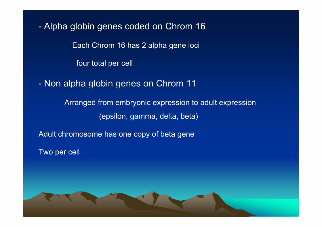
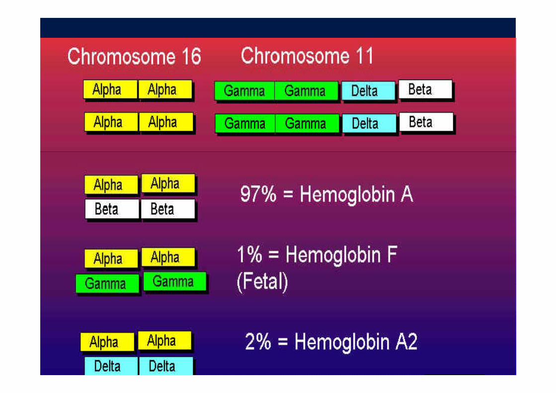
- Alpha globin genes coded on Chrom 16
Each Chrom 16 has 2 alpha gene loci
four total per cellfour total per cell
- Non alpha globin genes on Chrom 11p g g
Arranged from embryonic expression to adult expression
( il d lt b t )(epsilon, gamma, delta, beta)
Adult chromosome has one copy of beta gene
Two per cell


Hemoglobin AbnormalitiesHemoglobin Abnormalitiesgg
There are 3 main categories of inherited Hemoglobin abnormalities:
- Structural or qualitative: The amino acid sequence isStructural or qualitative: The amino acid sequence is altered because of incorrect DNA code (HBs).
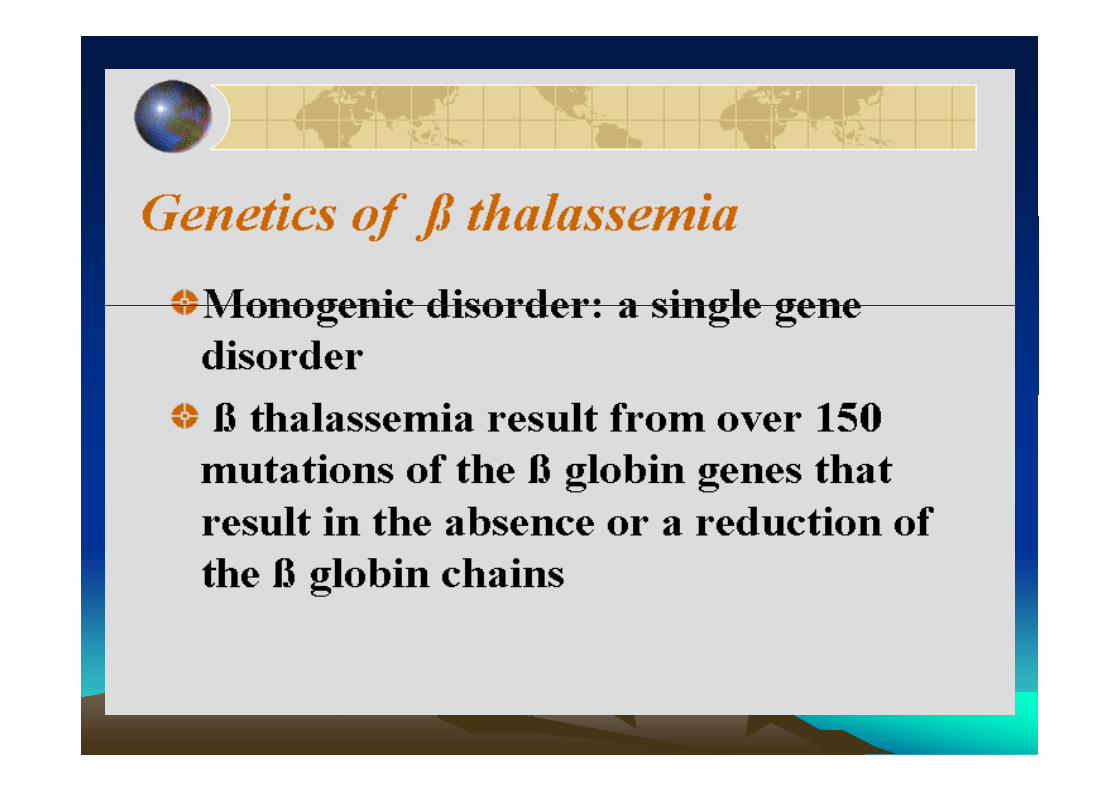
Quantitative: Production of one or more globin- Quantitative: Production of one or more globin chains is reduced or absent (Thalassemia).
- Hereditary persistence of Fetal Hemoglobin (HPFH): Complete or partial failure of γ globin to switch to βgglobin.

Laboratory Methods to evaluate Laboratory Methods to evaluate HemoglobinHemoglobin
Red cell morphologies:HbS: Sickle cells


Red cell morphologies:HbS: Sickle cellsHbC: Target cells, crystals after splenectomy


Red cell morphologies:HbS: Sickle cellsHbC: Target cells, crystals after splenectomyThalassemias: Microcystosis target cellsThalassemias: Microcystosis, target cells, basophilic stippling


ThalassemiaThalassemia
Variety of genetic defects in globin chain synthesis –decreased or absent
Classified according to globin chain thatClassified according to globin chain that
is affected – e.g. β-thalassemia vs. α thalassemia
Heterozygous: minor
Homozygous: majorHomozygous: major
The name is derived from the Greek words Thalasso = Sea" and "Hemia = Blood" in reference to anemia of the sea.

Pathophysiology
- If α chain is affected, excess of β chains produced. If βchain is affected, excess of α chains produced
- Imbalance in chain synthesis causes
-Decrease in total hemoglobin production
-Ineffective erythropoiesis-Ineffective erythropoiesis
-Chronic hemolysis
-Excess α chains are unstable – precipitate
within cell – precipitates bind to cell
membrane, causing membrane damage

Excess β chains combine to form Hb HExcess β chains combine to form Hb H-High oxygen affinity – poor oxygen transporter-unstableunstable


β- thalassemia Major –Cooley’s Anemia
Homozygous (β+/ β + or β 0/ β 0) or doubleHomozygous (β / β or β 0/ β 0) or double heterozygous (β +/ β 0) inheritance
Pathophysiology: dramatic reduction or complete absence of β chain synthesis β y
– Symptoms begin to manifest at age 6 months
-Increase in non β containing hemoglobins
- Excess α chains precipitate in cellsExcess α chains precipitate in cells
- hemolysis

Pathophsiology Pathophsiology
ɣ α βexcessexcess
α2 ɣ 2 dest of RBC
hb F hemolysis
level of hb F Splenomegaly ineff erythropoiesisof RBCof RBC
Anemia
High O2 affinity g ytissue hypoxia
Epop

anemiaEPO
Marrow expansionMarrow expansion
Skeletal def.Inc.mwtabolic rate Inc. Fe absorption transfusion Gout
Folate def. Fe overload
endocrine def.cardiac f
LC
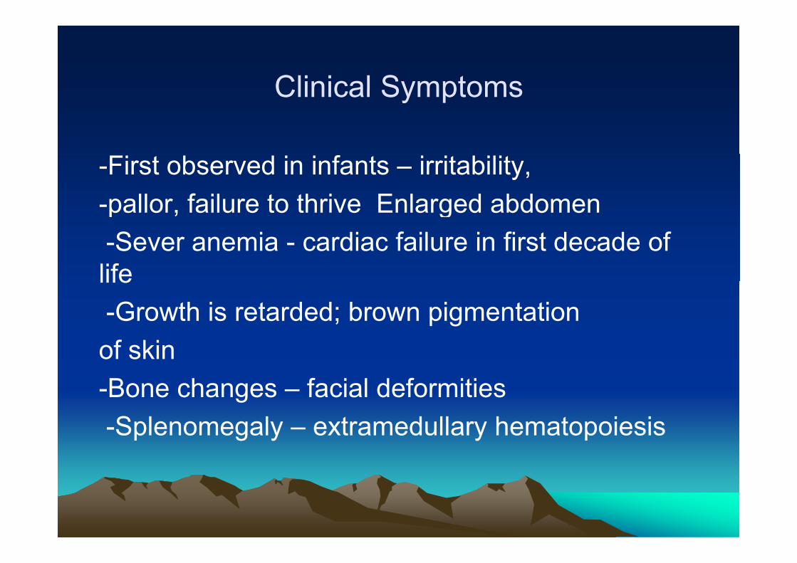
Clinical SymptomsClinical Symptoms
-First observed in infants – irritability,-pallor, failure to thrive Enlarged abdomenp g-Sever anemia - cardiac failure in first decade of lifelife-Growth is retarded; brown pigmentationof skinof skin-Bone changes – facial deformities-Splenomegaly – extramedullary hematopoiesis

Gallstones – due to increasedintravascular and extravascular hemolysisintravascular and extravascular hemolysis
-Skeletal abnormalities – expansion ofbone marrrow
-Pathological fractures – thinning of-Pathological fractures – thinning ofcalcified bone
-Iron toxicity – multiple transfusions

Laboratory Findings of tha majorLaboratory Findings of tha.major
Hemoglobin as low as 2-3 g/dL
-Markedly microcytic/hypochromic
Marked anisocytosis and poikilocytosis-Marked anisocytosis and poikilocytosis
-Basophilic stippling and polychromasia
-Hemoglobin electrophoresis – 90% Hb F and increased Hb A2
-Increased bilirubin, decreased haptoglobin
Increased serum iron and decreased TIBC-Increased serum iron and decreased TIBC


Thalassemia carriers (trait):Thalassemia carriers (trait):
Usually no signs or symptoms are Usua y o s g s o sy p o s a eapparent, except for a mild anemia.
Carriers are usually initially detected th h i h f ithrough screening, or when performing routine CBC Later it can be confirmedroutine CBC . Later it can be confirmed using hemoglobin electrophoresis.

Alpha ThalassemiasAlpha ThalassemiasAlpha ThalassemiasAlpha ThalassemiasThere are two α genes on each of two chromosome 16 structures (four α genes in the diploid state)
M t ti ff t f thMutations can affect one or more of the α
genes resulting in four levels of severity
Wh ll f d l t d-When all four genes deleted – no α
chains, hydrops fetalis or a-thalassemia
majormajor
- 3 of the four deleted, hemoglobin H
DiseaseDisease-2 of the 4 deleted, a-thalassemia minor-1 deletion, silent carrier

Classification & TerminologyClassification & TerminologyAlphaAlpha ThalassemiaThalassemia
•Normalαα/αα•Silent carrier- α/αα
•Minor-α/-α--/αα--/αα
•Hb H disease--/-α•Barts hydrops fetalis--/--