Embed Size (px)
Citation preview

HEMOGLOBIN
Learning Objectives:
1. List the steps in the biosynthesis of Hemoglobin
2.Describle the function of Hemoglobin
3. Describe the fate of Hemoglobin
4. List the Normal and Abnormal Hemoglobin
5. Discuss the types of Jaundice
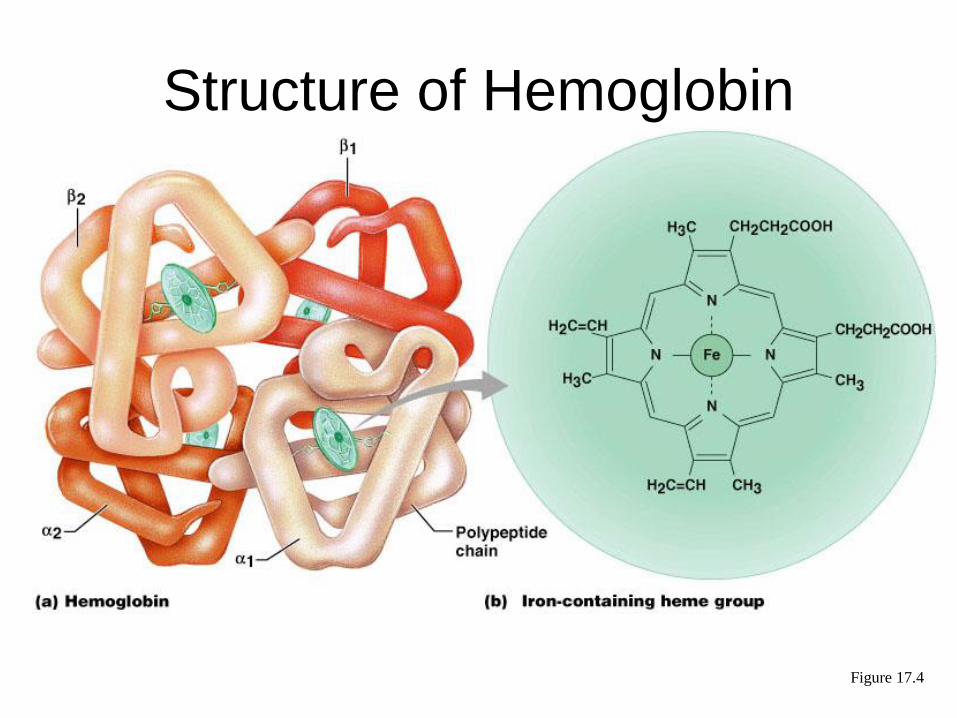
Structure of Hemoglobin
Figure 17.4
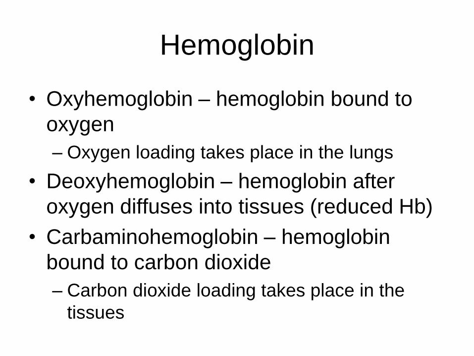
Hemoglobin
• Oxyhemoglobin – hemoglobin bound to
oxygen
– Oxygen loading takes place in the lungs
• Deoxyhemoglobin – hemoglobin after
oxygen diffuses into tissues (reduced Hb)
• Carbaminohemoglobin – hemoglobin
bound to carbon dioxide
– Carbon dioxide loading takes place in the
tissues
Synthesis of Haemoglobin (Hb)
• Haem & globin produced at two different sites in
the cells
• Haem in mitochondria
• Globin in polyribosomes
• Well synchronized
• Normal hemoglobin production is dependent
upon 3 processes: Adequate iron delivery and
supply, adequate synthesis of protoporphyrins
and adequate globin synthesis.
Hemoglobin Structure and
Function• Hemoglobin occupies 33% of the RBC
volume and 90-95% of the dry weight.
– 65% of the hemoglobin synthesis occurs in the nucleated stages of RBC maturation and 35% during the reticulocyte stage.
– Normal hemoglobin consists of 4 heme groups which contain a protoporphyrin ring plus iron and globin which is a tetramer of 2 pairs of polypeptide chains.
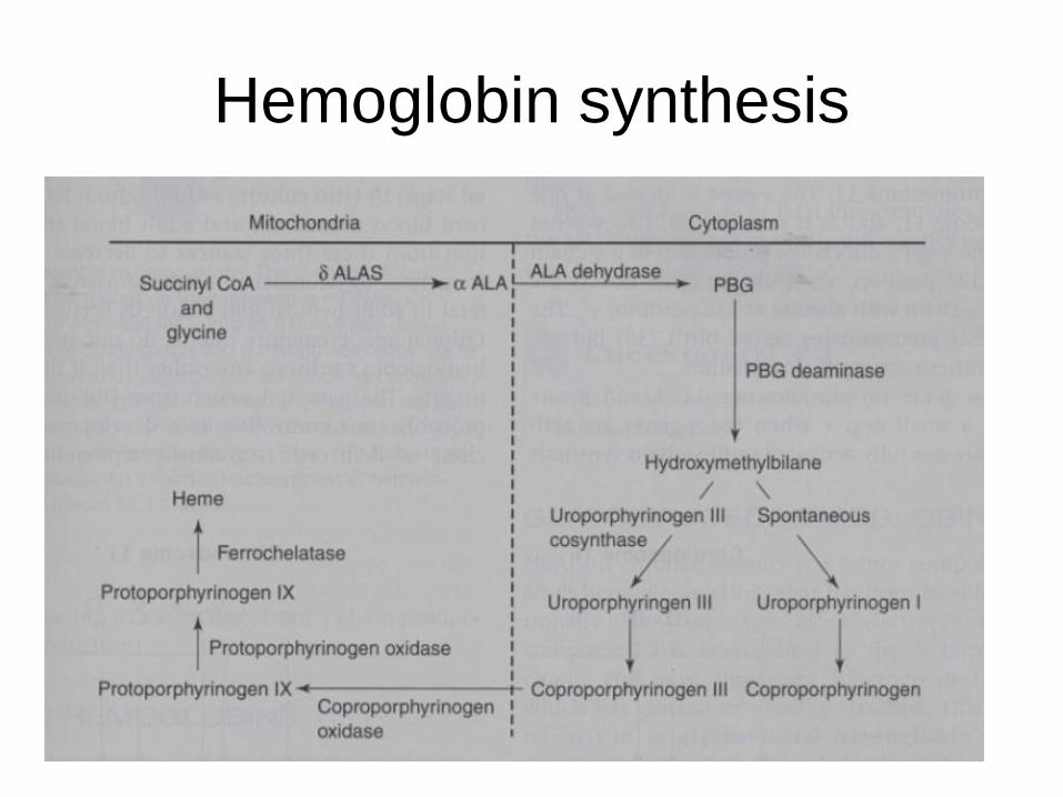
Hemoglobin synthesis
Porphyria
– Since porphyrinogens are readily oxidized to form porphyrins,
excess formation of porphyrins can occur if any of the normal
enzymatic steps in heme synthesis is blocked.
– Inherited
Erythropoietic porphyria - results from
excessive production of porphyrins in the bone
marrow.
Hepatic porphyria - results from excessive
production of porphyrins in the liver.
- Acquired
Lead intoxication - interferes with protoporphyrin
synthesis
Chronic alcoholic liver disease
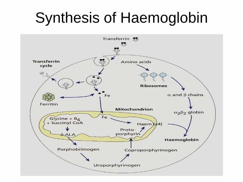
Synthesis of Haemoglobin
• Globin Synthesis
– In the fetus and the adult 4 types of hemoglobin chains may be formed: alpha ( ), beta ( ), gamma ( ), and delta ( ).
– Normal hemoglobin's contain 4 globin chains.
– Hemoglobin (hgb) F= 2 2 and is the predominant hgb formed during liver and bone marrow erythropoiesis in the fetus.
– A normal, full term baby has 50-85% hgbF.
– Near the end of the first year of life, normal adult hgb levels are reached.
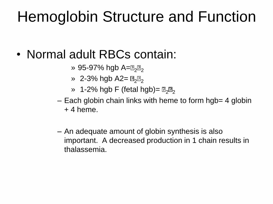
Hemoglobin Structure and Function
• Normal adult RBCs contain:» 95-97% hgb A= 2 2
» 2-3% hgb A2= 2 2
» 1-2% hgb F (fetal hgb)= 2 2
– Each globin chain links with heme to form hgb= 4 globin
+ 4 heme.
– An adequate amount of globin synthesis is also
important. A decreased production in 1 chain results in
thalassemia.
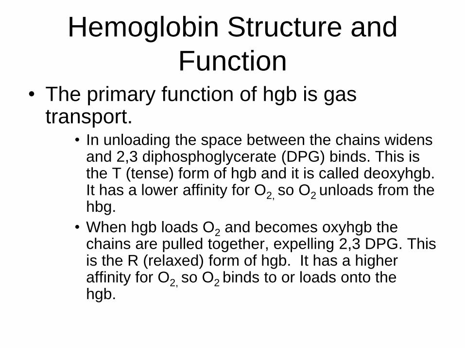
Hemoglobin Structure and
Function• The primary function of hgb is gas
transport. • In unloading the space between the chains widens
and 2,3 diphosphoglycerate (DPG) binds. This is the T (tense) form of hgb and it is called deoxyhgb. It has a lower affinity for O2, so O2 unloads from the hbg.
• When hgb loads O2 and becomes oxyhgb the chains are pulled together, expelling 2,3 DPG. This is the R (relaxed) form of hgb. It has a higher affinity for O2, so O2 binds to or loads onto the hgb.
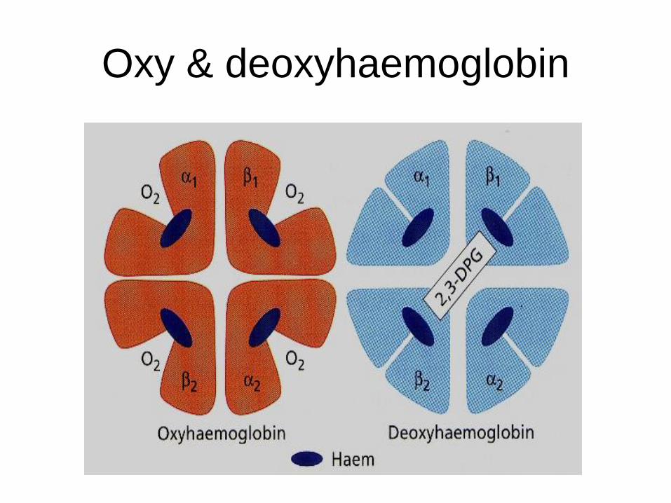
Oxy & deoxyhaemoglobin
Hemoglobin Structure and
Function• Acquired abnormal hgbs of clinical importance are
those that have been altered post- translationallyto produce hgbs that are unable to transport or deliver O2 and they include:
– Carboxyhgb - CO replaces O2 and binds 200X tighter than O2.
» This may be seen with heavy smokers
– Methgb - occurs when iron is oxidized to the +3 (ferric) state. In order for hgb to carry O2 the iron must be in the +2 (ferrous) state. In the body, normally~ 2% is formed and reducing systems prevent an increase beyond 2%.
» Increases above 2% can occur with the ingestion of strong oxidant drugs or
» As a result of enzyme deficiency.
Hemoglobin Structure and
Function» Methgb can be reduced by treatment with methylene
blue or ascorbic acid.
– Sulfhgb - occurs when the sulfur content of the blood
increases due to ingestion of sulfur containing drugs or to
chronic constipation. Unlike 1 and 2 this is an
irreversible change of hgb.
Erythrocyte destruction
• RBC destruction is normally the result of senescsence.– Each day ~ 1% of the RBCs are removed and
replaced.
– RBC aging is characterized by decreased glycolytic enzyme activity which leads to decreased energy production and subsequent loss of deformability and membrane integrity.
– 90% of aged RBC destruction is extravascular and occurs mainly in the phagocytic cells in the spleen, with a small amount occurring in the liver and bone marrow.
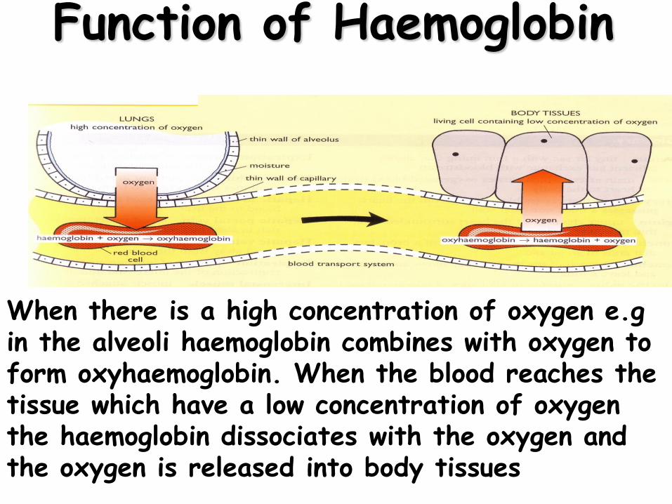
When there is a high concentration of oxygen e.g in the alveoli haemoglobin combines with oxygen to form oxyhaemoglobin. When the blood reaches the tissue which have a low concentration of oxygen the haemoglobin dissociates with the oxygen and the oxygen is released into body tissues
Function of Haemoglobin
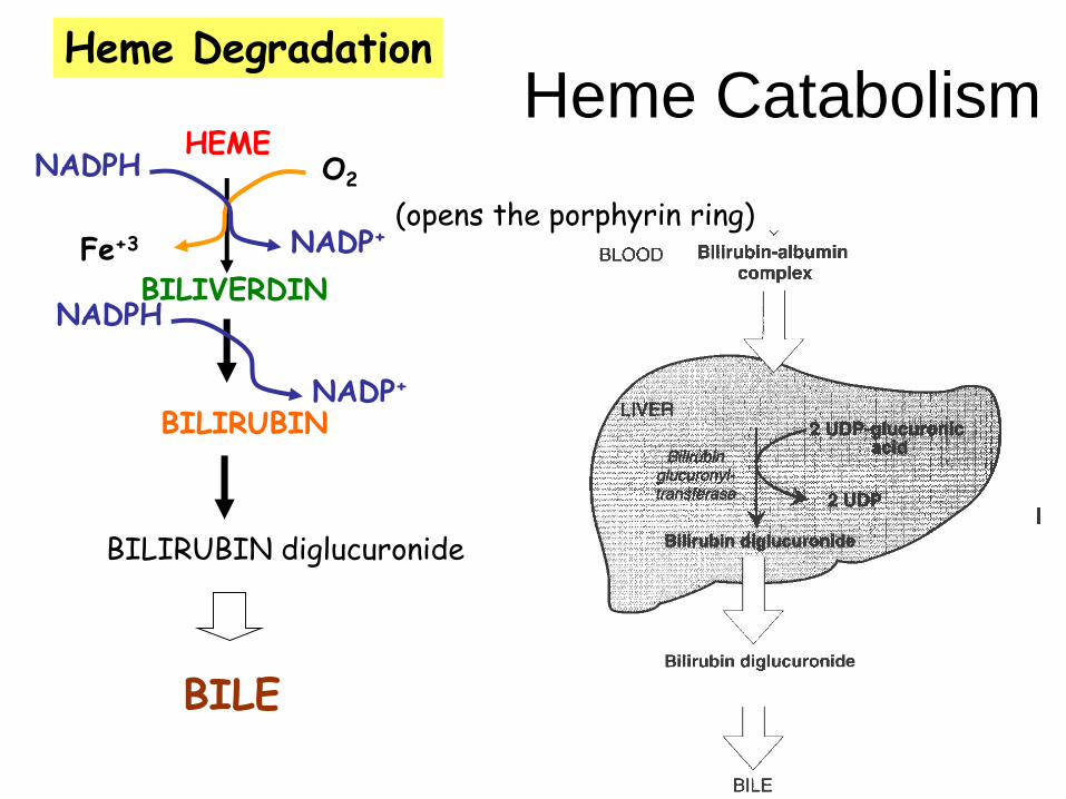
Heme CatabolismHeme Degradation
HEME
BILIVERDIN
O2
Fe+3
NADPH
NADP+(opens the porphyrin ring)
BILIRUBIN
NADPH
NADP+
BILIRUBIN diglucuronide
BILE
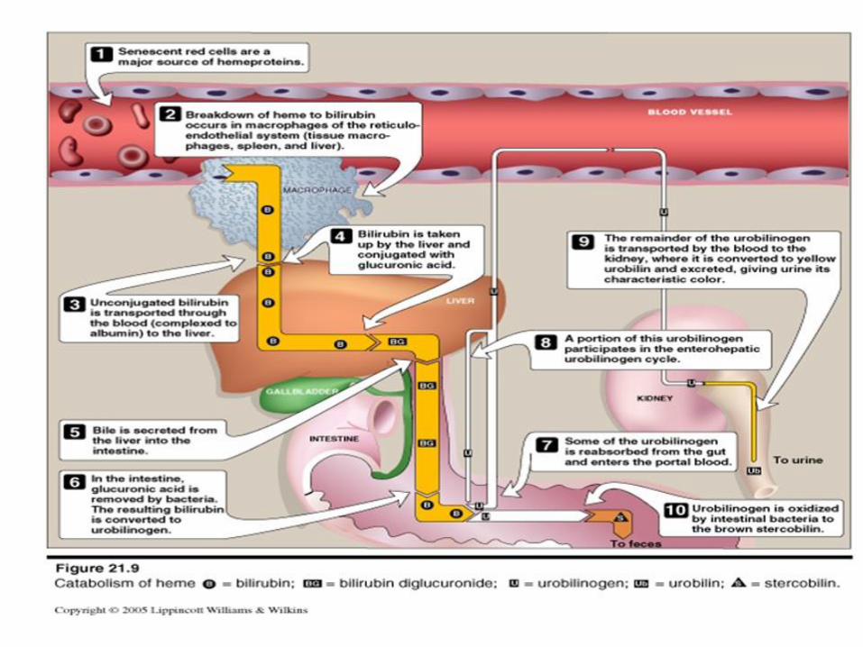
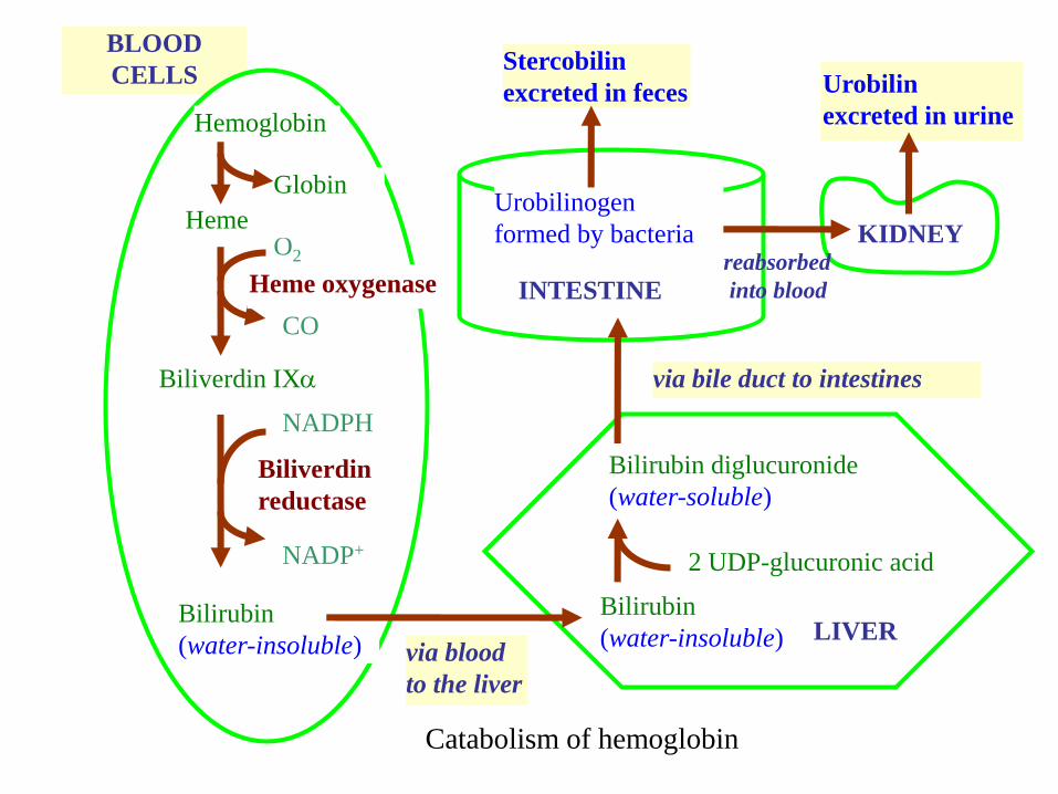
BLOOD
CELLS
LIVER
Bilirubin diglucuronide
(water-soluble)
2 UDP-glucuronic acid
via bile duct to intestines
Stercobilin
excreted in feces
Urobilinogen
formed by bacteria KIDNEY
Urobilin
excreted in urine
CO
Biliverdin IX
Heme oxygenase
O2
Bilirubin
(water-insoluble)
NADP+
NADPH
Biliverdin
reductase
Heme
Globin
Hemoglobin
reabsorbed
into blood
Bilirubin
(water-insoluble)via blood
to the liver
INTESTINE
Catabolism of hemoglobin
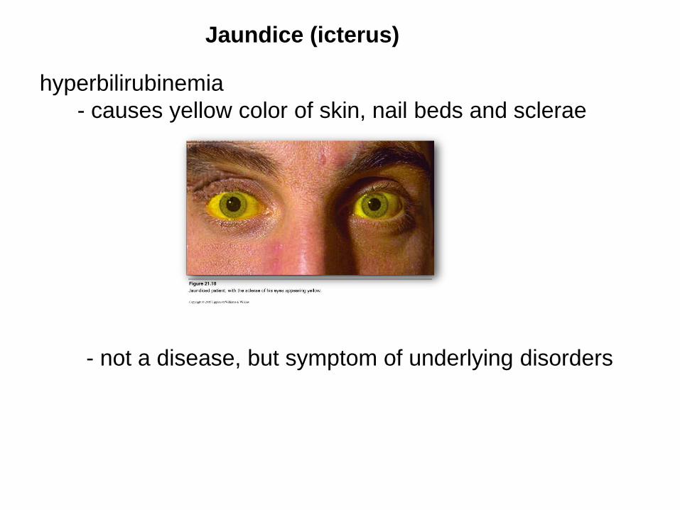
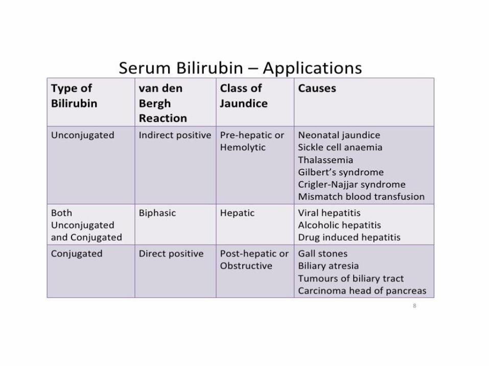
Jaundice (icterus)
hyperbilirubinemia
- causes yellow color of skin, nail beds and sclerae
- not a disease, but symptom of underlying disorders
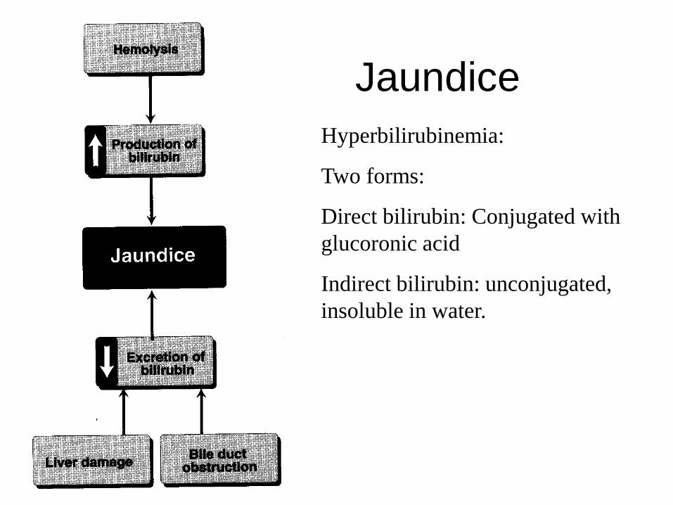
Jaundice
Hyperbilirubinemia:
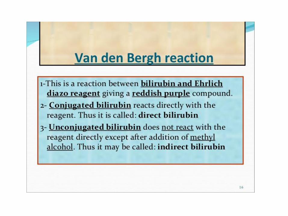
Two forms:
Direct bilirubin: Conjugated with
glucoronic acid
Indirect bilirubin: unconjugated,
insoluble in water.
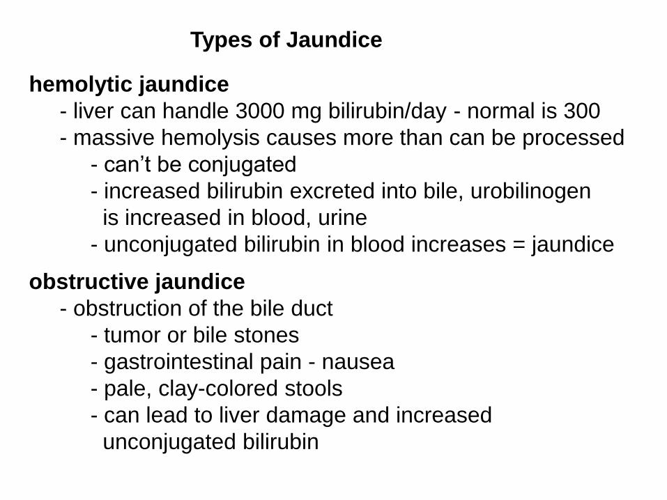
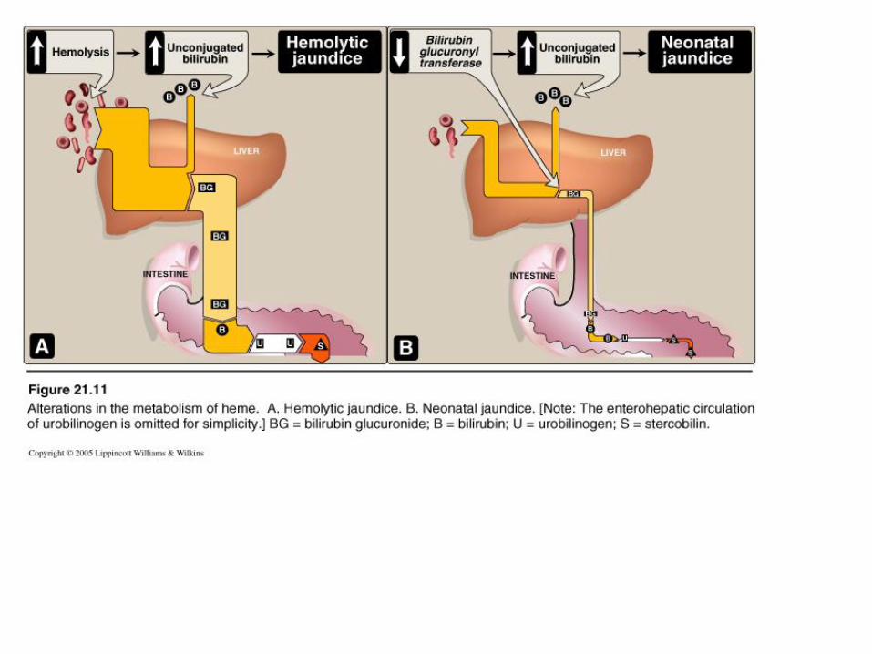
Types of Jaundice
hemolytic jaundice
- liver can handle 3000 mg bilirubin/day - normal is 300
- massive hemolysis causes more than can be processed
- can’t be conjugated
- increased bilirubin excreted into bile, urobilinogen
is increased in blood, urine
- unconjugated bilirubin in blood increases = jaundice
obstructive jaundice
- obstruction of the bile duct
- tumor or bile stones
- gastrointestinal pain - nausea
- pale, clay-colored stools
- can lead to liver damage and increased
unconjugated bilirubin
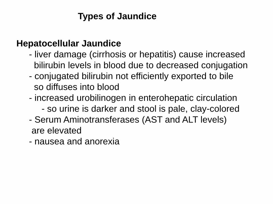
Hepatocellular Jaundice
- liver damage (cirrhosis or hepatitis) cause increased
bilirubin levels in blood due to decreased conjugation
- conjugated bilirubin not efficiently exported to bile
so diffuses into blood
- increased urobilinogen in enterohepatic circulation
- so urine is darker and stool is pale, clay-colored
- Serum Aminotransferases (AST and ALT levels)
are elevated
- nausea and anorexia
Types of Jaundice
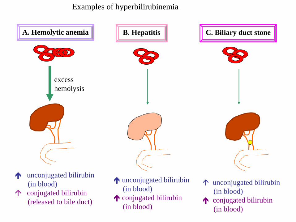
A. Hemolytic anemia
excess
hemolysis
unconjugated bilirubin
(in blood)
conjugated bilirubin
(released to bile duct)
B. Hepatitis
unconjugated bilirubin
(in blood)
conjugated bilirubin
(in blood)
C. Biliary duct stone
unconjugated bilirubin
(in blood)
conjugated bilirubin
(in blood)
Examples of hyperbilirubinemia
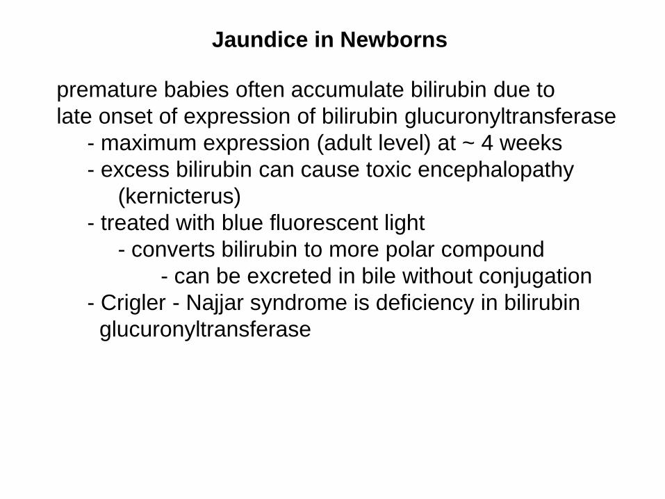
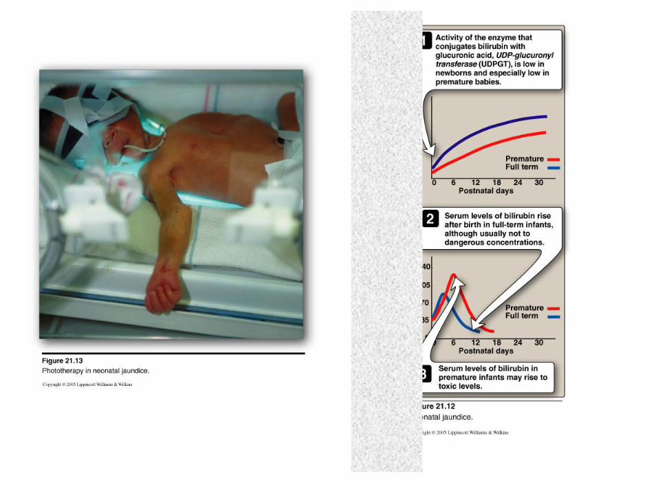
Jaundice in Newborns
premature babies often accumulate bilirubin due to
late onset of expression of bilirubin glucuronyltransferase
- maximum expression (adult level) at ~ 4 weeks
- excess bilirubin can cause toxic encephalopathy
(kernicterus)
- treated with blue fluorescent light
- converts bilirubin to more polar compound
- can be excreted in bile without conjugation
- Crigler - Najjar syndrome is deficiency in bilirubin
glucuronyltransferase