Embed Size (px)
Citation preview
Chapter 4
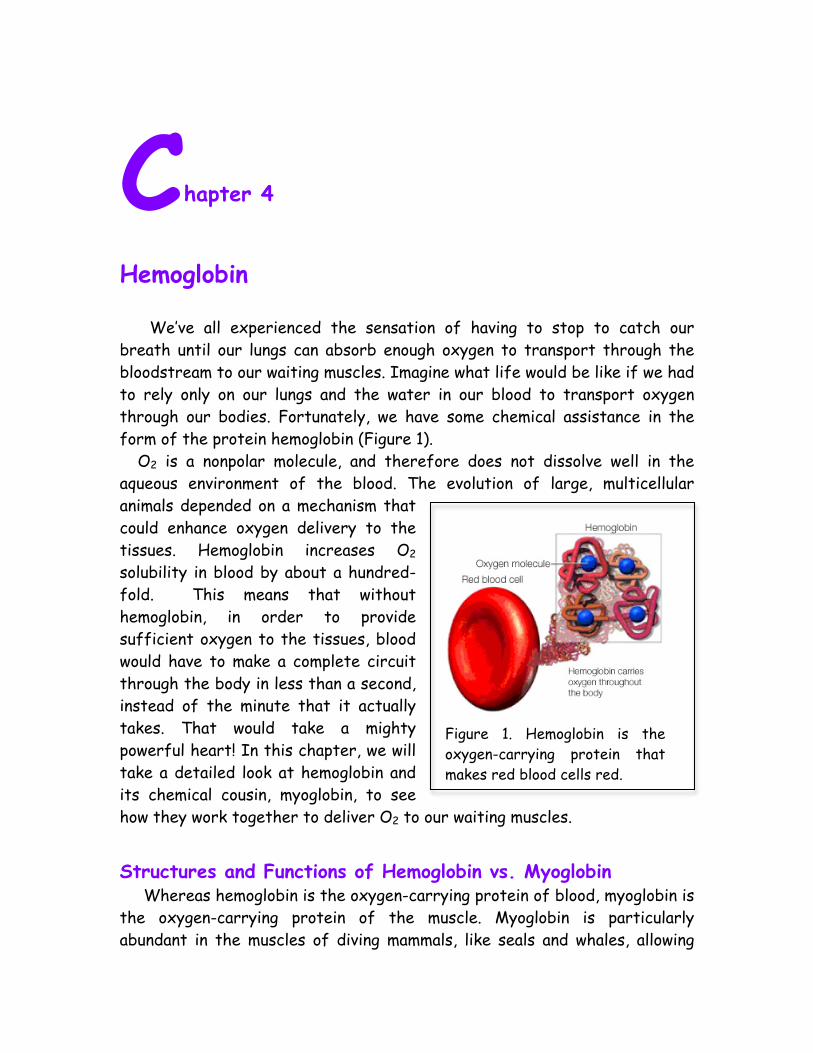
Hemoglobin We’ve all experienced the sensation of having to stop to catch our breath until our lungs can absorb enough oxygen to transport through the bloodstream to our waiting muscles. Imagine what life would be like if we had to rely only on our lungs and the water in our blood to transport oxygen through our bodies. Fortunately, we have some chemical assistance in the form of the protein hemoglobin (Figure 1). O2 is a nonpolar molecule, and therefore does not dissolve well in the aqueous environment of the blood. The evolution of large, multicellular animals depended on a mechanism that could enhance oxygen delivery to the tissues. Hemoglobin increases O2 solubility in blood by about a hundred-fold. This means that without hemoglobin, in order to provide sufficient oxygen to the tissues, blood would have to make a complete circuit through the body in less than a second, instead of the minute that it actually takes. That would take a mighty powerful heart! In this chapter, we will take a detailed look at hemoglobin and its chemical cousin, myoglobin, to see how they work together to deliver O2 to our waiting muscles.
Structures and Functions of Hemoglobin vs. Myoglobin Whereas hemoglobin is the oxygen-carrying protein of blood, myoglobin is the oxygen-carrying protein of the muscle. Myoglobin is particularly abundant in the muscles of diving mammals, like seals and whales, allowing
Figure 1. Hemoglobin is the oxygen-carrying protein that makes red blood cells red.
2
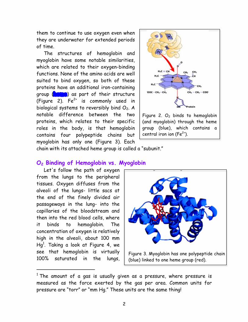
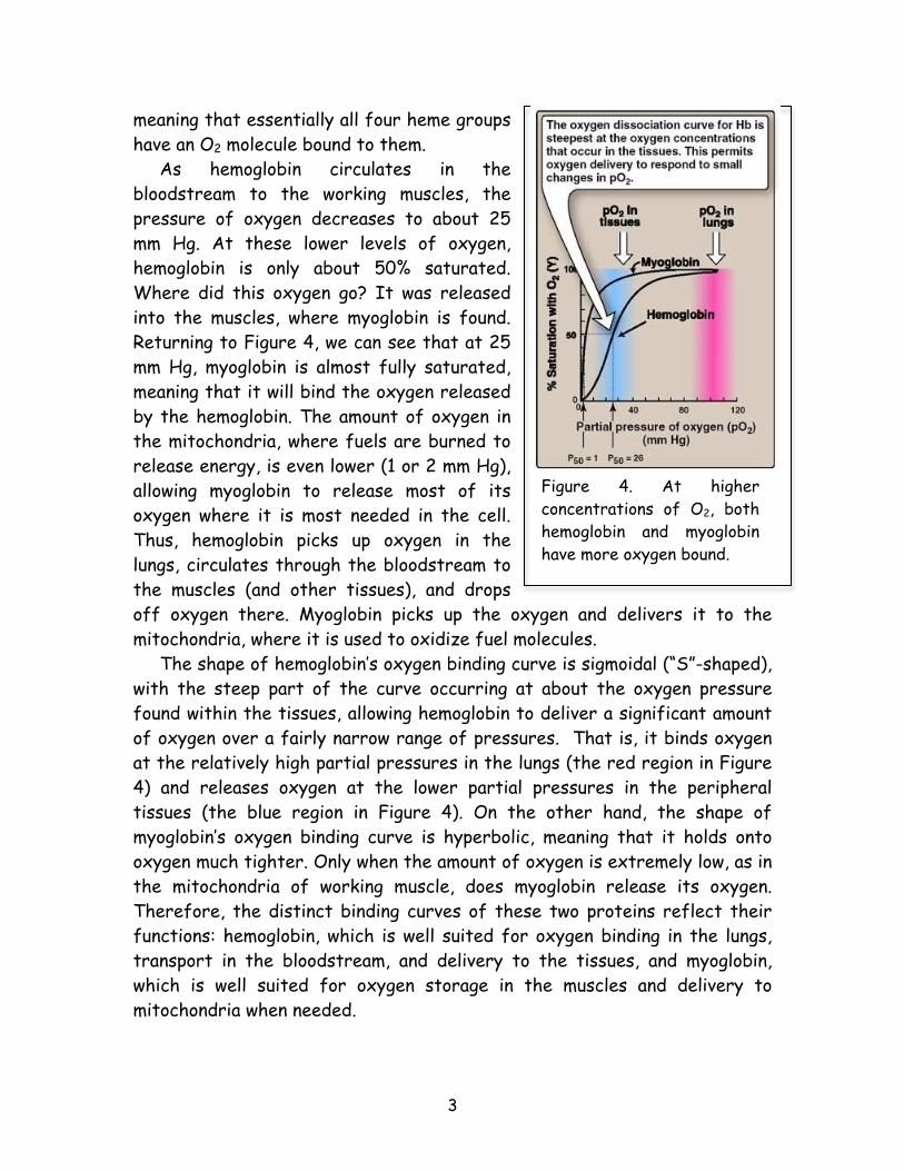
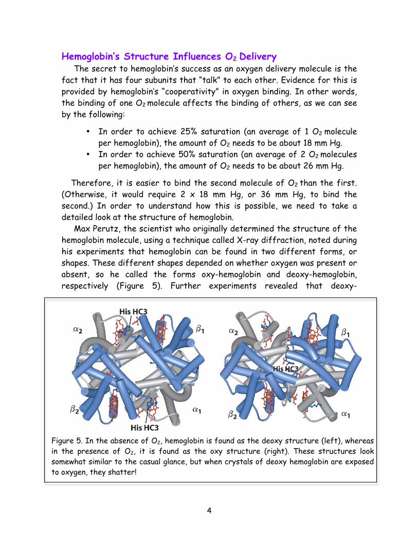
them to continue to use oxygen even when they are underwater for extended periods of time. The structures of hemoglobin and myoglobin have some notable similarities, which are related to their oxygen-binding functions. None of the amino acids are well suited to bind oxygen, so both of these proteins have an additional iron-containing group (heme) as part of their structure (Figure 2). Fe2+ is commonly used in biological systems to reversibly bind O2. A notable difference between the two proteins, which relates to their specific roles in the body, is that hemoglobin contains four polypeptide chains but myoglobin has only one (Figure 3). Each chain with its attached heme group is called a “subunit.” O2 Binding of Hemoglobin vs. Myoglobin Let's follow the path of oxygen from the lungs to the peripheral tissues. Oxygen diffuses from the alveoli of the lungs- little sacs at the end of the finely divided air passageways in the lung- into the capillaries of the bloodstream and then into the red blood cells, where it binds to hemoglobin. The concentration of oxygen is relatively high in the alveoli, about 100 mm Hg1. Taking a look at Figure 4, we see that hemoglobin is virtually 100% saturated in the lungs,
1 The amount of a gas is usually given as a pressure, where pressure is measured as the force exerted by the gas per area. Common units for pressure are “torr” or “mm Hg.” These units are the same thing!
Figure 2. O2 binds to hemoglobin (and myoglobin) through the heme group (blue), which contains a central iron ion (Fe2+).
Figure 3. Myoglobin has one polypeptide chain (blue) linked to one heme group (red).
3
meaning that essentially all four heme groups have an O2 molecule bound to them. As hemoglobin circulates in the bloodstream to the working muscles, the pressure of oxygen decreases to about 25 mm Hg. At these lower levels of oxygen, hemoglobin is only about 50% saturated. Where did this oxygen go? It was released into the muscles, where myoglobin is found. Returning to Figure 4, we can see that at 25 mm Hg, myoglobin is almost fully saturated, meaning that it will bind the oxygen released by the hemoglobin. The amount of oxygen in the mitochondria, where fuels are burned to release energy, is even lower (1 or 2 mm Hg), allowing myoglobin to release most of its oxygen where it is most needed in the cell. Thus, hemoglobin picks up oxygen in the lungs, circulates through the bloodstream to the muscles (and other tissues), and drops off oxygen there. Myoglobin picks up the oxygen and delivers it to the mitochondria, where it is used to oxidize fuel molecules. The shape of hemoglobin’s oxygen binding curve is sigmoidal (“S”-shaped), with the steep part of the curve occurring at about the oxygen pressure found within the tissues, allowing hemoglobin to deliver a significant amount of oxygen over a fairly narrow range of pressures. That is, it binds oxygen at the relatively high partial pressures in the lungs (the red region in Figure 4) and releases oxygen at the lower partial pressures in the peripheral tissues (the blue region in Figure 4). On the other hand, the shape of myoglobin’s oxygen binding curve is hyperbolic, meaning that it holds onto oxygen much tighter. Only when the amount of oxygen is extremely low, as in the mitochondria of working muscle, does myoglobin release its oxygen. Therefore, the distinct binding curves of these two proteins reflect their functions: hemoglobin, which is well suited for oxygen binding in the lungs, transport in the bloodstream, and delivery to the tissues, and myoglobin, which is well suited for oxygen storage in the muscles and delivery to mitochondria when needed.
Figure 4. At higher concentrations of O2, both hemoglobin and myoglobin have more oxygen bound.
4
Hemoglobin’s Structure Influences O2 Delivery The secret to hemoglobin’s success as an oxygen delivery molecule is the fact that it has four subunits that “talk” to each other. Evidence for this is provided by hemoglobin’s “cooperativity” in oxygen binding. In other words, the binding of one O2 molecule affects the binding of others, as we can see by the following:
• In order to achieve 25% saturation (an average of 1 O2 molecule per hemoglobin), the amount of O2 needs to be about 18 mm Hg.
• In order to achieve 50% saturation (an average of 2 O2 molecules per hemoglobin), the amount of O2 needs to be about 26 mm Hg.
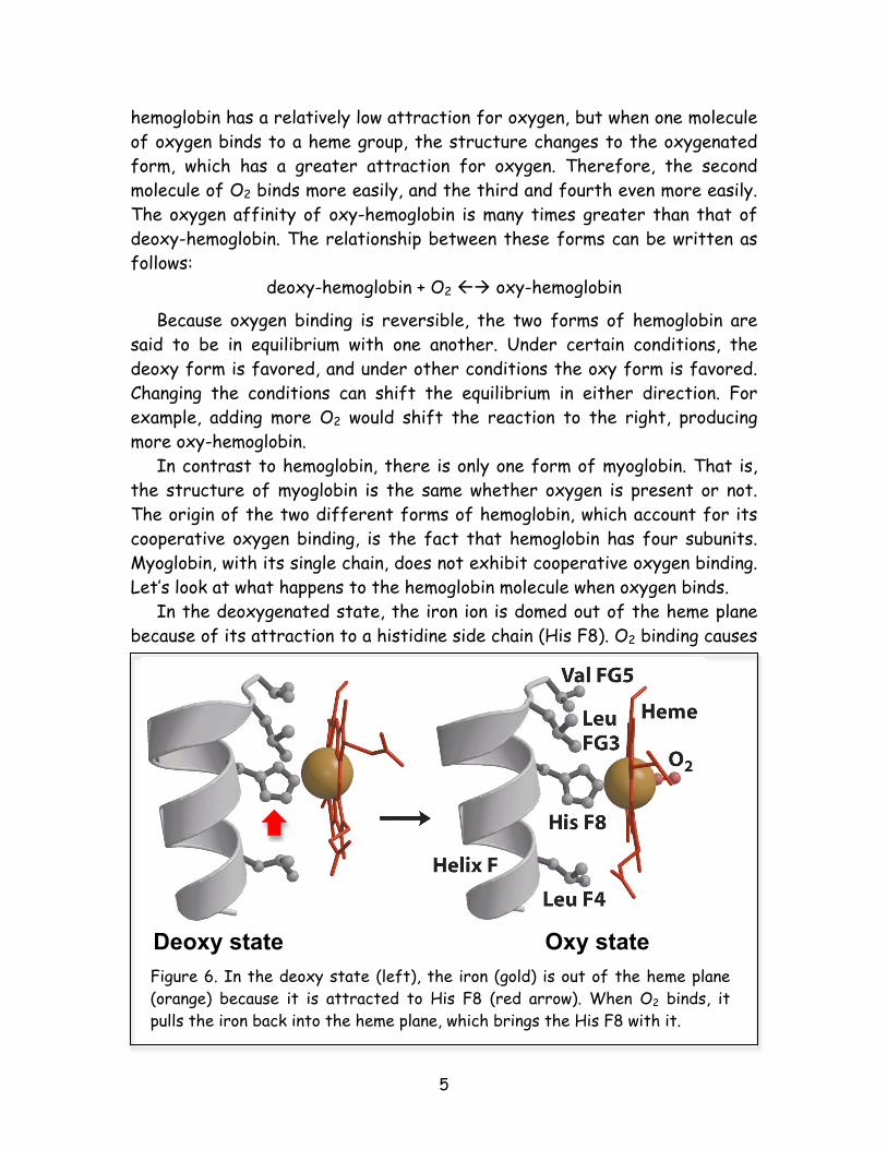
Therefore, it is easier to bind the second molecule of O2 than the first. (Otherwise, it would require 2 x 18 mm Hg, or 36 mm Hg, to bind the second.) In order to understand how this is possible, we need to take a detailed look at the structure of hemoglobin. Max Perutz, the scientist who originally determined the structure of the hemoglobin molecule, using a technique called X-ray diffraction, noted during his experiments that hemoglobin can be found in two different forms, or shapes. These different shapes depended on whether oxygen was present or absent, so he called the forms oxy-hemoglobin and deoxy-hemoglobin, respectively (Figure 5). Further experiments revealed that deoxy-
Figure 5. In the absence of O2, hemoglobin is found as the deoxy structure (left), whereas in the presence of O2, it is found as the oxy structure (right). These structures look somewhat similar to the casual glance, but when crystals of deoxy hemoglobin are exposed to oxygen, they shatter!
5
hemoglobin has a relatively low attraction for oxygen, but when one molecule of oxygen binds to a heme group, the structure changes to the oxygenated form, which has a greater attraction for oxygen. Therefore, the second molecule of O2 binds more easily, and the third and fourth even more easily. The oxygen affinity of oxy-hemoglobin is many times greater than that of deoxy-hemoglobin. The relationship between these forms can be written as follows:
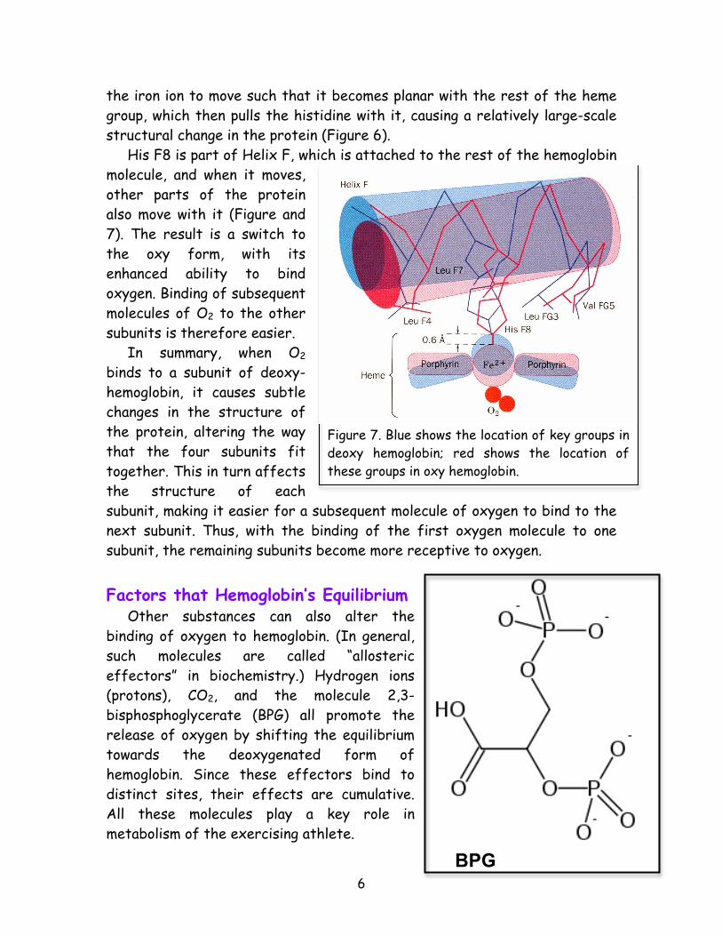
deoxy-hemoglobin + O2 oxy-hemoglobin Because oxygen binding is reversible, the two forms of hemoglobin are said to be in equilibrium with one another. Under certain conditions, the deoxy form is favored, and under other conditions the oxy form is favored. Changing the conditions can shift the equilibrium in either direction. For example, adding more O2 would shift the reaction to the right, producing more oxy-hemoglobin. In contrast to hemoglobin, there is only one form of myoglobin. That is, the structure of myoglobin is the same whether oxygen is present or not. The origin of the two different forms of hemoglobin, which account for its cooperative oxygen binding, is the fact that hemoglobin has four subunits. Myoglobin, with its single chain, does not exhibit cooperative oxygen binding. Let’s look at what happens to the hemoglobin molecule when oxygen binds. In the deoxygenated state, the iron ion is domed out of the heme plane because of its attraction to a histidine side chain (His F8). O2 binding causes
Deoxy state Oxy state Figure 6. In the deoxy state (left), the iron (gold) is out of the heme plane (orange) because it is attracted to His F8 (red arrow). When O2 binds, it pulls the iron back into the heme plane, which brings the His F8 with it.
6
the iron ion to move such that it becomes planar with the rest of the heme group, which then pulls the histidine with it, causing a relatively large-scale structural change in the protein (Figure 6). His F8 is part of Helix F, which is attached to the rest of the hemoglobin molecule, and when it moves, other parts of the protein also move with it (Figure and 7). The result is a switch to the oxy form, with its enhanced ability to bind oxygen. Binding of subsequent molecules of O2 to the other subunits is therefore easier. In summary, when O2 binds to a subunit of deoxy-hemoglobin, it causes subtle changes in the structure of the protein, altering the way that the four subunits fit together. This in turn affects the structure of each subunit, making it easier for a subsequent molecule of oxygen to bind to the next subunit. Thus, with the binding of the first oxygen molecule to one subunit, the remaining subunits become more receptive to oxygen. Factors that Hemoglobin’s Equilibrium Other substances can also alter the binding of oxygen to hemoglobin. (In general, such molecules are called “allosteric effectors” in biochemistry.) Hydrogen ions (protons), CO2, and the molecule 2,3-bisphosphoglycerate (BPG) all promote the release of oxygen by shifting the equilibrium towards the deoxygenated form of hemoglobin. Since these effectors bind to distinct sites, their effects are cumulative. All these molecules play a key role in metabolism of the exercising athlete.
BPG
Figure 7. Blue shows the location of key groups in deoxy hemoglobin; red shows the location of these groups in oxy hemoglobin.
7
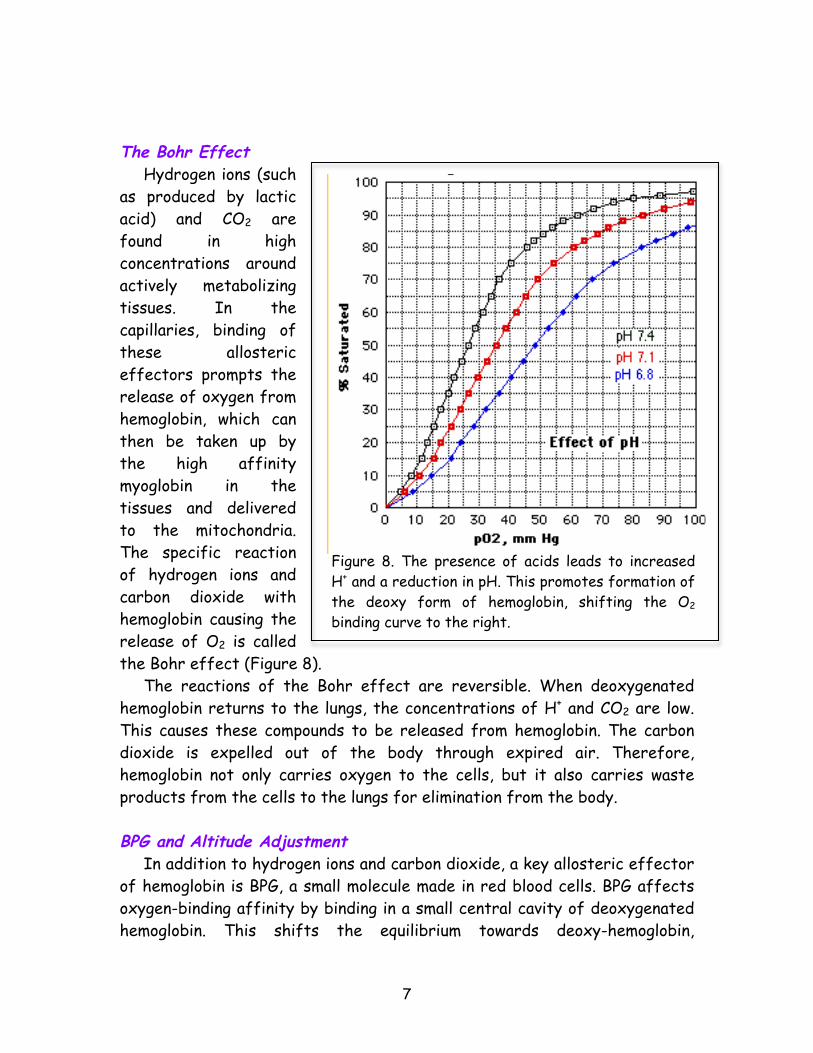
The Bohr Effect Hydrogen ions (such as produced by lactic acid) and CO2 are found in high concentrations around actively metabolizing tissues. In the capillaries, binding of these allosteric effectors prompts the release of oxygen from hemoglobin, which can then be taken up by the high affinity myoglobin in the tissues and delivered to the mitochondria. The specific reaction of hydrogen ions and carbon dioxide with hemoglobin causing the release of O2 is called the Bohr effect (Figure 8). The reactions of the Bohr effect are reversible. When deoxygenated hemoglobin returns to the lungs, the concentrations of H+ and CO2 are low. This causes these compounds to be released from hemoglobin. The carbon dioxide is expelled out of the body through expired air. Therefore, hemoglobin not only carries oxygen to the cells, but it also carries waste products from the cells to the lungs for elimination from the body. BPG and Altitude Adjustment In addition to hydrogen ions and carbon dioxide, a key allosteric effector of hemoglobin is BPG, a small molecule made in red blood cells. BPG affects oxygen-binding affinity by binding in a small central cavity of deoxygenated hemoglobin. This shifts the equilibrium towards deoxy-hemoglobin,
Figure 8. The presence of acids leads to increased H+ and a reduction in pH. This promotes formation of the deoxy form of hemoglobin, shifting the O2 binding curve to the right.
8
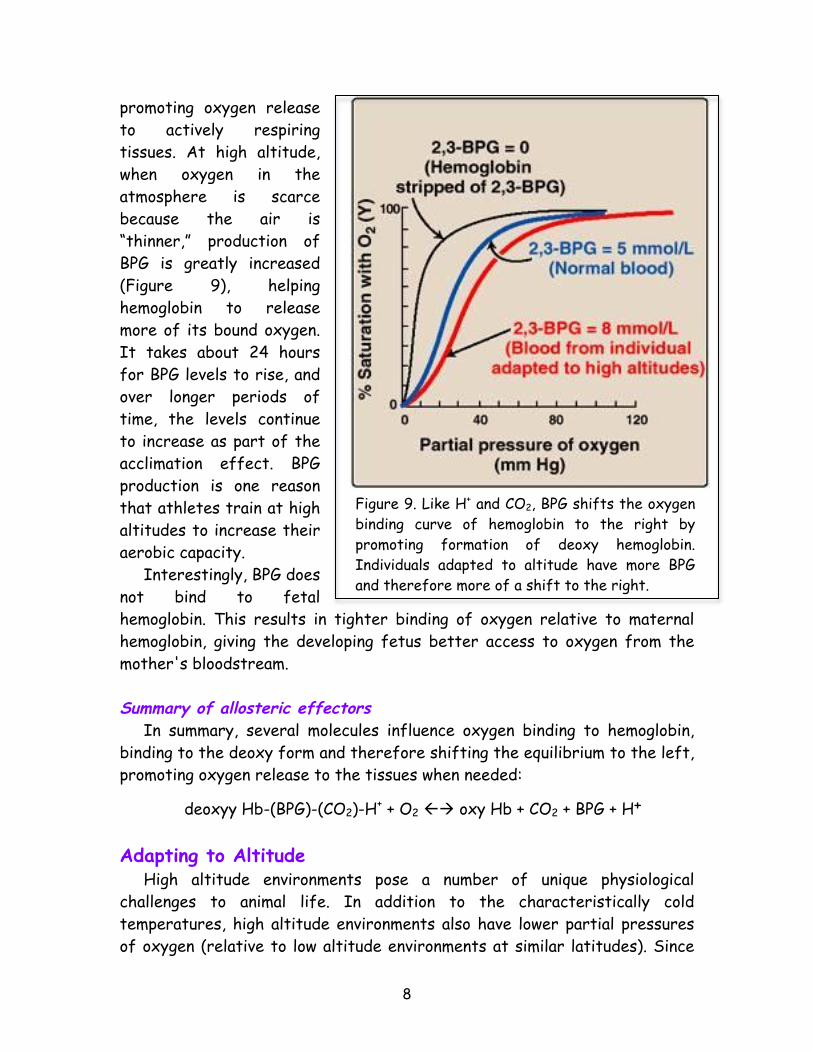
promoting oxygen release to actively respiring tissues. At high altitude, when oxygen in the atmosphere is scarce because the air is “thinner,” production of BPG is greatly increased (Figure 9), helping hemoglobin to release more of its bound oxygen. It takes about 24 hours for BPG levels to rise, and over longer periods of time, the levels continue to increase as part of the acclimation effect. BPG production is one reason that athletes train at high altitudes to increase their aerobic capacity. Interestingly, BPG does not bind to fetal hemoglobin. This results in tighter binding of oxygen relative to maternal hemoglobin, giving the developing fetus better access to oxygen from the mother's bloodstream. Summary of allosteric effectors In summary, several molecules influence oxygen binding to hemoglobin, binding to the deoxy form and therefore shifting the equilibrium to the left, promoting oxygen release to the tissues when needed:
deoxyy Hb-(BPG)-(CO2)-H+ + O2 oxy Hb + CO2 + BPG + H+ Adapting to Altitude High altitude environments pose a number of unique physiological challenges to animal life. In addition to the characteristically cold temperatures, high altitude environments also have lower partial pressures of oxygen (relative to low altitude environments at similar latitudes). Since
Figure 9. Like H+ and CO2, BPG shifts the oxygen binding curve of hemoglobin to the right by promoting formation of deoxy hemoglobin. Individuals adapted to altitude have more BPG and therefore more of a shift to the right.
9
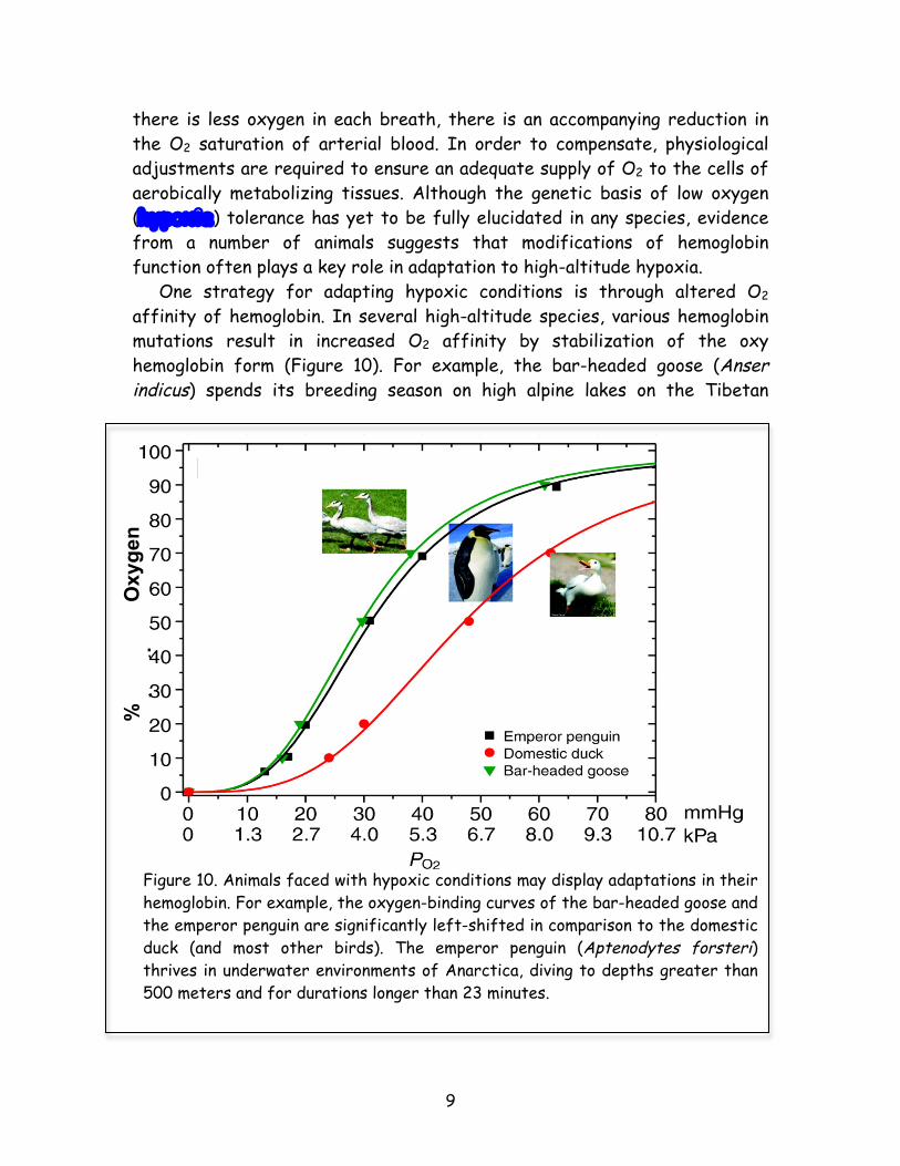
there is less oxygen in each breath, there is an accompanying reduction in the O2 saturation of arterial blood. In order to compensate, physiological adjustments are required to ensure an adequate supply of O2 to the cells of aerobically metabolizing tissues. Although the genetic basis of low oxygen (hypoxia) tolerance has yet to be fully elucidated in any species, evidence from a number of animals suggests that modifications of hemoglobin function often plays a key role in adaptation to high-altitude hypoxia. One strategy for adapting hypoxic conditions is through altered O2 affinity of hemoglobin. In several high-altitude species, various hemoglobin mutations result in increased O2 affinity by stabilization of the oxy hemoglobin form (Figure 10). For example, the bar-headed goose (Anser indicus) spends its breeding season on high alpine lakes on the Tibetan
%
Oxy
gen
satu
ratio
n
Figure 10. Animals faced with hypoxic conditions may display adaptations in their hemoglobin. For example, the oxygen-binding curves of the bar-headed goose and the emperor penguin are significantly left-shifted in comparison to the domestic duck (and most other birds). The emperor penguin (Aptenodytes forsteri) thrives in underwater environments of Anarctica, diving to depths greater than 500 meters and for durations longer than 23 minutes.
10
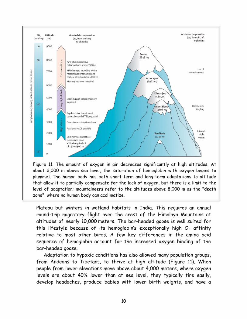
Plateau but winters in wetland habitats in India. This requires an annual round-trip migratory flight over the crest of the Himalaya Mountains at altitudes of nearly 10,000 meters. The bar-headed goose is well suited for this lifestyle because of its hemoglobin’s exceptionally high O2 affinity relative to most other birds. A few key differences in the amino acid sequence of hemoglobin account for the increased oxygen binding of the bar-headed goose. Adaptation to hypoxic conditions has also allowed many population groups, from Andeans to Tibetans, to thrive at high altitude (Figure 11). When people from lower elevations move above about 4,000 meters, where oxygen levels are about 40% lower than at sea level, they typically tire easily, develop headaches, produce babies with lower birth weights, and have a
Figure 11. The amount of oxygen in air decreases significantly at high altitudes. At about 2,000 m above sea level, the saturation of hemoglobin with oxygen begins to plummet. The human body has both short-term and long-term adaptations to altitude that allow it to partially compensate for the lack of oxygen, but there is a limit to the level of adaptation: mountaineers refer to the altitudes above 8,000 m as the "death zone", where no human body can acclimatize.
11
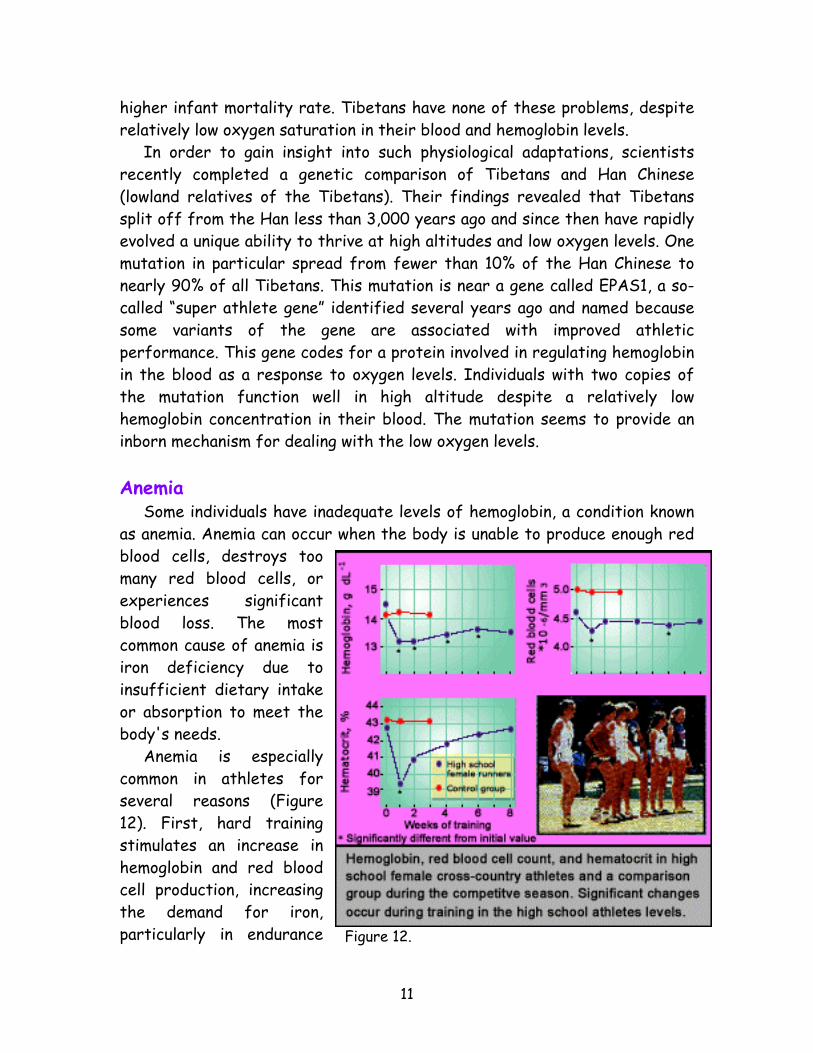
higher infant mortality rate. Tibetans have none of these problems, despite relatively low oxygen saturation in their blood and hemoglobin levels. In order to gain insight into such physiological adaptations, scientists recently completed a genetic comparison of Tibetans and Han Chinese (lowland relatives of the Tibetans). Their findings revealed that Tibetans split off from the Han less than 3,000 years ago and since then have rapidly evolved a unique ability to thrive at high altitudes and low oxygen levels. One mutation in particular spread from fewer than 10% of the Han Chinese to nearly 90% of all Tibetans. This mutation is near a gene called EPAS1, a so-called “super athlete gene” identified several years ago and named because some variants of the gene are associated with improved athletic performance. This gene codes for a protein involved in regulating hemoglobin in the blood as a response to oxygen levels. Individuals with two copies of the mutation function well in high altitude despite a relatively low hemoglobin concentration in their blood. The mutation seems to provide an inborn mechanism for dealing with the low oxygen levels. Anemia Some individuals have inadequate levels of hemoglobin, a condition known as anemia. Anemia can occur when the body is unable to produce enough red blood cells, destroys too many red blood cells, or experiences significant blood loss. The most common cause of anemia is iron deficiency due to insufficient dietary intake or absorption to meet the body's needs. Anemia is especially common in athletes for several reasons (Figure 12). First, hard training stimulates an increase in hemoglobin and red blood cell production, increasing the demand for iron, particularly in endurance Figure 12.
12
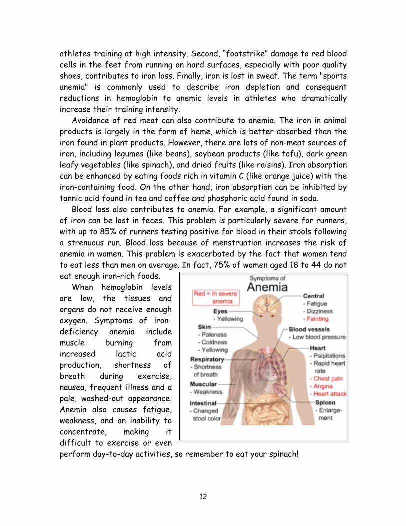
athletes training at high intensity. Second, “footstrike” damage to red blood cells in the feet from running on hard surfaces, especially with poor quality shoes, contributes to iron loss. Finally, iron is lost in sweat. The term "sports anemia" is commonly used to describe iron depletion and consequent reductions in hemoglobin to anemic levels in athletes who dramatically increase their training intensity. Avoidance of red meat can also contribute to anemia. The iron in animal products is largely in the form of heme, which is better absorbed than the iron found in plant products. However, there are lots of non-meat sources of iron, including legumes (like beans), soybean products (like tofu), dark green leafy vegetables (like spinach), and dried fruits (like raisins). Iron absorption can be enhanced by eating foods rich in vitamin C (like orange juice) with the iron-containing food. On the other hand, iron absorption can be inhibited by tannic acid found in tea and coffee and phosphoric acid found in soda. Blood loss also contributes to anemia. For example, a significant amount of iron can be lost in feces. This problem is particularly severe for runners, with up to 85% of runners testing positive for blood in their stools following a strenuous run. Blood loss because of menstruation increases the risk of anemia in women. This problem is exacerbated by the fact that women tend to eat less than men on average. In fact, 75% of women aged 18 to 44 do not eat enough iron-rich foods. When hemoglobin levels are low, the tissues and organs do not receive enough oxygen. Symptoms of iron-deficiency anemia include muscle burning from increased lactic acid production, shortness of breath during exercise, nausea, frequent illness and a pale, washed-out appearance. Anemia also causes fatigue, weakness, and an inability to concentrate, making it difficult to exercise or even perform day-to-day activities, so remember to eat your spinach!