Embed Size (px)
Citation preview
REVIEW ARTICLEpublished: 07 May 2012
doi: 10.3389/fphar.2012.00081
Heme oxygenase, inflammation, and fibrosis: the good,the bad, and the ugly?Ditte M. S. Lundvig1, Stephan Immenschuh2* and Frank A. D.T. G. Wagener 1*
1 Department of Orthodontics and Craniofacial Biology, Nijmegen Centre for Molecular Life Sciences, Radboud University Nijmegen Medical Centre, Nijmegen,Netherlands
2 Institute for Transfusion Medicine, Hannover Medical School, Hannover, Germany
Edited by:
Mahin D. Maines, University ofRochester School of Medicine, USA
Reviewed by:
Roberto Motterlini, Institut Nationalde la Santé et de la RechercheMédicale, FranceRene Schmidt, Freiburg UniversityMedical Center, Germany
*Correspondence:
Stephan Immenschuh, Institute forTransfusion Medicine, HannoverMedical School, Carl Neuberg-Street1, 30625 Hannover, Germany.e-mail: [email protected];Frank A. D. T. G. Wagener ,Department of Orthodontics andCraniofacial Biology, Nijmegen Centrefor Molecular Life Sciences, RadboudUniversity Nijmegen Medical Centre,PO Box 9101, 6500 HB Nijmegen,Netherlands.e-mail: [email protected]
Upon injury, prolonged inflammation and oxidative stress may cause pathological woundhealing and fibrosis, leading to formation of excessive scar tissue. Fibrogenesis can occurin most organs and tissues and may ultimately lead to organ dysfunction and failure. Theunderlying mechanisms of pathological wound healing still remain unclear, and are con-sidered to be multifactorial, but so far, no efficient anti-fibrotic therapies exist. Extra- andintracellular levels of free heme may be increased in a variety of pathological conditionsdue to release from hemoproteins. Free heme possesses pro-inflammatory and oxidativeproperties, and may act as a danger signal. Effects of free heme may be counteracted byheme-binding proteins or by heme degradation. Heme is degraded by heme oxygenase(HO) that exists as two isoforms: inducible HO-1 and constitutively expressed HO-2. HOgenerates the effector molecules biliverdin/bilirubin, carbon monoxide, and free iron/ferritin.HO deficiency in mouse and man leads to exaggerated inflammation following mild insults,and accumulating epidemiological and preclinical studies support the widely recognizednotion of the cytoprotective, anti-oxidative, and anti-inflammatory effects of the activity ofthe HO system and its effector molecules. In this review, we address the potential effects oftargeted HO-1 induction or administration of HO-effector molecules as therapeutic targetsin fibrotic conditions to counteract inflammatory and oxidative insults.This is exemplified byvarious clinically relevant conditions, such as hypertrophic scarring, chronic inflammatoryliver disease, chronic pancreatitis, and chronic graft rejection in transplantation.
Keywords: heme oxygenase, heme, HO-effector molecules, fibrosis, therapy
INTRODUCTIONTissue injury can occur after physical, chemical, or infectiousinsults, such as burns, trauma, and toxins. Wound healing is adynamic and complex process involving the coordinated action ofdifferent cell types. The quality of the wound healing process isdependent on different factors, including the severity of the insultand the inflammatory and redox status of the wound. Pathologicalwound healing can lead to two extreme cases: chronic, non-healingwounds or fibrosis, which ultimately may lead to excessive scar-ring. Both types of complications related to pathological woundhealing have major impact on patient quality of life in terms ofaesthetical and functional problems.
Numerous clinical and experimental observations have demon-strated that prolonged inflammation and oxidative stress maycause pathological wound healing and the development of fibrosis.However, the underlying mechanisms still remain largely unclear.Inflammation is a complex response of the innate immune systemin vascularized tissues initiated to protect the organism againstinvading pathogens as well as to restore tissue homeostasis at thewound site.
WOUND HEALING AND FIBROSIS – WHEN GOOD TURNSBADThe primary goal of tissue repair is to restore tissue integrityand homeostasis. Hereto, wounding initiates a complex cascade
of events to stop blood loss, to eliminate invading pathogens, and,ultimately, to promote tissue integrity and homeostasis (Chettibiet al., 1999; Hunt et al., 2000).
Wound healing occurs in three distinct, but overlapping phases:(1) inflammation, (2) regeneration, and (3) remodeling, andinvolves a well-coordinated sequence of cellular responses.
The inflammatory phase is preceded by blood clot formationand coagulation to stop blood loss and to reduce pathogen inva-sion. This fibrin-containing clot also serves as an early provisionalextracellular matrix (ECM) to provide structural support for cel-lular attachment and proliferation. Coagulation also triggers theproduction of pro-inflammatory agents, and activation of thecomplement system. This leads to increased blood vessel perme-ability, chemokine expression, vascular adhesion molecule expres-sion, and recruitment of immune cells. At the wound site, granulo-cytes and macrophages are pivotal for the innate immune system-mediated elimination of invading pathogens through the gener-ation of reactive oxygen species (ROS; Fialkow et al., 2007), andthe production of pro-inflammatory chemokines and cytokines(Ryan et al., 2004).
The temporal and spatial pattern of inflammation resolution iscrucial for proper wound healing and is characterized by reducedlevels of pro-inflammatory adhesion molecules and cytokinesand a decreasing number of inflammatory cells at the site ofinjury.
www.frontiersin.org May 2012 | Volume 3 | Article 81 | 1
Lundvig et al. The heme oxygenase system in anti-fibrotic therapy
This is followed by the initiation of the regenerative phasethat mediates re-epithelialization, neovascularization, and woundclosure. In particular, fibroblasts produce and deposit ECM com-ponents to substitute the provisional matrix. Also, recruitedkeratinocytes and endothelial cells are crucial to this process.
When the wound area is fully re-epithelialized, remodelingtakes over from regeneration. Fibroblasts differentiate into myofi-broblasts causing wound contraction and ECM reorganization bysynthesis and deposition of ECM components and by providingmatrix metalloproteinases (MMPs) and tissue inhibitors of MMPs(TIMPs), which ultimately leads to wound closure. At the end ofthis phase, myofibroblasts present in the granulation tissue disap-pear by apoptosis, producing a rather acellular scar. It is importantto realize that scar formation is a natural part of the wound healingprocess, as normal scar tissue, which mainly consists of connec-tive tissue, represents a stable restoration of the skin (for a recentreview, see Guo and Dipietro, 2010).
Prolonged inflammatory conditions accompanied by oxida-tive stress may interfere with the normal wound healing process,leading to an extended presence of myofibroblasts and excessivescar formation, a process known as fibrosis. Fibrosis is not onlyrestricted to dermal wound healing, but also occurs in palatal tis-sue, lungs, heart, liver, intestine, and joints, and causes major med-ical problems ranging from disfigurement to progressive disabilityand even death.
Despite the fact that disorders that are associated with fibro-sis account for up to 45% of deaths in the developed world, nosuccessful anti-fibrotic therapies exist (Wynn, 2008).
Extensive research has shown that the heme–heme oxy-genase (HO) system is closely involved in the regulation ofvarious (patho)physiological processes, in particular in cellu-lar adaptation to oxidative stress, and the anti-inflammatoryresponse.
In this review, we address the role of heme, HO-1 and HO-effector molecules in inflammation and fibrosis with an empha-sis on hypertrophic scarring, chronic inflammatory liver disease,chronic pancreatitis, and transplantation complications.
INFLAMMATION AND SCARRING – WHEN BAD TURNS UGLYA prolonged inflammatory phase is considered a major cause offibrosis and excessive scar formation. A clear link between inflam-mation and fibrosis has been revealed through embryonic studies.Mammalian embryos heal without scar formation, if wound heal-ing occurs without influx of inflammatory cells (Ashcroft et al.,1999), indicating that inflammatory cell recruitment to the woundsite is a prerequisite for scar formation. Wound healing stud-ies in different knockout and transgenic mouse models (Fathkeet al., 2004; Duffield et al., 2005; Oakley et al., 2005; Tabibiazaret al., 2006; Saito et al., 2008; Goren et al., 2009), athymic mice(Gawronska-Kozak et al., 2006), and in vivo antisense knockdownstudies (Mori et al., 2006, 2008; Mathew et al., 2007) further sup-port this notion. Also, an overwhelming amount of data haveshown that inflammatory cells emigrating from blood, such asgranulocytes, macrophages, and helper T cells, provide signalsthat promote granulation and fibrosis, including ROS, ECM depo-sition, and chemokines and cytokines (reviewed in Martin andLeibovich, 2005).
Clinical data demonstrate elevated inflammatory profiles inpatients with pathological scarring (Harty et al., 2003; Martin andLeibovich, 2005). Also, chronic wounds demonstrate impaired res-olution of the inflammatory phase, and thus remain in a chronicinflammatory state (Loots et al., 1998). Time is a critical factorfor abnormal scar formation, as long-lasting wound healing sig-nificantly increases the risk of hypertrophic or excessive scarring(Deitch et al., 1983).
The continuous presence of inflammatory mediators results inincreased secretion of growth factors, extending the proliferativephase via the prolonged presence of myofibroblasts (Singer andClark, 1999). This in turn causes excessive deposition of ECMcomponents, exaggerating the outcome of the proliferative phaseand contributing to hypertrophic scarring or fibrosis (Aarabi et al.,2007). Thus, the inflammatory cell-derived ROS generated duringthe early inflammatory phase must be tightly controlled by cellularanti-oxidative and anti-inflammatory systems, as impaired ROSclearance exacerbates wound healing (Frantz et al., 1993; Steilinget al., 1999).
The unifying hallmark of fibrotic disorders is abnormal andexcessive deposition of ECM components. As exemplified in thefollowing sections, the broad range of affected organs, the progres-sive and often irreversible nature of fibrosis, and the large numberof affected patients combined with ineffective treatment posesa challenge to the development of novel therapeutic approachestoward limiting fibrosis.
HYPERTROPHIC SCARRINGThe skin is the organ with the largest surface area in the body,functioning as a barrier against the external milieu, and is impor-tant in protecting the body against pathogens and dehydration.Other functions include temperature regulation, tactile sensation,and vitamin D synthesis.
Upon cutaneous injury, dermal wound healing is crucial forrestoration of the damaged skin barrier. In most cases, woundhealing will result in the formation of a visible scar. However,prolonged inflammation and high levels of oxidative stress (e.g.,due to infections) may result in excessive deposition of predomi-nantly collagen I (Sidgwick and Bayat, 2012), leading to fibrosisand excessive scarring that in skin is manifested as a hyper-trophic or a keloid scar. Both keloids and hypertrophic scars arecharacterized by excessive collagen deposition and are discoloredwith a rough, stiff appearance, but whereas hypertrophic scarsare confined to the original wound area, keloids overgrow theseboundaries. Hypertrophic scars may occur in persons of any ageand at any site, whereas keloids develop with higher risk in certainethnic groups (Brissett and Sherris, 2001). Almost all abnormalscars are associated with prolonged inflammation caused by for-eign body reaction, bacterial infections, tattoos, burns, injections,bites, cuts,vaccination, trauma, surgery,or infections. Importantly,immunological alterations have been reported in hypertrophicscars, including abnormal growth factor and cytokine levels (vander Veer et al., 2009).
Often patients report itching and pain as major issues besidesthe aesthetical and functional problems (Bock et al., 2006). Thesecomplications may require surgical corrections, which may thenlead to further scarring complications.
Frontiers in Pharmacology | Drug Metabolism and Transport May 2012 | Volume 3 | Article 81 | 2
Lundvig et al. The heme oxygenase system in anti-fibrotic therapy
FIBROSIS IN CHRONIC INFLAMMATORY LIVER DISEASE AND CHRONICPANCREATITISLiver and pancreas are two major abdominal organs which havespecific exocrine and endocrine functions within the gastrointesti-nal tract and in control of the body’s metabolic homeostasis.
The liver is not only a major regulator of systemic metab-olism but also has key functions in detoxification of endoge-nous and exogenous toxins. Hepatic fibrosis is mainly causedby chronic hepatitis B and C, alcoholic liver disease, and fattyliver disease/non-alcoholic steatohepatitis (extensively reviewed inHernandez-Gea and Friedman, 2011). Albeit from different ori-gins, these disorders have similar early pro-inflammatory features,which can progress into hepatic fibrosis and ultimately into endstage liver cirrhosis. Hepatic fibrosis can be regarded as a result ofthe hepatic wound healing response to repeated injury. If hepaticinjury sustains, failure of liver regeneration occurs and results infibrosis, in which activated hepatic stellate cells (HSCs) play a keyrole via excessive production of ECM components. More specifi-cally, HSC activation is the result of a complex sequence of eventssuch as secretion of pro-inflammatory cytokines by other hepaticcells such as liver tissue macrophages (Kupffer cells). Ultimately,hepatic fibrosis and cirrhosis lead to a replacement of healthyhepatic tissue with scars and regenerative nodules, causing lossof liver function.
The pancreas is a major gland organ and has crucial exocrinefunctions in the gastrointestinal digestive tract. In addition, it isa major endocrine organ and produces the glucose-regulatingpeptide hormones insulin and glucagon. Chronic pancreatitis isa progressive inflammatory disease (Witt et al., 2007), which ismost commonly caused by chronic alcoholism or is of idiopathicorigin. Similar to the liver, pancreatic stellate cells (PSCs) play akey role in pancreatic fibrogenesis (Omary et al., 2007). Normally,PSCs regulate the synthesis and degradation of ECM proteins and,thereby, the maintenance of the healthy tissue architecture. Uponpancreatic injury, PSCs differentiate into an activated phenotypewhich is secreting excessive amounts of ECM components andleads to the formation of fibrotic tissue (Omary et al., 2007). Per-sistent inflammation of the pancreas causes permanent structuraldeterioration with tissue fibrosis and ductal obstruction, and sub-sequently irreversible, declined exocrine and endocrine function(pancreatic insufficiency).
FIBROSIS ASSOCIATED WITH GRAFT REJECTIONDuring transplantation, cells, tissues, or organs are transferredbetween organisms from one individual to another or from onesite to another in the same individual to correct the loss or dys-function of an organ (e.g., kidney, liver, skin). A major problemin transplantation biology is the immunological barrier betweendonor and recipient, which may cause graft rejection. Based onclinical and pathological criteria, graft rejection has been classifiedinto three major forms: hyperacute, acute, and chronic graft rejec-tion (Azimzadeh et al., 2011). Hyperacute rejection starts withinminutes of transplantation and exhibits thrombosis of graft ves-sels and ischemic necrosis due to circulating recipient antibodies,and organ failure occurs hours after transplantation. Moreover,acute graft rejection generally occurs within days or weeks afterorgan transplantation and leads to graft failure within the first
year post-transplantation. This form of rejection has been primar-ily linked to a T cell-mediated reaction of graft destruction. Finally,chronic transplant rejection (CTR) is less well defined and resultsfrom multifactorial, pathological events, involving both immuno-logical and non-immunological factors. Immune cells such as Tcells and macrophages, chemokines, pro-inflammatory cytokines,and allo-antibodies have all been linked to the initiation and pro-gression of the rejection process (Cornell et al., 2008; Ashoorand Najafian, 2012). Organs subjected to CTR generally displayfibrotic scarring and vascular damage due to transplant vasculopa-thy (TV), which has similar features to atherosclerosis (Mitchell,2009; Racusen and Regele, 2010).
Together, impaired resolution of inflammation and/or elevatedlevels of ROS may facilitate fibrosis and scar formation. It is nowcommonly accepted that the heme–HO system is a key player inthe control of the inflammation and oxidative stress (Keyse andTyrrell, 1989; Willis et al., 1996; Rushworth and O’Connell, 2004;Pae et al., 2008).
THE HEME–HO SYSTEMThe microsomal enzyme heme oxygenase (HO) catalyzes theoxidative degradation of free heme, and generates carbon monox-ide (CO), ferrous iron (Fe2+), and biliverdin (Tenhunen et al.,1968; Maines, 1988). Two genetically distinct HO isoforms exist:an inducible form, HO-1, and the constitutively expressed HO-2(Immenschuh and Ramadori, 2000). Accumulating data demon-strate that the HO enzymes execute anti-inflammatory, anti-apoptotic, and anti-proliferative functions through the effectormolecules generated by heme catabolism (reviewed in Willis et al.,1996; Abraham and Kappas, 2008). Biliverdin is almost instanta-neously converted into bilirubin by biliverdin reductase, and thefree iron is rapidly scavenged by co-induced ferritin (Wageneret al., 2003a). CO, Fe2+/ferritin, and biliverdin/bilirubin affect dif-ferent biological processes (Abraham and Kappas, 2008), includingresolution of inflammation (Willoughby et al., 2000); however,the executed effects depend on the generated amounts and themicroenvironment (Wagener et al., 2003b).
HEME AS A MOLECULAR SWITCHHeme (iron protoporphyrin IX) is composed of an iron atomconjugated to a porphyrin group. Heme is synthesized by everymammalian cell, and enables in physiological concentrations awide range of essential biological functions, by acting as theprosthetic group for hemoproteins (hemoglobin, cyclooxygenases,peroxidases) and cellular signaling (reviewed in Wagener et al.,2003b).
However, upon injury, free heme is released from hemopro-teins, causing severe tissue damage (Nath et al., 1995; Balla et al.,2000; Ryter and Tyrrell, 2000; Jeney et al., 2002), predominantly bygenerating ROS through the Fenton reaction (Halliwell and Gut-teridge, 1984) and oxidative modifications of proteins (Nath et al.,1995; Jeney et al., 2002). Also, local accumulation of high levelsof free heme, e.g., in blood clots, or atherosclerotic lesions (Hasanand Schafer, 2008), overwhelms cellular detoxification systems byprolonged oxidative stress, which may cause ROS-dependent oxi-dation of lipids, proteins, and DNA, subsequently damaging cellsand tissues (Balla et al., 1991, 1993; Jeney et al., 2002).
www.frontiersin.org May 2012 | Volume 3 | Article 81 | 3
Lundvig et al. The heme oxygenase system in anti-fibrotic therapy
Furthermore, accumulating evidence shows that heme besidesbeing a pro-oxidant in high concentration also possesses pro-inflammatory properties, because heme increases the expressionof vascular adhesion molecules ICAM-1, VCAM-1, and E-selectin(Wagener et al., 1997, 1999, 2001a; Tolosano et al., 2002; Belcheret al., 2006), upregulates vascular permeability (Wagener et al.,2001b), promotes leukocyte recruitment (Wagener et al., 2001b;Porto et al., 2007), and induces the release of pro-inflammatorycytokines (Natarajan et al., 2007; Cosgrove et al., 2011; Hao et al.,2011). Administration of large amounts of heme has been demon-strated to not only result in oxidative and inflammatory stress(Wagener et al., 2001a,b; Tolosano et al., 2002), but also to exac-erbate different disease settings (Pamplona et al., 2007; Seixaset al., 2009; Larsen et al., 2010). Together, this heme-inducedinjury has been associated with pathological manifestations ofdifferent conditions, e.g., malaria (Pamplona et al., 2007). Interest-ingly, increased heme levels have also been associated with fibrosisformation (Tolosano et al., 2002; Kovtunovych et al., 2010).
Furthermore, independent studies have supported a role forheme in wound healing. In moderate concentrations, heme stim-ulates vasoconstriction, pro-coagulation, complement activation,platelet aggregation, and cell differentiation (Nakajima et al.,1999). Also, low concentrations of free heme down-regulates thelevels of pro-inflammatory cytokines, contributing to the resolu-tion of inflammation, likely by inducing HO-1 expression (Maet al., 2007; Cambos et al., 2010; Cambos and Scorza, 2011).
Consequently, heme can be considered as a molecular switch,since different concentrations of free heme generate different cellu-lar responses (Wagener et al., 2003a). In small amounts, it providesessential cellular functions and cytoprotection via HO-1 induc-tion, whereas in high concentrations, free heme can cause severetissue injury. It is thus of importance to control the levels of freeheme at sites of injury.
THE HEME–HO SYSTEM AND INFLAMMATION – FRIENDS OR FOE?The dose-dependency of the effects mediated by free heme under-scores the importance of proper control of the cellular levels hereofby the HO system.
High levels of free heme in the vascular system have beensuggested to be involved in the initiation and progression ofatherosclerosis, a chronic inflammatory vascular disease withfibrotic plaque pathology (Hasan and Schafer, 2008). Heme-induced oxidative stress induces expression of immediate earlygene early growth response (Egr)-1 protein (Hasan and Schafer,2008) that has been directly linked to vascular pathologies (McCaf-frey et al., 2000; Blaschke et al., 2004). Importantly, this inductionis inhibited by CO (Hasan and Schafer, 2008), underscoring theimportance of the heme–HO feedback loop as a mechanism tocounteract pathological levels of free heme. Heme also triggersvascular pro-inflammatory processes by promoting foam cell for-mation through oxidative modification of low-density lipoprotein(LDL) and apolipoprotein B100, which are major risk factors forthe development of atherosclerosis (Tsimikas and Miller, 2011).
In contrast, free heme is an inducer of the expression ofstress-sensitive genes, including HO-1, ferritin, Hsp70 as well aschemokines, and adhesion molecules (Theodorakis et al., 1989;Wagener et al., 1997; Kanakiriya et al., 2003; Iwasaki et al., 2006).
Also, the local release of large amounts of free heme upon injuryinduces inflammatory processes, suggesting that heme acts as adanger signal (Wagener et al., 2003a; Figueiredo et al., 2007).
Furthermore, the induction of HO-1 by increased levels ofheme at the site of injury also functions as a feedback systemby improving the anti-inflammatory response, as HO activitycounteracts a diverse range of cellular stresses (Abraham et al.,1988).
Recently, we and others have demonstrated a clear link betweenHO activity and diverse (pathological) cellular processes, as HOactivity has been shown to be cytoprotective and anti-apoptoticas well as to reduce oxidative stress and inflammation in a mul-titude of different cellular and rodent models (Willis et al., 1996;Soares et al., 1998; Brouard et al., 2000; Rucker et al., 2001; Wageneret al., 2003a, 2010; Ryter and Choi, 2009; Gozzelino et al., 2010).These effects are mediated through the actions of the effector mol-ecules generated by HO activity (Abraham and Kappas, 2008), asbiliverdin and bilirubin possess strong anti-oxidant properties andCO is implicated in different signaling cascades and vasodilatation(Siow et al., 1999).
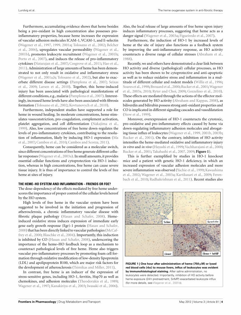
Moreover, overexpression of HO-1 counteracts the cytotoxic,pro-oxidative and pro-inflammatory effects caused by heme viadown-regulating inflammatory adhesion molecules and abrogat-ing tissue influx of leukocytes (Wagener et al., 1999, 2001b, 2003b;Rucker et al., 2001). On the contrary, inhibition of HO activityintensifies the heme-mediated oxidative and inflammatory injuryin vitro and in vivo (Hayashi et al., 1999; Vachharajani et al., 2000;Rucker et al., 2001; Takahashi et al., 2007, 2009; Figure 1).
This is further exemplified by studies in HO-1 knockoutmice and a patient with genetic HO-1 deficiency, in which anincreased expression of vascular adhesion molecules and moresevere inflammation was observed (Yachie et al., 1999; Kawashimaet al., 2002; Wagener et al., 2003a; Kartikasari et al., 2009; Feren-bach et al., 2010; Radhakrishnan et al., 2011). Recent studies also
FIGURE 1 | One hour after administration of heme (750 μM) or lysed
red blood cells (rbc) to mouse livers, influx of leukocytes was evident
by immunohistological staining. After saline administration, noleukocytes were detected. Importantly, inhibition of HO activity beforeheme exposure (24 h pretreatment, SnMP) exacerbated leukocyte influx(for more details, see Wagener et al., 2001b).
Frontiers in Pharmacology | Drug Metabolism and Transport May 2012 | Volume 3 | Article 81 | 4
Lundvig et al. The heme oxygenase system in anti-fibrotic therapy
suggest that HO-2 is important for the resolution of inflamma-tion, as HO-2 deficient mice demonstrate slower corneal epithelialwound healing and an amplified inflammatory response (Bellneret al., 2008, 2009, 2011; Halilovic et al., 2011; Marrazzo et al., 2011).
In contrast, transgenic HO-1 mice are better protected fromoxidative stress, inflammation, and vascular dysfunction as well asfibrotic tissue formation post-infarction (Wang et al., 2010).
Also, clinical studies have suggested that swift activation of HO-1 gene expression might be important to cope with cellular stresses.Several HO-1 gene promoter polymorphisms have been discov-ered, determining the levels of HO-1 induction in humans (Exneret al., 2004). The clinical relevance of the HO-1 promoter vari-ability has been demonstrated in independent studies and higherHO-1 induction has been associated with lower incidence forcardiovascular and inflammatory diseases (Wagener et al., 2008;Grochot-Przeczek et al., 2012).
Thus the degradation of the pro-inflammatory and pro-oxidative heme molecules by HO, the signaling actions of CO,the anti-oxidant properties of bilirubin combined with the scav-enging of the pro-oxidant free iron by ferritin may all contributeto the anti-inflammatory effects of the HO system.
By degrading heme, the HO system efficiently converts thepro-oxidative and pro-inflammatory heme molecule into anti-inflammatory and anti-oxidant molecules, thereby contributingto improved wound healing. This suggests that the heme–HOsystem executes regulatory functions in a diverse range of cel-lular processes with a direct influence on a timely execution of theinflammatory phase, including resolution of inflammation, apop-tosis, and proliferation. Differential protection against oxidativeand pro-inflammatory insults may also explain clinical differencesin fibrogenesis.
PHARMACOLOGICAL REGULATION OF HO-1 AND ITSEFFECTOR MOLECULES AS THERAPEUTIC TARGETS ININFLAMMATION AND FIBROSISA rapidly growing body of experimental evidence shows that spe-cific overexpression of HO-1 has protective effects in inflammationand fibrosis (Bauer et al., 2008). Accordingly, pharmacologicalinduction of HO-1 or administration of its effector moleculesmay be a promising therapeutic approach for the treatment ofinflammatory disorders associated with fibrosis in various organs.However, to apply HO-1 for specific therapeutic interventions anumber of important issues have to be taken into consideration.
ANTI-INFLAMMATORY EFFECTS OF HO-1 ARE MEDIATED IN A CELLTYPE-SPECIFIC MANNERHO-1 gene expression is up-regulated in a wide range of cell typesand tissues. The specific anti-inflammatory effects of HO-1, how-ever, seem to be dependent on its coordinate up-regulation in a celltype- and cell-context-specific manner. Notably, the regulation ofHO-1 expression in mononuclear/myeloid and in endothelial cellsappears to be of major importance for the immunomodulatoryand anti-inflammatory functions of this enzyme.
The key immunomodulatory role of HO-1 in mononuclearcells has been shown in conditional knockout mice, in which theHO-1 gene has been specifically deleted in myeloid cells. In thisreport, various pathological immune reactions in experimental
infectious and autoimmune conditions have been demonstratedin vivo (Tzima et al., 2009). HO-1 has also been demonstrated toplay a major role in the function of dendritic cells, which are a bonemarrow-derived myeloid cell population involved in mediatingadaptive immune responses (Remy et al., 2009; Park et al., 2010).Moreover, HO-1 appears to be of major significance for modu-lating inflammatory responses in endothelial cells. This has beenshown in the initial reports on HO-1 knockout mice (Poss andTonegawa, 1997) and in the first human case of genetic HO-1 defi-ciency (Yachie et al., 1999). In either case, the endothelium revealedspecific pathological alterations, which have been ascribed to anincreased endothelial sensitivity to oxidative stress. Accordingly,it was reported that genetic HO-1 deficiency exhibited pheno-typic alterations of the vascular endothelium, causing acceleratedformation of arterial thrombosis (True et al., 2007).
REGULATION OF HO-1 GENE EXPRESSIONTo apply HO-1 induction by specific pharmacological com-pounds for therapeutic interventions, it is not only important tounderstand the physiological functions of HO-1 and its products.
It is also important to understand the specific regulatory path-ways that up-regulate HO-1. Although primarily known to beinduced by oxidative stress, HO-1 is also up-regulated by a vari-ety of stress-independent stimuli including various well-knownpharmacologic compounds (Immenschuh and Ramadori, 2000;Li et al., 2007). Regulation of HO-1 is mainly, but not exclu-sively governed on the transcriptional level. Accordingly, a largevariety of transcription factors has been shown to be involved inHO-1 induction. In particular, redox-dependent transcription fac-tors such as Nrf2, which is a master regulator of the anti-oxidantresponse, activator protein-1 (AP-1) and nuclear factor (NF)-κB,but also other nuclear factors such as Egr-1, upstream regulatoryfactor-2 (USF-2), and specificity protein (SP)-1 have been shownto mediate HO-1 induction (Ryter et al., 2006; Paine et al., 2010).A key role in governing HO-1 expression has also been recognizedfor the transcriptional repressor Bach1. Bach1 is a heme-bindingprotein, the activity of which is primarily regulated via intracellu-lar levels of free heme (Ogawa et al., 2001; Igarashi and Sun, 2006).As both, Bach1 and Nrf2, interact with the anti-oxidant responseelement (ARE), which is the major cis-acting regulatory DNA ele-ment in the HO-1 gene promoter, the interplay of these two nuclearfactors appears to be of major importance for inducible HO-1gene expression. Whether and how Bach1 mediates the specificregulation of HO-1 if compared to other ARE-dependent stressresponse genes such as (NADPH):oxidoreductase-1 (NQO1) orglutathione-S-transferase (GST)-1 is a subject for currently on-going investigations (Dhakshinamoorthy et al., 2005; MacLeodet al., 2009; Okada et al., 2010). The complexity of HO-1 generegulation is further illustrated by the array of signaling cascadesincluding protein kinase Cs (PKCs) and mitogen-activated pro-tein kinases (MAPKs) that are involved in the regulation of thisgene (for a review, see Paine et al., 2010). In addition, some ofthese pathways such as p38 MAPK appear to have contradictorycell-specific regulatory effects on HO-1 gene expression (Naiduet al., 2009) indicating that HO-1 is governed by a highly com-plex interplay of various signaling modules. Macrophage-specificup-regulation of HO-1 gene expression in inflammation has been
www.frontiersin.org May 2012 | Volume 3 | Article 81 | 5
Lundvig et al. The heme oxygenase system in anti-fibrotic therapy
demonstrated in various experimental models (Paine et al., 2010).As an example, HO-1 in liver tissue macrophages (Kupffer cells) isup-regulated in response to the cytokine adiponectin via an IL-10dependent pathway (Mandal et al., 2010). Moreover, macrophage-specific induction of HO-1 by various toll-like receptors (TLR)ligands, including lipopolysaccharides (LPS), requires activationof the Tec kinase Bruton’s tyrosine kinase (Vijayan et al., 2011).
TARGETED PHARMACOLOGICAL HO-1 INDUCTION AS THERAPY FORINFLAMMATION AND FIBROSISNumerous compounds have been shown to provide therapeuticeffects via HO-1 induction. However, rather than giving an exhaus-tive list of compounds that may up-regulate HO-1 for therapeuticpurposes in inflammatory disorders, we refer to more specificoverviews on this topic (Li et al., 2007; Abraham and Kappas,2008; Vijayan et al., 2010).
Briefly, cobalt-protoporphyrin (CoPPIX), which has beenextensively applied in various experimental models to induce HO-1, does not seem an ideal compound for targeted therapeuticHO-1 up-regulation due to possible toxicity. Interestingly, sys-temic administration of heme (e.g., heme arginate) to healthyindividuals has been shown to increase HO-1 levels in serum(Doberer et al., 2010), and has been used in the clinics for years inthe treatment of porphyria (Tenhunen and Mustajoki, 1998; Maet al., 2011). More research is warranted to explore the potentialinduction of systemic HO activity in the human setting by hemearginate.
Natural products such as quercetin or theaflavin (Loke et al.,2008, 2010) are less toxic and may be more appropriate for poten-tial therapeutic applications in inflammatory disorders. Interest-ingly, Loke et al. (2010) recently demonstrated increased expres-sion of endothelial HO-1 in aortic lesions and subsequently atten-uation of aortic lesion formation in quercetin-fed ApoE knockoutmice, underscoring the potential of targeted HO-1 induction inclinical therapies.
Curcumin is another naturally occurring compound witha broad range of pharmacological activities, including anti-oxidative, anti-inflammatory, anti-carcinogenic, anti-diabetic, andanti-viral effects (Motterlini et al., 2000; McNally et al., 2007),mediating cellular protection against ROS (Barzegar and Moosavi-Movahedi, 2011; Wang et al., 2012; Yin et al., 2012) by up-regulating HO-1 through Nrf2 (Balogun et al., 2003). Recentstudies have shown beneficial effects of curcumin in a liver injurymodel (Cerny et al., 2011), and curcumin treatment reducedradiation-induced lung fibrosis (Lee et al., 2010).
Moreover, macrophage-specific (Wijayanti et al., 2005) HO-1 induction by defined compounds such as 4-(2-aminoethyl)-benezensulfonyl fluoride (AEBSF) and endothelial cell-specificHO-1 induction by statins (Grosser et al., 2004; Lee et al., 2004),might be useful for such therapeutic applications.
Another key issue that needs attention when applying HO-1induction for therapeutic applications deals with the question atwhat stage of the inflammatory phase HO-1 should be inducedto afford its salutary protective effects. Importantly, HO-1 induc-tion after the onset of an inflammatory disorder or in full-blowninflammation seems to be not effective in certain circumstances.This has been shown in an experimental model of dextran-sulfate-
induced colitis, in which HO-1 failed to have anti-inflammatoryeffects if induced after the onset of this disease (Paul et al., 2005)or in an experimental model of acute pancreatitis (Nakamichiet al., 2005). Therefore, the time point of targeted HO-1 inductionfor therapeutic applications needs further attention. Alternatively,administration of HO-effector molecules may mediate more directanti-inflammatory effects.
EXPOSURE TO HO-EFFECTOR MOLECULES CO AND BILIRUBIN:POSSIBLE AMELIORATING EFFECTS ON INFLAMMATION AND FIBROSISA wide array of preclinical and epidemiological evidence sug-gests that the protective properties of HO can also be medi-ated via its effector molecules bilirubin, CO, and co-inducedferritin. In inflammatory and fibrotic models both CO and biliru-bin demonstrated significant protection (recently reviewed inGrochot-Przeczek et al., 2012).
CO exerts its cytoprotective effects through different mech-anisms, including anti-flammatory (Neto et al., 2006; Choraet al., 2007), vasodilatory (Sammut et al., 1998), anti-coagulative(Chlopicki et al., 2006), anti-apoptotic (Wang et al., 2007), andanti-fibrotic pathways (Neto et al., 2006). CO deliverance isachieved through inhalation (Mayr et al., 2005; Moore et al., 2005),CO saturated physiological solutions (Nakao et al., 2006), andCO-releasing molecules (CO-RMs; Motterlini et al., 2002).
Interestingly, there are recent attempts to translate the promis-ing findings with CO inhalation and administration of CO-RMstoward the human setting (Motterlini and Otterbein, 2010). TheCO-RMs have been demonstrated to provide cytoprotective effectsin different in vitro (Clark et al., 2003; Sandouka et al., 2005;Sawle et al., 2005; Srisook et al., 2006; Zobi et al., 2010) andin vivo (Vera et al., 2005; Sandouka et al., 2006; Zhou et al., 2009;Wei et al., 2010) models, which urge for translation toward theclinic.
However, the potential safety issues with these compoundsmust be thoroughly addressed. CO-RMs are complexes of heavymetal, like nickel, cobalt, iron, or ruthenium surrounded by car-bonyl groups that are released as CO under the appropriate con-ditions (Motterlini et al., 2002, 2003). Ruthenium-based CO-RMshave recently been suggested to generate cytotoxic, CO-depletedby-products in vitro (Winburn et al., 2012). However, the devel-opment of water-soluble CO-RMs (Clark et al., 2003; Foresti et al.,2004), the use of other transition metals, e.g., manganese, in thechemical structure of CO-RMs (Motterlini et al., 2002; Crook et al.,2011), and even the use of CO-RMs devoid of transition metals(Motterlini et al., 2005) may promote the development of clinicallysafe CO-RMs due to less confounding effects.
Bilirubin is another generated HO-effector molecule withpromising clinical applications. Besides being a powerful anti-oxidant (Stocker et al., 1987), several studies have shown thatbiliverdin/bilirubin administration has cytoprotective effects dur-ing ischemia–reperfusion injury and graft rejection after trans-plantation (Clark et al., 2000; Fondevila et al., 2004). Also, bilirubinexerts immunomodulatory and anti-inflammatory effects (Williset al., 1996) by down-regulating expression of inflammatorycytokines and adhesion molecules (Nakao et al., 2004), as well asby reducing immune cell infiltration (Hayashi et al., 1999; Nakaoet al., 2004).
Frontiers in Pharmacology | Drug Metabolism and Transport May 2012 | Volume 3 | Article 81 | 6
Lundvig et al. The heme oxygenase system in anti-fibrotic therapy
Individuals with Gilbert’s syndrome have a polymorphism inthe UGT1A1 promoter and are protected against cardiovascu-lar complications (Schwertner and Vitek, 2008). This polymor-phism results in slower glucuronidation and therefore dimin-ished excretion of bilirubin, leading to elevated bilirubin levels.Unfortunately, bilirubin is not available for human use. We andothers postulated that experimentally induced mild hyperbiliru-binemia would mimic the positive effects seen in Gilbert’s syn-drome (McCarty, 2007; Dekker et al., 2011). In a double-blind,placebo-controlled crossover design, we demonstrated that ele-vating bilirubin levels using the HIV protease inhibitor atazanavirindeed ameliorates vascular function in type 2 diabetes mellituspatients (Dekker et al., 2011). In addition, the redox status in thesepatients was improved following induction of mild hyperbiliru-binemia (Dekker et al., 2011). This demonstrates for the first timethe potential beneficial power of bilirubin as a novel mechanismin humans. We expect that these clinically significant protectiveeffects of HO-effector molecules in humans strongly encouragemore translational research toward the protective properties of HOand its effector molecules in inflammatory and fibrotic conditions.
Finally, three examples of inflammatory disorders, which arelinked to organ fibrosis and in which HO-1 and its effectormolecules may be of therapeutic use, are discussed in more detail.
The heme–HO system in hypertrophic scarringExcept for fast healing wounds, such as shallow scratches, mostwounds will result in visible scar formation, which is a naturalpart of the wound healing process. However, in some individuals –depending on geographic and ethnographic distribution as well asthe character of the injury – excessive deposition of ECM occurs,resulting in the formation of a raised, discolored hypertrophicscar.
The involvement of the HO system is evident from knockoutmouse models being devoid of HO-1 or HO-2, respectively. Bothmodels demonstrate delayed wound healing due to poor responsetoward inflammatory and oxidative insults (Braun et al., 2002; Setaet al., 2006; Deshane et al., 2007; Bellner et al., 2008, 2009, 2011;Patil et al., 2008; Kovtunovych et al., 2010; Halilovic et al., 2011).Also, pharmacological inhibition of HO activity has a negativeimpact on dermal wound healing (Grochot-Przeczek et al., 2009).
Also in dermal wound healing, the duality of heme is evident.Local accumulation of free heme liberated by spontaneous degra-dation of hemoglobin at the wound site may promote free radicalformation and oxidative damage (Balla et al., 1991, 2000; Jeneyet al., 2002). We have postulated that this injury-derived hememay be the trigger that initiates the inflammatory and the innateimmunity response (Wagener et al., 2003a). To rapidly cope withoxidative stress, dermal fibroblasts are capable to rapidly induceHO-1 expression as a feedback response, and keratinocytes containhigh levels of HO-2 (Applegate et al., 1995). When HO-1 levels areincreased at the wound site resolution of inflammation will takeplace and the wound healing cascade can enter the next phase.
Pre-induction of HO by daily heme administration(30 mg/kg/day i.p.) in a mouse excisional wound healing modelenhances wound contraction by increasing cellular prolifera-tion and collagen synthesis (Ahanger et al., 2010). HO-1 induc-tion also resulted in reduced transcription of pro-inflammatorycytokines, e.g., TNFα, and an up-regulated transcription of the
anti-inflammatory cytokine interleukin-10 (Ahanger et al., 2010).This demonstrates that low levels of heme may promote woundhealing via induction of HO-1, whereas high levels of heme mayprolong oxidative and inflammatory stress and result in a pro-fibrotic environment. Indeed, up-regulation of HO-1 expressionin keratinocytes in transgenic mice bearing the HO-1 gene underthe control of keratin 14 promoter resulted in improved neovascu-larization and accelerated wound healing (Grochot-Przeczek et al.,2009). Also, adenoviral mediated HO-1 gene delivery significantlyimproved dermal wound healing (Grochot-Przeczek et al., 2009).
Together, these data suggest that increased levels of HO-1 mayimprove wound healing and reduce dermal scarring.
Also, (myo)fibroblast apoptosis is essential in normal andhypertrophic scarring, as fibroblasts in pathological wound heal-ing remain in the wound area and deposit excessive amounts ofECM components, contributing to fibrogenesis. At low concentra-tions, curcumin has been shown to induce HO expression, whereasHO-1 expression and activity was negligible at high curcumindoses (Scharstuhl et al., 2009). Despite this, HO-1 expressionhas been demonstrated to protect against fibroblast apoptosisinduced by high concentrations of curcumin through the actionsof effector molecules biliverdin/bilirubin (Scharstuhl et al., 2009).Importantly, pre-induction of HO-1 with low doses of curcuminalso protected fibroblasts against curcumin-induced apoptosis(Scharstuhl et al., 2009). This suggest that curcumin in high dosesmay affect pathological scar formation through affecting fibroblastapoptosis, while HO-1 and its effector molecules can fine-tune thisresponse.
The heme–HO system as therapeutic target in hepatic andpancreatic fibrosisThe current therapeutic options in specific treatment of bothhepatic (Popov and Schuppan, 2009) as well as pancreatic fibro-sis (Braganza et al., 2011) are only limited and novel therapeuticoptions are urgently needed. Interestingly, independent groupshave demonstrated that specific overexpression of HO-1 had aninhibitory effect on chronic viral hepatitis (Zhu et al., 2008; Houet al., 2010) as one of the major causes of hepatic fibrosis. Exposureto high levels of heme has been demonstrated to result in inflam-matory leukocyte influx and fibrosis into the liver (Wagener et al.,2001b; Tolosano et al., 2002). Moreover, in a more recent studypharmacological HO-1 induction via CoPPIX has been shown toprevent liver fibrosis in a Mdr2 knockout mice (Barikbin et al.,2012). Importantly, in this report up-regulation of HO-1 not onlyreduced activation of HSCs, but also reversed established hepaticfibrosis in this mouse model.
Heme exposure has also shown to strongly promote inflam-mation in the pancreas, as illustrated by increased vascular per-meabilization (Wagener et al., 2001b). HO-1 might also serve as atherapeutic target in pancreatic fibrosis. Schwer et al. (2008) haveshown that up-regulation of HO-1 by curcumin inhibited PSCproliferation, which plays a crucial role in the progression of pan-creatic fibrosis. These findings have been extended in a more recentreport, in which PSC proliferation was inhibited via activation ofthe p38 MAPK/HO-1 pathway (Schwer et al., 2010). Moreover,these authors also indicated that the protective effect on PSC maybe mediated via HO-1-dependent up-regulation of the cell cycleinhibitor p21/CIP-1 (Schwer et al., 2010).
www.frontiersin.org May 2012 | Volume 3 | Article 81 | 7
Lundvig et al. The heme oxygenase system in anti-fibrotic therapy
The heme–HO system in CTRA late but severe clinical complication after solid organ transplan-tation is CTR, which is currently considered the major limitingfactor for long-term graft survival in particular in kidney and hearttransplantation (Mitchell, 2009). A hallmark of CTR is TV, alsotermed graft vascular disease or allograft arteriopathy, in whichthe success rate of current therapeutic regimens including treat-ment with immunosuppressive drugs is very poor (Cornell et al.,2008). Pathologic features of TV include diffuse concentric inti-mal hyperplasia and adventitial sclerosis due to proliferation ofvarious cell types of the vascular wall.
Survival of cardiac xenografts has been shown to be criticallydependent on the expression of endothelial HO-1 (Soares et al.,1998). Accordingly, targeted overexpression of HO-1 via a genetherapy approach has been demonstrated to protect against TVand to cause a prolonged survival of chronic allogenic rejection ofaortic vascular transplants in a rat model (Chauveau et al., 2002).Additionally, HO-1 activity has been shown to reduce atheroscle-rosis (Bouche et al., 2002; Ferran, 2006; Du et al., 2007), whichcould further contribute to a higher transplantation success rate.
Although the underlying mechanisms of how HO-1 mediatesits protective effects are not well understood, one major beneficialeffect of HO-1 may be its anti-proliferative effects in the vascula-ture (Valenzuela and Reed, 2011), which has previously also beenshown in an experimental angioplasty rat model (Duckers et al.,2001). In corroboration, an increasing number of studies haveproven HO-1 activity to be important for transplant graft sur-vival (Ollinger and Pratschke,2010),as HO-1 expression inductionimproves recipient immune tolerance (see reviews of Soares andBach, 2007; Blancou et al., 2011) and donor graft survival afterischemia/reperfusion injury (for recent reviews, see Ferenbachet al., 2010; Sass et al., 2012).
The beneficial effects of HO-1 activity have been linked tothe induction level of HO-1, as HO-1 gene promoter poly-morphisms affect transplantation outcome (Immenschuh andRamadori, 2000; Baan et al., 2004; Geuken et al., 2005; Holweget al., 2005; Courtney et al., 2007; Gerbitz et al., 2008). However, it isnot only the direct effect of HO-1 on limiting heme levels in injurysites that mediates the beneficial outcome of HO-1 activity. Also,the cytoprotective and anti-inflammatory effects of the end prod-ucts generated by heme degradation, CO and biliverdin/bilirubin,have proven pivotal in improving the success of graft transplanta-tion (Soares et al., 1998; Immenschuh and Ramadori, 2000; Katoet al., 2003; Nakao et al., 2004; Lee et al., 2007; Brugger et al., 2010).
The implication of heme-mediated regulation of the expres-sion of the transcription factor and inflammatory mediator Egr-1
in atherosclerosis may have a significant impact on the success rateof graft survival after transplantation, as atherosclerosis is one ofthe causative agents for graft rejection and transplantation failure.Interestingly, HO-1 effects have been associated with its functionalinteraction with the cyclin-dependent kinase inhibitor p21/CIP-1,which belongs to the Cip/Kip protein family of cell prolifera-tion inhibitors (Duckers et al., 2001). In addition, the functionof p21/CIP-1 has been demonstrated to up-regulate the cellularanti-oxidant response via direct binding to the master HO-1 genetranscriptional regulator Nrf2 (Chen et al., 2009). These findingsare in keeping with a recent report showing a major regulatoryrole for p21/CIP-1 in endothelial cell proliferation in atheroscle-rosis (Obikane et al., 2010), and may suggest that the interplay ofp21/CIP-1 with HO-1 and/or Nrf2 is involved in the pathogenesisof TV.
Together, this exemplifies that the HO system is of major inter-est for the development of future therapeutic approaches that takeadvantage of pharmacological induction of HO-1 in endothe-lial cells. As a precedent, it has recently been demonstrated in arodent atherosclerosis model that HO-1 induction by the pharma-cological compound probucol had salutary effects via inhibitionof vascular smooth muscle cell proliferation (Wu et al., 2006).Also, the importance of transcriptional regulation of HO-1 fur-ther underscores the clinical significance of the HO-1 promoterpolymorphisms on therapy success, and the need for personalizedmedicine in the future.
FUTURE PERSPECTIVESIn this review we showed examples clearly demonstratingthat excess levels of heme may promote pro-oxidant, pro-inflammatory, and subsequently fibrotic processes, in which theHO system may have counter-regulatory effects. These effects mayoccur via degradation of pro-inflammatory free heme and by thecytoprotective and anti-inflammatory effects of the HO-effectormolecules CO and biliverdin/bilirubin. Promising (pre)clinicaldata support the utilization of HO and its effector molecules asnovel therapeutic targets to reduce tissue inflammation, oxida-tive stress, and fibrosis. However, in order to translate thistoward the clinical setting, safe and potent inducers of HO-1 areneeded.
ACKNOWLEDGMENTSSupported by a grant from the Dutch Burns Foundation (#09.110),the Radboud University Nijmegen Medical Centre, and Van Leer-sum Foundation (Frank Wagener) and by a grant from the BMBF(FKZ 0316045B; Stephan Immenschuh).
REFERENCESAarabi, S., Longaker, M. T., and Gurt-
ner, G. C. (2007). Hypertrophicscar formation following burnsand trauma: new approachesto treatment. PLoS Med. 4,e234. doi:10.1371/journal.pmed.0040234
Abraham, N. G., and Kappas,A. (2008). Pharmacologicaland clinical aspects of heme
oxygenase. Pharmacol. Rev. 60,79–127.
Abraham, N. G., Lin, J. H., Schwartz-man, M. L., Levere, R. D., andShibahara, S. (1988). The phys-iological significance of hemeoxygenase. Int. J. Biochem. 20,543–558.
Ahanger, A. A., Prawez, S., Leo,M. D., Kathirvel, K., Kumar, D.,Tandan, S. K., and Malik, J.
K. (2010). Pro-healing potentialof hemin: an inducer of hemeoxygenase-1. Eur. J. Pharmacol. 645,165–170.
Applegate, L. A., Noel,A.,Vile, G., Frenk,E., and Tyrrell, R. M. (1995). Twogenes contribute to different extentsto the heme oxygenase enzyme activ-ity measured in cultured humanskin fibroblasts and keratinocytes:implications for protection against
oxidant stress. Photochem. Photobiol.61, 285–291.
Ashcroft, G. S., Yang, X., Glick, A.B., Weinstein, M., Letterio, J. L.,Mizel, D. E., Anzano, M., Greenwell-Wild, T., Wahl, S. M., Deng, C.,and Roberts, A. B. (1999). Micelacking Smad3 show acceleratedwound healing and an impairedlocal inflammatory response. Nat.Cell Biol. 1, 260–266.
Frontiers in Pharmacology | Drug Metabolism and Transport May 2012 | Volume 3 | Article 81 | 8
Lundvig et al. The heme oxygenase system in anti-fibrotic therapy
Ashoor, I. F., and Najafian, N. (2012).Rejection and regulation: a tight bal-ance. Curr. Opin. Organ Transplant17, 1–7.
Azimzadeh, A. M., Lees, J. R., Ding,Y., and Bromberg, J. S. (2011).Immunobiology of transplantation:impact on targets for large and smallmolecules. Clin. Pharmacol. Ther. 90,229–242.
Baan, C., Peeters, A., Lemos, F., Uit-terlinden, A., Doxiadis, I., Claas,F., Ijzermans, J., Roodnat, J., andWeimar, W. (2004). Fundamentalrole for HO-1 in the self-protectionof renal allografts. Am. J. Transplant.4, 811–818.
Balla, G., Jacob, H. S., Eaton, J. W.,Belcher, J. D., and Vercellotti, G.M. (1991). Hemin: a possible phys-iological mediator of low densitylipoprotein oxidation and endothe-lial injury. Arterioscler. Thromb. 11,1700–1711.
Balla, J., Balla, G., Jeney, V., Kakuk,G., Jacob, H. S., and Vercellotti,G. M. (2000). Ferriporphyrins andendothelium: a 2-edged sword-promotion of oxidation and induc-tion of cytoprotectants. Blood 95,3442–3450.
Balla, J., Jacob, H. S., Balla, G., Nath,K., Eaton, J. W., and Vercellotti, G.M. (1993). Endothelial-cell hemeuptake from heme proteins: induc-tion of sensitization and desensiti-zation to oxidant damage. Proc. Natl.Acad. Sci. U.S.A. 90, 9285–9289.
Balogun, E., Hoque, M., Gong, P.,Killeen, E., Green, C. J., Foresti,R., Alam, J., and Motterlini, R.(2003). Curcumin activates thehaem oxygenase-1 gene via regula-tion of Nrf2 and the antioxidant-responsive element. Biochem. J. 371,887–895.
Barikbin, R., Neureiter, D., Wirth, J.,Erhardt, A., Schwinge, D., Kluwe, J.,Schramm, C., Tiegs, G., and Sass,G. (2012). Induction of heme oxy-genase 1 prevents progression ofliver fibrosis in Mdr2 knockout mice.Hepatology 55, 553–562.
Barzegar, A., and Moosavi-Movahedi,A. A. (2011). Intracellular ROSprotection efficiency and freeradical-scavenging activity ofcurcumin. PLoS ONE 6, e26012.doi:10.1371/journal.pone.0026012
Bauer, M., Huse, K., Settmacher, U.,and Claus, R. A. (2008). The hemeoxygenase-carbon monoxide sys-tem: regulation and role in stressresponse and organ failure. IntensiveCare Med. 34, 640–648.
Belcher, J. D., Mahaseth, H., Welch,T. E., Otterbein, L. E., Hebbel, R.P., and Vercellotti, G. M. (2006).
Heme oxygenase-1 is a modulator ofinflammation and vaso-occlusion intransgenic sickle mice. J. Clin. Invest.116, 808–816.
Bellner, L., Martinelli, L., Halilovic, A.,Patil, K., Puri, N., Dunn, M. W.,Regan, R. F., and Schwartzman, M.L. (2009). Heme oxygenase-2 dele-tion causes endothelial cell acti-vation marked by oxidative stress,inflammation, and angiogenesis. J.Pharmacol. Exp. Ther. 331, 925–932.
Bellner, L., Vitto, M., Patil, K. A., Dunn,M. W., Regan, R., and Laniado-Schwartzman, M. (2008). Exacer-bated corneal inflammation andneovascularization in the HO-2 nullmice is ameliorated by biliverdin.Exp. Eye Res. 87, 268–278.
Bellner, L., Wolstein, J., Patil, K.A., Dunn, M. W., and Laniado-Schwartzman, M. (2011). Biliverdinrescues the HO-2 null mouse pheno-type of unresolved chronic inflam-mation following corneal epithelialinjury. Invest. Ophthalmol. Vis. Sci.52, 3246–3253.
Blancou, P., Tardif, V., Simon, T., Remy,S., Carreno, L., Kalergis, A., andAnegon, I. (2011). Immunoregula-tory properties of heme oxygenase-1. Methods Mol. Biol. 677, 247–268.
Blaschke, F., Bruemmer, D., and Law, R.E. (2004). Egr-1 is a major vascularpathogenic transcription factor inatherosclerosis and restenosis. Rev.Endocr. Metab. Disord. 5, 249–254.
Bock, O., Schmid-Ott, G., Malewski, P.,and Mrowietz, U. (2006). Qualityof life of patients with keloid andhypertrophic scarring. Arch. Derma-tol. Res. 297, 433–438.
Bouche, D., Chauveau, C., Roussel, J. C.,Mathieu, P., Braudeau, C., Tesson,L., Soulillou, J. P., Iyer, S., Buelow,R., and Anegon, I. (2002). Inhibi-tion of graft arteriosclerosis devel-opment in rat aortas following hemeoxygenase-1 gene transfer. Transpl.Immunol. 9, 235–238.
Braganza, J. M., Lee, S. H., Mccloy,R. F., and Mcmahon, M. J. (2011).Chronic pancreatitis. Lancet 377,1184–1197.
Braun, S., Hanselmann, C., Gassmann,M. G., Auf Dem Keller, U., Born-Berclaz, C., Chan, K., Kan, Y. W.,and Werner, S. (2002). Nrf2 tran-scription factor, a novel target ofkeratinocyte growth factor actionwhich regulates gene expressionand inflammation in the healingskin wound. Mol. Cell. Biol. 22,5492–5505.
Brissett, A. E., and Sherris, D. A.(2001). Scar contractures, hyper-trophic scars, and keloids. FacialPlast. Surg. 17, 263–272.
Brouard, S., Otterbein, L. E., Anrather,J., Tobiasch, E., Bach, F. H., Choi, A.M., and Soares, M. P. (2000). Car-bon monoxide generated by hemeoxygenase 1 suppresses endothelialcell apoptosis. J. Exp. Med. 192,1015–1026.
Brugger, J., Schick, M. A., Brock,R. W., Baumann, A., Muellenbach,R. M., Roewer, N., and Wunder,C. (2010). Carbon monoxide hasantioxidative properties in the liverinvolving p38 MAP kinase path-way in a murine model of systemicinflammation. Microcirculation 17,504–513.
Cambos, M., Bazinet, S., Abed, E.,Sanchez-Dardon, J., Bernard, C.,Moreau, R., Olivier, M., and Scorza,T. (2010). The IL-12p70/IL-10interplay is differentially regu-lated by free heme and hemozoinin murine bone-marrow-derivedmacrophages. Int. J. Parasitol. 40,1003–1012.
Cambos, M., and Scorza, T. (2011).Robust erythrophagocytosis leads tomacrophage apoptosis via a hemin-mediated redox imbalance: role inhemolytic disorders. J. Leukoc. Biol.89, 159–171.
Cerny, D., Lekic, N., Vanova, K.,Muchova, L., Horinek, A., Kmon-ickova, E., Zidek, Z., Kamenikova,L., and Farghali, H. (2011). Hepato-protective effect of curcumin inlipopolysaccharide/-galactosaminemodel of liver injury in rats: rela-tionship to HO-1/CO antioxidantsystem. Fitoterapia 82, 786–791.
Chauveau, C., Bouchet, D., Roussel,J. C., Mathieu, P., Braudeau, C.,Renaudin, K., Tesson, L., Soulillou,J. P., Iyer, S., Buelow, R., and Ane-gon, I. (2002). Gene transfer of hemeoxygenase-1 and carbon monoxidedelivery inhibit chronic rejection.Am. J. Transplant. 2, 581–592.
Chen, W., Sun, Z., Wang, X. J., Jiang, T.,Huang,Z.,Fang,D.,and Zhang,D. D.(2009). Direct interaction betweenNrf2 and p21(Cip1/WAF1) upreg-ulates the Nrf2-mediated antiox-idant response. Mol. Cell 34,663–673.
Chettibi, S., Ferguson, W. J., Gallin, J. L.,and Snyderman, R. (1999). “Woundrepair: an overview,” in Inflamma-tion: Basic Principles and ClinicalCorrelates eds J. Gallin and R. Sny-derman (Philadelphia: LippincottWilliams & Wilkins), 865–881.
Chlopicki, S., Olszanecki, R.,Marcinkiewicz, E., Lomnicka,M., and Motterlini, R. (2006). Car-bon monoxide released by CORM-3inhibits human platelets by amechanism independent of soluble
guanylate cyclase. Cardiovasc. Res.71, 393–401.
Chora, A. A., Fontoura, P., Cunha, A.,Pais, T. F., Cardoso, S., Ho, P. P.,Lee, L. Y., Sobel, R. A., Steinman,L., and Soares, M. P. (2007). Hemeoxygenase-1 and carbon monox-ide suppress autoimmune neuroin-flammation. J. Clin. Invest. 117,438–447.
Clark, J. E., Foresti, R., Sarathchandra,P., Kaur, H., Green, C. J., and Motter-lini, R. (2000). Heme oxygenase-1-derived bilirubin ameliorates postis-chemic myocardial dysfunction. Am.J. Physiol. Heart Circ. Physiol. 278,H643–H651.
Clark, J. E., Naughton, P., Shurey, S.,Green, C. J., Johnson, T. R., Mann,B. E., Foresti, R., and Motterlini, R.(2003). Cardioprotective actions bya water-soluble carbon monoxide-releasing molecule. Circ. Res. 93,e2–e8.
Cornell, L. D., Smith, R. N., and Colvin,R. B. (2008). Kidney transplantation:mechanisms of rejection and accep-tance. Annu. Rev. Pathol. 3, 189–220.
Cosgrove, S., Chotirmall, S. H., Greene,C. M., and Mcelvaney, N. G. (2011).Pulmonary proteases in the cysticfibrosis lung induce interleukin 8expression from bronchial epithelialcells via a heme/meprin/epidermalgrowth factor receptor/Toll-likereceptor pathway. J. Biol. Chem. 286,7692–7704.
Courtney,A. E.,Mcnamee,P. T.,Middle-ton, D., Heggarty, S., Patterson, C. C.,and Maxwell, A. P. (2007). Associa-tion of functional heme oxygenase-1gene promoter polymorphism withrenal transplantation outcomes. Am.J. Transplant. 7, 908–913.
Crook, S. H., Mann, B. E., Meijer, A.J., Adams, H., Sawle, P., Scapens,D., and Motterlini, R. (2011).[Mn(CO)4S2CNMe(CH2CO2H)],a new water-soluble CO-releasingmolecule. Dalton Trans. 40,4230–4235.
Deitch, E. A., Wheelahan, T. M., Rose,M. P., Clothier, J., and Cotter, J.(1983). Hypertrophic burn scars:analysis of variables. J. Trauma 23,895–898.
Dekker, D., Dorresteijn, M. J., Pij-nenburg, M., Heemskerk, S.,Rasing-Hoogveld, A., Burger, D.M., Wagener, F. A., and Smits, P.(2011). The bilirubin-increasingdrug atazanavir improves endothe-lial function in patients with type2 diabetes mellitus. Arterioscler.Thromb. Vasc. Biol. 31, 458–463.
Deshane, J., Chen, S., Caballero, S.,Grochot-Przeczek, A., Was, H., Li,C. S., Lach, R., Hock, T. D., Chen,
www.frontiersin.org May 2012 | Volume 3 | Article 81 | 9
Lundvig et al. The heme oxygenase system in anti-fibrotic therapy
B., Hill-Kapturczak, N., Siegal, G. P.,Dulak, J., Jozkowicz,A., Grant, M. B.,and Agarwal,A. (2007). Stromal cell-derived factor 1 promotes angio-genesis via a heme oxygenase 1-dependent mechanism. J. Exp. Med.204, 605–618.
Dhakshinamoorthy, S., Jain, A.K., Bloom, D. A., and Jaiswal,A. K. (2005). Bach1 competeswith Nrf2 leading to negativeregulation of the antioxidantresponse element (ARE)-mediatedNAD(P)H:quinone oxidoreductase1 gene expression and inductionin response to antioxidants. J. Biol.Chem. 280, 16891–16900.
Doberer, D., Haschemi, A., Andreas,M., Zapf, T. C., Clive, B., Jeitler,M., Heinzl, H., Wagner, O., Wolzt,M., and Bilban, M. (2010). Haemarginate infusion stimulates haemoxygenase-1 expression in healthysubjects. Br. J. Pharmacol. 161,1751–1762.
Du, D., Chang, S., Chen, B., Zhou, H.,and Chen, Z. K. (2007). Adenovirus-mediated heme oxygenase transferinhibits graft arteriosclerosis in rataortic transplants. Transplant. Proc.39, 3446–3448.
Duckers, H. J., Boehm, M., True, A. L.,Yet, S. F., San, H., Park, J. L., Clin-ton Webb, R., Lee, M. E., Nabel, G.J., and Nabel, E. G. (2001). Hemeoxygenase-1 protects against vascu-lar constriction and proliferation.Nat. Med. 7, 693–698.
Duffield, J. S., Forbes, S. J., Constandi-nou, C. M., Clay, S., Partolina, M.,Vuthoori, S., Wu, S., Lang, R., andIredale, J. P. (2005). Selective deple-tion of macrophages reveals distinct,opposing roles during liver injuryand repair. J. Clin. Invest. 115, 56–65.
Exner, M., Minar, E., Wagner, O., andSchillinger, M. (2004). The role ofheme oxygenase-1 promoter poly-morphisms in human disease. FreeRadic. Biol. Med. 37, 1097–1104.
Fathke, C., Wilson, L., Hutter, J., Kapoor,V., Smith, A., Hocking, A., and Isik,F. (2004). Contribution of bonemarrow-derived cells to skin: colla-gen deposition and wound repair.Stem Cells 22, 812–822.
Ferenbach, D. A., Kluth, D. C., andHughes, J. (2010). Hemeoxygenase-1 and renal ischaemia-reperfusioninjury. Nephron Exp. Nephrol. 115,e33–e37.
Ferran, C. (2006). Protective genes inthe vessel wall: modulators of graftsurvival and function. Transplanta-tion 82, S36–S40.
Fialkow, L., Wang, Y., and Downey, G. P.(2007). Reactive oxygen and nitro-gen species as signaling molecules
regulating neutrophil function. FreeRadic. Biol. Med. 42, 153–164.
Figueiredo, R. T., Fernandez, P. L.,Mourao-Sa, D. S., Porto, B. N.,Dutra, F. F., Alves, L. S., Oliveira, M.F., Oliveira, P. L., Graca-Souza, A.V., and Bozza, M. T. (2007). Char-acterization of heme as activator ofToll-like receptor 4. J. Biol. Chem.282, 20221–20229.
Fondevila, C., Shen, X. D., Tsuchiyashi,S., Yamashita, K., Csizmadia, E.,Lassman, C., Busuttil, R. W., Kupiec-Weglinski, J. W., and Bach, F.H. (2004). Biliverdin therapy pro-tects rat livers from ischemia andreperfusion injury. Hepatology 40,1333–1341.
Foresti, R., Hammad, J., Clark, J. E.,Johnson, T. R., Mann, B. E., Friebe,A., Green, C. J., and Motterlini,R. (2004). Vasoactive properties ofCORM-3, a novel water-soluble car-bon monoxide-releasing molecule.Br. J. Pharmacol. 142, 453–460.
Frantz, F. W., Bettinger, D. A., Haynes,J. H., Johnson, D. E., Harvey, K. M.,Dalton, H. P., Yager, D. R., Diegel-mann, R. F., and Cohen, I. K. (1993).Biology of fetal repair: the presenceof bacteria in fetal wounds inducesan adult-like healing response. J.Pediatr. Surg. 28, 428–433.
Gawronska-Kozak, B., Bogacki, M.,Rim, J. S., Monroe, W. T., andManuel, J. A. (2006). Scarless skinrepair in immunodeficient mice.Wound Repair Regen. 14, 265–276.
Gerbitz, A., Hillemanns, P., Schmid, C.,Wilke, A., Jayaraman, R., Kolb, H.J., Eissner, G., and Holler, E. (2008).Influence of polymorphism withinthe heme oxygenase-I promoter onoverall survival and transplantation-related mortality after allogeneicstem cell transplantation. Biol. BloodMarrow Transplant. 14, 1180–1189.
Geuken, E., Buis, C. I., Visser, D. S.,Blokzijl, H., Moshage, H., Nemes,B., Leuvenink, H. G., De Jong, K.P., Peeters, P. M., Slooff, M. J., andPorte, R. J. (2005). Expression ofheme oxygenase-1 in human liv-ers before transplantation correlateswith graft injury and function aftertransplantation. Am. J. Transplant. 5,1875–1885.
Goren, I., Allmann, N., Yogev, N.,Schurmann, C., Linke, A., Hold-ener, M., Waisman, A., Pfeilschifter,J., and Frank, S. (2009). A trans-genic mouse model of induciblemacrophage depletion: effects ofdiphtheria toxin-driven lysozymeM-specific cell lineage ablation onwound inflammatory, angiogenic,and contractive processes. Am. J.Pathol. 175, 132–147.
Gozzelino, R., Jeney, V., and Soares,M. P. (2010). Mechanisms of cellprotection by heme oxygenase-1.Annu. Rev. Pharmacol. Toxicol. 50,323–354.
Grochot-Przeczek, A., Dulak, J.,and Jozkowicz, A. (2012). Haemoxygenase-1: non-canonical roles inphysiology and pathology. Clin. Sci.122, 93–103.
Grochot-Przeczek, A., Lach, R.,Mis, J., Skrzypek, K., Gozdecka,M., Sroczynska, P., Dubiel, M.,Rutkowski, A., Kozakowska, M.,Zagorska, A., Walczynski, J., Was,H., Kotlinowski, J., Drukala, J.,Kurowski, K., Kieda, C., Herault,Y., Dulak, J., and Jozkowicz, A.(2009). Heme oxygenase-1 accel-erates cutaneous wound healingin mice. PLoS ONE 4, e5803.doi:10.1371/journal.pone.0005803
Grosser, N., Hemmerle, A., Berndt,G., Erdmann, K., Hinkelmann, U.,Schurger, S., Wijayanti, N., Immen-schuh, S., and Schroder, H. (2004).The antioxidant defense proteinheme oxygenase 1 is a novel targetfor statins in endothelial cells. FreeRadic. Biol. Med. 37, 2064–2071.
Guo, S., and Dipietro, L. A. (2010). Fac-tors affecting wound healing. J. Dent.Res. 89, 219–229.
Halilovic, A., Patil, K. A., Bellner, L.,Marrazzo, G., Castellano, K., Cullaro,G., Dunn, M. W., and Schwartz-man, M. L. (2011). Knockdown ofheme oxygenase-2 impairs cornealepithelial cell wound healing. J. Cell.Physiol. 226, 1732–1740.
Halliwell, B., and Gutteridge, J. M.(1984). Oxygen toxicity, oxygen rad-icals, transition metals and disease.Biochem. J. 219, 1–14.
Hao, K., Hanawa, H., Ding, L., Ota,Y., Yoshida, K., Toba, K., Ogura, M.,Ito, H., Kodama, M., and Aizawa, Y.(2011). Free heme is a danger signalinducing expression of proinflam-matory proteins in cultured cellsderived from normal rat hearts. Mol.Immunol. 48, 1191–1202.
Harty, M., Neff, A. W., King, M. W.,and Mescher, A. L. (2003). Regen-eration or scarring: an immunologicperspective. Dev. Dyn. 226, 268–279.
Hasan, R. N., and Schafer, A. I. (2008).Hemin upregulates Egr-1 expres-sion in vascular smooth muscle cellsvia reactive oxygen species ERK-1/2-Elk-1 and NF-kappaB. Circ. Res. 102,42–50.
Hayashi, S., Takamiya, R., Yamaguchi,T., Matsumoto, K., Tojo, S. J.,Tamatani, T., Kitajima, M., Makino,N., Ishimura, Y., and Suematsu,M. (1999). Induction of hemeoxygenase-1 suppresses venular
leukocyte adhesion elicited byoxidative stress: role of bilirubingenerated by the enzyme. Circ. Res.85, 663–671.
Hernandez-Gea, V., and Friedman, S.L. (2011). Pathogenesis of liverfibrosis. Annu. Rev. Pathol. 6,425–456.
Holweg, C. T., Balk, A. H., Uitterlinden,A. G., Niesters, H. G., Maat, L. P.,Weimar, W., and Baan, C. C. (2005).Functional heme oxygenase-1 pro-moter polymorphism in relation toheart failure and cardiac transplan-tation. J. Heart Lung Transplant. 24,493–497.
Hou, W., Tian, Q., Zheng, J.,and Bonkovsky, H. L. (2010).MicroRNA-196 represses Bach1protein and hepatitis C virus geneexpression in human hepatoma cellsexpressing hepatitis C viral proteins.Hepatology 51, 1494–1504.
Hunt, T. K., Hopf, H., and Hussain, Z.(2000). Physiology of wound heal-ing. Adv. Skin Wound Care 13, 6–11.
Igarashi, K., and Sun, J. (2006). Theheme-Bach1 pathway in the regula-tion of oxidative stress response anderythroid differentiation. Antioxid.Redox Signal. 8, 107–118.
Immenschuh, S., and Ramadori, G.(2000). Gene regulation of hemeoxygenase-1 as a therapeutic target.Biochem. Pharmacol. 60, 1121–1128.
Iwasaki, K., Mackenzie, E. L., Haile-mariam, K., Sakamoto, K., and Tsuji,Y. (2006). Hemin-mediated regu-lation of an antioxidant-responsiveelement of the human ferritin Hgene and role of Ref-1 during ery-throid differentiation of K562 cells.Mol. Cell. Biol. 26, 2845–2856.
Jeney, V., Balla, J., Yachie, A., Varga, Z.,Vercellotti, G. M., Eaton, J. W., andBalla, G. (2002). Pro-oxidant andcytotoxic effects of circulating heme.Blood 100, 879–887.
Kanakiriya, S. K., Croatt, A. J., Hag-gard, J. J., Ingelfinger, J. R., Tang,S. S., Alam, J., and Nath, K. A.(2003). Heme: a novel inducer ofMCP-1 through HO-dependent andHO-independent mechanisms. Am.J. Physiol. Renal Physiol. 284, F546–F554.
Kartikasari, A. E., Wagener, F. A., Yachie,A., Wiegerinck, E. T., Kemna, E.H., and Winkels, D. W. (2009).Hepcidin suppression and defectiveiron recycling account for dysregu-lation of iron homeostasis in hemeoxygenase-1 deficiency. J. Cell. Mol.Med. 13, 3091–3102.
Kato, Y., Shimazu, M., Kondo, M.,Uchida, K., Kumamoto, Y., Wak-abayashi, G., Kitajima, M., and Sue-matsu, M. (2003). Bilirubin rinse:
Frontiers in Pharmacology | Drug Metabolism and Transport May 2012 | Volume 3 | Article 81 | 10
Lundvig et al. The heme oxygenase system in anti-fibrotic therapy
a simple protectant against the ratliver graft injury mimicking hemeoxygenase-1 preconditioning. Hepa-tology 38, 364–373.
Kawashima, A., Oda, Y., Yachie, A.,Koizumi, S., and Nakanishi, I.(2002). Heme oxygenase-1 defi-ciency: the first autopsy case. Hum.Pathol. 33, 125–130.
Keyse, S. M., and Tyrrell, R. M. (1989).Heme oxygenase is the major 32-kDa stress protein induced in humanskin fibroblasts by UVA radiation,hydrogen peroxide, and sodiumarsenite. Proc. Natl. Acad. Sci. U.S.A.86, 99–103.
Kovtunovych, G., Eckhaus, M. A.,Ghosh, M. C., Ollivierre-Wilson, H.,and Rouault, T. A. (2010). Dysfunc-tion of the heme recycling systemin heme oxygenase 1-deficient mice:effects on macrophage viability andtissue iron distribution. Blood 116,6054–6062.
Larsen, R., Gozzelino, R., Jeney, V.,Tokaji, L., Bozza, F. A., Japiassu, A.M., Bonaparte, D., Cavalcante, M.M., Chora, A., Ferreira, A., Marguti,I., Cardoso, S., Sepulveda, N., Smith,A., and Soares, M. P. (2010). A cen-tral role for free heme in the patho-genesis of severe sepsis. Sci. Transl.Med. 2, 51ra71.
Lee, J. C., Kinniry, P. A., Arguiri, E.,Serota, M., Kanterakis, S., Chatter-jee, S., Solomides, C. C., Javvadi,P., Koumenis, C., Cengel, K. A.,and Christofidou-Solomidou, M.(2010). Dietary curcumin increasesantioxidant defenses in lung,ameliorates radiation-inducedpulmonary fibrosis, and improvessurvival in mice. Radiat. Res. 173,590–601.
Lee, S. S., Gao, W., Mazzola, S., Thomas,M. N., Csizmadia, E., Otterbein, L.E., Bach, F. H., and Wang, H. (2007).Heme oxygenase-1, carbon monox-ide, and bilirubin induce tolerancein recipients toward islet allograftsby modulating T regulatory cells.FASEB J. 21, 3450–3457.
Lee,T. S.,Chang,C. C.,Zhu,Y., and Shyy,J. Y. (2004). Simvastatin inducesheme oxygenase-1: a novel mecha-nism of vessel protection. Circula-tion 110, 1296–1302.
Li, C., Hossieny, P., Wu, B. J., Qawas-meh, A., Beck, K., and Stocker, R.(2007). Pharmacologic induction ofheme oxygenase-1. Antioxid. RedoxSignal. 9, 2227–2239.
Loke, W. M., Proudfoot, J. M.,Hodgson, J. M., Mckinley, A. J.,Hime, N., Magat, M., Stocker, R.,and Croft, K. D. (2010). Spe-cific dietary polyphenols attenuateatherosclerosis in apolipoprotein
E-knockout mice by alleviatinginflammation and endothelial dys-function. Arterioscler. Thromb. Vasc.Biol. 30, 749–757.
Loke, W. M., Proudfoot, J. M., Stew-art, S., Mckinley, A. J., Needs, P.W., Kroon, P. A., Hodgson, J. M.,and Croft, K. D. (2008). Meta-bolic transformation has a profoundeffect on anti-inflammatory activityof flavonoids such as quercetin: lackof association between antioxidantand lipoxygenase inhibitory activity.Biochem. Pharmacol. 75, 1045–1053.
Loots, M. A., Lamme, E. N., Zeege-laar, J., Mekkes, J. R., Bos, J. D., andMiddelkoop, E. (1998). Differencesin cellular infiltrate and extracellu-lar matrix of chronic diabetic andvenous ulcers versus acute wounds.J. Invest. Dermatol. 111, 850–857.
Ma, E., Mar, V., Varigos, G., Nicoll, A.,and Ross, G. (2011). Haem arginateas effective maintenance therapyfor hereditary coproporphyria. Aus-tralas. J. Dermatol. 52, 135–138.
Ma, J. L., Yang, P. Y., Rui, Y. C.,Lu, L., Kang, H., and Zhang, J.(2007). Hemin modulates cytokineexpressions in macrophage-derivedfoam cells via heme oxygenase-1induction. J. Pharmacol. Sci. 103,261–266.
MacLeod, A. K., Mcmahon, M., Plum-mer, S. M., Higgins, L. G., Pen-ning, T. M., Igarashi, K., and Hayes,J. D. (2009). Characterization ofthe cancer chemopreventive NRF2-dependent gene battery in humankeratinocytes: demonstration thatthe KEAP1-NRF2 pathway, and notthe BACH1-NRF2 pathway, controlscytoprotection against electrophilesas well as redox-cycling compounds.Carcinogenesis 30, 1571–1580.
Maines, M. D. (1988). Heme oxyge-nase: function, multiplicity, regula-tory mechanisms, and clinical appli-cations. FASEB J. 2, 2557–2568.
Mandal, P., Park, P. H., MacMullen,M. R., Pratt, B. T., and Nagy, L.E. (2010). The anti-inflammatoryeffects of adiponectin are mediatedvia a heme oxygenase-1-dependentpathway. Hepatology 51, 1420–1429.
Marrazzo, G., Bellner, L., Halilovic, A.,Li Volti, G., Drago, F., Dunn, M. W.,and Schwartzman, M. L. (2011). Therole of neutrophils in corneal woundhealing in HO-2 null mice. PLoSONE 6, e21180. doi:10.1371/jour-nal.pone.0021180
Martin, P., and Leibovich, S. J. (2005).Inflammatory cells during woundrepair: the good, the bad and theugly. Trends Cell Biol. 15, 599–607.
Mathew, L. K., Sengupta, S., Kawakami,A., Andreasen, E. A., Lohr, C. V.,
Loynes, C. A., Renshaw, S. A., Peter-son, R. T., and Tanguay, R. L. (2007).Unraveling tissue regeneration path-ways using chemical genetics. J. Biol.Chem. 282, 35202–35210.
Mayr, F. B., Spiel, A., Leitner, J., Mar-sik, C., Germann, P., Ullrich, R.,Wagner, O., and Jilma, B. (2005).Effects of carbon monoxide inhala-tion during experimental endotox-emia in humans. Am. J. Respir. Crit.Care Med. 171, 354–360.
McCaffrey, T. A., Fu, C., Du, B., Eksi-nar, S., Kent, K. C., Bush, H. Jr.,Kreiger, K., Rosengart, T., Cybulsky,M. I., Silverman, E. S., and Collins,T. (2000). High-level expression ofEgr-1 and Egr-1-inducible genes inmouse and human atherosclerosis. J.Clin. Invest. 105, 653–662.
McCarty, M. F. (2007). “IatrogenicGilbert syndrome” – a strategyfor reducing vascular and cancerrisk by increasing plasma unconju-gated bilirubin. Med. Hypotheses 69,974–994.
McNally, S. J., Harrison, E. M., Ross,J. A., Garden, O. J., and Wig-more, S. J. (2007). Curcumin inducesheme oxygenase 1 through gener-ation of reactive oxygen species,p38 activation and phosphataseinhibition. Int. J. Mol. Med. 19,165–172.
Mitchell, R. N. (2009). Graft vascu-lar disease: immune response meetsthe vessel wall. Annu. Rev. Pathol. 4,19–47.
Moore, B. A., Overhaus, M., Whitcomb,J., Ifedigbo, E., Choi, A. M., Otter-bein, L. E., and Bauer, A. J. (2005).Brief inhalation of low-dose car-bon monoxide protects rodents andswine from postoperative ileus. Crit.Care Med. 33, 1317–1326.
Mori, R., Power, K. T., Wang, C.M., Martin, P., and Becker, D. L.(2006). Acute downregulation ofconnexin43 at wound sites leads toa reduced inflammatory response,enhanced keratinocyte proliferationand wound fibroblast migration. J.Cell. Sci. 119, 5193–5203.
Mori, R., Shaw, T. J., and Martin, P.(2008). Molecular mechanisms link-ing wound inflammation and fibro-sis: knockdown of osteopontin leadsto rapid repair and reduced scarring.J. Exp. Med. 205, 43–51.
Motterlini, R., Clark, J. E., Foresti,R., Sarathchandra, P., Mann, B.E., and Green, C. J. (2002). Car-bon monoxide-releasing molecules:characterization of biochemical andvascular activities. Circ. Res. 90, E17–E24.
Motterlini, R., Foresti, R., Bassi, R., andGreen, C. J. (2000). Curcumin, an
antioxidant and anti-inflammatoryagent, induces heme oxygenase-1and protects endothelial cells againstoxidative stress. Free Radic. Biol.Med. 28, 1303–1312.
Motterlini, R., Mann, B. E., John-son, T. R., Clark, J. E., Foresti, R.,and Green, C. J. (2003). Bioactivityand pharmacological actions of car-bon monoxide-releasing molecules.Curr. Pharm. Des. 9, 2525–2539.
Motterlini, R., and Otterbein, L. E.(2010). The therapeutic potential ofcarbon monoxide. Nat. Rev. DrugDiscov. 9, 728–743.
Motterlini, R., Sawle, P., Hammad, J.,Bains, S., Alberto, R., Foresti, R., andGreen, C. J. (2005). CORM-A1: anew pharmacologically active car-bon monoxide-releasing molecule.FASEB J. 19, 284–286.
Naidu, S., Vijayan, V., Santoso, S., Kiet-zmann, T., and Immenschuh, S.(2009). Inhibition and genetic defi-ciency of p38 MAPK up-regulatesheme oxygenase-1 gene expres-sion via Nrf2. J. Immunol. 182,7048–7057.
Nakajima, O., Takahashi, S., Harigae,H., Furuyama, K., Hayashi, N.,Sassa, S., and Yamamoto, M. (1999).Heme deficiency in erythroid lin-eage causes differentiation arrest andcytoplasmic iron overload. EMBO J.18, 6282–6289.
Nakamichi, I., Habtezion, A., Zhong,B., Contag, C. H., Butcher, E. C.,and Omary, M. B. (2005). Hemin-activated macrophages home to thepancreas and protect from acutepancreatitis via heme oxygenase-1 induction. J. Clin. Invest. 115,3007–3014.
Nakao, A., Otterbein, L. E., Over-haus, M., Sarady, J. K., Tsung, A.,Kimizuka, K., Nalesnik, M. A., Kaizu,T., Uchiyama, T., Liu, F., Murase, N.,Bauer, A. J., and Bach, F. H. (2004).Biliverdin protects the functionalintegrity of a transplanted syngeneicsmall bowel. Gastroenterology 127,595–606.
Nakao, A., Schmidt, J., Harada, T.,Tsung, A., Stoffels, B., Cruz, R. J. Jr.,Kohmoto, J., Peng, X., Tomiyama, K.,Murase, N., Bauer, A. J., and Fink,M. P. (2006). A single intraperi-toneal dose of carbon monoxide-saturated ringer’s lactate solutionameliorates postoperative ileus inmice. J. Pharmacol. Exp. Ther. 319,1265–1275.
Natarajan, R., Fisher, B. J., and Fowler,A. A. III. (2007). Hypoxia induciblefactor-1 modulates hemin-inducedIL-8 secretion in microvascularendothelium. Microvasc. Res. 73,163–172.
www.frontiersin.org May 2012 | Volume 3 | Article 81 | 11
Lundvig et al. The heme oxygenase system in anti-fibrotic therapy
Nath, K. A., Balla, J., Croatt, A. J.,and Vercellotti, G. M. (1995). Hemeprotein-mediated renal injury: aprotective role for 21-aminosteroidsin vitro and in vivo. Kidney Int. 47,592–602.
Neto, J. S., Nakao, A., Toyokawa, H.,Nalesnik, M. A., Romanosky, A. J.,Kimizuka, K., Kaizu, T., Hashimoto,N., Azhipa, O., Stolz, D. B., Choi,A. M., and Murase, N. (2006). Low-dose carbon monoxide inhalationprevents development of chronicallograft nephropathy. Am. J. Physiol.Renal Physiol. 290, F324–F334.
Oakley, F., Mann, J., Nailard, S.,Smart, D. E., Mungalsingh, N.,Constandinou, C., Ali, S., Wilson,S. J., Millward-Sadler, H., Iredale,J. P., and Mann, D. A. (2005).Nuclear factor-kappaB1 (p50) lim-its the inflammatory and fibrogenicresponses to chronic injury. Am. J.Pathol. 166, 695–708.
Obikane, H., Abiko, Y., Ueno, H.,Kusumi, Y., Esumi, M., andMitsumata, M. (2010). Effectof endothelial cell prolifera-tion on atherogenesis: a role ofp21(Sdi/Cip/Waf1) in monocyteadhesion to endothelial cells.Atherosclerosis 212, 116–122.
Ogawa, K., Sun, J., Taketani, S., Naka-jima, O., Nishitani, C., Sassa, S.,Hayashi, N., Yamamoto, M., Shiba-hara, S., Fujita, H., and Igarashi,K. (2001). Heme mediates derepres-sion of Maf recognition elementthrough direct binding to transcrip-tion repressor Bach1. EMBO J. 20,2835–2843.
Okada, S., Muto, A., Ogawa, E.,Nakanome, A., Katoh, Y., Ikawa, S.,Aiba, S., Igarashi, K., and Okuyama,R. (2010). Bach1-dependent and-independent regulation of hemeoxygenase-1 in keratinocytes. J. Biol.Chem. 285, 23581–23589.
Ollinger, R., and Pratschke, J. (2010).Role of heme oxygenase-1 intransplantation. Transpl. Int. 23,1071–1081.
Omary, M. B., Lugea, A., Lowe, A. W.,and Pandol, S. J. (2007). The pan-creatic stellate cell: a star on the risein pancreatic diseases. J. Clin. Invest.117, 50–59.
Pae, H. O., Lee, Y. C., and Chung, H.T. (2008). Heme oxygenase-1 andcarbon monoxide: emerging thera-peutic targets in inflammation andallergy. Recent Pat. Inflamm. AllergyDrug Discov. 2, 159–165.
Paine, A., Eiz-Vesper, B., Blasczyk, R.,and Immenschuh, S. (2010). Signal-ing to heme oxygenase-1 and its anti-inflammatory therapeutic potential.Biochem. Pharmacol. 80, 1895–1903.
Pamplona, A., Ferreira, A., Balla, J.,Jeney, V., Balla, G., Epiphanio, S.,Chora, A., Rodrigues, C. D., Gre-goire, I. P., Cunha-Rodrigues, M.,Portugal, S., Soares, M. P., and Mota,M. M. (2007). Heme oxygenase-1 and carbon monoxide suppressthe pathogenesis of experimentalcerebral malaria. Nat. Med. 13,703–710.
Park, D. J., Agarwal, A., and George, J.F. (2010). Heme oxygenase-1 expres-sion in murine dendritic cell sub-populations: effect on CD8+ den-dritic cell differentiation in vivo. Am.J. Pathol. 176, 2831–2839.
Patil, K., Bellner, L., Cullaro, G.,Gotlinger, K. H., Dunn, M. W., andSchwartzman, M. L. (2008). Hemeoxygenase-1 induction attenuatescorneal inflammation and acceler-ates wound healing after epithelialinjury. Invest. Ophthalmol. Vis. Sci.49, 3379–3386.
Paul, G., Bataille, F., Obermeier, F.,Bock, J., Klebl, F., Strauch, U.,Lochbaum, D., Rummele, P., Farkas,S., Scholmerich, J., Fleck, M., Rogler,G., and Herfarth, H. (2005). Analy-sis of intestinal haem-oxygenase-1(HO-1) in clinical and experimen-tal colitis. Clin. Exp. Immunol. 140,547–555.
Popov, Y., and Schuppan, D. (2009).Targeting liver fibrosis: strategiesfor development and validation ofantifibrotic therapies. Hepatology50, 1294–1306.
Porto, B. N., Alves, L. S., Fernandez,P. L., Dutra, T. P., Figueiredo, R.T., Graca-Souza, A. V., and Bozza,M. T. (2007). Heme induces neu-trophil migration and reactive oxy-gen species generation through sig-naling pathways characteristic ofchemotactic receptors. J. Biol. Chem.282, 24430–24436.
Poss, K. D., and Tonegawa, S. (1997).Reduced stress defense in hemeoxygenase 1-deficient cells.Proc. Natl. Acad. Sci. U.S.A. 94,10925–10930.
Racusen, L. C., and Regele, H. (2010).The pathology of chronic allograftdysfunction. Kidney Int. Suppl. 119,S27–S32.
Radhakrishnan, N., Yadav, S. P.,Sachdeva, A., Pruthi, P. K., Sawh-ney, S., Piplani, T., Wada, T., andYachie, A. (2011). Human hemeoxygenase-1 deficiency presentingwith hemolysis, nephritis, andasplenia. J. Pediatr. Hematol. Oncol.33, 74–78.
Remy, S., Blancou, P., Tesson, L., Tardif,V., Brion, R., Royer, P. J., Mot-terlini, R., Foresti, R., Painchaut,M., Pogu, S., Gregoire, M., Bach,
J. M., Anegon, I., and Chau-veau, C. (2009). Carbon monoxideinhibits TLR-induced dendritic cellimmunogenicity. J. Immunol. 182,1877–1884.
Rucker, M., Schafer, T., Roesken, F.,Spitzer, W. J., Bauer, M., and Menger,M. D. (2001). Reduction of inflam-matory response in composite flaptransfer by local stress conditioning-induced heat-shock protein 32.Surgery 129, 292–301.
Rushworth, S. A., and O’Connell, M.A. (2004). Haem oxygenase-1 ininflammation. Biochem. Soc. Trans.32, 1093–1094.
Ryan, K. A., Smith, M. F. Jr., Sanders, M.K., and Ernst, P. B. (2004). Reactiveoxygen and nitrogen species differ-entially regulate Toll-like receptor 4-mediated activation of NF-kappa Band interleukin-8 expression. Infect.Immun. 72, 2123–2130.
Ryter, S. W., Alam, J., and Choi,A. M. (2006). Heme oxygenase-1/carbon monoxide: from basic sci-ence to therapeutic applications.Physiol. Rev. 86, 583–650.
Ryter, S. W., and Choi, A. M. (2009).Heme oxygenase-1/carbon monox-ide: from metabolism to moleculartherapy. Am. J. Respir. Cell Mol. Biol.41, 251–260.
Ryter, S. W., and Tyrrell, R. M. (2000).The heme synthesis and degradationpathways: role in oxidant sensitivity.Heme oxygenase has both pro- andantioxidant properties. Free Radic.Biol. Med. 28, 289–309.
Saito, Y., Hasegawa, M., Fujimoto, M.,Matsushita, T., Horikawa, M., Take-naka, M., Ogawa, F., Sugama, J., Stee-ber, D. A., Sato, S., and Takehara, K.(2008). The loss of MCP-1 attenu-ates cutaneous ischemia-reperfusioninjury in a mouse model of pres-sure ulcer. J. Invest. Dermatol. 128,1838–1851.
Sammut, I. A., Foresti, R., Clark, J. E.,Exon, D. J., Vesely, M. J., Sarathchan-dra,P.,Green,C. J., and Motterlini,R.(1998). Carbon monoxide is a majorcontributor to the regulation of vas-cular tone in aortas expressing highlevels of haeme oxygenase-1. Br. J.Pharmacol. 125, 1437–1444.
Sandouka, A., Balogun, E., Foresti,R., Mann, B. E., Johnson, T. R.,Tayem, Y., Green, C. J., Fuller, B.,and Motterlini, R. (2005). Carbonmonoxide-releasing molecules (CO-RMs) modulate respiration in iso-lated mitochondria. Cell. Mol. Biol.(Noisy-le-grand) 51, 425–432.
Sandouka, A., Fuller, B. J., Mann, B. E.,Green, C. J., Foresti, R., and Motter-lini, R. (2006). Treatment with CO-RMs during cold storage improves
renal function at reperfusion. KidneyInt. 69, 239–247.
Sass, G., Barikbin, R., and Tiegs, G.(2012). The multiple functions ofheme oxygenase-1 in the liver. Z.Gastroenterol. 50, 34–40.
Sawle, P., Foresti, R., Mann, B. E.,Johnson, T. R., Green, C. J., andMotterlini, R. (2005). Carbonmonoxide-releasing molecules(CO-RMs) attenuate the inflam-matory response elicited bylipopolysaccharide in RAW264.7murine macrophages. Br. J.Pharmacol. 145, 800–810.
Scharstuhl, A., Mutsaers, H. A., Pen-nings, S. W., Szarek, W. A., Russel,F. G., and Wagener, F. A. (2009).Curcumin-induced fibroblast apop-tosis and in vitro wound contrac-tion are regulated by antioxidantsand heme oxygenase: implicationsfor scar formation. J. Cell. Mol. Med.13, 712–725.
Schwer, C. I., Guerrero, A. M., Humar,M., Roesslein, M., Goebel, U., Stoll,P., Geiger, K. K., Pannen, B. H.,Hoetzel, A., and Schmidt, R. (2008).Heme oxygenase-1 inhibits the pro-liferation of pancreatic stellate cellsby repression of the extracellu-lar signal-regulated kinase1/2 path-way. J. Pharmacol. Exp. Ther. 327,863–871.
Schwer, C. I., Mutschler, M., Stoll, P.,Goebel, U., Humar, M., Hoetzel, A.,and Schmidt, R. (2010). Carbonmonoxide releasing molecule-2 inhibits pancreatic stellatecell proliferation by activatingp38 mitogen-activated proteinkinase/heme oxygenase-1 signaling.Mol. Pharmacol. 77, 660–669.
Schwertner, H. A., and Vitek, L. (2008).Gilbert syndrome, UGT1A1∗28allele, and cardiovascular diseaserisk: possible protective effectsand therapeutic applications ofbilirubin. Atherosclerosis 198, 1–11.
Seixas, E., Gozzelino, R., Chora, A.,Ferreira, A., Silva, G., Larsen, R.,Rebelo, S., Penido, C., Smith,N. R., Coutinho, A., and Soares,M. P. (2009). Heme oxygenase-1 affords protection against non-cerebral forms of severe malaria.Proc. Natl. Acad. Sci. U.S.A. 106,15837–15842.
Seta, F., Bellner, L., Rezzani, R., Regan,R. F., Dunn, M. W., Abraham,N. G., Gronert, K., and Laniado-Schwartzman, M. (2006). Hemeoxygenase-2 is a critical determinantfor execution of an acute inflamma-tory and reparative response. Am. J.Pathol. 169, 1612–1623.
Sidgwick, G. P., and Bayat, A. (2012).Extracellular matrix molecules
Frontiers in Pharmacology | Drug Metabolism and Transport May 2012 | Volume 3 | Article 81 | 12
Lundvig et al. The heme oxygenase system in anti-fibrotic therapy
implicated in hypertrophic andkeloid scarring. J. Eur. Acad.Dermatol. Venereol. 26, 141–152.
Singer, A. J., and Clark, R. A. (1999).Cutaneous wound healing. N. Engl.J. Med. 341, 738–746.
Siow, R. C., Sato, H., and Mann, G. E.(1999). Heme oxygenase-carbonmonoxide signalling pathway inatherosclerosis: anti-atherogenicactions of bilirubin and carbonmonoxide? Cardiovasc. Res. 41,385–394.
Soares, M. P., and Bach, F. H.(2007). Heme oxygenase-1 in organtransplantation. Front. Biosci. 12,4932–4945.
Soares, M. P., Lin, Y., Anrather, J., Csiz-madia, E., Takigami, K., Sato, K.,Grey, S. T., Colvin, R. B., Choi, A. M.,Poss, K. D., and Bach, F. H. (1998).Expression of heme oxygenase-1 candetermine cardiac xenograft sur-vival. Nat. Med. 4, 1073–1077.
Srisook, K., Han, S. S., Choi, H.S., Li, M. H., Ueda, H., Kim,C., and Cha, Y. N. (2006). COfrom enhanced HO activity or fromCORM-2 inhibits both O2- andNO production and downregulatesHO-1 expression in LPS-stimulatedmacrophages. Biochem. Pharmacol.71, 307–318.
Steiling, H., Munz, B., Werner, S., andBrauchle, M. (1999). Different typesof ROS-scavenging enzymes areexpressed during cutaneous woundrepair. Exp. Cell Res. 247, 484–494.
Stocker, R., Yamamoto, Y., Mcdonagh,A. F., Glazer, A. N., and Ames, B. N.(1987). Bilirubin is an antioxidant ofpossible physiological importance.Science 235, 1043–1046.
Tabibiazar, R., Cheung, L., Han, J.,Swanson, J., Beilhack, A., An, A.,Dadras, S. S., Rockson, N., Joshi,S., Wagner, R., and Rockson, S.G. (2006). Inflammatory manifes-tations of experimental lymphaticinsufficiency. PLoS Med. 3, e254.doi:10.1371/journal.pmed.0030254
Takahashi, T., Shimizu, H., Mori-matsu, H., Inoue, K., Akagi, R.,Morita, K., and Sassa, S. (2007).Heme oxygenase-1: a fundamen-tal guardian against oxidative tissueinjuries in acute inflammation. MiniRev. Med. Chem. 7, 745–753.
Takahashi, T., Shimizu, H., Morimatsu,H., Maeshima, K., Inoue, K., Akagi,R., Matsumi, M., Katayama, H., andMorita, K. (2009). Heme oxygenase-1 is an essential cytoprotective com-ponent in oxidative tissue injuryinduced by hemorrhagic shock. J.Clin. Biochem. Nutr. 44, 28–40.
Tenhunen, R., Marver, H. S., andSchmid, R. (1968). The enzymatic
conversion of heme to bilirubin bymicrosomal heme oxygenase. Proc.Natl. Acad. Sci. U.S.A. 61, 748–755.
Tenhunen, R., and Mustajoki, P. (1998).Acute porphyria: treatment withheme. Semin. Liver Dis. 18, 53–55.
Theodorakis, N. G., Zand, D. J.,Kotzbauer, P. T., Williams, G. T.,and Morimoto, R. I. (1989). Hemin-induced transcriptional activationof the HSP70 gene during ery-throid maturation in K562 cells isdue to a heat shock factor-mediatedstress response. Mol. Cell. Biol. 9,3166–3173.
Tolosano, E., Fagoonee, S., Hirsch,E., Berger, F. G., Baumann, H.,Silengo, L., and Altruda, F. (2002).Enhanced splenomegaly and severeliver inflammation in haptoglo-bin/hemopexin double-null miceafter acute hemolysis. Blood 100,4201–4208.
True, A. L., Olive, M., Boehm, M., San,H., Westrick, R. J., Raghavachari,N., Xu, X., Lynn, E. G., Sack, M.N., Munson, P. J., Gladwin, M. T.,and Nabel, E. G. (2007). Hemeoxygenase-1 deficiency acceleratesformation of arterial thrombosisthrough oxidative damage to theendothelium, which is rescued byinhaled carbon monoxide. Circ. Res.101, 893–901.
Tsimikas, S., and Miller, Y. I.(2011). Oxidative modificationof lipoproteins: mechanisms, rolein inflammation and potentialclinical applications in cardiovas-cular disease. Curr. Pharm. Des. 17,27–37.
Tzima, S., Victoratos, P., Kranidioti, K.,Alexiou, M., and Kollias, G. (2009).Myeloid heme oxygenase-1 regulatesinnate immunity and autoimmunityby modulating IFN-beta produc-tion. J. Exp. Med. 206, 1167–1179.
Vachharajani, T. J., Work, J., Issekutz,A. C., and Granger, D. N. (2000).Heme oxygenase modulates selectinexpression in different regional vas-cular beds. Am. J. Physiol. Heart Circ.Physiol. 278, H1613–1617.
Valenzuela, N. M., and Reed, E. F.(2011). The link between major his-tocompatibility complex antibodiesand cell proliferation. Transplant.Rev. (Orlando) 25, 154–166.
van der Veer, W. M., Bloemen, M. C.,Ulrich, M. M., Molema, G., VanZuijlen, P. P., Middelkoop, E., andNiessen, F. B. (2009). Potential cel-lular and molecular causes of hyper-trophic scar formation. Burns 35,15–29.
Vera, T., Henegar, J. R., Drummond, H.A., Rimoldi, J. M., and Stec, D. E.(2005). Protective effect of carbon
monoxide-releasing compounds inischemia-induced acute renal fail-ure. J. Am. Soc. Nephrol. 16, 950–958.
Vijayan, V., Baumgart-Vogt, E., Naidu,S., Qian, G., and Immenschuh, S.(2011). Bruton’s tyrosine kinase isrequired for TLR-dependent hemeoxygenase-1 gene activation via Nrf2in macrophages. J. Immunol. 187,817–827.
Vijayan, V., Mueller, S., Baumgart-Vogt,E., and Immenschuh, S. (2010).Heme oxygenase-1 as a therapeu-tic target in inflammatory disordersof the gastrointestinal tract. World J.Gastroenterol. 16, 3112–3119.
Wagener, F. A., Abraham, N. G., Van,K. Y., De, W. T., and Figdor, C. G.(2001a). Heme-induced cell adhe-sion in the pathogenesis of sickle-celldisease and inflammation. TrendsPharmacol. Sci. 22, 52–54.
Wagener, F. A., Eggert, A., Boerman,O. C., Oyen, W. J., Verhofstad, A.,Abraham, N. G., Adema, G., Van,K. Y., De, W. T., and Figdor, C. G.(2001b). Heme is a potent inducer ofinflammation in mice and is coun-teracted by heme oxygenase. Blood98, 1802–1811.
Wagener, F. A., Da Silva, J. L., Farley, T.,De, W. T., Kappas, A., and Abraham,N. G. (1999). Differential effects ofheme oxygenase isoforms on hememediation of endothelial intracel-lular adhesion molecule 1 expres-sion. J. Pharmacol. Exp. Ther. 291,416–423.
Wagener, F. A., Feldman, E., De, W. T.,and Abraham, N. G. (1997). Hemeinduces the expression of adhesionmolecules ICAM-1, VCAM-1, andE selectin in vascular endothelialcells. Proc. Soc. Exp. Biol. Med. 216,456–463.
Wagener, F. A., Scharstuhl, A., Tyrrell,R. M., Von Den Hoff, J. W., Jozkow-icz, A., Dulak, J., Russel, F. G.,and Kuijpers-Jagtman, A. M. (2010).The heme-heme oxygenase systemin wound healing; implications forscar formation. Curr. Drug Targets11, 1571–1585.
Wagener, F. A., Toonen, E. J., Wigman,L., Fransen, J., Creemers, M. C., Rad-stake, T. R., Coenen, M. J., Barrera,P., Van Riel, P. L., and Russel, F.G. (2008). HMOX1 promoter poly-morphism modulates the relation-ship between disease activity andjoint damage in rheumatoid arthri-tis. Arthritis Rheum. 58, 3388–3393.
Wagener, F. A., Van Beurden, H. E., VonDen Hoff, J. W., Adema, G. J., andFigdor, C. G. (2003a). The heme-heme oxygenase system: a molecularswitch in wound healing. Blood 102,521–528.
Wagener, F. A., Volk, H. D., Willis,D., Abraham, N. G., Soares, M.P., Adema, G. J., and Figdor, C.G. (2003b). Different faces of theheme-heme oxygenase system ininflammation. Pharmacol. Rev. 55,551–571.
Wang, G., Hamid, T., Keith, R. J.,Zhou, G., Partridge, C. R., Xiang, X.,Kingery, J. R., Lewis, R. K., Li, Q.,Rokosh, D. G., Ford, R., Spinale, F.G., Riggs, D. W., Srivastava, S., Bhat-nagar, A., Bolli, R., and Prabhu, S. D.(2010). Cardioprotective and anti-apoptotic effects of heme oxygenase-1 in the failing heart. Circulation 121,1912–1925.
Wang, N., Wang, G., Hao, J., Ma,J., Wang, Y., Jiang, X., and Jiang,H. (2012). Curcumin ameliorateshydrogen peroxide-induced epithe-lial barrier disruption by upregu-lating heme oxygenase-1 expressionin human intestinal epithelial cells.Dig. Dis. Sci. doi: 10.1007/s10620-012-2094-7. [Epub ahead of print].
Wang, X., Wang, Y., Kim, H. P.,Nakahira, K., Ryter, S. W., andChoi, A. M. (2007). Carbon monox-ide protects against hyperoxia-induced endothelial cell apopto-sis by inhibiting reactive oxygenspecies formation. J. Biol. Chem. 282,1718–1726.
Wei, Y., Chen, P., De Bruyn, M.,Zhang, W., Bremer, E., andHelfrich, W. (2010). Carbonmonoxide-releasing molecule-2(CORM-2) attenuates acute hepaticischemia reperfusion injury inrats. BMC Gastroenterol. 10, 42.doi:10.1186/1471-230X-10-42
Wijayanti, N., Kietzmann, T., andImmenschuh, S. (2005). Hemeoxygenase-1 gene activation by theNAD(P)H oxidase inhibitor 4-(2-aminoethyl) benzenesulfonyl fluo-ride via a protein kinase B, p38-dependent signaling pathway inmonocytes. J. Biol. Chem. 280,21820–21829.
Willis, D., Moore, A. R., Frederick,R., and Willoughby, D. A. (1996).Heme oxygenase: a novel targetfor the modulation of the inflam-matory response. Nat. Med. 2,87–90.
Willoughby, D. A., Moore, A. R.,Colville-Nash, P. R., and Gilroy,D. (2000). Resolution of inflamma-tion. Int. J. Immunopharmacol. 22,1131–1135.
Winburn, I. C., Gunatunga, K.,Mckernan, R. D., Walker, R. J.,Sammut, I. A., and Harrison, J.C. (2012). Cell damage follow-ing carbon monoxide releasingmolecule exposure: implications
www.frontiersin.org May 2012 | Volume 3 | Article 81 | 13
Lundvig et al. The heme oxygenase system in anti-fibrotic therapy
for therapeutic applications. BasicClin. Pharmacol. Toxicol. doi:10.1111/j.1742-7843.2012.00856.x.[Epub ahead of print].
Witt, H., Apte, M. V., Keim, V., andWilson, J. S. (2007). Chronic pan-creatitis: challenges and advancesin pathogenesis, genetics, diagnosis,and therapy. Gastroenterology 132,1557–1573.
Wu, B. J., Kathir, K., Witting, P. K.,Beck, K., Choy, K., Li, C., Croft, K.D., Mori, T. A., Tanous, D., Adams,M. R., Lau, A. K., and Stocker, R.(2006). Antioxidants protect fromatherosclerosis by a heme oxygenase-1 pathway that is independent of freeradical scavenging. J. Exp. Med. 203,1117–1127.
Wynn, T. A. (2008). Cellular and mol-ecular mechanisms of fibrosis. J.Pathol. 214, 199–210.
Yachie, A., Niida, Y., Wada, T., Igarashi,N., Kaneda, H., Toma, T., Ohta, K.,Kasahara,Y., and Koizumi, S. (1999).Oxidative stress causes enhancedendothelial cell injury in humanheme oxygenase-1 deficiency. J. Clin.Invest. 103, 129–135.
Yin, W., Zhang, X., Shi, X., and Li, Y.(2012). Curcumin protects SH-SY5Y cells from oxidative stressby up-regulating HO-1 via Phos-phatidylinositol 3 Kinase/Akt/Nrf-2and down-regulating HO-2.Mol Neurodegener 7(Suppl. 1),S14.
Zhou, J. L., Li, G., Hai, Y., Guan, L.,Huang, X. L., and Sun, P. (2009).Protection of carbon monoxide-releasing molecule against lunginjury induced by limb ischemia-reperfusion. Chin. J. Traumatol. 12,71–76.
Zhu, Z., Wilson, A. T., Mathahs, M.M., Wen, F., Brown, K. E., Luxon,B. A., and Schmidt, W. N. (2008).Heme oxygenase-1 suppresseshepatitis C virus replication andincreases resistance of hepatocytesto oxidant injury. Hepatology 48,1430–1439.
Zobi, F., Degonda, A., Schaub, M.C., and Bogdanova, A. Y. (2010).CO releasing properties andcytoprotective effect of cis-trans-[Re(II)(CO)2Br2L2]n complexes.Inorg. Chem. 49, 7313–7322.
Conflict of Interest Statement: Theauthors declare that the research wasconducted in the absence of any com-mercial or financial relationships thatcould be construed as a potential con-flict of interest.
Received: 01 February 2012; paperpending published: 27 February 2012;accepted: 13 April 2012; published online:07 May 2012.Citation: Lundvig DMS, ImmenschuhS and Wagener FADTG (2012) Hemeoxygenase, inflammation, and fibro-sis: the good, the bad, and theugly? Front. Pharmacol. 3:81. doi:10.3389/fphar.2012.00081This article was submitted to Frontiersin Drug Metabolism and Transport, aspecialty of Frontiers in Pharmacology.Copyright © 2012 Lundvig , Immen-schuh and Wagener. This is an open-access article distributed under the termsof the Creative Commons AttributionNon Commercial License, which per-mits non-commercial use, distribution,and reproduction in other forums, pro-vided the original authors and source arecredited.
Frontiers in Pharmacology | Drug Metabolism and Transport May 2012 | Volume 3 | Article 81 | 14