Embed Size (px)
Citation preview
Ann. uppl. Biol. (1980), 94, 83-90 Printed in Great Britain
83
Hedgerow hawthorn (Crataegus spp.) and blackthorn (Prunus spinosa) as hosts of fruit tree viruses in Britain
BY J. B. SWEET
Long Ashton Research Station, University of Bristol, BS18 9AF
(Accepted 20 August 1979)
S U M M A R Y
Apple chlorotic leafspot virus (CLSV) was detected in 27 of 109 hawthorn and three of 67 blackthorn plants sampled in various parts of Britain. The CLSV isolates possessed similar properties to those isolated from other rosaceous species but differed in the severity of symptoms they induced in woody indicators. No seed or aphid transmission of CLSV was detected.
Prunus necrotic ringspot (PNRV) and prune dwarf (PDV) viruses were detected in four and three respectively of 67 blackthorn plants. The PNRV and PDV isolates were serologically closely related to isolates from cherry. Arabis mosaic virus was detected in one blackthorn plant, but plum pox virus was not found in any of the tested plants.
I N T R O D U C T I O N
Hawthorn and blackthorn seedlings were widely planted and are among the commonest woody hedgerow plants in Britain. The hawthorns are a mixture of Crataegus oxyacanthoides Thuill., C. monogyna Jacq. and interspecific hybrids (Clapham, Tutin & Warburg, 1962).
Posnette (1957) described a chlorotic ring pattern disease on the leaves of hawthorn associated with infection with pear ring pattern mosaic virus (= apple chlorotic leafspot virus, CLSV - Cropley, 1 9 6 9 ~ ) . Subsequently CLSV was detected in hawthorn plants as well as in some exotic Chaenomeles, Crataegus, Cydonia, Mespilus and Sorbus spp. (Sweet, 1976a; Sweet & Campbell, 1976). CLSV (R/1 : 2.3/5 :E/E :S/*; clostero-virus group) has been widely reported in Malus and Pyrus species and cultivars in many parts of the world (Lister, 1970), mainly as a result of the distribution of infected vegetatively propagated mother plants and rootstocks (Cropley, 1969a).
CLSV has been detected in Prunus species (Cropley, 19693) but more frequently this genus has been found infected with prunus necrotic ring-spot and plum line pattern viruses of sub-group B of the isometric labile ringspot viruses (ilarviruses) (Fulton, 1968a; Shepherd et a f . , 1976), and prune dwarf virus (PDV) (Fulton, 1970). In East Germany, Schimanski, Schmelzer, Kegler & Albrecht (1975) reported that 8.8% of wild blackthorn, 2.3% of wild bird cherry (P. padus L.) and 1 - 1% of wild black cherry (P. serotina Ehrh.) were infected with ilarviruses. In the two latter species, only prunus necrotic ringspot virus (PNRV) was detected, whereas 80% of the infected blackthorn plants contained PDV and 30% contained PNRV.
In the USA Fulton (1961) detected only PNRV in wild Prunus species and in England Sweet (19763) described a chlorotic ringspot disease of blackthorn associated with a graft trans- missible virus which induced PNRV type symptoms in woody indicators.
Blackthorn (P. spinosa L.) has also been found infected with plum pox virus (PPV) in Yugoslavia (Jordovic, 1969). This virus has been found in Britain (Cropley, 19686).
Hawthorn and blackthorn are members of the Pomoideae and Prunoideae respectively which @ 1980 Association of Applied Biologists
84 J . B. S W E E T
also contain several species of horticultural importance, many of which are susceptible to CLSV, PNRV and PDV. The role of hedgerow hawthorn and blackthorn in the epidemiology of these viruses in Britain has not been studied. This paper describes a study of the distribution of these viruses and their possible means of spread in hedgerow and hawthorn.
MATERIALS A N D METHODS
Graft transmission tests Hawthorn and blackthorn shoots were collected from trees in England, Scotland and Wales.
Leaves and budsticks from 109 hawthorns and 67 blackthorns were sent to Long Ashton Research Station in 1975 and 1976 where buds were double-budded with apple and pear virus indicators (Table 1) on apple and pear seedling rootstocks (Sweet & Campbell, 1976), or with Prunus virus indicators (Table 2) as described by Sweet (1976a). The test material was also budded directly on to P. persica (L.) Batsch GF 305 seedlings and P. serrulata Lindl. cv. Shiro- fugen plants.
Sap transmission tests
into herbaceous indicators (Sweet, 1975; Cooper & Sweet, 1976). Hawthorn and blackthorn leaves were examined for symptoms, macerated and sap inoculated
Table 1. Symptoms induced by CLSV isolates from one blackthorn and four hawthorns in apple andpear virus indicators
Indicator
Malus platycarpa
M. sylvestris R12740-7A
M. sylvestrfs spy 221
M. sylvestris Virginia Crab
Cydonia oblonga (Quince) C7/1
Pyronia veftchii
Pyrus communis
P. communis cv. LA 62
Jules d’Airolles
Blackthorn
chlorotic rings
chlorotic rings and lines
chlorotic spots
no symptoms
chlorotic rings
mottle
chlorotic rings
chlorotic rings
Hawthorn A
chlorotic rings and lines
chlorotic leaf spot and stem pitting
chlorotic spots and stem pitting
stem pitting
chlorotic spots
chlorosis and
chlorotic rings stunt
chlorotic rings
Hawthorn B
chlorotic rings and lines
no symptoms
chlorosis and stunt
no symptoms
chlorotic spots
no symptoms
no symptoms
no symptoms
Hawthorn C
no symptoms
chlorotic leaf spot and stem pitting
very mild chlorotic spots
no symptoms
chlorotic spots
no symptoms
no symptoms
no symptoms
Hawthorn D
no symptoms
no symptoms
no symptoms
no symptoms
chlorotic spots
no symptoms
no symptoms
no symptoms
Table 2. Symptoms induced by PNR V and PDV isolatesflorn blackthorn in Prunus indicators
Indicator PNRV PDV (Source X) PDV
Prunus avium necrotic spots and holes mottle mottle P. avium cv. Sam mosaic and necrotic spots yellow mottle yellow mottle P. avium cv. Mazzard F 12/ 1 necrotic spots no symptoms mottle P. domestica cv. Italian Prune necrotic spots no symptoms stunt P. persica GF 305 necrotic spots and chlorotic lines stunt stunt P. serruluta cv. Shirofugen gumosis and necrosis necrotic spots and holes gummosis and necrosis
Viruses in hawthorn and blackthorn 85
Serological tests Sap from CLSV infected Chenopodium quinoa Willd. was clarified by centrifugation at 6000
rev./min for 10 min and concentrated by adding polyethylene glycol (PEG, mol. wt = 6000) to a concentration of 0.5% w/v, stirred vigorously for 30 min at 2-3"C, and centrifuged at 10 000 rev./min for 20 min. The precipitate was suspended in a minimal amount of 0.1 M phosphate buffered saline (PBS) at pH 7.2 prior to its use in double-diffusion tests in 0.7% agar with CLSV antisera. The identity of ilarviruses was determined by the reaction of infected cucumber sap with antisera to PNRV and PDV strains in double diffusion tests. In addition the presence of CLSV and PNRV in some of the hawthorn and blackthorn was determined by enzyme-linked immuno-sorbent assay (ELISA) (Clark & Adams, 1976; Flegg & Clark, 1979).
Seed transmission tests Seed from infected hawthorns, stratified for 3 months at 1-2 OC, were germinated in the
spring. The seedlings were tested for CLSV by double-budding with apple and pear virus indicators at the end of their first growing season.
Aphid transmission tests Virus-tested Myzus persicae Sulz. were placed on CLSV infected hawthorn and allowed to
probe and feed for periods of 10 min to 24 h before transference for 24 h to C. quinoa plants at the 4-6 leaf stage.
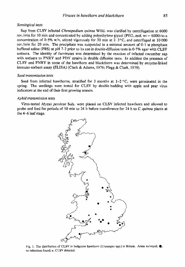
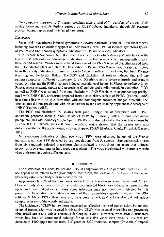
Fig. 1. The distribution of CLSV in hedgerow hawthorn (Crataegus spp.) in Britain. Areas surveyed, 0, no infections found; *, CLSV detected.
86 J . B. SWEET
R E S U L T S
Apple chlorotic leclfspot virus Chlorotic ring pattern leaf symptoms, similar to those described by Posnette (1957) and Sweet
& Campbell (1976) were observed in four of the 109 hawthorn samples. These four diseased plants and 23 symptomless hawthorns induced symptoms indicative of CLSV in the apple and pear virus indicators, though mostly the symptoms tended to be milder than those of typical apple and pear isolates of CLSV. Three symptomless hawthorns (B, C and D) induced milder symptoms or no symptoms in some CLSV indicator plants. The symptoms induced by these inocula and one of the diseased plants (hawthorn A) are described in Table 1. CLSV was detected in 18 of 26 sites in England, at one of three in Wales but at neither of two sites in Scotland (Fig. 1).
A hawthorn hedge on a Hereford nursery was examined and approximately 75% of the plants had chlorotic rings and lines on their leaves. Six hawthorns, selected at random, were indexed and were all found to be infected with CLSV, including one that was symptomless.
An adjacent hedge running at right angles,to the other contained many hawthorns though none showed leaf symptoms. Random tests on five plants detected no CLSV infection.
An isolate of CLSV was obtained from hawthorn A by sap inoculation to C. quinoa and eight further isolates by sap inoculation from the woody indicators quince C7/1 or Russian apple seedling R 12740/7A to which eight symptomless hawthorns had been itidexed. The symptoms induced in C. quinou and C. amaranticolor Coste & Reyn. varied in severity but were similar to those induced by typical apple and pear isolates of CLSV (Cropley, 1969~ ; Lister, 1970).
Stem pitting was induced in some Malus indicators (Table 1) by 17 of the 27 infected hawthorns, but not by any of the hawthorns without CLSV. Hawthorn A and five others induced stem pitting symptoms in Virginia Crab, indicating infection with the pathogen (SPV) causing stem pitting (Posnette & Cropley, 1963). SPV has not been characterised and is assumed to be a virus distinct from CLSV (Cropley, 1 9 6 8 ~ ; Mink, Shay, Gilmer & Stouffer, 197 1).
Three of the 67 tested blackthorns, including one tree showing chlorotic ring and line pattern leaf symptoms (Sweet, 19766), induced symptoms indicative of CLSV infection in the apple and pear virus indicators, though in repeated graft inoculations approximately one in three grafts of infected P. spinosa material failed to induce symptoms in the woody indicators. CLSV was not isolated by sap inoculation from the three blackthorns but PNRV was isolated from the tree with symptoms. However, CLSV could be transmitted by sap from graft infected indicators quince C7/1 and Mulus sylvestris R12740/7A. These CLSV isolates also induced typical symptoms in the herbaceous indicators.
CLSV isolates in C. quinoa sap formed single precipitin lines with antisera to CLSV-A, an apple isolate described by Lister & Hadidi (1971) and CLSV-S, an antiserum prepared from the soluble antigen of CLSV-A (Chairez & Lister, 1973). The precipitates were continuous with those of CLSV isolates from apple and pear with no spur formation or crossing over of the precipitate lines.
CLSV-S antiserum reacted in ELISA with sap from six infected hawthorns but not with sap from two hawthorns that failed to induce symptoms in the woody indicators. Two of the three infected blackthorns gave a positive ELISA reaction with CLSV-S antiserum, but the plant with chlorotic ring and line pattern symptoms reacted only with PNRV antiserum.
Inoculations by grafting C. quinoa shoots infected with a CLSV isolate from apple to five hawthorn and five blackthorn seedlings failed to induce symptoms, but CLSV was detected in two hawthorns and one blackthorn by indexing on woody indicators 1 yr after inoculation. CLSV was not graft transmitted from any of 55 seedlings raised from the seed of two infected hawthorn plants.
Viruses in hawthorn and blackthorn 81
No symptoms appeared in C. quinoa seedlings after a total of 74 transfers of groups of six aphids following variable feeding periods on CLSV-infected hawthorn, though M. persicae probed, fed and reproduced on infected hawthorn.
Ilaruiruses Seven of 67 blackthorns induced symptoms in Prunus indicators (Table 2). Four blackthorns,
including two with chlorotic ringspots on their leaves (Sweet, 19763) induced symptoms typical of PNRV and two induced symptoms indicative of PDV in the woody indicators.
The seventh blackthorn (plant X) induced necrotic spots which developed into holes in the leaves of P. serrulata cv. Shirofugen indicators in the first season which subseqeuntly died in their second season. Viruses were isolated from one of the PNRV infected blackthorns and from the PDV infected plant and plant X. In addition PDV and PNRV were readily sap-transmitted from the woody indicator P. avium cv. Sam to Cucumis sativus L. cvs Ohio, Butcher’s Disease Resisting and Perfection Ridge. The PDV and blackthorn A isolates induced ring and line pattern symptoms in Nicotiana tabacum L. cv. Xanthi-nc and a severe chlorosis and stunt in cucumber whereas the PNRV isolates induced necrotic local lesions in Phaseolus vulgaris L. cv. Prince, severe systemic mottle and necrosis in C. quinoa and a mild mosaic in cucumber. PDV as well as PNRV was isolated from one blackthorn. PNRV isolates in cucumber sap precipi- tated with PNRV-RA antiserum prepared from a sour cherry isolate of PNRV (Fulton, 19683) in a single line with no spur formation with the homologous antiserum/antigen precipitin line. The isolates did not precipitate with an antiserum to the East Malling apple mosaic serotype of PNRV (Fulton, 19686).
The PDV and blackthorn X isolates each gave a single line of precipitate with PDV-B antiserum prepared from a plum isolate of PDV by Fulton (1968~) forming continuous precipitate lines with homologous precipitin. PNRV was also detected in the four blackthorns by ELISA (D. J. Barbara, personal communication) which showed that the viruses were also distantly related to the apple mosaic virus serotype of PNRV (Barbara, Clark, Thrush & Casper, 1978).
No symptoms indicative of plum pox virus (PPV) were observed in any of the Prunus indicators, nor was PPV detected by sap inoculations from P. spinosa. Sap transmission tests from six randomly selected blackthorn plants isolated a virus from one plant that induced nepovirus-type symptoms in herbaceous test plants. The virus precipitated with arabis mosaic virus antiserum in double diffusion tests.
D I S C U S S I O N
The distribution of CLSV, PNRV and PDV in hedgerows was in no particular pattern and did not appear to be related to the proximity of fruit crops, the location or the aspect of the hedge. No newly established hedges or trees were tested.
Approximately 25% of the hawthorns and 4% of the blackthorns were infected with CLSV. However, only about two thirds of the grafts from infected blackthorn induced symptoms in the apple and pear indicators and thus some infections may not have been detected by this procedure. In addition the mildness of some of the virus isolates suggested that, as in the USA (Gilmer & Mink, 1971), there may also have been some CLSV isolates that did not induce symptoms in any of the woody indicators.
The incidence of CLSV in hawthorn suggested an effective means of transmission, but no seed or aphid transmission was detected in this study. CLSV was detected in seedling and previously virus-tested apple and quince (Posnette & Cropley, 1964). However, when EMLA fruit trees which had been on commercial holdings for at least five years were tested, CLSV was not detected in 1400 apple mother trees, 715 pears or 3200 rootstock samples (Posnette, Campbell
88 J . B. SWEET
& Cropley, 1976). Sweet (1978) tested some 200-yr-old apple and pear trees originally propagated from seed and detected CLSV in two of 25 apples, but Campbell (196 1) failed to detect CLSV in the original 150-yr-old Bramley seedling apple. So if natural spread of CLSV does occur in some fruit trees it is extremely slow, whereas the distribution of CLSV in hawthorns suggests that in these plants natural transmission is quicker.
The distribution of CLSV in one of the Hereford hawthorn hedges was consistent with virus spread along the hedge, perhaps by rootgrafting, from initial sources of infection. There was no plant contact between this hedge and the adjacent one in which no CLSV infection was detected, which again supports the view that plant contact may be the main method of secondary spread of CLSV in hawthorn. However, no means of establishment of the primary foci of CLSV infection can be suggested.
Sweet & Campbell (1976) and Sweet (1978) detected CLSV in only one of 14 clonal ornamental Crataegus species which have been commercially propagated on seedling C. rnonogyna or C. oxyacanthoides rootstocks for many years. This suggests that few seedling hawthorns are likely to be infected with CLSV.
Marenaud, Mazy & Chaffurin (1976) reported some spread of CLSV in plum, perhaps associated with the aphid-borne spread of plum pox virus, and Bem & Murant (1978) reported that the morphologically similar heracleum latent virus was also aphid transmitted but only from Heracleurn sphondylium L. and not other Umbelliferae. The aphid transmission of CLSV between hawthorns is now being investigated.
The incidence of CLSV in blackthorn suggested that it was not an important source of infection for cultivated trees. The incidence of PNRV and PDV in blackthorn (5% and 4%) was similar to that found in East Germany by Schimanski et al. (1975). These two viruses are pollen and seed-transmitted in cherry (Gilmer & Way, 1960) and PDV is pollen-transmitted in peach (Smith & Stubbs, 1976). However, Schimanski et al. (1975) detected no seed transmission of PNRV in blackthorn or bird cherry (P. padus), and thought these species played only a minor role in the epidemiology of PNRV and PDV in stone fruit crops. Seed and pollen transmission of PNRV and PDV has been recorded in the sub-genus Prunophora (Cameron, Milbrath & Tate, 1973) in the USA but no natural spread has been recorded in Britain. However, it seems probable that some infection of P. spinosa with PNRV and PDV is due to seed or pollen transmission, but it is unlikely that these plants play a significant role in the epidemiology of PNRV and PDV in cultivated Prunus in Britain. The rate at which fruit trees become infected with CLSV is very slow and the presence of CLSV in hawthorn appears to be of little significance in the epidemiology of CLSV in fruit trees, or related ornamentals.
Arabis mosaic virus (AMV) is common in woody rosaceous plants (Sweet, 1975; Cooper & Sweet, 1976) and viruliferous nematodes are prevalent in many hedgerows (Murant, 1970) hence the recovery of AMV from a P. spinosa plant was not unexpected.
The author thanks Mr A. R. Carter, other ADAS officers, Dr J. I. Cooper, Dr A. T. Jones and Mrs Dorothy Hinchcliffe for collecting plant material. Thanks are also due to Professor I$ W. Fulton and Dr R. M. Lister for supplying antisera, Drs Caroline Flegg, M. F. Clark and D. J. Barbara for carrying out ELISA tests, Ms Caroline Carter for cartography, Mrs Joan Llewellyn for typing the manuscripts and Mr C. G. Thomas and his staff for help in the nursery.
REFERENCES
BARBARA, D. J., CLARK, M. I?, THRESH, J. M. Br CASPER, R. (1978). Rapid detection and serotyping of prunus necrotic ringspot virus in perennial crops by enzyme-linked immunosorbent assay. Anna Is of Applied Biology 90, 395-399.
BEM, F. 8t MURANT, A. ~:(1978). Viruses from Heracleum sphondylium. Report of the Scottish Horticultural Research Institute for 1977, 100 pp.
Viruses in hawthorn and blackthorn 89
CAMERON, H. R., MILBRATH, J. A. L TATE, L. A. (1973). Pollen transmission of prunus ringspot virus in prune and sour cherry orchards. Plant Disease Reporter 57,241-243.
CAMPBELL, A. I. (1961). Virus diseases of fruit trees. VII. Latent virus in apple varieties and rootstocks. Report of the Long Ashton Research Station for 1960, pp. 5 1-55.
CHAIREZ, R. & LISTER, R. M. (1973). Soluble antigens associated with infection with apple chlorotic leafspot virus. Virology 54,506-5 14.
CLAPHAM, A. R., TUTIN, T. G. & WARBURG, E. F. (1962). Flora of the British Isles. 2nd Edition, Cambridge University Press, 1269 pp.
CLARK, M. F. & ADAMS, A. N. (1977). Characteristics of the microplate method of enzyme-linked immuno- sorbent assay (ELISA) for the detection of plant viruses. Journal of General Virology 34 475-483.
COOPER, J. I. & SWEET, J. B. (1976). The detection of viruses with nematode vectors in six woody hosts. Forestry 49, 73-78.
CROPLEY, R. (1968~) . Comparison of some apple latent viruses. Annals of Applied Biology 61, 36 1-372. CROPLEY, R. (19686). The identification of plum pox (Sharka) virus in England. Plant Patholoy 17, 66-
70. CROPLEY, R. (19694. Apple chlorotic leafspot virus. Technical Communication of the Commonwealth
Bureau of Horticulture No. 30, Suppl. 21314.10. CROPLEY, R. (19696). Virus diseases in ornamental Prunus plants. Nurseryman and Garden Centre 149,
357-359. FLEGG, c . L. & CLARK, M. F. (1979). The detection of apple chlorotic leafspot virus by a modified
procedure of enzyme-linked immunosorbent assay (ELISA). Annals ofApplied Biology 91,6 1-65. FULTON, R. w. (1961). Characteristics of a virus endemic in wild Prunus. Tid'skrift for Planteavl 65,
FULTON, R. w . (1968a). Relationships among the ringspot viruses of Prunus. Tagungsberichte. Deutsche
FULTON, R. w. (19686). Serology of viruses causing cherry necrotic ringspot, plum-line pattern, rose
PULTON, R. w. (1970). Prune dwarf virus. C.M.I.1AA.B. Descriptions of Plant Viruses No. 19. GILMER, R. M. & MINK, G. I. (1971). Latent viruses of apple. 111. Indexing with Chenopodium quinoa.
GILMER, R. M. ~i WAY, R. D. (1960). Pollen transmission of necrotic ringspot and prune dwarf virus in sour
JORDOVIC, M. (1969). Ispitivanja Prunus spinosa L. kao domacina virusa sarke. Zaftita BiGa Beograd
LISTER, R. M. (1970). Apple chlorotic leafspot virus. C.M.I.1AA.B. Descriptions of Plant viruses No. 30. LISTER, R. M. & HADIDI, A. F. (197 1). Some properties of apple chlorotic leafspot virus and their relation to
MARENAUD, c., MAZY, K. & CHAFFURIN, M. (1976). Observations sur la diffusion et le detection du virus de
MINK, G. I., SHAY, J. R., GILMER, R. M. & STOUFFER, R. F. (1971). Latent viruses of apple. 11. Symptoms in
MURANT, A. F. (1970). The importance of wild plants in the ecology of nematode transmitted viruses.
POSNETTE, A. F. (1957). Virus diseases of pears in England. Journal of Horticultural Science 32, 53-61. POSNETTE, A. F., CAMPBELL, A. I. & CROPLEY, R. (1976). The reindexing of EMLA scionwood trees and
rootstocks growing on special stock nurseries. Acta Horticulturae 67,275-278. POSNETTE, A. F. & CROPLEY, R. (1963). Apple stem pitting. Commonwealth Agricultural Bureau,
Technical Communication no. 30, pp. 77-78. POSNETTE, A. F. & CROPLEY, R. (1964). Natural spread of chlorotic leafspot virus in apple and quince.
Report of the East Mulling Research Station for 1963, pp. 115-1 16. SCHIMANSKI, H-H., SCHMELZER, K., KEGLER, H. ALBRECHT, H. J. (1975). Wildwachsende Prunus arten
der untergattungen Prunophora und Padus als naturliche wirtspflanzen fur kirschanringflecken-viren. Zentralblatt fur Bakteriologie, Parasitenkunde, Znfectionskrankheiten und Hygiene 30, 109- 120.
AINHA, R. c., TREMAINE, J. H., VALENTA, v. L WETTE, c. (1976). New groups of plant viruses approved by the International Committee on Taxonomy of Viruses, September 1975. htervirology 6, 181-184.
147-150.
Akademie der Landwirtschaftswissenschaften zu Berlin 97, 123-138.
mosaic and apple mosaic. Phytopathology 58, 635-638.
Search Agriculture 1, 15-2 1.
cherries. Phytopathology 50,624-625.
105,253-259.
purification problems. Virology 45,240-25 1.
le sharka dans un verger pichers d'Aquitaine. Phytoma 28,20-24.
woody indicators and strain variation. Search Agriculture 1,9-15.
Outlook on Agriculture 6, 1 14-1 2 1.
SHEPHERD, R. J., FRANCKI, R. I. B., HIRTH, L., HOLLINGS, M., INOWE, T., MACLOED, R., PURCIFULL, D. E.,

90 J . B . S W E E T
SMITH, P. R. & STUBBS, L. L. (1976). Transmission of prune dwarf virus by peach pollen and latent infection in peach trees. Australian Journal of Agricultural Research 21, 839-843.
SWEET, J. B. (1975). Soil-borne viruses occurring in nursery soils and infecting some ornamental species of Rosaceae. Annals of Applied Biology 19,49-54.
SWEET, J. B. ( 1 9 7 6 ~ ) . Virus diseases of some ornamental and indigenous trees and shrubs in Britain. Acta Horticulturae 59,83-92.
SWEET, J. B. (1976b). Prunus necrotic ringspot virus in ornamental and indigenous Prunus spp. Plant Pathology 25,55.
SWEET, J. 8. (1978). Studies of three groups of viruses infecting woody amenity plants. Ph.D. thesis, University of Bristol.
SWEET, J. B. CAMPBELL, A. I. (1976). Pome fruit virus infections of some woody ornamental and indigenous species of Rosaceae. Journal of Horticultural Science 5 1,9 1-97.
(Received 2 April 1979)