Embed Size (px)
Citation preview
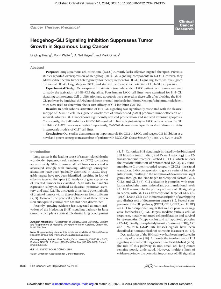
Cancer Therapy: Preclinical
Hedgehog–GLI Signaling Inhibition Suppresses TumorGrowth in Squamous Lung Cancer
Lingling Huang1, Vonn Walter2, D. Neil Hayes2, and Mark Onaitis1
AbstractPurpose: Lung squamous cell carcinoma (LSCC) currently lacks effective targeted therapies. Previous
studies reported overexpression of Hedgehog (HH)–GLI signaling components in LSCC. However, they
addressed neither the tumor heterogeneity nor the requirement forHH–GLI signaling.Here,we investigated
the role of HH–GLI signaling in LSCC, and studied the therapeutic potential of HH–GLI suppression.
ExperimentalDesign:Gene expression datasets of two independent LSCCpatient cohortswere analyzed
to study the activation of HH–GLI signaling. Four human LSCC cell lines were examined for HH–GLI
signaling components. Cell proliferation and apoptosis were assayed in these cells after blocking the HH–
GLI pathway by lentiviral-shRNAknockdownor small-molecule inhibitors. Xenografts in immunodeficient
mice were used to determine the in vivo efficacy of GLI inhibitor GANT61.
Results: In both cohorts, activation of HH–GLI signaling was significantly associated with the classical
subtype of LSCC. In cell lines, genetic knockdown of Smoothened (SMO) produced minor effects on cell
survival, whereas GLI2 knockdown significantly reduced proliferation and induced extensive apoptosis.
Consistently, the SMO inhibitor GDC-0449 resulted in limited cytotoxicity in LSCC cells, whereas the GLI
inhibitor GANT61was very effective. Importantly, GANT61 demonstrated specific in vivo antitumor activity
in xenograft models of GLIþ cell lines.
Conclusion:Our studies demonstrate an important role for GLI2 in LSCC, and suggest GLI inhibition as a
novel andpotent strategy to treat a subset of patientswith LSCC.ClinCancer Res; 20(6); 1566–75.�2014AACR.
IntroductionLung cancer is the leading cause of cancer-related deaths
worldwide. Squamous cell carcinoma (LSCC) comprisesapproximately 30% of non–small cell lung cancers and isassociated strongly with smoking. Although oncogenicalterations have been gradually described in LSCC, drug-gable targets have not been identified, resulting in lack ofeffective targeted therapies (1). Analysis of gene expressionof resected tumors has classified LSCC into four mRNAexpression subtypes, defined as classical, primitive, secre-tory, andbasal (2). Theoncogenic drivers andpotential cellsof origin of tumors within these subtypes are likely different(2, 3). However, the practical application of these expres-sion subtypes in clinical care has not been determined.
Recently, growing evidence has suggested aberrant acti-vation of the Hedgehog (HH) signaling pathway in lungcancer, which plays a critical role during lung development
(4, 5). Canonical HH signaling is initiated by the binding ofHH ligands (Sonic, Indian, and Desert Hedgehog) to a 12-transmembrane receptor Patched (PTCH), which relievesthe catalytic inhibition of Smoothened (SMO), a 7-trans-membrane G-protein-coupled receptor (GPCR)–like signaltransducer. SMO de-repression triggers a series of intracel-lular events, resulting in the activation of downstream targetgenes through the zinc-finger transcription factors GLI1,GLI2, and GLI3 (6). GLI activation is complex, with regu-lationatboththetranscriptionalandposttranslational levels(7). GLI2 seems to be the primary activator of HH signalingin cancer, with GLI1 as a transcriptional target of GLI2 (8–10).GLI2 andGLI1 also induce transcriptionof overlappingand distinct sets of downstream targets (11). Several com-ponents of theHHpathway (PTCH,GLI1, GLI2, andHHIP)are GLI transcriptional targets that induce positive or neg-ative feedbacks (7). GLI targets mediate various cellularresponses, notably enhanced cell proliferation and survivalby upregulating D-type cyclins and antiapoptotic proteins(12–14).Finally,phosphatidylinositol3-kinase(PI3K)/AKTand RAS–MEK (MAP–ERK kinase) signals have beendescribed as noncanonicalHHactivators in cancer (15–17).
Dysregulation of the HH pathway has been implicated ina variety of cancers (18). Although the involvement of HHsignaling in small-cell lung cancer is well established (4, 5),the role of this pathway in non–small cell lung cancerremains poorly understood. However, multiple lines ofevidence point to the potential importance of HH signaling
Authors' Affiliations: 1Department of Surgery, Duke University, Durhamand 2Department of Medicine, University of North Carolina, Chapel Hill,North Carolina
Note: Supplementary data for this article are available at Clinical CancerResearch Online (http://clincancerres.aacrjournals.org/).
Corresponding Author: Mark Onaitis, Duke University, DUMC Box 3305,Durham, NC 27710. Phone: 919-684-6974; Fax: 919-684-8508; E-mail:[email protected]
doi: 10.1158/1078-0432.CCR-13-2195
�2014 American Association for Cancer Research.
ClinicalCancer
Research
Clin Cancer Res; 20(6) March 15, 20141566
on March 21, 2020. © 2014 American Association for Cancer Research. clincancerres.aacrjournals.org Downloaded from
Published OnlineFirst January 14, 2014; DOI: 10.1158/1078-0432.CCR-13-2195
in LSCC. Immunohistochemical studies in patient speci-mens reported the overexpression of HH signaling compo-nents (19, 20). Microarray analysis identified hyperactiveHH signaling in LSCC (21). HH–GLI signaling has alsobeen implicated in squamous cancer of other organs (22–24). Despite these studies, very little is known about thespecific role of HH signaling in regulating cellular survivaland proliferation in LSCC.Targeted inhibitors of the HH pathway have become
available recently. Because of its accessibility on themembrane and its importance in regulation of the path-way, SMO has been the primary focus in the developmentof small-molecule inhibitors of the HH pathway. GDC-0449 (vismodegib; Genentech) is an orally administeredagent that selectively suppresses SMO activity and was thefirst SMO inhibitor to progress to clinical trials. It hasproduced promising antitumor responses in patients withadvanced basal cell carcinoma and medulloblastoma (25,26), but resistance has been reported (27, 28). The resis-tance to SMO inhibitors highlights the therapeutic needto target downstream effectors to maintain robust on-target responses. In a cell-based screen of GLI-mediatedtranscription (29), the small molecule GANT61 wasidentified as a specific inhibitor of GLI1 and GLI2. Itsuppresses the DNA-binding capacity of GLIs and inhibitsGLI-mediated transcription. GANT61 reduces prolifera-tion and induces apoptosis in a GLI-specific fashion inprostate cancer (29), colon carcinoma (30, 31), oralsquamous cell carcinoma (23), pancreatic cancer (32),neuroblastoma (33), and chronic lymphocytic leukemia(34). In this study, we investigate the role of HH–GLIsignaling in LSCC and assess the clinical feasibility ofusing GDC-0449 or GANT61 to treat LSCC.
Materials and MethodsRNAseq and microarray analysisRSEM values (35) for 178 tumor samples from The
Cancer Genome Atlas (TCGA) LSCC study (3) were con-
verted to expression measurements by replacing valuesequal to zero with the smallest nonzero value, taking alog2 transformation. After median centering by gene, heat-maps of the expression values from TCGA (3) and micro-array data from 56 LSCC samples collected at the Universityof North Carolina (ref. 2; UNC cohort) were produced withR 2.15.1 (36) and the gplots package. Hypotheses weresubsequently tested: one-sided Wilcoxon rank sum testswere used to test the null hypothesis that the mean expres-sion levels of PTCH1, GLI1, GLI2, GLI3, and SUFU are thesame in the classical subtype as all other subtypes com-bined. For PTCH1, GLI1, and GLI2, the alternative hypoth-esis was that the expression levels are higher in the classicalsubtype, whereas for GLI3 and SUFU, the alternativehypothesis was that the expression levels are lower in theclassical subtype. A Bonferroni adjustment was applied tocorrect for multiple comparisons.
Two-sided Wilcoxon rank sum tests were used to test thenull hypothesis that GLI1 and GLI2 expression values wereequal in the TCGA andUNC cohorts. Spearman correlationcoefficients were computed on the basis of the uncenteredexpression values of GLI1, GLI2, TP63, PIK3CA, and SOX2in both cohorts. The resultingunadjustedP valueswereusedto assess the significance of these associations (Supplemen-tary Table S2).
Gene expression data from 20 LSCC cell lines wereobtained from the Cancer Cell Line Encyclopedia (CCLE;ref. 37). After median centering the expression values bygene, the centroid classifier from ref. (2) was used to predictexpression subtypes for each line by finding the nearestcentroid using a distance metric equal to one minus thePearson correlation coefficient. Gene expression heatmapswere then produced using R2.15.1 (36) and the gplotspackage.
Cell culture and reagentsNCI-H520, NCI-H2170, NCI-H226, and SK-MES-1 cells
were obtained from the American Type Culture Collection.Cell lines were routinely verified by morphology andgrowth characteristics, and verified biannually to be myco-plasma-free. NCI-H520, NCI-H2170, and NCI-H226 cellswere maintained in the RPMI-1640 medium containing10% FBS. SK-MES-1 cells were maintained in minimumessential medium (MEM) containing 10% FBS, 0.1mmol/Lnonessential amino acids, and 1.0 mmol/L sodium pyru-vate. Antibodies used were SHH, PTCH, and SMO (SantaCruz Biotechnology); GLI2 and GAPDH (Abcam); GLI1(Novus Biologicals); cleaved caspase-3 and cleaved PARP(Cell Signaling Technology); and CCND1 (BD Bios-ciences). Compounds used were GANT61 (Sigma) andGDC-0449 (Chemietek).
Lentiviral production and transductionLentiviral short hairpin RNA (shRNA) clones (Sigma;
MISSION RNAi) targeting SMO, GLI2, and the nontargetingcontrol (SHC002) were purchased from Sigma-Aldrich.293T cells were plated in 10-cm plates 24 hours beforetransfection inDulbecco’sModified EagleMedium (DMEM)
Translational RelevanceTargeted therapeutics for lung squamous cell carcino-
ma (LSCC) are currently lacking. In this study, we haveanalyzed molecular subtypes of LSCC and identifiedoverexpression of Hedgehog family members in theclassical subtype. In representative LSCCcell lines, genet-ic deletion of Smoothened (SMO) produced minoreffects on cell survival, whereas GLI2 knockdown greatlyreduced cell viability and induced extensive apoptosis.Using both in vitro and in vivo approaches, we evaluatedtherapeutic efficacy of GDC-0449, a clinically availableSMO inhibitor, as well as GANT61, a targeted GLIinhibitor. GANT61 was significantly more effective thanGDC-0449 in reduction of proliferation and inductionof apoptosis. We report SMO-independent regulation ofGLI in LSCC, and present a potential strategy of targetingGLIs to treat a subset of patients with LSCC.
Hedgehog Signaling in Squamous Cell Lung Cancer
www.aacrjournals.org Clin Cancer Res; 20(6) March 15, 2014 1567
on March 21, 2020. © 2014 American Association for Cancer Research. clincancerres.aacrjournals.org Downloaded from
Published OnlineFirst January 14, 2014; DOI: 10.1158/1078-0432.CCR-13-2195
containing 10% FBS without antibiotics; 5 mg of shRNAplasmid, 4 mg psPAX2, and 1 mg pCI-VSVGpackaging vectors(Addgene) were cotransfected into 293T cells using Lipofec-tamine 2000 Reagent (Invitrogen). Viral supernatants werecollected, centrifuged, and filtered with 0.45-mm PES sterileSyringe filter. Target cells were plated and incubated at 37�C,5% CO2 overnight, and changed to medium containinglentivirus and 8 mg/mL polybrene. Control plates wereincubated with medium containing 8 mg/mL polybrene.Cells were changed to fresh culture medium 24 hours afterinfection. Puromycin selection (5 mg/mL) was started 48hours after infection and continued for 4 to 5 days until noviable cells were observed in control plates. Once decreasedexpression of the targeted gene was confirmed, cells wereused for subsequent experiments. Stable expression of non-targeting control, SMO, or GLI2 shRNAs was ensured byculturing cells in the presence of puromycin.
The shRNA sequences were as follows:
SMO sh1 (50-CCGGCCTGATGGACACAGAACTCATCTCG-AGATGAGTTCTG TGTCCATCAGGTTTTT-30)
SMO sh2 (50-CCGGCATCTTTGTCATCGTGTACTACTCG-AGTAGTACACGA TGACAAAGATGTTTTT-30)
SMO sh3 (50-CCGGGTGGAGAAGATCAACCTGTTTCTCG-AGAAACAGGTTG ATCTTCTCCACTTTTT-30)
GLI2 sh1 (50-CCGGCCAACGAGAAACCCTACATCTCTCG-AGAGATGTAGGGT TTCTCGTTGGTTTTTG-30)
GLI2 sh2 (50-CCGGCACTCAAGGATTCCTGCTCATCTCG-AGATGAGCAGGAA TCCTTGAGTGTTTTTG-30)
GLI2 sh3 (50-CCGGGCTCTACTACTACGGCCAGATCTCG-AGATCTGGCCGTA GTA GTAGAGCTTTTTG-30)
Assessment of cell viability and caspase-3/7 activityCell viability and caspase-3/7 activity were determined by
using ApoLive-GloMultiplex Assay (Promega) according tothemanufacturer’s instructions. Briefly, cells were seeded in96-well clear-bottomwhite plates at a density of 10,000 cellsper well and incubated with completemedium overnight at37�C, 5% CO2. The following day, cells were changed into0.5% FBS-containing medium with either dimethyl sulfox-ide (DMSO) control or drugs at designated concentrations(0.1% final DMSO concentration) as triplicates and treatedfor 96 hours. At the end of treatment, viability reagent wasadded into all wells and gently mixed. After 1.5-hourincubation at 37�C, fluorescence was measured at thewavelength set 355EX/520EM by a FLUOstar Omega Micro-plate reader. Later, Caspase-Glo 3/7 Reagent was added toall wells and gently mixed. Luminescence was measuredafter 1-hour incubation at room temperature. The readingof the blank control was subtracted from readings of otherwells as the background in the data analysis.
RNA isolation and quantitative PCRTotal RNAwas isolatedusing theQiagenRNeasyMiniKit,
treated with DNase I (Invitrogen), and converted to cDNAusing iScript cDNA Synthesis Kit (Bio-Rad). Real-time PCRwas performed using TaqMan Gene Expression Master Mix
on an Eppendorf Mastercycler, and raw data were analyzedby Realplex software. TaqMan probes for SHH, PTCH1,SMO,GLI1,GLI2,HHIP, andGAPDHwere purchased fromApplied Biosystems.
Western blot analysisTotal cellular lysates were prepared by using RIPA buffer
(Sigma) with protease inhibitor cocktail (Sigma) and Phos-STOP (Roche). Protein concentrations were determined bythe Micro BCA Protein Assay Kit (Thermo Scientific). Pro-teins were separated on the NuPAGE 4%–12% Bis-Tris Gel(Life Technologies) and transferred using Invitrolon PVDF/Filter Paper Sandwich. Membranes were blocked with 5%nonfat drymilk or 5%bovine serumalbumin (BSA) in0.1%Tris-buffered saline with 0.1% Tween 20 (TBST) for 1 hourat room temperature and then incubated with primaryantibody overnight at 4�C. They were subsequently washedwith 0.1%TBST and incubatedwith the secondary antibodyfor 1 hour at room temperature. Western Lightning -ECL(PerkinElmer) was used to develop the membranes.
Xenograft and tumor treatmentOfnote, 106NCI-H520 cells, 106NCI-H2170 cells, or 5�
106NCI-H226 cellswere suspended in a total volumeof 100mL of a 1:1 mixture of RPMI-1640 medium: Matrigel (BDBiosciences). Cellswere injected subcutaneously in the rightposterior flank of 6- to 8-week C.129S7 (B6)-Rag1tm1Mom/ J(Rag1�/�) mice. Tumors were grown until they reached amedian size of approximately 250 mm3 (NCI-H520),approximately 230 mm3 (NCI-H2170), and approximately150 mm3 (NCI-H226). Animals were randomly dividedinto groups and treated with solvent only (corn oil:ethanol,4:1) or GANT61 in solvent (50 mg/kg). Treatments weregiven every other day for 20 days by intraperitoneal injec-tion. Tumor volumes were calculated by the formula 0.52�length � (width)2. At the end of treatment, tumors wereremoved, weighed, and processed for subsequent analysis.All animal experimentswere approvedby and conformed tothe policies and regulations of the Institutional AnimalCareand Use Committees at Duke University.
ResultsActivation of HH–GLI signaling is associated with theclassical subtype of human LSCC
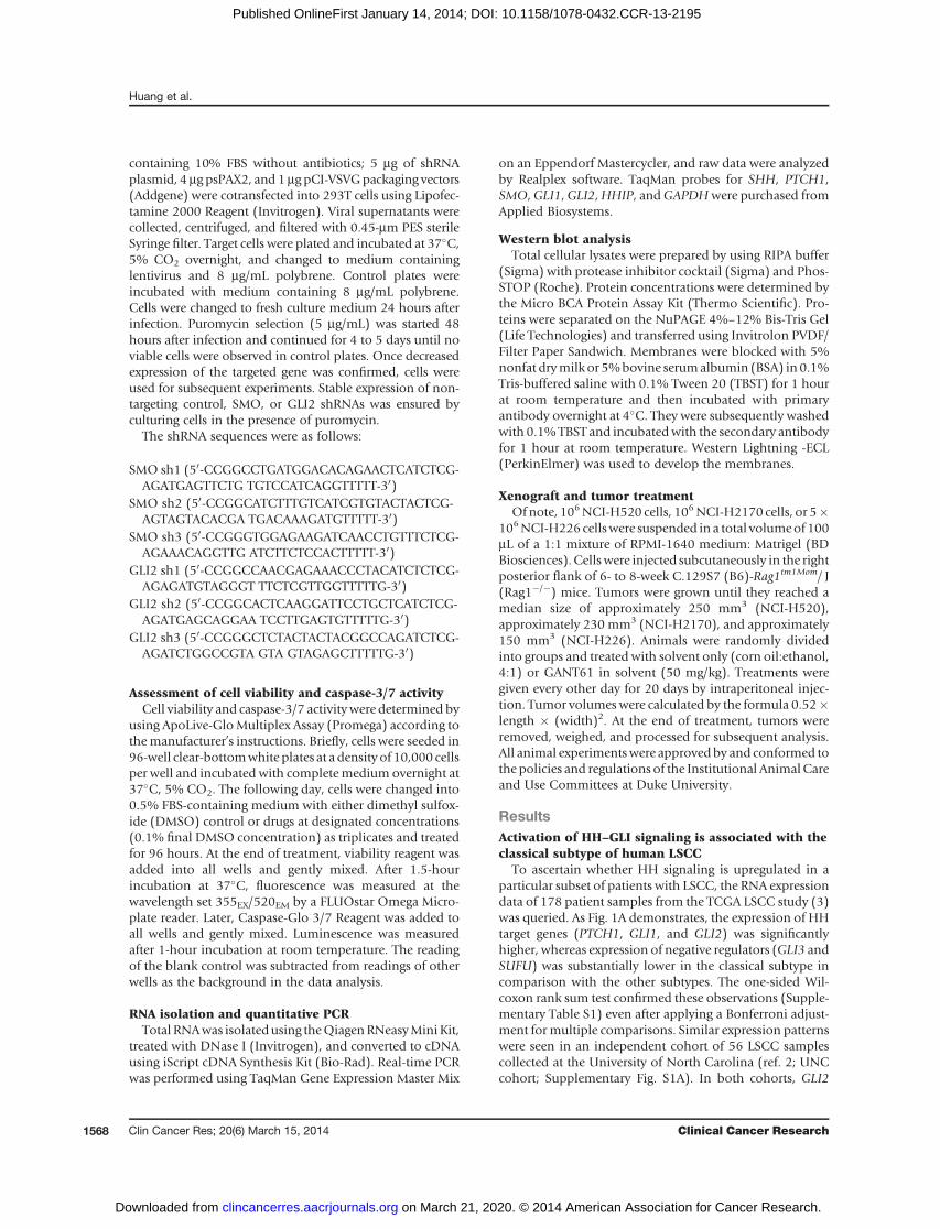
To ascertain whether HH signaling is upregulated in aparticular subset of patients with LSCC, the RNA expressiondata of 178 patient samples from the TCGA LSCC study (3)was queried. As Fig. 1A demonstrates, the expression of HHtarget genes (PTCH1, GLI1, and GLI2) was significantlyhigher, whereas expression of negative regulators (GLI3 andSUFU) was substantially lower in the classical subtype incomparison with the other subtypes. The one-sided Wil-coxon rank sum test confirmed these observations (Supple-mentary Table S1) even after applying a Bonferroni adjust-ment formultiple comparisons. Similar expression patternswere seen in an independent cohort of 56 LSCC samplescollected at the University of North Carolina (ref. 2; UNCcohort; Supplementary Fig. S1A). In both cohorts, GLI2
Huang et al.
Clin Cancer Res; 20(6) March 15, 2014 Clinical Cancer Research1568
on March 21, 2020. © 2014 American Association for Cancer Research. clincancerres.aacrjournals.org Downloaded from
Published OnlineFirst January 14, 2014; DOI: 10.1158/1078-0432.CCR-13-2195
mRNA level was significantly higher thanGLI1 (Fig. 1B andSupplementary Fig. S1B). Samples with high GLI2 expres-sion were mainly found in the classical subtype, althoughoccasionally in other subtypes. When taking the 75th per-centile of all GLI2 expression values in a given cohort as thethreshold for high GLI2, 55% (TCGA cohort) and 52%(UNC cohort) of all classical subtype samples exhibitedhighGLI2 expression (Fig. 1C and Supplementary Fig. S1C).Strong positive correlations betweenGLI2 and the prom-
inent markers for the classical subtype (SOX2, TP63, andPIK3CA) on chromosome 3qwere observed in both cohorts(Fig. 1D–F and Supplementary Fig. S1D–S1F). However,GLI1 was only associated with classical chr3q genes in theTCGA cohort (Supplementary Fig. S1G–I), suggesting thatGLI2 is highly likely to be the major signaling transducer inLSCC. Spearman correlation coefficients between GLI2/GLI1 and the classical subtype markers with correspondingP values are provided in Supplementary Table S2.
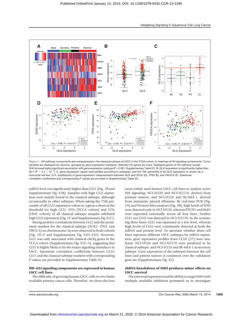
HH–GLI signaling components are expressed in humanLSCC cell linesThe difficulty of growing human LSCC cells in vitro limits
available primary cancer cells. Therefore, we chose the four
most widely used human LSCC cell lines to analyze activeHH signaling: NCI-H520 and NCI-H2170, derived fromprimary tumors; and NCI-H226 and SK-MES-1, derivedfrom metastatic pleural effusions. By real-time PCR (Fig.2A) andWestern blot analyses (Fig. 2B), high levels of SHHwere detected only inNCI-H520, whereas PTCH1 and SMOwere expressed universally across all four lines. NeitherGLI1 nor GLI2 was detected in NCI-H2170. In the remain-ing three lines, GLI1 was expressed at a low level, whereashigh levels of GLI2 were consistently detected at both themRNA and protein level. To ascertain whether these celllines represent different LSCC subtypes by mRNA expres-sion, gene expression profiles from CCLE (37) were ana-lyzed. NCI-H520 and NCI-H2170 were predicted to beclassical subtype, andNCI-H226 and SK-MES-1 as secretorysubtype. Gene expression of the subtypes between the celllines and patient tumors is consistent over the validationgene set (Supplementary Fig. S2).
shRNA knockdown of SMO produces minor effects onLSCC survival
TheuniversalexpressionandtheabilitytotargetSMOwithmultiple available inhibitors prompted us to investigate
Basal Secretory Primitive Classical
SHH
PTCH1
SMO
SUFU
GLI1
GLI2
GLI3
Color key
–1 –0.5 0 0.5 1
Value
D E F
A B C
–4–2
02
4G
ene
expr
essi
on
–4–2
02
4G
LI2
expr
essi
on
BasalSecretoryPrimitiveClassical
BasalSecretoryPrimitiveClassical
BasalSecretoryPrimitiveClassical
BasalSecretoryPrimitiveClassical
GLl1 GLl2
P = 4.2×10⁻⁵
BA SE PR CL
–50
SO
X2
expr
essi
on
TP
63 e
xpre
ssio
n
PIK
3CA
exp
ress
ion
5
–4 –2 0GLl2 expression GLl2 expression GLl2 expression
2 4 –4 –2 0 2 4 –4 –2 0 2 4
–4–2
02
46
8
12
34
5
Cor. = 0.45, P = 2.8×10⁻¹⁰Cor. = 0.56, P < 2.2×10⁻¹⁶Cor. = 0.48, P = 4.6×10⁻¹²
Figure 1. HH pathway components are overexpressed in the classical subtype of LSCC in the TCGA cohort. A, heatmap of HH signaling components. Tumorsamples are displayed as columns, grouped by gene expression subtypes. Selected HH genes are rows. Displayed genes of HH pathway exceptSHH showed highly significant associationwith gene expression subtype (P < 0.001; Supplementary Table S1). B,GLI2 expression is significantly higher thanGLI1 (P ¼ 4.2 � 10�5). C, gene expression values were plotted according to subtypes, and the 75th percentile of all GLI2 expression is shown as ahorizontal red line. D–F, scatterplots of gene expression measurements between GLI2 and SOX2 (D), TP63 (E), and PIK3CA (F). Spearmancorrelation coefficients and corresponding P values are provided in Supplementary Table S2.
Hedgehog Signaling in Squamous Cell Lung Cancer
www.aacrjournals.org Clin Cancer Res; 20(6) March 15, 2014 1569
on March 21, 2020. © 2014 American Association for Cancer Research. clincancerres.aacrjournals.org Downloaded from
Published OnlineFirst January 14, 2014; DOI: 10.1158/1078-0432.CCR-13-2195
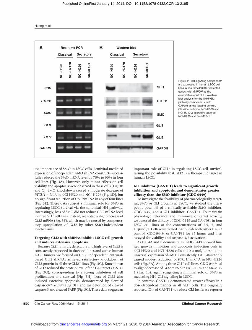
the importance of SMO in LSCC cells. Lentiviral-mediatedexpression of independent SMO shRNA constructs success-fully reduced the SMOmRNA level by 70% to 90% in fourcell lines (Fig. 3A). However, only minor effects on cellviability and apoptosis were observed in these cells (Fig. 3Band C). SMO knockdown caused a moderate decrease ofPTCH1 mRNA in NCI-H520 and NCI-H226 (Fig. 3D), butno significant reduction ofHHIPmRNA in any of four lines(Fig. 3E). These data suggest a minimal role for SMO inregulating LSCC survival via the canonical HH pathway.Interestingly, loss of SMO did not reduce GLI2mRNA levelin threeGLIþ cell lines. Instead,we noted a slight increase ofGLI2 mRNA (Fig. 3F), which may be caused by compensa-tory upregulation of GLI2 by other SMO-independentmechanisms.
Targeting GLI2 with shRNAs inhibits LSCC cell growthand induces extensive apoptosis
BecauseGLI1 is hardly detectable andhigh level ofGLI2 isconsistently expressed in three cell lines and across humanLSCC tumors, we focused on GLI2. Independent lentiviral-based GLI2 shRNAs achieved satisfactory knockdown ofGLI2 protein in all three GLI2þ lines (Fig. 3G). Knockdownof GLI2 reduced the protein level of the GLI target CCND1(Fig. 3G), corresponding to a strong inhibition of cellproliferation and survival (Fig. 3H). Loss of GLI2 alsoinduced extensive apoptosis, demonstrated by elevatedcaspase-3/7 activity (Fig. 3I), and the detection of cleavedcaspase-3 and cleaved PARP (Fig. 3G). These data suggest an
important role of GLI2 in regulating LSCC cell survival,raising the possibility that GLI2 is a therapeutic target inhuman LSCC.
GLI inhibitor (GANT61) leads to significant growthinhibition and apoptosis, and demonstrates greaterefficacy than the SMO inhibitor (GDC-0449)
To investigate the feasibility of pharmacologically target-ing SMO or GLI proteins in LSCC, we studied the thera-peutic potential of a clinically available SMO inhibitor,GDC-0449, and a GLI inhibitor, GANT61. To maintainphysiologic relevance and minimize off-target toxicity,we assessed the efficacy of GDC-0449 and GANT61 in fourLSCC cell lines at the concentrations of 2.5, 5, and10mmol/L. Cellswere treated in triplicatewith eitherDMSOcontrol, GDC-0449, or GANT61 for 96 hours, and thenassayed for viability and caspase-3/7 activation.
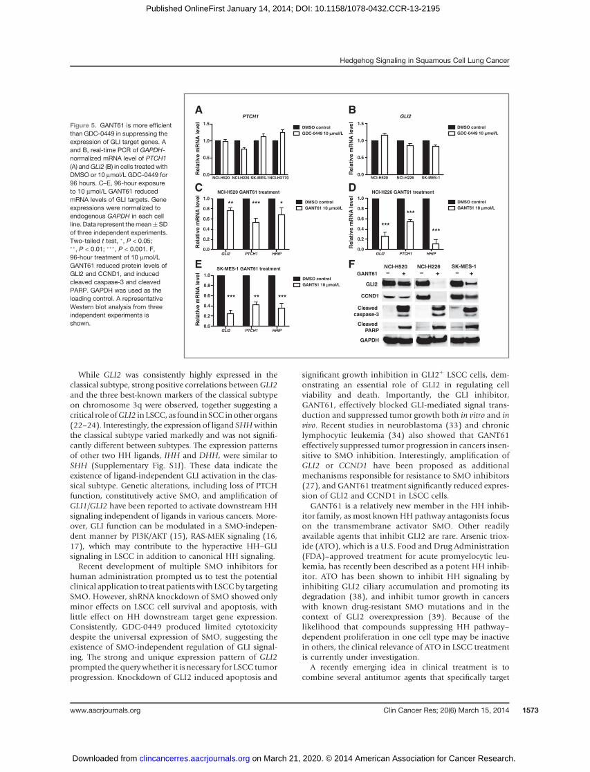
As Fig. 4A and B demonstrates, GDC-0449 showed lim-ited growth inhibition and apoptosis induction only inNCI-H520 and NCI-H226 cells at 10 m mol/L despite theuniversal expression of SMO.Consistently, GDC-0449 onlycaused modest reduction of PTCH1 mRNA in NCI-H226cells (Fig. 5A). Among three GLIþ cell lines, GDC-0449 ledto slight decrease ofGLI2mRNA inNCI-H226 and SK-MES-1 (Fig. 5B), again suggesting a minimal role of SMO inmediating HH–GLI signaling in LSCC.
In contrast, GANT61 demonstrated greater efficacy in adose-dependent manner in all GLIþ cells. The originallyreported IC50 of GANT61 to reduce GLI-luciferase reporter
SHH
PTCH1
SMO
GLI1
GLI2
GAPDH
GLI1
GLI2
GAPDH
Real-time PCR Western blotA B
NC
I-H
52
0
NC
I-H
21
70
NC
I-H
22
6
SK
-ME
S-1
NC
I-H
52
0
NC
I-H
21
70
NC
I-H
22
6
SK
-ME
S-1
SHH
PTCH1
SMO
Classical ClassicalSecretory Secretory
Figure 2. HH signaling componentsare expressed in human LSCC celllines. A, real-timePCR for indicatedgenes, with GAPDH as thequantitative control. B, Westernblot analysis for the SHH–GLIpathway components, withGAPDH as the loading control.Classical subtype, NCI-H520 andNCI-H2170; secretory subtype,NCI-H226 and SK-MES-1.
Huang et al.
Clin Cancer Res; 20(6) March 15, 2014 Clinical Cancer Research1570
on March 21, 2020. © 2014 American Association for Cancer Research. clincancerres.aacrjournals.org Downloaded from
Published OnlineFirst January 14, 2014; DOI: 10.1158/1078-0432.CCR-13-2195

activity is approximately 5 mmol/L (29), and 5 to 30 mmol/Lis commonly used (29–31). In our studies, the IC50 ofgrowth inhibition for three GLIþ lines was approximately5mmol/L (Fig. 4C). BothNCI-H520 andNCI-H226 showeda 55% reduction at 5 mmol/L and a 90% reduction at 10mmol/L in cell survival. SK-MES-1 displayed approximate40% and 60% decrease in viability at 5 and 10 mmol/L,respectively. As expected, GANT61 exhibited little cytotox-icity in GLI� NCI-H2170 cells. Consistently, increasedapoptosis was seen in GLIþ cell lines at correspondingGANT61 concentrations: NCI-H520 (1.8–2.3 fold), NCI-H226 (2.8–4 fold), and SK-MES-1 (2.4–2.6 fold), but not inGLI� NCI-H2170 (Fig. 4D).Real-time PCR demonstrated significant reduction of HH
downstream targets (GLI2, PTCH1, and HHIP) in NCI-H520 with a greater decrease in NCI-H226 and SK-MES-1in comparison with DMSO control (Fig. 5C–E). Westernblot analysis confirmed the reduction of GLI2 protein inGANT61-treated cells (Fig. 5F). Cleaved caspase-3 andcleaved PARP were detected in cells receiving GANT61 (Fig.
5F). The protein level of CCND1 was also decreased byGANT61 treatment, indicating impaired cell proliferationin addition to increased cell death (Fig. 5F).
Taken together, our results suggest that targeting HHsignaling at the level of GLI proteins may be more effectivethan targeting either the ligand SHHor the receptor SMO inLSCC, potentially due to the existence of the ligand orreceptor-independent pathway activation.
GANT61 suppresses GLIþ tumor progression in vivoCurrently, there are no available transgenic murine mod-
els that faithfully recapitulate human LSCC. Recently,patient-derived xenograft models of LSCC have shownpromise, but have not yet achieved satisfactory progress.Therefore, we used a xenograft model of representativehuman LSCC cell lines to determine the efficacy of GANT61in vivo. GLIþNCI-H520orNCI-H226 andGLI�NCI-H2170cancer cells were injected subcutaneously into the rightflank of immune-deficient Rag1�/� mice. Mice were ran-domly divided into two groups when tumors reached a
SMO
0.0
0.2
0.4
0.6
0.8
1.0
Re
lati
ve
mR
NA
Le
ve
l shNT
SMO sh1
SMO sh2
SMO sh3
NCI-H520 NCI-H226 SK-MES-1 NCI-H2170
***
***
*** ***
******
***
***
PTCH1
0.0
0.5
1.0
1.5
2.0
Rela
tive m
RN
A level
shNT
SMO sh1
SMO sh2
SMO sh3
NCI-H520 NCI-H226 SK-MES-1 NCI-H2170
SMO knockdown viability
0
25
50
75
100
125
Su
rviv
al fr
acti
on
(%
co
ntr
ol)
shNT
SMO sh1
SMO sh2
SMO sh3
NCI-H520 NCI-H226 SK-MES-1 NCI-H2170
HHIP
0.0
0.5
1.0
1.5
2.0
Rela
tive m
RN
A level
shNT
SMO sh1
SMO sh2
SMO sh3
NCI-H520 NCI-H226 SK-MES-1 NCI-H2170
SMO knockdown apoptosis
0.0
0.5
1.0
1.5
2.0
Casp
ase-3
/7 a
cti
vit
y
(fo
ld c
han
ge)
shNT
SMO sh1
SMO sh2
SMO sh3
NCI-H520 NCI-H226 SK-MES-1 NCI-H2170
GLI2
0
1
2
3
Rela
tive m
RN
A level
shNT
SMO sh1
SMO sh2
NCI-H520 NCI-H226 SK-MES-1
A B C
D E Fsh
NT
SK-MES-1
GLI2
Cleaved
caspase-3
Cleaved PARP
sh
NT
GL
I2 s
h1
GL
I2 s
h3
NCI-H520
sh
NT
GL
I2 s
h2
GL
I2 s
h3
NCI-H226
GL
I2 s
h2
GL
I2 s
h3
CCND1
GAPDH
G
GLI2 knockdown viability
0
20
40
60
80
100
Su
rviv
al fr
acti
on
(% c
on
tro
l)
shNT
NCI-H520 NCI-H226 SK-MES-1
GLI2 sh1
GLI2 sh2
GLI2 sh3***
***
***
***
******
GLI2 knockdown apoptosis
0
2
4
6
8
Casp
ase-3
/7 a
cti
vit
y
(fo
ld c
han
ge)
shNT
NCI-H520 NCI-H226 SK-MES-1
GLI2 sh1
GLI2 sh2
GLI2 sh3***
******
***
***
**
H I
Figure 3. SMO plays a minor role, whereas GLI2 is required for LSCC survival in vitro. A, change of GAPDH-normalized SMO mRNA level by real-time PCRfollowing lentiviral shRNA knockdown. Nontargeting shRNA control (shNT) or two independent shRNAs (SMO sh1, 2, 3) targeting SMO were usedin each cell line. B and C, measurements of viability (B) and apoptosis (C) in cells after SMO knockdown. Data were normalized to shNT control and representthe mean � SD of three independent experiments. D–F, real-time PCR showing the GAPDH-normalized mRNA level of PTCH1 (D), HHIP (E), and GLI2(F) after SMO knockdown. G, Western blot analyses showing GLI2 knockdown by independent shRNAs (GLI2 sh1, 2, 3). GLI2 shRNAs significantly reducedGLI2 protein levels, in comparison with the shNT control. GLI2 knockdown also caused induction of cleaved caspase-3, cleaved PARP, andreduction of CCND1. GAPDH was used as the loading control. A representative Western blot analysis from three independent experiments is shown.Knockdown of GLI2 in LSCC cells significantly reduced proliferation (H) and induced apoptosis (I). Data were normalized to shNT control andrepresent the mean � SD of three independent experiments. Two-tailed t test, ��, P < 0.01; ���, P < 0.001.
Hedgehog Signaling in Squamous Cell Lung Cancer
www.aacrjournals.org Clin Cancer Res; 20(6) March 15, 2014 1571
on March 21, 2020. © 2014 American Association for Cancer Research. clincancerres.aacrjournals.org Downloaded from
Published OnlineFirst January 14, 2014; DOI: 10.1158/1078-0432.CCR-13-2195
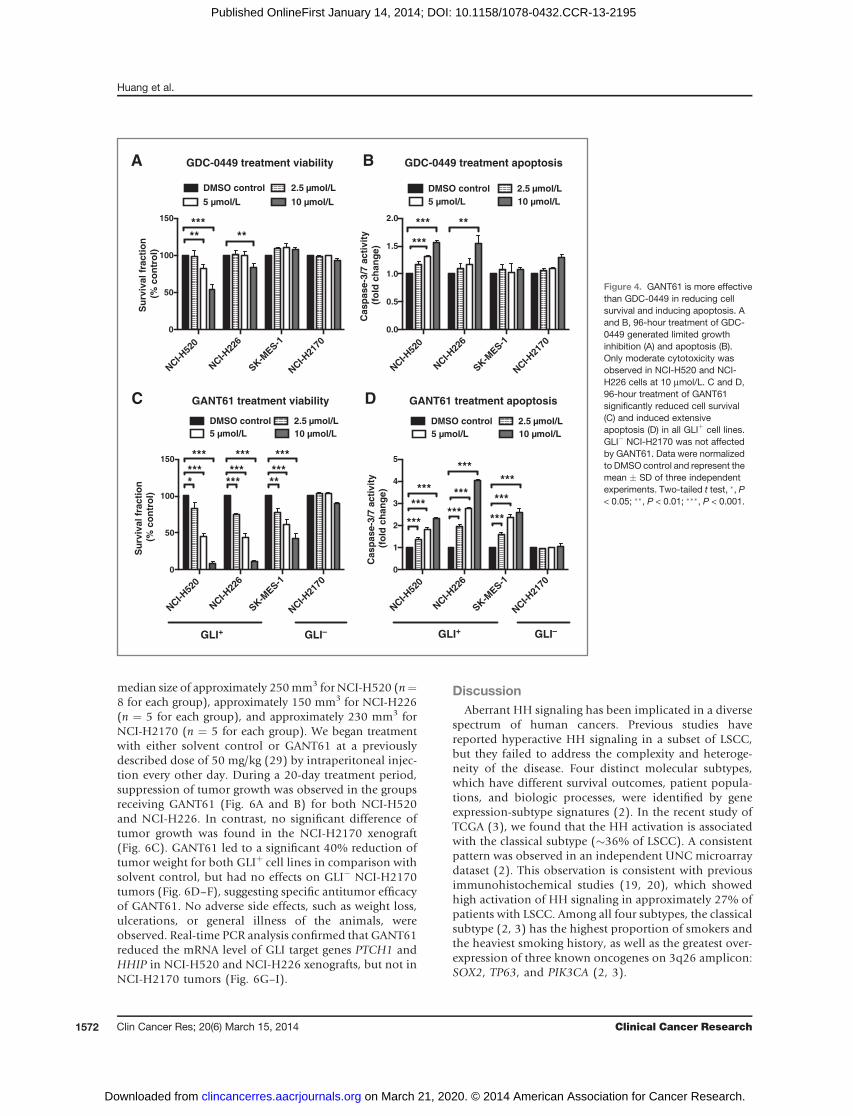
median size of approximately 250mm3 for NCI-H520 (n¼8 for each group), approximately 150 mm3 for NCI-H226(n ¼ 5 for each group), and approximately 230 mm3 forNCI-H2170 (n ¼ 5 for each group). We began treatmentwith either solvent control or GANT61 at a previouslydescribed dose of 50 mg/kg (29) by intraperitoneal injec-tion every other day. During a 20-day treatment period,suppression of tumor growth was observed in the groupsreceiving GANT61 (Fig. 6A and B) for both NCI-H520and NCI-H226. In contrast, no significant difference oftumor growth was found in the NCI-H2170 xenograft(Fig. 6C). GANT61 led to a significant 40% reduction oftumor weight for both GLIþ cell lines in comparison withsolvent control, but had no effects on GLI� NCI-H2170tumors (Fig. 6D–F), suggesting specific antitumor efficacyof GANT61. No adverse side effects, such as weight loss,ulcerations, or general illness of the animals, wereobserved. Real-time PCR analysis confirmed that GANT61reduced the mRNA level of GLI target genes PTCH1 andHHIP in NCI-H520 and NCI-H226 xenografts, but not inNCI-H2170 tumors (Fig. 6G–I).
DiscussionAberrant HH signaling has been implicated in a diverse
spectrum of human cancers. Previous studies havereported hyperactive HH signaling in a subset of LSCC,but they failed to address the complexity and heteroge-neity of the disease. Four distinct molecular subtypes,which have different survival outcomes, patient popula-tions, and biologic processes, were identified by geneexpression-subtype signatures (2). In the recent study ofTCGA (3), we found that the HH activation is associatedwith the classical subtype (�36% of LSCC). A consistentpattern was observed in an independent UNC microarraydataset (2). This observation is consistent with previousimmunohistochemical studies (19, 20), which showedhigh activation of HH signaling in approximately 27% ofpatients with LSCC. Among all four subtypes, the classicalsubtype (2, 3) has the highest proportion of smokers andthe heaviest smoking history, as well as the greatest over-expression of three known oncogenes on 3q26 amplicon:SOX2, TP63, and PIK3CA (2, 3).
A B
DC GANT61 treatment viability
NCI-H
520
NCI-H
226
SK-M
ES-1
NCI-H
2170
0
50
100
150
Su
rviv
al
fra
cti
on
(%
co
ntr
ol)
DMSO control 2.5 mol/L
5 mol/L 10 mol/L
GLI+ GLI–
****
***
******
***
*****
***
GANT61 treatment apoptosis
NCI-H
520
NCI-H
226
SK-M
ES-1
NCI-H
2170
0
1
2
3
4
5
Ca
sp
as
e-3
/7 a
cti
vit
y
(fo
ld c
ha
ng
e)
DMSO control 2.5 mol/L
5 mol/L 10 mol/L
GLI+ GLI–
***
***
***
***
***
***
***
***
***
GDC-0449 treatment viability
NCI-H
520
NCI-H
226
SK-M
ES-1
NCI-H
2170
0
50
100
150
Su
rviv
al
fra
cti
on
(%
co
ntr
ol)
DMSO control 2.5 mol/L
5 mol/L 10 mol/L
*****
**
GDC-0449 treatment apoptosis
NCI-H
520
NCI-H
226
SK-M
ES-1
NCI-H
2170
0.0
0.5
1.0
1.5
2.0
Ca
sp
as
e-3
/7 a
cti
vit
y
(fo
ld c
ha
ng
e)
DMSO control 2.5 mol/L
5 mol/L 10 mol/L
*** **
***
Figure 4. GANT61 is more effectivethan GDC-0449 in reducing cellsurvival and inducing apoptosis. Aand B, 96-hour treatment of GDC-0449 generated limited growthinhibition (A) and apoptosis (B).Only moderate cytotoxicity wasobserved in NCI-H520 and NCI-H226 cells at 10 mmol/L. C and D,96-hour treatment of GANT61significantly reduced cell survival(C) and induced extensiveapoptosis (D) in all GLIþ cell lines.GLI� NCI-H2170 was not affectedby GANT61. Data were normalizedto DMSO control and represent themean � SD of three independentexperiments. Two-tailed t test, �, P< 0.05; ��, P < 0.01; ���, P < 0.001.
Huang et al.
Clin Cancer Res; 20(6) March 15, 2014 Clinical Cancer Research1572
on March 21, 2020. © 2014 American Association for Cancer Research. clincancerres.aacrjournals.org Downloaded from
Published OnlineFirst January 14, 2014; DOI: 10.1158/1078-0432.CCR-13-2195
While GLI2 was consistently highly expressed in theclassical subtype, strong positive correlations betweenGLI2and the three best-known markers of the classical subtypeon chromosome 3q were observed, together suggesting acritical roleofGLI2 in LSCC, as found inSCC inotherorgans(22–24). Interestingly, the expression of ligand SHHwithinthe classical subtype varied markedly and was not signifi-cantly different between subtypes. The expression patternsof other two HH ligands, IHH and DHH, were similar toSHH (Supplementary Fig. S1J). These data indicate theexistence of ligand-independent GLI activation in the clas-sical subtype. Genetic alterations, including loss of PTCHfunction, constitutively active SMO, and amplification ofGLI1/GLI2 have been reported to activate downstream HHsignaling independent of ligands in various cancers. More-over, GLI function can be modulated in a SMO-indepen-dent manner by PI3K/AKT (15), RAS-MEK signaling (16,17), which may contribute to the hyperactive HH–GLIsignaling in LSCC in addition to canonical HH signaling.Recent development of multiple SMO inhibitors for
human administration prompted us to test the potentialclinical application to treat patientswith LSCCby targetingSMO. However, shRNA knockdown of SMO showed onlyminor effects on LSCC cell survival and apoptosis, withlittle effect on HH downstream target gene expression.Consistently, GDC-0449 produced limited cytotoxicitydespite the universal expression of SMO, suggesting theexistence of SMO-independent regulation of GLI signal-ing. The strong and unique expression pattern of GLI2prompted the querywhether it is necessary for LSCC tumorprogression. Knockdown of GLI2 induced apoptosis and
significant growth inhibition in GLI2þ LSCC cells, dem-onstrating an essential role of GLI2 in regulating cellviability and death. Importantly, the GLI inhibitor,GANT61, effectively blocked GLI-mediated signal trans-duction and suppressed tumor growth both in vitro and invivo. Recent studies in neuroblastoma (33) and chroniclymphocytic leukemia (34) also showed that GANT61effectively suppressed tumor progression in cancers insen-sitive to SMO inhibition. Interestingly, amplification ofGLI2 or CCND1 have been proposed as additionalmechanisms responsible for resistance to SMO inhibitors(27), and GANT61 treatment significantly reduced expres-sion of GLI2 and CCND1 in LSCC cells.
GANT61 is a relatively new member in the HH inhib-itor family, as most known HH pathway antagonists focuson the transmembrane activator SMO. Other readilyavailable agents that inhibit GLI2 are rare. Arsenic triox-ide (ATO), which is a U.S. Food and Drug Administration(FDA)–approved treatment for acute promyelocytic leu-kemia, has recently been described as a potent HH inhib-itor. ATO has been shown to inhibit HH signaling byinhibiting GLI2 ciliary accumulation and promoting itsdegradation (38), and inhibit tumor growth in cancerswith known drug-resistant SMO mutations and in thecontext of GLI2 overexpression (39). Because of thelikelihood that compounds suppressing HH pathway–dependent proliferation in one cell type may be inactivein others, the clinical relevance of ATO in LSCC treatmentis currently under investigation.
A recently emerging idea in clinical treatment is tocombine several antitumor agents that specifically target
NCI-H520 GANT61 treatment
GLI2 PTCH1 HHIP0.0
0.2
0.4
0.6
0.8
1.0R
ela
tive m
RN
A level
DMSO control
GANT61 10 mmol/L** *** *
NCI-H226 GANT61 treatment
GLI2 PTCH1 HHIP0.0
0.2
0.4
0.6
0.8
1.0DMSO control
GANT61 10 mmol/L
***
***
***
SK-MES-1 GANT61 treatment
GLI2 PTCH1 HHIP0.0
0.2
0.4
0.6
0.8
1.0
Rela
tive m
RN
A level
DMSO control
GANT61 10 mmol/L
** ******
C D
E
PTCH1
NCI-H520 NCI-H226 SK-MES-1NCI-H21700.0
0.5
1.0
1.5
Rela
tive m
RN
A level
DMSO control
GDC-0449 10 mmol/L
AGLI2
NCI-H520 NCI-H226 SK-MES-10.0
0.5
1.0
1.5
Rela
tive m
RN
A level
Rela
tive m
RN
A level
DMSO control
GDC-0449 10 mmol/L
B
F SK-MES-1
GLI2
NCI-H520 NCI-H226
GANT61 + + +
GAPDH
Cleaved
PARP
Cleaved
caspase-3
CCND1
Figure 5. GANT61 is more efficientthan GDC-0449 in suppressing theexpression of GLI target genes. Aand B, real-time PCR of GAPDH-normalized mRNA level of PTCH1(A) andGLI2 (B) in cells treated withDMSO or 10 mmol/L GDC-0449 for96 hours. C–E, 96-hour exposureto 10 mmol/L GANT61 reducedmRNA levels of GLI targets. Geneexpressions were normalized toendogenous GAPDH in each cellline. Data represent themean� SDof three independent experiments.Two-tailed t test, �, P < 0.05;��, P < 0.01; ���, P < 0.001. F,96-hour treatment of 10 mmol/LGANT61 reduced protein levels ofGLI2 and CCND1, and inducedcleaved caspase-3 and cleavedPARP. GAPDH was used as theloading control. A representativeWestern blot analysis from threeindependent experiments isshown.
Hedgehog Signaling in Squamous Cell Lung Cancer
www.aacrjournals.org Clin Cancer Res; 20(6) March 15, 2014 1573
on March 21, 2020. © 2014 American Association for Cancer Research. clincancerres.aacrjournals.org Downloaded from
Published OnlineFirst January 14, 2014; DOI: 10.1158/1078-0432.CCR-13-2195

different signaling pathways. It has been reported that GLIfunction can be modulated in a SMO-independent mannerby PI3K/AKT signaling (15). Pharmacologic inhibition ofPI3K/AKT signaling reduced tumor growth in GDC-0449–resistant medulloblastoma (27). Several inhibitors of thePI3K pathway are undergoing clinical evaluation. PIK3CAcopy-number gains and loss of PTEN function are prevalentin the classical subtype of LSCC, where the activation of HHsignaling was observed. This coexistence raises the possi-bility that a combined therapymay bemore beneficial thana monotherapy to enhance efficiency and overcome drugresistance.
Treatment options for LSCC overall are disappointing.Different from standard-of-care chemotherapy or small-molecule inhibitionof kinase signaling cascades,wepresenta potential strategy to treat a subset of patients with LSCCbytargeting the GLI transcriptional network. Our studies alsohighlight the need for agents that suppress GLI effectorswith high efficacy and selectivity.
Disclosure of Potential Conflicts of InterestNo potential conflicts of interest were disclosed.
Authors' ContributionsConception and design: L. Huang, M. OnaitisDevelopment of methodology: L. Huang, D.N. Hayes, M. OnaitisAcquisitionofdata (provided animals, acquired andmanagedpatients,provided facilities, etc.): L. Huang, D.N. Hayes, M. OnaitisAnalysis and interpretation of data (e.g., statistical analysis, biosta-tistics, computational analysis): L. Huang, V. Walter, D.N. Hayes,M. OnaitisWriting, review, and/or revision of the manuscript: L. Huang, V. Walter,D.N. Hayes, M. OnaitisAdministrative, technical, or material support (i.e., reporting or orga-nizing data, constructing databases): L. Huang, M. OnaitisStudy supervision: M. Onaitis
AcknowledgmentsThe authors thank Jing Zhang and Lixia Luo for animal husbandry.
Grant SupportThisstudywassupportedbyHowardHughesMedical InstituteEarlyCareer
Grant, Department of Defence Promising Young Investigator Grant, andThoracic Surgery Foundation for Research and Education Research Grant.
The costs of publicationof this articlewere defrayed inpart by thepaymentof page charges. This articlemust therefore be herebymarked advertisement inaccordance with 18 U.S.C. Section 1734 solely to indicate this fact.
Received August 9, 2013; revised December 13, 2013; accepted December17, 2013; published OnlineFirst January 14, 2014.
5 10 15 200
250
500
750
1,000
1,250
1,500
NCI-H520 xenograft
Tu
mo
r v
olu
me
(m
m3)
Tu
mo
r v
olu
me
(m
m3)
Tu
mo
r v
olu
me
(m
m3)
Solvent control GANT61 50 mg/kg
** ***** *** **
D
*** *** *** ***
NCI-H520 xenograft
0.0
0.5
1.0
1.5
2.0
2.5
Tu
mo
r w
eig
ht
(g)
Solvent control GANT61 50 mg/kg
P < 0.005
NCI-H226 xenograft
5 10 15 200
200
400
600
800
1,000
D
Solvent control GANT61 50 mg/kg
** * ** *** ** ** ***
NCI-H226 xenograft
0.0
0.2
0.4
0.6
0.8
Tu
mo
r w
eig
ht
(g)
Solvent control GANT61 50 mg/kg
P < 0.05
NCI-H2170 xenograft
5 10 15 200
200
400
600
800
1,000
D
Solvent control GANT61 50 mg/kg
NCI-H2170 xenograft
0.0
0.5
1.0
1.5
Tu
mo
r w
eig
ht
(g)
Solvent control GANT61 50 mg/kg
A B C
D E F
HG
NS
NCI-H520 xenograft
PTCH1 HHIP0.0
0.5
1.0
1.5
2.0
Re
lati
ve
mR
NA
le
ve
l Control tumor GANT61 tumor
*
*
NCI-H226 xenograft
PTCH1 HHIP0.0
0.5
1.0
1.5
Re
lati
ve
mR
NA
le
ve
l Control tumor GANT61 tumor
***
NCI-H2170 xenograft
PTCH1 HHIP0.0
0.5
1.0
1.5
2.0
Re
lati
ve
mR
NA
le
ve
l Control tumor GANT61 tumor
NS NS
I
Figure 6. GANT61 treatment suppressed GLIþ tumor progression in vivo. A–C, growth of NCI-H520 (A; n¼ 8), NCI-H226 (B; n¼ 5), and NCI-H2170 (C; n¼ 5)xenografts (mean � SD). D–F, measurement of tumor weight at the end of treatment for NCI-H520 (D; n ¼ 8), NCI-H226 (E; n ¼ 5), and NCI-H2170(F; n ¼ 5). Shown is the mean � SD. G–I, quantification of PTCH1 and HHIP mRNA by real-time PCR in treated tumors for NCI-H520 (G; n ¼ 4), NCI-H226(H; n ¼ 5), and NCI-H2170 (I; n ¼ 5). Values were normalized against GAPDH. Shown is the mean � SD of independent tumors in each group. A–I,data were analyzed by a two-tailed t test. �, P < 0.05; ��, P <0.01; ���, P < 0.001; NS, not significant.
Huang et al.
Clin Cancer Res; 20(6) March 15, 2014 Clinical Cancer Research1574
on March 21, 2020. © 2014 American Association for Cancer Research. clincancerres.aacrjournals.org Downloaded from
Published OnlineFirst January 14, 2014; DOI: 10.1158/1078-0432.CCR-13-2195

References1. Drilon A, Rekhtman N, Ladanyi M, Paik P. Squamous-cell carcinomas
of the lung: emerging biology, controversies, and the promise oftargeted therapy. Lancet Oncol 2012;13:e418-26.
2. Wilkerson MD, Yin X, Hoadley KA, Liu Y, Hayward MC, Cabanski CR,et al. Lung squamous cell carcinoma mrna expression subtypes arereproducible, clinically important, and correspond to normal cell types.Clin Cancer Res 2010;16:4864–75.
3. Cancer Genome Atlas Research Network. Comprehensive genomiccharacterization of squamous cell lung cancers. Nature 2012;489:519–25.
4. Watkins DN, Berman DM, Burkholder SG, Wang B, Beachy PA, BaylinSB. Hedgehog signalling within airway epithelial progenitors and insmall-cell lung cancer. Nature 2003;422:313–7.
5. Park K-S,Martelotto LG, PeiferM, SosML, Karnezis AN,MahjoubMR,et al. A crucial requirement for Hedgehog signaling in small cell lungcancer. Nat Med 2011;17:1504–8.
6. Lum L, Beachy PA. The Hedgehog response network: sensors,switches, and routers. Science 2004;304:1755–9.
7. Ruiz i Altaba A, Mas C, Stecca B. The Gli code: an information nexusregulating cell fate, stemness and cancer. Trends Cell Biol 2007;17:438–47.
8. Thiyagarajan S, Bhatia N, Reagan-Shaw S, Cozma D, Thomas-Tikhonenko A, Ahmad N, et al. Role of GLI2 transcription factor ingrowth and tumorigenicity of prostate cells. Cancer Res 2007;67:10642–6.
9. Ikram MS, Neill GW, Regl G, Eichberger T, Frischauf A-M, Aberger F,et al. GLI2 is expressed in normal human epidermis and BCC andinduces GLI1 expression by binding to its promoter. J Inves Dermatol2004;122:1503–9.
10. Bai CB, AuerbachW, Lee JS, Stephen D, Joyner AL. Gli2, but not Gli1,is required for initial Shh signaling and ectopic activation of the Shhpathway. Development 2002;129:4753–61.
11. Eichberger T, Sander V, Schnidar H, Regl G, KasperM, SchmidC, et al.Overlapping and distinct transcriptional regulator properties of theGLI1 and GLI2 oncogenes. Genomics 2006;87:616–32.
12. Duman-Scheel M, Weng L, Xin S, Du W. Hedgehog regulates cellgrowth and proliferation by inducing Cyclin D and Cyclin E. Nature2002;417:299–304.
13. Bigelow RLH, Chari NS, Und�en AB, Spurgers KB, Lee S, Roop DR,et al. Transcriptional regulation of bcl-2 mediated by the sonichedgehog signaling pathway through gli-1. J Biol Chem 2004;279:1197–205.
14. Regl G, Kasper M, Schnidar H, Eichberger T, Neill GW, Philpott MP,et al. Activation of the BCL2 promoter in response to Hedgehog/GLISignal Transduction Is Predominantly Mediated by GLI2. Cancer Res2004;64:7724–31.
15. Riob�o NA, Lu K, Ai X, Haines GM, Emerson CP. Phosphoinositide 3-kinase and Akt are essential for Sonic Hedgehog signaling. Proc NatlAcad Sci U S A 2006;103:4505–10.
16. Stecca B, Mas C, Clement V, Zbinden M, Correa R, Piguet V, et al.Melanomas require HEDGEHOG-GLI signaling regulated by interac-tions between GLI1 and the RAS-MEK/AKT pathways. Proc Natl AcadSci 2007;104:5895–900.
17. Nolan-Stevaux O, Lau J, Truitt ML, Chu GC, Hebrok M, Fern�andez-Zapico ME, et al. GLI1 is regulated through smoothened-independentmechanisms in neoplastic pancreatic ducts and mediates PDAC cellsurvival and transformation. Genes Dev 2009;23:24–36.
18. Katoh Y, Katoh M. Hedgehog target genes: mechanisms of carcino-genesis induced by aberrant Hedgehog signaling activation. Curr MolMed 2009;9:873–86.
19. Gialmanidis IP, Bravou V, Amanetopoulou SG, Varakis J, Kourea H,Papadaki H. Overexpression of hedgehog pathway molecules andFOXM1 in non-small cell lung carcinomas. Lung Cancer 2009;66:64–74.
20. Raz G, Allen KE, Kingsley C, Cherni I, Arora S, Watanabe A, et al.Hedgehog signaling pathway molecules and ALDH1A1 expression
in early-stage non-small cell lung cancer. Lung Cancer 2012;76:191–6.
21. Shi I, Sadraei NH, Duan Z-H, Shi T, Shi I, Sadraei NH, et al. Aberrantsignaling pathways in squamous cell lung carcinoma. Cancer Inform2011;10:273–85.
22. SnijdersAM,Schmidt BL, Fridlyand J,DekkerN, Pinkel D, JordanRCK,et al. Rare amplicons implicate frequent deregulation of cell fatespecification pathways in oral squamous cell carcinoma. Oncogene2005;24:4232–42.
23. Yan M, Wang L, Zuo H, Zhang Z, Chen W, Mao L, et al. HH/GLIsignalling as a new therapeutic target for patients with oral squamouscell carcinoma. Oral Oncol 2011;47:504–9.
24. Yang L, Wang L-S, Chen XL, Gatalica Z, Qiu S, Liu Z, et al. Hedgehogsignaling activation in the development of squamous cell carcinomaand adenocarcinoma of esophagus. Int J Biochem Mol Biol 2012;3:46–57.
25. Rudin CM, Hann CL, Laterra J, Yauch RL, Callahan CA, Fu L, et al.Treatment of medulloblastoma with Hedgehog pathway inhibitorGDC-0449. N Engl J Med 2009;361:1173–8.
26. Von Hoff DD, LoRusso PM, Rudin CM, Reddy JC, Yauch RL, Tibes R,et al. Inhibition of the Hedgehog pathway in advanced basal-cellcarcinoma. N Engl J Med 2009;361:1164–72.
27. Dijkgraaf GJP, AlickeB,Weinmann L, Januario T,West K,Modrusan Z,et al. Small molecule inhibition of GDC-0449 refractory smoothenedmutants and downstreammechanisms of drug resistance. Cancer Res2011;71:435–44.
28. Yauch RL, Dijkgraaf GJP, Alicke B, Januario T, Ahn CP, Holcomb T,et al.Smoothenedmutationconfers resistance toaHedgehogpathwayinhibitor in medulloblastoma. Science 2009;326:572–4.
29. Lauth M, Bergstr€om Å, Shimokawa T, Toftga�rd R. Inhibition of GLI-
mediated transcription and tumor cell growth by small-moleculeantagonists. Proc Natl Acad Sci 2007;104:8455–60.
30. Mazumdar T, DeVecchio J, Shi T, Jones J, Agyeman A, Houghton JA.Hedgehog signaling drives cellular survival in human colon carcinomacells. Cancer Res 2011;71:1092–102.
31. Mazumdar T, DeVecchio J, Agyeman A, Shi T, Houghton JA. BlockingHedgehog survival signaling at the level of the GLI genes induces DNAdamage and extensive cell death in human colon carcinoma cells.Cancer Res 2011;71:5904–14.
32. Fu J, Rodova M, Roy SK, Sharma J, Singh KP, Srivastava RK, et al.GANT-61 inhibits pancreatic cancer stem cell growth in vitro and inNOD/SCID/IL2R gamma null mice xenograft. Cancer Lett 2013;330:22–32.
33. Wickstr€om M, Dyberg C, Shimokawa T, Milosevic J, Baryawno N,Fuskeva
�g OM, et al. Targeting the hedgehog signal transduction
pathway at the level of GLI inhibits neuroblastoma cell growth in vitroand in vivo. Int J Cancer 2013;132:1516–24.
34. Desch P, Asslaber D, Kern D, Schnidar H, Mangelberger D, Alinger B,et al. Inhibition of GLI, but not Smoothened, induces apoptosis inchronic lymphocytic leukemia cells. Oncogene 2010;29:4885–95.
35. Li B, Dewey C. RSEM: accurate transcript quantification from RNA-Seq data with or without a reference genome. BMC Bioinformatics2011;12:323.
36. R Core Team (2012). R: A language and environment for statisticalcomputing. Austria: R Foundation for Statistical Computing; 2009.
37. Barretina J, Caponigro G, StranskyN, Venkatesan K,Margolin AA, KimS, et al. The Cancer Cell Line Encyclopedia enables predic-tive modelling of anticancer drug sensitivity. Nature 2012;483:603–7.
38. Kim J, Lee JJ, Kim J, Gardner D, Beachy PA. Arsenic antagonizes theHedgehog pathway by preventing ciliary accumulation and reducingstability of the Gli2 transcriptional effector. Proc Natl Acad Sci2010;107:13432–7.
39. Kim J, Aftab Blake T, Tang Jean Y, Kim D, Lee Alex H, Rezaee M, et al.Itraconazole and arsenic trioxide inhibit Hedgehog pathway activationand tumor growth associatedwith acquired resistance to smoothenedantagonists. Cancer Cell 2013;23:23–34.
Hedgehog Signaling in Squamous Cell Lung Cancer
www.aacrjournals.org Clin Cancer Res; 20(6) March 15, 2014 1575
on March 21, 2020. © 2014 American Association for Cancer Research. clincancerres.aacrjournals.org Downloaded from
Published OnlineFirst January 14, 2014; DOI: 10.1158/1078-0432.CCR-13-2195
2014;20:1566-1575. Published OnlineFirst January 14, 2014.Clin Cancer Res Lingling Huang, Vonn Walter, D. Neil Hayes, et al. Squamous Lung Cancer
GLI Signaling Inhibition Suppresses Tumor Growth in−Hedgehog
Updated version
10.1158/1078-0432.CCR-13-2195doi:
Access the most recent version of this article at:
Material
Supplementary
http://clincancerres.aacrjournals.org/content/suppl/2014/01/14/1078-0432.CCR-13-2195.DC1
Access the most recent supplemental material at:
Cited articles
http://clincancerres.aacrjournals.org/content/20/6/1566.full#ref-list-1
This article cites 38 articles, 14 of which you can access for free at:
Citing articles
http://clincancerres.aacrjournals.org/content/20/6/1566.full#related-urls
This article has been cited by 4 HighWire-hosted articles. Access the articles at:
E-mail alerts related to this article or journal.Sign up to receive free email-alerts
Subscriptions
Reprints and
To order reprints of this article or to subscribe to the journal, contact the AACR Publications Department at
Permissions
Rightslink site. Click on "Request Permissions" which will take you to the Copyright Clearance Center's (CCC)
.http://clincancerres.aacrjournals.org/content/20/6/1566To request permission to re-use all or part of this article, use this link
on March 21, 2020. © 2014 American Association for Cancer Research. clincancerres.aacrjournals.org Downloaded from
Published OnlineFirst January 14, 2014; DOI: 10.1158/1078-0432.CCR-13-2195