Embed Size (px)
Citation preview

Hazard assessment of nanomaterials in consumer products
Environmental project No. 1637, 2015

2 Hazard assessment of nanomaterials in consumer products
Title:
Hazard assessment of nanomaterials in consumer
products
Editing:
Anne Thoustrup Saber
Sonja Hagen Mikkelsen
Henrik Rye Lam
Karin Sørig Hougaard
Poul Bo Larsen
Frans Christensen
Ulla Vogel
Published by:
The Danish Environmental Protection Agency
Strandgade 29
1401 Copenhagen K
Denmark
www.mst.dk/english
Year:
2015
ISBN no.
978-87-93283-58-9
Disclaimer:
When the occasion arises, the Danish Environmental Protection Agency will publish reports and papers concerning
research and development projects within the environmental sector, financed by study grants provided by the Danish
Environmental Protection Agency. It should be noted that such publications do not necessarily reflect the position or
opinion of the Danish Environmental Protection Agency.
However, publication does indicate that, in the opinion of the Danish Environmental Protection Agency, the content
represents an important contribution to the debate surrounding Danish environmental policy.
Sources must be acknowledged.
Hazard assessment of nanomaterials in consumer products 3
Contents
Preface ........................................................................................................................... 6
Abbreviations and acronyms ......................................................................................... 8
Conclusion and Summary ........................................................................................... 10
Dansk sammenfatning.................................................................................................. 17
1. Introduction ......................................................................................................... 25 1.1 Background .......................................................................................................................... 25 1.2 Objective ............................................................................................................................... 25
2. Biokinetics ........................................................................................................... 26 2.1 Absorption of nanomaterials ............................................................................................... 26
2.1.1 Pulmonary absorption .......................................................................................... 26 2.1.2 Gastrointestinal absorption ...................................................................................27 2.1.3 Dermal absorption ................................................................................................ 29 2.1.4 Eye absorption....................................................................................................... 32 2.1.5 Olfactory absorption ............................................................................................. 32
2.2 Distribution of nanomaterials ............................................................................................. 34 2.2.1 General (blood/organs) ........................................................................................ 34 2.2.2 CNS .........................................................................................................................35 2.2.3 Fetus .......................................................................................................................35
2.3 Metabolism........................................................................................................................... 36 2.4 Excretion/accumulation ...................................................................................................... 36 2.5 Summary .............................................................................................................................. 38
3. Adverse effects of pristine nanomaterials ........................................................... 39 3.1 Respiratory system .............................................................................................................. 39 3.2 Cardiovascular system ......................................................................................................... 40 3.3 Gastrointestinal tract ............................................................................................................ 41 3.4 CNS ....................................................................................................................................... 42 3.5 Other organs......................................................................................................................... 43 3.6 Skin ....................................................................................................................................... 44 3.7 Eyes....................................................................................................................................... 44 3.8 Developmental and reproductive system............................................................................ 46 3.9 Genotoxicity and cancer ...................................................................................................... 48 3.10 Immunotoxicity.................................................................................................................... 49
4. Adverse effects of nanomaterials when part of a matrix ..................................... 50 4.1 Composites (solid) ............................................................................................................... 50 4.2 Sprays ................................................................................................................................... 52 4.3 Liquids ...................................................................................................................................53 4.4 Food ...................................................................................................................................... 54
5. Physico-chemical factors of importance for toxicity ............................................ 59 5.1 Size........................................................................................................................................ 59 5.2 Agglomeration/aggregation ................................................................................................ 59 5.3 Specific surface area ............................................................................................................ 59

4 Hazard assessment of nanomaterials in consumer products
5.4 Shape .................................................................................................................................... 60 5.5 Crystallinity .......................................................................................................................... 60 5.6 Dissolution rate .................................................................................................................... 60 5.7 Chemical composition ......................................................................................................... 60 5.8 Surface modifications ........................................................................................................... 61 5.9 Surface charge ....................................................................................................................... 61 5.10 Behavior in biological media ................................................................................................ 61
6. Hazard evaluation of a selection of nanomaterials .............................................. 62 6.1 Carbon black (CB) ................................................................................................................ 62
6.1.1 Introduction .......................................................................................................... 62 6.1.2 Biokinetics ............................................................................................................. 62 6.1.3 Adverse effects of CB ............................................................................................. 63 6.1.4 Adverse effect of carbon black when part of a matrix ......................................... 65 6.1.5 Physico-chemical properties of importance for toxicity ...................................... 66 6.1.6 Summary ............................................................................................................... 66 6.1.7 Input to risk assessment ....................................................................................... 68
6.2 Carbon nanotubes (CNT)..................................................................................................... 68 6.2.1 Introduction .......................................................................................................... 68 6.2.2 Biokinetics ............................................................................................................. 69 6.2.3 Adverse effects of carbon nanotubes .................................................................... 70 6.2.4 Adverse effect of CNT when part of a matrix ........................................................72 6.2.5 Physico-chemical properties of importance for toxicity .......................................72 6.2.6 Summary ................................................................................................................73 6.2.7 Input to risk assessment ........................................................................................74
6.3 Amorphous silica (nano-SAS) ..............................................................................................74 6.3.1 Introduction ...........................................................................................................74 6.3.2 Biokinetics .............................................................................................................. 75 6.3.3 Adverse effects of SAS ............................................................................................76 6.3.4 Adverse effect of SAS when part of a matrix .........................................................79 6.3.5 Physico-chemical properties of importance for toxicity ...................................... 80 6.3.6 Summary ................................................................................................................ 81 6.3.7 Input to risk assessment ....................................................................................... 84
6.4 Nano silver (nano-Ag) ......................................................................................................... 85 6.4.1 Introduction .......................................................................................................... 85 6.4.2 Biokinetics ............................................................................................................. 85 6.4.3 Adverse effects of nano-Ag ................................................................................... 86 6.4.4 Adverse effect of nano-Ag when part of a matrix ................................................. 91 6.4.5 Physico-chemical properties of importance for toxicity ...................................... 92 6.4.6 Summary and conclusions .................................................................................... 92 6.4.7 Input to risk assessment ....................................................................................... 95
6.5 Nano titanium dioxide (nano-TiO2) .................................................................................... 96 6.5.1 Introduction .......................................................................................................... 96 6.5.2 Biokinetics ............................................................................................................. 96 6.5.3 Adverse effects of nano-TiO2 ................................................................................ 99 6.5.4 Adverse effect of TiO2 when part of a matrix ......................................................103 6.5.5 Physico-chemical properties of importance for toxicity .....................................104 6.5.6 Summary ..............................................................................................................104 6.5.7 Input to risk assessment ......................................................................................106
6.6 Nano zink oxide (nano-ZnO) .............................................................................................. 107 6.6.1 Introduction ......................................................................................................... 107 6.6.2 Biokinetics ............................................................................................................ 107 6.6.3 Adverse effects of nano-ZnO .............................................................................. 108 6.6.4 Adverse effect of nano-ZnO when part of a matrix ............................................. 112

Hazard assessment of nanomaterials in consumer products 5
6.6.5 Physico-chemical properties of importance for toxicity ..................................... 112 6.6.6 Summary and conclusions ................................................................................... 113 6.6.7 Input to risk assessment ...................................................................................... 115
6.7 Nano zirconium dioxide (nano-ZrO2) ............................................................................... 116 6.7.1 Introduction ......................................................................................................... 116 6.7.2 Biokinetics ............................................................................................................ 117 6.7.3 Adverse effects of nano- ZrO2 ............................................................................. 118 6.7.4 Adverse effect of ZrO2 when part of a matrix ...................................................... 121 6.7.5 Physico-chemical properties of importance for toxicity ..................................... 122 6.7.6 Summary .............................................................................................................. 122 6.7.7 Input to risk assessment ...................................................................................... 123
7. Bridging between hazard and risk assessment ...................................................124
8. Summary ............................................................................................................. 127
References ..................................................................................................................134

6 Hazard assessment of nanomaterials in consumer products
Preface
Nanomaterials are applied in a wide range of consumer products, and the commercial use of
nanomaterials in both amounts and diversity is anticipated to increase rapidly in the near future. It
is increasingly recognised that nanomaterials can have unique properties as compared to their bulk
substances favouring the use of nanomaterials in products, articles and technologies. At the same
time concerns in relation to the possible health and environmental properties and impacts of
nanomaterials have surfaced.
On this background, the Danish government and the Red-Green Alliance (a.k.a. Enhedslisten) have
signed an agreement for four years (2012-2015) that focuses on the use of nanomaterials in
products on the Danish market and their consequences on consumers and the environment. The
Danish Environmental Protection Agency (EPA) has initiated a series of projects with the aim of
further clarifying possible risks to consumers and the environment.
The current project addresses consumer exposure and risk assessment of nanomaterials in products
on the Danish market. It runs from third quarter 2013 through second quarter 2015.
The project is foreseen to result in four reports:
Occurrence and exposure assessment of nanomaterials in consumer products and review of
available risk assessment tools
Hazard assessment of nanomaterials in consumer products (the current report)
Human exposure to nanomaterials in the environment – as a reference to nanomaterials
exposure from consumer products
Consumer risk assessment and overall conclusions (final report)
The first three reports will be finalised during 2014, whereas the final report with the consumer risk
assessment and overall conclusions will be finalised during the second quarter of 2015.
The project has been implemented with support from a reference group:
Susan Dekker, National Institute for Public Health and the Environment (RIVM), The
Netherlands
Andrea Haase, Bundesinstitut für Risikobewertung (BfR), Germany
Gregory Moore, Swedish Chemicals Agency (KEMI), Sweden
Derk Brouwer, Netherlands Organisation for Applied Scientific Research (TNO), The
Netherlands
Lena Høglund (The Danish EPA), Denmark
Katrine Bom (The Danish EPA), Denmark
Anne Mette Boisen (The Danish EPA), Denmark
Kim Petersen (The Danish EPA), Denmark
The reference group has assisted with comments and ideas but is not responsible for the content of
the project reports.
Karen Bo Frydendall, National Research Centre for the Working Environment, Denmark, is
gratefully acknowledged for careful proofreading of the report.

Hazard assessment of nanomaterials in consumer products 7

8 Hazard assessment of nanomaterials in consumer products
Abbreviations and acronyms
3T3 NIH 3T3 rodent embryonic fibroblasts
5-HT 5-hydroxy-tryptamine / Serotonin (neurotransmitter)
ACGIH American Conference of Governmental Industrial Hygienists
ADME Absorption-Distribution-Metabolism-Excretion
BAL BronchoAlveolar Lavage
BALB Bagg Albino (inbread mouse strain)
BALF BronchoAlveolar Lavage Fluid
BBB Blood-Brain Barrier
bw Body Weight
CB Carbon Black
CNS Central Nervous System
CNT Carbon NanoTubes
CRP C-Reactive Protein (acute phase protein)
DA DopAmine (neurotransmitter)
DNEL Derived No Effect Level
DNT Developmental NeuroToxicity
ECETOC European Centre for Ecotoxicology and Toxicology Of Chemicals
ECHA European CHemicals Agency
EFSA European Food Safety Agency
ENP Engineered NanoParticles
EPA Environmental Protection Agency
GI Gastro Intestinal
HARN High Aspect Ratio Nanomaterials
IARC International Agency for Research on Cancer
IL InterLeukin
INEL Indicative No-Effect Level
ICP-MS Inductively Coupled Plasma Mass Spectrometry
LD50 Lethal Dose, 50 %
LDH Lactic Acid Dehydrogenase
LOAEC Lowest Observed Adverse Effect Concentration
LOAEL Lowest Observed Adverse Effect Level

Hazard assessment of nanomaterials in consumer products 9
MCMC Mean Cell Haemoglobin Concentration
Mg-PSZ Magnesia Partially Stabilized Zirconia
MNC Montmorillonite NanoComposite
MnO Manganese Oxide
MRC-5 Medical Research Council cell strain 5
MRI Magnetic Resonance Imaging
MTT assay Monocyte mediated cytotoxicity assay
MWCNT MultiWall Carbon Nanotubes
NA NorAdrenaline (neurotransmitter)
Nano- Abbreviation used in words such as nano-sized or nano-Ag
NICNAS National Industrial Chemicals Notification and Assessment Scheme
NIOSH National Institute for Occupational Safety and Health
NIST National Institute of Standards and Technology
NOAEC No Observed Adverse Effect Concentration
NOAEL No Observed Adverse Effect Level
OECD Organisation for Economic Co-operation and Development
OEL Occupational Exposure Limit
OSHA Occupational Safety and Health Administration
PBS Phosphate Buffered Saline
PEG PolyEthyleneGlycol
REACH Registration, Evaluation, Authorisation and Restriction of Chemicals
ROS Reactive Oxygen Species
SAS Synthetic Amorphous Silica
SCCS Scientific Committee on Consumer Safety
SCENIHR Scientific Committee on Emerging and Newly Identified Health Risks
SWCNT SingleWalled Carbon NanoTubes
TEM Transmission Electron Microscopy
TGF-β Transforming Growth Factor beta
TODA 3,6,9-TriOxaDecanoic Acid
US EPA United States Environmental Protection Agency
UV Ultraviolet
WHO World Health Organization
Y-TZP Yttrium-stabilized Tetragonal Zirconia Polycrystals
10 Hazard assessment of nanomaterials in consumer products
Conclusion and Summary
Background
Under the Agreement "Better Control of Nanomaterials" (“Bedre styr på nanomaterialer”), the
Danish Environmental Protection Agency (Danish EPA) has commissioned a number of projects
aiming to investigate and generate new knowledge on the presence of nanomaterials in products on
the Danish market and to assess the possible associated risks to consumers and the environment.
This report is part of a series of four from a project which addresses consumer exposure and risk
assessment of nanomaterials in products on the Danish market.
The aim of this report is to perform a hazard assessment of nanomaterials in consumer products.
The consumer is potentially exposed to nanomaterials in their final, intended use, i.e. when the
nanomaterials are part of a matrix, and therefore, this report focuses on the hazard of nano-
materials when part of a consumer matrix. However, free nanomaterials may be liberated during
the use phase and therefore the hazard of pristine nanomaterials is also described.
The aim of the report is to refer consensus rather than isolated findings. Since the focus of the
hazard evaluation is on nanomaterials during their intended use, the referred literature concerning
pristine nanomaterials consists primarily of reviews, while all identified original studies of hazard
related to nanomaterials as part of a product are described.
The structure of the report is that we review the data relevant to assessing the hazard of nano-
materials in consumer products, i.e. biokinetics of nanomaterials (Chapter 2), adverse effects of
pristine nanomaterials (Chapter 3), adverse effects of nanomaterials when part of a matrix (Chapter
4), physico-chemical factors of importance for toxicity (Chapter 5), perform a hazard assessment of
a selection of nanomaterials (Chapter 6), discuss how to bridge between hazard and risk assessment
(Capter 7) and finally we summarise and conclude (Chapter 8).
Biokinetics
For the consumer, the lungs, gastrointestinal tract and the skin are considered to be the primary
absorption routes for nanomaterials. In addition, uptake may occur through the eyes and the
olfactory system. We did not identify any studies covering how biokinetics are modified when the
nanomaterial is part of a matrix.
Inhalation of particles results in deposition of particles in the respiratory tract (nasopharyngeal,
tracheobronchial and alveolar regions). The fate of the particles after deposition depends on a
combination of physico-chemical properties of the particles and responses both locally in the lung
and in other parts of the body. The larger particles are trapped by the mucociliary system in the
upper airways and are removed relatively fast (hours to days). In contrast, the smaller particles
deposit in the alveolar region where they can stay for years in humans. A low degree of translocation
of particles from the lung to the circulation has been described.
In 2013 the Danish EPA published in 2013 a report on systemic absorption of nanomaterials by oral
exposure which gives an overview of the existing literature (Danish EPA, 2013c). In general, the
reviewed literature indicates that the gastrointestinal absorption is low. It is concluded that
physico-chemical characteristics such as size, agglomeration and crystal structure may affect
absorption. The report concludes that the number of publications on gastrointestinal absorption is
increasing, but only a few of these have been well performed. In particular it is of concern that only

Hazard assessment of nanomaterials in consumer products 11
a few of the physico-chemical parameters that are expected to influence absorption of nanoparticles
were reported.
In 2013 the Danish EPA has published a report on dermal absorption of nanomaterials which
provides an overview and evaluation of the knowledge base regarding dermal absorption of nano-
materials (Danish EPA, 2013b). The overall conclusion in the report is that absorption of particles
in the nano-range through the skin is possible although it seems to occur to a very low degree.
However, the level of penetration, depending on chemistry and experimental conditions, may be
greater for particles in the nano-range than for larger particles.
The present knowledge about absorption of nanomaterials into the eye and the eye toxicity is too
limited to be assessed and evaluated in general for the hazard assessment.
Olfactory absorption of nanomaterials has been demonstrated in laboratory animals and may also
be relevant for humans (Elder et al., 2006). At present, consistent knowledge is too limited for
hazard assessment in general or specifically.
Only a limited number of studies have investigated the potential metabolism of nanomaterials.
Where such information exists, most of the studies show that the nanomaterials, and in particular
insoluble nanomaterials, are not metabolised (Landsiedel et al., 2012).
Upon absorption following pulmonary, dermal, eye or gastrointestinal exposure, nanomaterials
may reach the blood circulation and/or the lymphatic system. The liver is the major distribution
organ and 40 nm Au particles have been shown to accumulate in the Kupffer cells of the liver rather
than being excreted (Sadauskas et al., 2009a). The placenta constitutes the nutritional interface
between mother and fetus. Studies of the passage of engineered nanoparticles (ENP) across the
placenta are still few in number, but rodent studies describe transplacental transport of nano-sized
particles, with rates ranging from almost negligible to high (several percent of the administered
dose).
Adverse effects of pristine nanomaterials
Pulmonary exposure to nanomaterials may cause pulmonary inflammation, fibrosis, DNA damage
and cancer. Concern has been raised that pulmonary exposure may also result in adverse
cardiovascular effects as seen for human exposure to particles from the ambient air. The
mechanisms behind the cardiovascular effects are suggested to be a systemic inflammatory and
acute phase response, particle translocation or respiratory reflexes.
The knowledge on the toxicity of orally administered nanomaterials in the gastrointestinal tract is
limited and it is therefore not possible to identify any overall conclusions regarding the toxicity of
nanomaterials for intended use in food and food-related products (Card et al., 2011). However, the
toxicity of the nanoformulation of a specific ingredient or material was not always consistently
increased as compared to the non-nanoformulation.
Overall, there is little evidence of dermal toxicity following topical application of nanomaterials. In
general, the uptake through intact healthy skin is very low. However, if nanomaterials are able to
penetrate the skin and enter the bloodstream, they may exert a number of adverse effects.
Adverse effects of nanomaterials when part of a matrix
Only a very limited number of studies have focused on the hazard of nanocomposites. Most of these
have focused on the hazard following pulmonary deposition of dust obtained by sanding different
types of nanocomposites (paint, cement and thermoplastic). The published studies on the
toxicological effects of sanding dusts from nanocomposites are in good agreement with each other.
No additional toxicity (inflammation and genotoxicity) have been detected for any of the nano-

12 Hazard assessment of nanomaterials in consumer products
composites compared to the corresponding products without nanomaterials. A few studies on the
toxicological effects of sprays containing nanomaterials have been published. A rat inhalation study
of a commercial spray containing nano titanium dioxide (nano-TiO2) particles indicated similar
toxicity of nano-TiO2 when part of the product and when nano-TiO2 was tested alone. Testing of a
commercial Ag spray product in rats resulted in moderate cardiovascular effects that were not
observed in rats exposed to a standard Ag reference product. A third study showed no increased
toxicity of a polymer dispersion compared to the non-nano product. Thus, based on the very few
available studies it seems that the toxicity of nanomaterials following pulmonary exposure are
masked in solid matrices while the toxicity of nanomaterials in some cases remains in spray
products.
A few studies have investigated penetration of nanomaterials in sunscreen products. The
penetration of nanomaterial containing cosmetics through skin was considered minimal, and there
was no evidence that nano-sized or submicronised TiO2 penetrated the intact epidermis to any
significant extent or evidence of systemic absorption.
The oral safety of food-related nanomaterials that have potential use from in vitro and in vivo
studies in laboratory was reviewed by Card et al. No adverse toxic effects were revealed in any of the
very few studies with well performed physico-chemical characterisation of the nanomaterials. None
of the studies characterised the nanomaterials in the diet or in any organ (Card et al., 2011).
No Derived No Effect Levels (DNELs) on hazard of nanomaterials as part of a matrix have been
identified. More studies are needed to make conclusions within this area.
Physico-chemical factors of importance for toxicity
A number of physico-chemical properties (size, shape, surface properties, composition, solubility,
aggregation/agglomeration, nanomaterial uptake, presence of mutagens and transition metals
associated with the nanomaterials etc.) have been suggested to be important for toxicity of nano-
materials. For insoluble so-called inert particles such as TiO2 and carbon black (CB), the specific
surface area of the particles has been shown to be correlated to the inflammatory response. The fact
that multiwall carbon nanotubes (MWCNT) and asbestos fibres have been shown to induce similar
genotoxic effects in rodents is an example of the effect of particle shape for genotoxity. For soluble
nanomaterials, such as for example zinc oxide (ZnO), the liberation of Zn ions is important for the
potential toxicity. The importance of physico-chemical factors for toxicity stresses the need for
toxicological studies using well-characterised nanomaterials.
Hazard assessment of seven nanomaterials
A specific hazard assessment has been performed for seven nanomaterials with relevance for
consumer exposure to serve as input for the consumer risk assessment of 20 scenarios. The
consumer risk assessment will be published in the final report. The nanomaterials have been chosen
to represent a diverse group of nanomaterials, i.e. 1) “inert” insoluble particles such as TiO2 and CB,
2) soluble nanoparticles such as ZnO and Ag and 3) high aspect ratio particles such as carbon
nanotubes (CNT) since nanomaterials constitute a very broad group which differs with respect to
chemistry, solubility, coating etc. The main conclusions on the hazard assessment of the seven
nanomaterials are inserted below.
Carbon black (CB)
The hazard assessment of CB in a matrix is primarily based on the recent Scientific Committee on
Consumer Safety (SCCS) opinion on CB (SCCS, 2014c). The aim of this SCCS opinion on CB was
specifically to decide if CB is safe for use as a colorant with a concentration up to 10 % in cosmetic
products, and is therefore considered highly relevant for the present purpose, namely to serve as an
input for the hazard part of the risk assessment of CB in mascara. The focus in the present
assessment was on hazard related to skin and eye exposure because these routes of exposure are

Hazard assessment of nanomaterials in consumer products 13
considered to be the most relevant in relation to consumer use of mascara. Three studies on skin
absorption of cosmetic formulations containing CB (all 20-30 nm in size) were evaluated by the
SCCS and did not indicate any skin absorption. As emphasized by the SCCS, the conclusion on no
risk of adverse effects of up to 10 % as CB as a colorant in cosmetic products is only valid when the
skin is intact and the CB particles are 20 nm or larger. No studies on eye absorption of CB were
evaluated by the SCCS. Therefore, hazard associated with eye absorption cannot be evaluated even
though it is highly relevant for the hazard assessment of consumer use of mascara containing CB.
The risk of eye irritation of CB cannot be excluded (SCCS, 2014c). We agree with the final
concluding remarks of the SCCS opinion stressing that the skin absorption studies have only been
done for CB sizes above 20 nm and that it is therefore not possible to conclude on cosmetic
products containing smaller sized CB.
Carbon nanotubes (CNT)
The hazard assessment of CNT will serve as background documentation for the risk assessment of
use (wear and tear) and sanding of a golf club containing CNT. With regard to the intended use of a
golf club, dermal exposure is considered to be the only relevant exposure route. No studies were
identified on the dermal toxicity of CNT incorporated into a solid matrix. If the CNT containing golf
club is sanded, sanding dust containing CNT and potentially free CNT may be liberated and exert
toxicity primarily by the pulmonary and dermal routes. The hazard assessment of free CNT is based
on a recently published report on risk assessment of CNT by the Danish EPA (Danish EPA, 2015a).
Animal studies have shown that pulmonary exposure to CNT consistently give asbestos-like
toxicological response characterised by persistent inflammation, granulomas and fibrosis with low
no-effect levels. Chronic human indicative no-effect levels (INELs) for the general public have been
suggested to be 0.25 µg/m3 (inhalation) and 0.78 and 2.3 mg/person (dermal) for two different
scenarios (Aschberger et al., 2010). Two studies were identified on the toxicity of sanding dusts
from different types of CNT composites (Wohlleben et al., 2011; Wohlleben et al., 2013). None of the
studies showed increased toxicity of sanding dust from the CNT materials compared to the
conventional products without CNT. However, it has not yet been tested if sanding dust from
ultraviolet (UV)-exposed or otherwise weathered materials have a different toxicity profile due to a
potentially increased liberation of free CNT.
Nano-sized synthetic amorphous silica (nano-SAS)
The hazard assessment of synthetic amorphous silica (SAS) as part of a matrix is primarily based on
recent reviews by Dekkers et al (Dekkers et al., 2011; Dekkers et al., 2013), a review of the hazard of
SAS by (Fruijtier-Pôlloth, 2012), and reports on SAS by the International Agency for Research on
Cancer (IARC) (IARC Monographs on the evaluation of carcinogenic risks to humans, 1997) and by
the European Centre for Ecotoxicology and Toxicology of Chemicals (ECETOC) through the Joint
Assessment of Commodity Chemicals (JACC) program (European Centre for Ecotoxicology and
Toxicology of Chemicals (ECETOC), 2006). The purpose of a hazard assessment is to serve as an
input for the hazard part of the risk assessment of SAS in 1) food items and food containers, 2) face
powders, and 3) “easy to clean” impregnation. Thus, the focus is put on the potential hazards
associated with exposure by the gastrointestinal route (relevant for the food items and food
container scenarios) and exposure by the dermal and the inhalation routes (relevant for the face
powder and the “easy to clean” impregnation product). The critical effect following oral exposure is
assessed as the hepatic effect. The No Observed Adverse Effect Level (NOAEL) has been suggested
to be 1,500 mg/kg Body Weight (bw)/day (Dekkers et al., 2011). The critical effect following
pulmonary exposure is pulmonary inflammation. Based on the evaluation by ECETOC, the Lowest
Observed Adverse Effect Levels (LOAELs) and NOAELs were typically 1-50 mg/m3 and 0.5-10
mg/m3, respectively (European Centre for Ecotoxicology and Toxicology of Chemicals (ECETOC),
2006). These differences were by ECETOC evaluated as particle size-dependent: i.e. in general the
NOAEL/LOAEL decreased by particle size. Our literature search identified several recent studies
showing that the NOAEL/LOAEL was affected by size and surface modification, highlighting that
the physico-chemical properties have to be taken into account. No studies were identified by

14 Hazard assessment of nanomaterials in consumer products
ECETOC on the dermal or oral absorption of SAS (European Centre for Ecotoxicology and
Toxicology of Chemicals (ECETOC), 2006). For that reason no NOAEL/LOAEL has been suggested.
Thus, the overall conclusion by ECETOC is that “SAS is essentially non-toxic in humans via the
oral, dermal/ocular and inhalation routes of exposure and no data exist on systemic effects in
humans”.
Nano-sized silver (Nano-Ag)
The purpose of this hazard assessment is to serve as an input for the hazard part of the risk
assessment of Ag when used in food supplements, paints for spraying, nano-filtering, disinfectant
pump and propellant sprays, textiles and wound dressings. Thus, pulmonary, gastrointestinal and
dermal exposures are all relevant exposure routes for the chosen risk scenarios.
The hazard assessment is mainly based on recent reports and references therein on nano-Ag by The
Scientific Committee on Emerging and Newly Identified Health Risks (SCENIHR) and The Danish
EPA (Scientific Committee on Emerging and Newly Identified Health Risks (SCENIHR), 2014a;
Danish EPA, 2011) and recent reviews (Hadrup & Lam, 2014; Johnston et al., 2010a). Ag has no
known essential function in man. The daily human intake has been estimated to be 0.007-0.5 µg/kg
bw/day as the sum from all routes of exposure. Nano-Ag dissolves in solution and releases Ag+.
There is substantial evidence suggesting that the released Ag+ are responsible for toxicological
effects (Hadrup & Lam, 2014). The best described adverse effects in humans caused by long-term
exposure to Ag is a permanent bluish-grey discoloration (argyria or agyrosis) of the skin and/or
eyes (Scientific Committee on Emerging and Newly Identified Health Risks (SCENIHR), 2014a).
Human risk assessment of Ag is most often based on epidemiological studies showing development
of argyria. World Health Organisation (WHO) has set a NOAEL of 5 µg/kg bw/day as the sum of all
routes of exposure (WHO, 2003). This NOAEL has been adopted by The European Food Safety
Agency (EFSA) (EFSA Panel on Food Contact Materials, 2011). Likewise, The United States
Environmental Protection Agency (US EPA) has published an oral reference dose of 5 µg Ag/kg
bw/day in relation to a life-time exposure to Ag (U.S.Environmental Protection Agency, 1996).
Dermal absorption through damaged skin has been reported in humans applying wound dressings
containing nano-Ag. No toxic effects were reported in these test persons and less than <0.1 % of
dose was estimated to be absorbed (Vlachou et al., 2007; Danish EPA, 2011; Moiemen et al., 2011;
Walker & Parsons, 2014). The DNEL, as set by the REACH registrant, is 0.1 mg Ag/m3 and 0.04 mg
Ag/m3 in the air for workers and the general population, respectively (European Chemicals Agency
(ECHA), 2014). SCENIHR concluded that the toxicity of Ag, including nanoparticles of Ag, to
humans is generally low. Futhermore, it is concluded that more data is needed to understand 1) the
bacterial response to ionic Ag as well as Ag nanoparticles and 2) the hazard associated with the
dissemination of resistance (Scientific Committee on Emerging and Newly Identified Health Risks
(SCENIHR), 2014a). Overall, we agree in these thresholds and consider them applicable for the risk
assessment.
Nano-sized titanium dioxide (Nano-TiO2)
The hazard assessment of nano-sized TiO2 is intended to serve as background documentation for
the risk assessment of TiO2 used in chewing gum, sunscreen, sunscreen lipstick, paint and cement
and in relation to sanding a surface painted with nano-TiO2-containing paint. The different
applications of TiO2 involve exposure by the oral, dermal, eye and pulmonary route and focus is
therefore on the hazards associated with these exposure routes.
The SCCS has concluded that nano-TiO2 at concentrations up to 25 % (wt/wt) and containing
maximum 5 % anatase in sunscreens for dermal application are considered safe for the consumers.
Use of TiO2 nanoparticles in sprayable products is not recommended (SCCS, 2014d). Based on the
available information, lung toxicity following inhalation appears to be the most critical effect in
relation to long-term exposure to nano-TiO2. Deposition in the lung depends on the dose and
particles may be retained in the lung for a long time. Inflammation appears to be determined by the
surface area of the particles. A NOAEL of 62.5 mg/kg bw/day was established based on a 30 days

Hazard assessment of nanomaterials in consumer products 15
oral (gavage) study in mice exposed to anatase TiO2 nanomaterials with a primary particle size of 5
nm. A LOAEL of 5 mg/kg bw/day was derived based on impaired neurofunction and behaviour
from a 60 days oral gavage study in mice exposed to anatase TiO2 nanomaterials with primary
particle size 5 nm. The occupational exposure limit (8-hour average) for TiO2 in all forms
(calculated as Ti) is 6 mg/m3 in Denmark corresponding to 10 mg/m3 TiO2. The National Institute
for Occupational Safety and Health (NIOSH) has recommended a threshold of 2.5 mg/m3 for fine
TiO2 and 0.3 mg/m3 for ultrafine TiO2 (< 100 nm) for up to 10 hours/day during a 40-hour work
week (NIOSH, 2011).
Nano-sized zinc oxide (Nano-ZnO)
The purpose of this hazard assessment of ZnO in nanoform (nano-ZnO) is to serve as background
documentation for the risk assessment of sunscreen pump sprays containing nano-ZnO. The most
relevant exposure route associated with consumer use of nano-ZnO sunscreen pump spray is
dermal exposure. However, oral and pulmonary exposure may also occur but to a lesser extent.
Nano-ZnO dissolves in biological fluids including artificial gastrointestinal fluid and lung fluid to
form Zn2+ that seems to be distributed systematically to organs. This hazard assessment of nano-
ZnO in a matrix is primarily based on the recent SCCS opinion on ZnO (SCCS, 2012) and two
addendums related to this opinion (SCCS, 2014a; SCCS, 2014b). The aim of these SCCS opinions on
ZnO was specifically to decide if ZnO in nanoform is safe for use as an UV-filter with a
concentration up to 25 % in cosmetic products and is therefore considered highly relevant for the
present purpose. It is concluded by the SCCS that the use of the different forms of ZnO
nanoparticles as specified in the opinions at a concentration up to 25% as a UV-filter in sunscreens
can be considered not to pose a risk of adverse effects in humans after dermal application. However,
this does not apply to other applications that might lead to inhalation exposure to ZnO
nanoparticles (such as sprayable products) because pulmonary exposure induces serious pulmonary
effects. Therefore, the SCCS concludes that the use of ZnO nanoparticles in spray products that
could lead to exposure of the consumer’s lungs to nano-ZnO by inhalation cannot be considered
safe.
Nano-sized zirconia (Nano-ZrO2)
The hazard assessment of nano-sized zirconium dioxide (ZrO2) is intended to serve as background
documentation for the risk assessment of ZrO2 used in dental fillings (implants). This particular use
involves exposures of the consumer related to the application process (with spatula), to sanding and
polishing, and to contact with the material migrating from the dental filling to the oral mucosa and
saliva. Focus will be on the hazards associated with exposure by the inhalation route and exposure
by the gastrointestinal route. In addition, exposure to the eye will be covered. ZrO2 is generally
described as a substance of low toxicity. There are very few in vivo studies available investigating
the toxicity of ZrO2. ZrO2 has extremely low solubility in water and absorption is expected to be low
from all exposure routes. When deposited in the alveolar region, particles are expected to be
engulfed by alveolar macrophages and only a small amount to end up in the blood via the lymphatic
system (European Chemicals Agency (ECHA), 2014). No specific adverse effects have been
identified for the substance in the more recent literature. In older literature, immunostimulating
effects are reported following injections in the thorax cavity and the peritoneum of mice. In
addition, an ability to cause axilliary granulomas when applied in deoderants is described (Health
Council of the Netherlands, 2002). The general occupational exposure limit (8-hour average) for Zr
compounds (calculated as Zr) is 5 mg/m3 in Denmark. The Occupational Safety and Health
Administration (OSHA) in the US have applied the same value.
Bridging between hazard and risk assessment
Only a few studies on the hazard of nanomaterials as part of a matrix have been published and no
DNELs for nanomaterials when part of matrix have been identified. Although consumers are not
assumed to be exposed to free nanomaterials to the same extent as workers, the requirements for
sufficient hazard data are relevant in relation to setting DNELs for consumers as well. There are a

16 Hazard assessment of nanomaterials in consumer products
number of important barriers for the establishment of health-based exposure limits for nano-
materials: 1) There is a lack of hazard data, 2) there is a limited understanding of how the physico-
chemical properties affect the toxicity of a nanomaterial, 3) there is an uncertainty regarding the
most appropriate dose and exposure metric, and 4) there is still a lack of standardised and validated
methods for measuring air concentrations of nanomaterials. As described above there are several
barriers for setting exposure threshold limits for nanomaterials and to our knowledge presently no
legally binding specific exposure limits for nanomaterials exist. However, some initiatives have been
taken. For example, NIOSH has proposed specific Occupational Exposure Limits (OELs) for 1) all
CNT, and 2) for nano-sized TiO2 compared to larger sized TiO2.
Overall, the main uncertainties related to hazard assessment and derivation of DNELs for nano-
materials in consumers products for further risk assessment are:
Hazard data for pristine nanomaterials are applied in absence of hazard data for the nano-
materials in matrix.
Results are not always available for the most relevant exposure routes to be addressed.
As detailed characteristics of the nanomaterials are often not available, the information used
for evaluation of nanomaterials today may be based on data generated for different forms of
the particles with varying surface area, coating, and size distribution. There is little data on
how the physicochemical parameters influence toxicity.
The quality of data varies significantly and test results with insufficient characterisation of the
nanoform may be included in the evaluations but may be less useful.
It is not known if the generally applied assessment factors are valid for nanomaterials.
Conclusion
The aim of this report is to perform a hazard assessment of nanomaterials in consumer products.
The consumer is potentially exposed to nanomaterials in their final, intended use, i.e. when the
nanomaterials are part of a matrix, and therefore, this report focuses on the hazard of nano-
materials when part of a consumer matrix. Only a few studies on the hazard of nanomaterials as
part of a matrix have been published and no DNELs for nanomaterials when part of matrix have
been identified. A few studies on pulmonary exposure to sanding dust from solid matrices and spray
products containing nanomaterials have been published. No additional hazard was observed when
nanomaterials were part of a solid matrix, and the matrix was most important for the toxicity. A few
studies indicate that the hazard of nanomaterials may not always be masked when part of a liquid
matrix (e.g. spray products). Dermal application of CB containing mascara and nano-ZnO
containing sun screen resulted in no to very moderate dermal absorption. The studies on gastro-
intestinal absorption and toxicity are too few for conclusion. More research is needed to
characterise the hazard to consumers exposed to nanomaterials.
Due to the lack of information on hazard of nanomaterials when part of a consumer product, the
hazard of free nanomaterials can be used to predict the hazard in a worst-case scenario. However,
use of hazard data for free nanomaterials may also be challenging because of knowledge gaps. In
general, there is a lack of long-term toxicity studies. The continuous introduction of new nano-
materials (with new surface modifications etc) makes it impossible to test all nanomaterials.
Therefore recent research strategies have emphasised that grouping and ranking are necessary tools
to predict hazard.

Hazard assessment of nanomaterials in consumer products 17
Dansk sammenfatning
Baggrund
Under overskriften "Bedre styr på nanomaterialer" har den danske Miljøstyrelse iværksat en række
projekter, der sigter på at undersøge og generere ny viden om forekomsten af nanomaterialer i
produkter på det danske marked og vurdere potentielle risici for forbrugerne og miljøet. Denne
rapport er en del af en serie på fire i et projekt, som omhandler forbrugereksponering og
risikovurdering af nanomaterialer i produkter på det danske marked.
Formålet med denne rapport er at foretage en farevurdering af nanomaterialer i
forbrugerprodukter. Forbrugeren er potentielt eksponeret for nanomaterialer når disse er en del af
en matrice. Derfor fokuserer denne rapport på faren forbundet med nanomaterialer som en del af et
forbrugerprodukt. Eftersom frie nanomaterialer kan frigøres i forbindelse med brug af produktet,
bliver faren af frie nanomaterialer også beskrevet.
Formålet med denne rapport er at referere konsensus frem for enkeltstående fund. Eftersom fokus
for farevurderingen er på nanomaterialer i brugsfasen, består den refererede litteratur vedrørende
pristine1 nanomaterialer primært af reviews, hvorimod alle identificerede originale farestudier af
nanomaterialer, som indgår i et produkt, er beskrevet.
Rapporten er struktureret således, at data, som er relevante for at vurdere faren af nanomaterialer i
forbrugerprodukter, gennemgås, herunder nanomaterialers biokinetik (Kapitel 2), nanomaterialers
sundhedsskadelige effekter (Kapitel 3), de sundhedsskadelige effekter af nanomaterialer i matricer
(Kapitel 4), de fysisk-kemiske faktorer af betydning for nanomaterialers toksicitet (Kapitel 5) samt
farevurderinger af udvalgte nanomaterialer (Kapitel 6). Endvidere præsenteres en diskussion af,
hvordan man bygger bro mellem fare og risikovurdering (Kapitel 7) afsluttende med en
opsummering og konklusion (Kapitel 8).
Biokinetik
For forbrugeren anses lungerne, mave-tarm-kanalen og huden for at være de primære
absorptionsveje for nanomaterialer. Desuden kan nanomaterialer optages via øjnene og det
olfaktoriske system. Vi identificerede ikke nogen studier, som dækker, hvordan biokinetikken
modificeres, når nanomaterialet indgår i en matrice.
Indånding af partikler fører til partikeldeponering i luftvejene (næse-svælg, luftrør, bronkier og
alveoler). Hvad der sker med partiklerne efter deponering, afhænger af en kombination af
partiklernes fysiske-kemiske egenskaber og responset både lokalt i lungerne og i andre dele af
kroppen. De større partikler opfanges af det mukociliære system i de øvre luftveje og fjernes relativt
hurtigt (timer til dage). I modsætning hertil deponeres mindre partikler i den alveolære region, hvor
partiklerne hos mennesker kan blive i årevis. En begrænset translokation af partikler fra lunger til
kredsløb er blevet beskrevet.
I 2013 publicerede den danske Miljøstyrelse en rapport om absorption af nanomaterialer ved oral
eksponering, som giver et overblik over den eksisterende litteratur. Generelt indikerer den
1 Ved pristine nanomaterialer forstås i denne rapport nanomaterialer, som endnu ikke er en del af et produkt (komposit, spray
eller lignende).

18 Hazard assessment of nanomaterials in consumer products
gennemgåede litteratur, at absorption fra mave-tarm-kanalen er lille. Rapporten konkluderer, at
fysiske-kemiske egenskaber som størrelse, agglomerering og krystalstruktur kan påvirke
absorption. Det konkluderes endvidere, at antallet af publikationer om absorption fra mave-tarm-
kanalen er stigende, men at kun få af disse studier er veludførte. Det vakte især bekymring, at kun
få af de fysike-kemiske parametre, som forventes at påvirke absorptionen, blev rapporteret (Danish
EPA, 2013c).
I 2013 publicerede den danske Miljøstyrelse en rapport om absorption af nanomaterialer gennem
huden, som giver et overblik og en vurdering af den eksisterende viden om optagelse af
nanomaterialer gennem huden. Rapportens overordnede konklusion er, at hudabsorption af
partikler i nanostørrelse er muligt, omend det ser ud til at forekomme i et meget lille omfang.
Omfanget af penetration, afhængigt af kemi og eksperimentelle forhold, kan være større for
partikler i nanostørrelse end for større partikler (Danish EPA, 2013b).
Den eksisterende viden om øjenabsorption af nanomaterialer og øjentoksicitet er for lille til at blive
vurderet og generelt evalueret til en farevurdering.
Olfaktorisk absorption af nanomaterialer er blevet vist i laboratoriedyr og kan også være relevant
for mennesker (Elder et al., 2006). Den nuværende viden er for begrænset til at foretage en
farevurdering.
Den potentielle metabolisme af nanomaterialer er kun undersøgt i et begrænset antal studier. Hvor
sådan information findes, viser de fleste studier, at nanomaterialer, og i særdeleshed uopløselige
nanomaterialer, ikke metaboliseres (Landsiedel et al., 2012).
Efter absorption fra lunger, hud, øjne eller mave-tarm-kanal, kan nanomaterialer nå
blodcirkulationen og/eller det lymfatiske system. Leveren er det primære distribueringsorgan, og
det har vist sig, at 40 nm Au nanopartikler snarere akkumulerer i leverens Kupffer celler end bliver
udskilt (Sadauskas et al., 2009a). Placenta udgør den ernæringsmæssige grænseflade mellem mor
og foster. Der er stadig kun få studier af passagen af nanopartikler på tværs af placentaen, men
studier i gnavere beskriver transplacential transport af partikler i nanostørrelse, som spænder fra
næsten negligerbart til højt (adskillige procent af den administrerede dosis).
Pristine nanomaterialers skadelige effekter
Eksponering af lunger for nanomaterialer kan medføre lungeinflammation, fibrose, DNA skade og
kræft. Der er blevet rejst bekymring for, at eksponering af lungerne også kan resultere i
hjertekareffekter, ligesom det er set ved human eksponering for partikler fra den omgivende luft.
Det er blevet foreslået, at mekanismerne bag hjertekareffekter er systemisk inflammation og
akutfaserespons, translokation af partikler og respiratoriske reflekser.
Der er begrænset viden om toksiciteten af oralt givne nanomaterialer i mave-tarm-kanalen, og det
er derfor ikke muligt at give nogle generelle konklusioner vedrørende toksiciteten af nanomaterialer
beregnet til anvendelse i fødevarer eller fødevare-relaterede produkter (Card et al., 2011). Dog var
toksiciteten af nanoformuleringen af en specifik ingrediens eller et specifikt nanomateriale ikke
altid forhøjet i sammenligning med en ingrediens eller et nanomateriale, der ikke var
nanoformuleret.
Samlet set er der kun lille evidens for dermal toksicitet ved hudpåførsel af nanomaterialer. Generelt
er optaget af nanomaterialer gennem intakt sund hud meget lille. Hvis nanomaterialer er i stand til
at penetrere huden og få adgang til blodbanen, vil de imidlertid kunne forårsage forskellige
skadelige effekter.

Hazard assessment of nanomaterials in consumer products 19
Skadelige effekter af nanomaterialer i en matrice
Kun et meget begrænset antal studier har fokuseret på faren ved nanokompositter, dvs. kompositter
som indeholder nanomaterialer. De fleste af disse studier har fokuseret på faren efter
lungedeponering af støv fremkommet ved slibning af forskellige typer nanokompositter (maling,
cement og termoplastik). De publicerede studier er i god overensstemmelse med hinanden. Der blev
ikke detekteret nogen yderligere toksicitet (inflammation og genotoksicitet) for nanokompositterne
sammenlignet med tilsvarende kompositmaterialer uden nanomaterialer. Et rotteinhalationsstudie
af en kommerciel spray indeholdende nano titaniumdioxid (nano-TiO2) partikler indikerede
tilsvarende toksicitet, når nano-TiO2 indgik i et produkt, som når nano-TiO2 blev testet alene.
Testning af en kommerciel Ag spray i rotter resulterede i moderate hjertekareffekter, som ikke blev
observeret i rotter eksponeret for et standard Ag reference produkt. Et tredje studie viste ikke øget
toksicitet af en polymerdispergering sammenlignet med det tilsvarende ”non-nano” produkt. De
meget få tilgængelige studier tyder altså på, at toksiciteten ved lungeeksponering maskeres i faste
matricer, mens toksiciteten af nanomaterialer i visse tilfælde bevares i sprayprodukter.
Enkelte studier har undersøgt hudpenetrationen af nanomaterialer i solcremer. Penetration af
huden af solcreme indeholdende nanomateriale blev antaget at være minimal, og der var ikke nogen
evidens for at TiO2 i nano-størrelse eller submikroniseret TiO2 penetrerede intakt epidermis i noget
betydeligt omfang. Der var heller ikke evidens for større systemisk absorption.
In vivo og in vitro studier af nanomaterialer med potentiel anvendelse i fødevarer blev gennemgået
i et review af Card et al. Der blev ikke fundet nogen skadelige effekter i de få studier, som indeholdt
veludført karakterisering af nanomaterialer. Ingen af studierne karakteriserede nanomaterialer i
fødevarer eller i organer (Card et al., 2011).
Der blev ikke identificeret ”Derived No Effect Levels” (DNELs) for faren for nanomaterialer som del
af en matrice. Det er nødvendigt med flere studier for at foretage konklusioner på dette område.
Fysisk-kemiske faktorer af betydning for toksicitet
En række fysisk-kemiske egenskaber såsom størrelse, form, overfladeegenskaber, sammensætning,
opløselighed, aggregering/agglomerering, nanomaterialeoptag, tilstedeværelse af mutagener og
overgangsmetaller associeret med nanomaterialerne osv. kan være vigtige for toksiciteten. For
uopløselige, såkaldte inerte, partikler som TiO2 og carbon black (CB), har partiklernes specifikke
overfladeareal vist sig at være korreleret til det inflammatoriske respons. Det faktum, at
flervæggede kulstofnanorør (MWCNT2) og asbestfibre har vist sig at inducere sammenlignelige
genotoksiske effekter i gnavere, er et eksempel på partikelformens betydning for genotoksicitet. For
opløselige nanomaterialer som f.eks. zinkoxid (ZnO) er frigørelsen af Zn ioner vigtig for den
potentielle toksicitet. Betydningen af de fysiske-kemiske faktorer for toksiciteten understreger
behovet for, at de undersøgte nanomaterialer i toksikologiske studier er velkarakteriserede.
Farevurdering af syv nanomaterialer
Der er blevet foretaget en specifik farevurdering af syv nanomaterialer, som har relevans for
forbrugereksponering, og denne skal fungere som baggrundsdata ved risikovurderingen af 20
forbrugerscenarier. Forbrugerrisikovurderingen vil blive publiceret i den endelige rapport.
Eftersom nanomaterialerne udgør en meget bred gruppe, som adskiller sig med hensyn til kemi,
opløselighed, coating m.m., er nanomaterialerne udvalgt, så de repræsenterer denne
forskelligartethed, dvs. 1) inerte. uopløselige partikler som TiO2 og CB, 2) opløselige partikler som
ZnO og Ag, og 3) ”high aspect ratio”3 partikler som kulstofnanorør (CNT).
2 MWCNT (multiwalled carbon nanotubes) 3 High aspect ratio (stort længdebreddeforhold)

20 Hazard assessment of nanomaterials in consumer products
Carbon black (CB)
Farevurderingen af CB i en matrice er primært baseret på EUs videnskabelige komité for
forbrugersikkerheds (SCCS) ”Opinion4 on Carbon Black (nano form)” (SCCS, 2014c). Formålet med
denne SCCS holdning var helt specifikt at afgøre, om det er sikkert at anvende CB som farvestof
med et indhold på op til 10 % i kosmetiske produkter. Denne holdning antages derfor at være
særdeles relevant for det nærværende formål, nemlig at fungere som baggrundsviden for faredelen
af risikovurderingen af CB i mascara. I denne vurdering er fokus på hud- og øjeneksponering,
eftersom disse eksponeringsveje anses at være de mest relevante i relation til forbrugeranvendelse
af mascara. SCCS vurderede tre studier med hudabsorption af CB fra kosmetiske formuleringer (CB
størrelse: 20-30 nm) og ingen af disse indikerede hudabsorption. Som det understreges af SCCS, er
konklusionen, om at der ikke er nogen risiko forbundet med brugen af kosmetiske produkter med
op til 10 % CB som farve, kun gyldig, når huden er intakt, og CB partiklerne er 20 nm eller større.
SCCS vurderede ikke nogen øjenabsorptionsstudier af CB. Derfor kan faren forbundet med
øjeneksponering ikke vurderes, selvom det er særdeles relevant for farevurderingen af forbrugeres
anvendelse af mascara indeholdende CB. Risikoen for øjenirritation kan ikke udelukkes (SCCS,
2014c). Vi er enige i de endelige konkluderende bemærkninger i SCCSs holdning, som understreger,
at hudabsorptionsstudierne kun er blevet udført for CB størrelser over 20 nm, og at det derfor ikke
er muligt at konkludere på kosmetiske produkter med CB i mindre partikelstørrelse.
Kulstofnanorør (CNT)
Farevurderingen af CNT vil fungere som baggrundsdokumentation for risikovurderingen af brug
(slitage) og slibning af en golfkølle indeholdende CNT. Hudeksponering antages at være den eneste
relevante eksponeringsvej i forbindelse med den sædvanlige brug af en golfkølle. Der blev ikke
identificeret nogen studier af den dermale toksicitet af CNT indbygget i en fast matrice. Ved slibning
af en golfkølle indeholdende CNT vil der potentielt kunne frigives både slibestøv indeholdende CNT
og frie CNT, som vil kunne medføre toksicitet via lunge- og hudeksponering. Farevurderingen af frie
CNT er baseret på en rapport om risikovurdering af CNT, som er under udarbejdelse ved den
danske Miljøstyrelse (Danish EPA, 2015a). Dyrestudier har vist, at eksponering af lunger for CNT
giver et konsistent asbestose-lignende toksikologisk respons, som er karakteriseret ved vedvarende
inflammation, granulomer og fibrose med lave ”no-effect” niveauer. Det er blevet foreslået, at
kroniske humane ”Indicative No-Effect Levels” (INELs) for den almindelige befolkning er 0,25
µg/m3 (indånding) og 0,78 mg/person (hud) (Aschberger et al., 2010). Der blev identificeret to
studier omhandlende toksiciteten af slibestøv fra forskellige typer CNT kompositter (Wohlleben et
al., 2013; Wohlleben et al., 2011). Ingen af disse studier viste øget toksicitet af slibestøv fra CNT
materialer sammenlignet med de konventionelle produkter uden CNT. Det er dog ikke blevet testet,
om slibestøv fra materiale, som er blevet ultraviolet (UV)-eksponeret eller forvitret på anden måde,
har en anden toksicitetsprofil pga. potentiel frigørelse af frie CNT.
Syntetisk amorft silica i nanostørrelse (nano-SAS)
Farevurderingen af syntetisk amorft silica (SAS) som del af en matrice er primært baseret på nylige
reviews af Dekkers et al (Dekkers et al., 2013; Dekkers et al., 2011), et review af faren forbundet med
SAS (Fruijtier-Pôlloth, 2012) og rapporter om SAS skrevet af WHOs canceragentur IARC (IARC
Monographs on the evaluation of carcinogenic risks to humans, 1997) og ECETOC5 (European
Centre for Ecotoxicology and Toxicology of Chemicals (ECETOC), 2006). Formålet med
farevurderingen er at udgøre baggrunden for faredelen af risikovurderingen af SAS i 1) fødevarer og
fødevareemballage, 2) ansigtspudder og 3) ”easy to clean” imprægnering. Således er fokus på den
potentielle fare forbundet med eksponering via mave-tarm-kanalen (relevant for fødevarer og
fødevareemballage) og eksponering via hud og indånding (relevant for ansigtspudder og ”easy to
clean” imprægneringsprodukter). Den kritiske effekt ved oral eksponering er vurderet til at være
effekt på leveren. ’No Observed Adverse Effect Level’ (NOAEL) er blevet foreslået at være 1,500
4 ”Opinion” omtales herefter som holdning i denne rapport 5 European Centre for Ecotoxicology and Toxicology of Chemicals

Hazard assessment of nanomaterials in consumer products 21
mg/kg kropsvægt/dag (Dekkers et al., 2011). Den kritiske effekt ved lungeeksponering er lunge
inflammation. Baseret på ECETOCs vurdering er ’Lowest Observed Adverse Effect Levels (LOAELs)
og NOAELs typisk henholdsvis 1-50 mg/m3 og 0,5 mg/m3 (European Centre for Ecotoxicology and
Toxicology of Chemicals (ECETOC), 2006). ECETOC vurderede disse forskelligheder som
partikelstørrelsesafhængige idet NOAEL/LOAEL generelt faldt med partikelstørrelse. Vores
litteratursøgning identificerede adskillige nyere studier, som viste, at NOAEL/LOAEL var påvirket
af størrelse og overflademodifikation, hvilket understreger, at de fysisk-kemiske egenskaber skal
tages i betragtning. ECETOC identificerede ingen studier om dermal og oral absorption (European
Centre for Ecotoxicology and Toxicology of Chemicals (ECETOC), 2006) og der er på denne
baggrund ikke foreslået nogen NOAEL/LOAEL. ECETOCs overordnede konklusion er, at SAS i al
væsentlighed ikke er toksisk for mennesker hverken via mave-tarm-kanalen, hud, øjne eller via
inhalation, og der eksisterer ingen data på systemiske effekter i mennesker.
Sølv i nanoform (nano-Ag)
Farevurderingen af nano-Ag har til formål at danne baggrund for risikovurderingen af nano-Ag ved
anvendelse i kosttilskud, i sprøjtemaling, ved nano-filtrering, som desinfektionsmiddel i pumpe- og
drivmiddel-sprays, i tekstiler og i sårbandager. Eksponering via lunger, mave-tarm-kanal og hud er
alle relevante eksponeringsveje for de nævnte risikoscenarier.
Nærværende farevurdering er hovedsageligt baseret på rapporter fra SCENIHR6 og den danske
Miljøstyrelse (Scientific Committee on Emerging and Newly Identified Health Risks (SCENIHR),
2014a; Danish EPA, 2011) og på referencer heri samt på nyere review-artikler (Hadrup & Lam,
2014; Johnston et al., 2010a). Ag har ingen kendt livsvigtig funktion i mennesket. Daglig indtagelse
er estimeret til 0,007 - 0,5 µg/kg legemsvægt/dag. Nano-Ag opløses i vandigt miljø og frigiver
herved Ag+ ioner. Der er overvejende sandsynlighed for, at disse frigivne Ag+ ioner er ansvarlige for
sølvs toksikologiske virkninger (Hadrup & Lam, 2014). De bedst beskrevne alvorlige effekter i
mennesker som følge af langvarig udsættelse for Ag er permanent blålig-grå misfarvning (argyri
eller agyrose) af hud og/eller øjne (Scientific Committee on Emerging and Newly Identified Health
Risks (SCENIHR), 2014a). Risikovurdering baseres oftest på sådanne undersøgelser i mennesker.
Verdenssundhedsorganisationen WHO har fastsat en NOAEL på 5 µg/kg legemsvægt/dag som
summen af alle eksponeringsveje (WHO, 2003). Denne NOAEL er blevet tilsluttet af Den
Europæiske Fødevaresikkerhedsautoritet (EFSA) (EFSA Panel on Food Contact Materials, 2011).
Ligeledes har USAs miljøstyrelse (US EPA), anvist en oral referencedosis på 5 µg Ag/kg
legemsvægt/dag ved livsvarig udsættelse for Ag (U.S.Environmental Protection Agency, 1996).
Absorption gennem beskadiget hud er i forsøg blevet set hos mennesker, der påsattes sårbandager
indeholdende nano-Ag. Ingen giftvirkninger blev rapporteret hos disse testpersoner, og mindre end
0,1 % af den påsatte dosis blev anslået at blive absorberet (Walker & Parsons, 2014; Moiemen et al.,
2011; Danish EPA, 2011; Vlachou et al., 2007). Af en REACH-registrant er DNEL i indåndingsluft
sat til 0,1 mg Ag/m3 for arbejdere og 0,04 mg Ag/m3 for den almindelige befolkning (European
Chemicals Agency (ECHA), 2014). SCENIHR har konkluderet, at toksiciteten af Ag, herunder nano-
Ag, i mennesker generelt er lav. Herudover konkluderer SCENIHR, at der er behov for flere data for
at kunne forstå 1) det bakterielle respons på ionisk Ag og nano- Ag og 2) faren i forbindelse med
udbredelsen af bakteriel resistens (Scientific Committee on Emerging and Newly Identified Health
Risks (SCENIHR), 2014a).
Vi er enige i ovenfor nævnte tærskelværdier og anser dem for brugbare for vores risikovurdering
ved de nævnte scenarier.
TiO2 i nanostørrelse (nano-TiO2)
Farevurderingen af nano-TiO2 har til formål at danne baggrund for risikovurderingen af nano-TiO2
ved anvendelse i tyggegummi, solcreme, læbepomade med solbeskyttelse, maling og cement, samt i
relation til slibning af en overflade malet med nano-TiO2-holdig maling. De forskellige anvendelser
6 EUs videnskabelige komité vedrørende fremtidige og nyligt identificerede farer

22 Hazard assessment of nanomaterials in consumer products
af TiO2 involverer eksponering via henholdsvis indtagelse, i hud, i øjne og i lunger, og fokus er
derfor på farer forbundet med disse eksponeringsveje.
EUs videnskabelige komité for forbrugersikkerhed (SCCS) har konkluderet, at nano-TiO2 i
koncentrationer på op til 25 % (v/v) og med indhold af højst 5 % anatase7 i solcremer til påføring på
huden kan anses for sikre for forbrugerne. Anvendelse af TiO2 nanopartikler i produkter til spray-
påføring anbefales ikke (SCCS, 2014d). På baggrund af den tilgængelige viden synes lungetoksicitet
efter inhalation at være den kritiske effekt i relation til langvarig udsættelse for nano-TiO2. Aflejring
i lungerne afhænger af dosis, og partiklerne kan tilbageholdes i lungerne i lang tid. Inflammationen
ser ud til at være bestemt af overfladearealet af partiklerne. En NOAEL på 62,5 mg/kg
kropsvægt/dag er fastlagt baseret på et 30 dages oralt (sonde) studie i mus eksponeret for nano-
anatase TiO2 med en primær partikelstørrelse på 5 nm. En LOAEL på 5 mg/kg kropsvægt/dag blev
afledt, baseret på nedsat funktion af nervesystemet og adfærdspåvirkning fra et 60 dages oralt
studie i mus udsat for nano-anatase TiO2 med primær partikelstørrelse 5 nm i føden.
Grænseværdien (8 timers gennemsnit) i arbejdsmiljøet for TiO2 i alle former (beregnet som Ti) er 6
mg/m3 i Danmark svarende til 10 mg/m3 TiO2. National Institute for Occupational Safety and
Health (NIOSH) har anbefalet en grænseværdi på 2,5 mg/m3 for fine partikler af TiO2 og 0,3 mg/m3
for ultrafine TiO2-partikler (<100 nm) i op til 10 timer/dag ved en arbejdsuge på 40 timer (NIOSH,
2011).
ZnO i nanostørrelse (nano-ZnO)
Farevurdering af nano-ZnO har til formål at kunne bruges som baggrund for risikovurderingen af
nano-ZnO ved anvendelse af solcreme pumpe sprays som indeholder nano-ZnO. Her er den mest
relevante eksponeringsvej eksponering af hud. Herudover kan oral eksponering og eksponering ved
indånding også forekomme, omend i mindre omfang. Nano-ZnO opløses i biologiske væsker,
herunder kunstig mave- og lungevæske, hvorved der dannes Zn2 + ioner, som efterfølgende fordeles
til organismens forskellige organer. Farevurdering af nano-ZnO i denne matrix er primært baseret
på en nylig fremkommet holdning fra EUs videnskabelige komité for forbrugersikkerhed (SCCS)
(SCCS, 2012) og to tilføjelser hertil (SCCS, 2014a; SCCS, 2014b). Formålet med disse SCCS
holdninger var netop at vurdere, om nano-ZnO er sikkert til brug som UV-filter ved en
koncentration op til 25 % i kosmetiske produkter, og kan således betragtes som yderst relevante til
nærværende formål. SCCS konkluderer, at anvendelsen af nano-ZnO som UV-filter i solcremer i en
koncentration på op til 25 % af de forskellige former for nano-ZnO (nærmere specificeret i SCCSs
holdninger), ikke anses at kunne udgøre en risiko for alvorlige effekter i mennesker efter påsmøring
af hud. Denne vurdering gælder ikke for andre anvendelser, som kan føre til eksponering ved
indånding (f.eks. spraybare produkter), fordi en sådan eksponering kan inducere alvorlige effekter i
lungene. Derfor konkluderer SCCS, at brugen af nano-ZnO i produkter, som kan føre til
eksponering af lungerne ved indånding, ikke kan betragtes som sikre.
ZrO2 i nanostørrelse (nano- ZrO2)
Farevurderingen af ZrO2 i nanostørrelse har til formål at danne baggrund for risikovurderingen af
ZrO2 brugt i tandfyldninger (implantater). Denne anvendelse involverer eksponering af forbrugeren
i forbindelse med påføringsprocessen (med spatel), ved slibning og polering og ved kontakt med
stof, der migrerer ud af tandfyldningsmaterialet til mundslimhinden og spyt. Fokus er derfor på
farer forbundet med eksponering ved indånding og ved indtagelse. Desuden vil eksponering af
øjene blive dækket. ZrO2 beskrives generelt som et stof med lav toksicitet. Der er meget få in vivo
studier tilgængelige til belysning af toksiciteten af ZrO2. ZrO2 har meget lav opløselighed i vand, og
det forventes at absorptionen er lav ved alle eksponeringsveje. Når partiklerne aflejres i den
alveolære region, forventes de at blive fagocyteret af alveolære makrofager, og kun en lille mængde
vil ende i blodet via lymfesystemet (European Chemicals Agency (ECHA), 2014). Der er ikke
identificeret specifikke negative effekter af stoffet i den nyere litteratur. Ældre litteratur har
7 En naturlig form af TiO2

Hazard assessment of nanomaterials in consumer products 23
rapporteret om immunostimulerende effekter efter injektion i brystkassen (thorax) og bughinden
(peritoneum) af mus. Desuden har stoffet forårsaget axillære granulomer i forbindelse med tidligere
anvendelse i deoderanter (Health Council of the Netherlands, 2002). Den generelle grænseværdi (8
timers gennemsnit) for Zr-forbindelser (beregnet som Zr) er 5 mg/m3 i Danmark. Arbejdstilsynet i
USA (OSHA) har anvendt samme værdi.
Brobygning mellem fare og risikovurdering
Der er kun blevet publiceret få studier om faren af nanomaterialer som del af en matrice, og der er
ikke blevet identificeret nogen DNELs for disse. Selvom det ikke antages, at forbrugere bliver
eksponeret for nanomaterialer i samme omfang som arbejdere, er det alligevel relevant at have
tilstrækkelige faredata i relation til fastsættelse af DNELs for forbrugere. Der er en stribe afgørende
barrierer i forhold til at etablere helbredsbaserede eksponeringsgrænser for nanomaterialer: 1) Der
er mangel på faredata, 2) der er begrænset forståelse for, hvordan de fysiske-kemiske egenskaber
påvirker nanomaterialets toksicitet, 3) der er usikkerhed mht. den mest passende dosis og
eksponeringsmål og 4) der er stadig en mangel på standardiserede og validerede metoder til måling
af luftkoncentrationer af nanomaterialer. Som beskrevet er der adskillige barrierer for at sætte
grænseværdier for nanomaterialer. Dog er der taget nogle initiativer, idet fx NIOSH har foreslået
specifikke erhvervsmæssige grænseværdier for CNT og for nano-TiO2 sammenlignet med større
TiO2.
Samlet set er de største usikkerheder for risikovurdering, som er relateret til farevurdering og
udledning af DNELs for nanomaterialer i forbrugerprodukter, følgende:
Faredata for pristine nanomaterialer anvendes i mangel på faredata for nanomaterialer i
kompositmaterialer
Der er ikke altid tilgængelige resultater for den mest relevante eksponeringsvej
Eftersom der sjældent foreligger detaljerede karakteriseringer af nanomaterialer, anvendes
der i dag ofte data genereret fra forskellige partikelformer med varierende overfladeareal,
coating og størrelsesfordeling. Der er få data på, hvordan fysiske-kemiske parametre
påvirker toksiciteten
Kvaliteten af data varierer betydeligt, og testresultater med utilstrækkelig karakterisering
af nanoformen kan blive inkluderet i vurderingerne, men kan være mindre brugbare
Det vides ikke, om de generelt brugte sikkerhedsfaktorer er gyldige for nanomaterialer
Konklusion
Formålet med rapporten er at foretage en farevurdering af nanomaterialer i forbrugerprodukter.
Forbrugeren er potentielt eksponeret for nanomaterialer i brugsfasen, dvs. når nanomaterialerne
indgår i et forbrugerprodukt, og derfor fokuserer denne rapport på faren af nanomaterialer, når
disse indgår i en forbrugermatrice. Der er kun publiceret få studier om faren af nanomaterialer som
del af en matrice, og ingen DNELs for disse er blevet identificeret. Der er blevet publiceret få studier
om lungeeksponering for slibestøv fra henholdsvis faste matricer og sprayprodukter med
nanomaterialer. Der blev ikke observeret yderligere toksicitet, når nanomaterialer indgik i en fast
matrice, og matricen var af størst betydning for toksiciteten. Få studier indikerer, at
nanomaterialers fare ikke altid maskeres, når nanomaterialer er del af en væskematrice (f.eks.
sprayprodukter). Hudpåførsel af mascara indeholdende CB og solcreme indeholdende nano-ZnO
resulterede i ingen til meget moderat hudabsorption. Der er for få studier omhandlende optag af
nanomaterialer og toksicitet i mave-tarm-kanalen til, at man kan konkludere noget herpå. Der er
behov for mere forskning til at være i stand til at karakterisere forbrugeres fare ved eksponering for
nanomaterialer.
I mangel på information på faren af nanomaterialer, når disse indgår i et forbrugerprodukt, kan
faren for de frie nanomaterialer anvendes til at prædiktere faren i et ”worst-case scenario”. Dog kan
anvendelsen af faredata for frie nanomaterialer også være udfordrende pga. huller i den
tilgængelige viden. Der er generelt en mangel på langtidstoksicitetsstudier. Den fortsatte

24 Hazard assessment of nanomaterials in consumer products
introduktion af nye nanomaterialer (med nye overflademodifikationer osv.) gør det umuligt at teste
alle nanomaterialer. Derfor har nyere forskningsstrategier understreget behovet for gruppering og
rangordning som nødvendige værktøjer til at kunne prædiktere fare.

Hazard assessment of nanomaterials in consumer products 25
1. Introduction
1.1 Background
There is an increasing use of nanomaterial-containing products. Nanomaterials are added to very
diverse product groups to achieve improved properties. These product groups include skin care
products, food packaging, paints and other chemical products. Therefore more and more consumers
are at potential risk of nanomaterial exposure.
1.2 Objective
The overall aim of this report is to perform a hazard assessment of nanomaterials in consumer
products. The consumer is potentially exposed to nanomaterials in their final, intended use, i.e.
when the nanomaterials are part of a matrix. Thus, the report is focusing on the following:
1. Identification of the hazard of free/pristine nanomaterials, including comparison with the
macro counterpart where relevant,
2. Identification of the hazard of nanomaterials when part of a consumer matrix rather than the
hazard of the pristine nanoparticle, where possible, and
3. Identification of the influence on hazard of physical chemical characteristics such as size,
surface coating, shape etc.
The aim of the report is to refer consensus rather than isolated findings. Since the focus of the
hazard evaluation is on nanomaterials during their intended use, the referred literature concerning
pristine nanomaterials consists primarily of reviews, while all identified original studies of hazard
related to nanomaterials as part of a product are described.
In contrast to the pristine nanomaterials, the number of publications regarding the toxicological
effects of nanomaterials when part of consumer products is very limited. Therefore all such studies
identified in the literature search have been included.
For seven nanomaterials with relevance for consumer exposure, a specific hazard assessment has
been performed. Because nanomaterials constitute a very broad group which differs with respect to
chemistry, solubility, coating etc., the nanomaterials have been chosen to represent a diverse group
of nanomaterials, i.e. 1) “inert” insoluble particles such as TiO2 and CB, 2) soluble nanoparticles
such as ZnO and silver (Ag) and 3) high aspect ratio particles such as CNT.
Nanomaterials as pharmaceutical drug-carriers and tattoo colours are outside the scope of this
report.

26 Hazard assessment of nanomaterials in consumer products
2. Biokinetics
The term biokinetics covers the fate of a foreign material in the body. It includes the following
sequence of processes: absorption, distribution, metabolism and excretion (ADME). In this chapter
these different parts of the biokinetics for nanomaterials are discussed. No studies covering how
biokinetics is modified when the nanomaterial is part of a matrix were identified.
2.1 Absorption of nanomaterials
For the consumer, the lungs, gastrointestinal tract and the skin are considered to be the primary
absorption routes of nanomaterials. In addition, uptake may occur through the eyes and the
olfactory system. The different absorption routes will be dealt with in the following paragraphs.
2.1.1 Pulmonary absorption
Inhalation of particles results in deposition of particles in the respiratory tract (nasopharyngeal,
tracheobronchial and alveolar regions). The fate of the particles after deposition depends on a
combination of physico-chemical properties of the particles and responses locally in the lung and in
other parts of the body. This paragraph is based on three recent reviews on the biokinetics of
inhaled particles (Geiser & Kreyling, 2010; Kreyling et al., 2013; Landsiedel et al., 2012) and a
review covering most parts of nanotoxicology (Oberdörster et al., 2005 and references therein).
The deposition pattern of particles in the different parts of the respiratory tract is strongly
dependent on size of the aerosolised particle agglomerate – that it the size of the inhaled
agglomerate of particles. The fractional deposition of inhaled particles has been provided by results
from The Human Respiratory Tract Model of the International Commission of Radiological
Protection. As can be seen from Figure 1, the particles reaching the alveolar region consist primarily
of nano-sized particles and to a smaller extent on particles in sizes up to 10 µm. However, it is also
evident that inhaled nanoparticles deposit in the entire respiratory tract. In contrast, most of the
larger particles (> 1-2 µm) deposit in the upper airways.
Particles may be removed from the respiratory tract by several different clearance mechanisms.
Basically, the clearance of particles will either take place by chemical clearance or by physical
translocation of particles. Chemical clearance of particles or particle compounds occurs when the
particles or the coating of the particles are dissolved in body fluids. Chemical clearance may occur in
all parts of the respiratory tract and is followed by absorption of solutes by a process similar to
soluble chemicals. In contrast, the efficiency of physical translocation differs between the different
regions of the respiratory tract. The bronchi, bronchioles and nose are covered by a blanket of
mucus overlying the cilia. In these regions of the respiratory tract, deposited particles are removed
by the mucociliary escalator. Deposited particles are moved upwards by the mucociliary escalator
and are subsequently swallowed. This process usually takes hours to days. In the alveoli, the
primary mechanism for particle clearance is macrophage mediated phagocytosis of particles.
Hereafter the particle-loaded macrophages are moved toward the mucociliary escalator. This
clearance mechanism is very slow and the half-time of particles in the alveolar region is months in
rats and years in humans.

Hazard assessment of nanomaterials in consumer products 27
FIGURE 1
PREDICTED FRACTIONAL DEPOSITION OF INHALED PARTICLES IN THE NASOPHARYNGEAL, TRACHEOBRONCHIAL
AND ALVEOLAR REGION OF THE HUMAN RESPIRATORY TRACT DURING NOSE BREATHING. REPRODUCED WITH
PERMISSION FROM (OBERDÖRSTER ET AL., 2005) AND ACCORDING TO THE PUBLISHED ADDENDUM
((OBERDÖRSTER ET AL., 2010)).
The very slow removal of particles from the alveolar region increases retention time and the
possibility for uptake of particles in alveolar cells and systemic translocation. Translocation of
ultrafine particles from the lung into the blood circulation has been described in humans as well as
in animals. Nemmar et al. reported from a study in humans, that particles could be detected in the
blood circulation shortly after inhalation of 99m Tc-labeled ultrafine carbon particles (Technegas)
(Nemmar et al., 2002). This indicates that translocation of particles from the lungs into the
cardiovascular system takes place. Similar findings have been reported for other species. In rats
exposed to ultrafine carbon, particles accumulated in the liver after 24 hours, indicating trans-
location from the respiratory system to the blood circulation (Oberdörster et al., 2002). Similar
findings have been reported in hamsters (Nemmar et al., 2002).In contrast, other studies in
humans with 99mTc-labeled carbon particles (Technegas) by Brown et al. (Brown et al., 2002) and
by Mills et al. (Mills et al., 2006) did not confirm such uptake into the liver. In summary, the
literature, especially in humans, on the translocation of small particles from the lungs into the blood
circulation is limited and still conflicting. However, summarising the existing evidence from animal
and human studies, this suggests that particle translocation from lung to circulation also exists in
humans. As reviewed by Geiser & Kreyling, human studies have shown that the translocated
nanoparticle mass fraction is less than 1 % of the dose delivered to the lungs (Geiser & Kreyling,
2010). Furthermore, particle size and surface characteristics/chemistry of the particles seem to be
important determinants for the degree of extrapulmonary translocation.
2.1.2 Gastrointestinal absorption
Gastrointestinal exposure may occur, both directly by intentional intake of food or beverages
containing nanomaterials, or indirectly by e.g. leakage of nanomaterials from food packaging to
food. In addition, gastrointestinal exposure may occur following translocation of inhaled particles
through e.g. the mucociliary escalator which are subsequently swallowed.

28 Hazard assessment of nanomaterials in consumer products
The gastrointestinal tract is composed of the oral cavity, the esophagus, the stomach, and the small
and large intestine. To be absorbed from the gastrointestinal tract particles have to 1) diffuse
through the mucus covering the surface of the gastrointestinal tract and 2) either be taken up by
enterocytes or M-cells (phagocytizing enterocytes), or be absorbed by paracellular transport (Bergin
& Witzmann, 2013).
As recently reviewed by Landsiedel et al., some studies report no absorption or only limited
absorption while others report quite high levels of gastrointestinal absorption (Landsiedel et al.,
2012). For example, no or minimal (~0.2 %) absorption was detected in rodents exposed orally to 192Ir, 14C-taurine-MWCNT or fullerene (C60). In contrast, when rats were exposed to large (130 nm)
and small (48 nm) polystyrene particles by oral gavage, respectively, 26 % and 34 % of the doses
were absorbed. A recent study on gastrointestinal uptake of different TiO2 nanoparticles in rats
showed that gastrointestinal absorption occurs to a very limited extent (Geraets et al., 2014).
The Danish EPA published in 2013 a report on systemic absorption of nanomaterials by oral
exposure which gives an overview of the existing literature. In general, it was concluded that only a
very limited number of well performed studies on gastrointestinal absorption exists and that more
studies are needed to be able to perform a risk assessment of exposure of humans to nanomaterials
(Danish EPA, 2013c).
The report gives an overview of physicochemical properties which were identified to affect the
absorption of nanomaterials.
Size
Size may affect the absorption of nanomaterials. For example, a higher absorption of smaller
particles compared to the absorption of larger sized particles of same chemical composition has
been documented for Au, Ag, Fe and ZnO particles. It should be noted that the higher absorption of
small Ag and ZnO particles may be due to higher dissolution of small particles compared to larger
particles.
Agglomeration
Agglomeration of anatase nano-TiO2 has in one study been shown to decrease absorption.
Crystal structure
A single study on the effect of crystal structure on the gastrointestinal absorption was identified.
The study showed that rutile TiO2 was better absorbed than anatase TiO2.
In addition to size, agglomeration and crystal structure, the following physicochemical
characteristics are mentioned as having a possible effect on absorption: Coatings/stabilisers,
surface charge and surface reactivity. However, no documentation for these parameters was
identified.
In general it is recommended by EFSA to perform an ADME study in animals if there is any
potential exposure via the oral route (Danish EPA, 2013c and references therein). Currently, no in
vitro methods for the hazard assessment of gastrointestinal absorption of nanomaterials have been
validated (Danish EPA, 2013c). Most of the referred studies have been conducted in rodents.
Important interspecies differences exist between rodents and humans. An example is that the
rodent gastrointestinal tract is characterised by a much higher proportion of M-cells in the Peyer’s
Patches compared to humans (Landsiedel et al., 2012 and references therein). Other differences are
that rats do not have a gall bladder (Fröhlich & Roblegg, 2012 and refererences therein), and in the
human stomach the pH is about 1-2, whereas it is 3-4 in the rat stomach (McConnell et al., 2008;
Fröhlich & Roblegg, 2012). These inter-species differences should be taken into account when
extrapolating from rat to humans, especially when considering soluble nanomaterials. In addition,

Hazard assessment of nanomaterials in consumer products 29
it is stressed that it is important that techniques for determination of nanomaterials in tissues are
available (Danish EPA, 2013c).
In general, the report concludes that the number of publications on gastrointestinal absorption is
increasing, but only a few of these have been well performed. The gastrointestinal uptake of
nanomaterials is dependent on physico-chemical properties such as particle size and surface
chemistry. A recent study on gastrointestinal uptake of different TiO2 nanomaterials in rats showed
very limited absorption. Following absorption from the gastrointestinal tract, nanomaterials may
reach the systemic circulation and be distributed to the rest of the body.
2.1.3 Dermal absorption
Dermal exposure can occur in all phases of the life cycle of a nanomaterial and the products
containing the nanomaterial. Some application areas such as cosmetics result in very direct dermal
exposure and often daily applications whereas exposure to nanomaterials from other application
areas, e.g. where the materials are bound in a solid matrix/polymeric composite material as in the
case of a tennis racket, is expected to be more limited. Following dermal exposure, the possible
distribution of nanoparticles in the skin and the extent to which the particles reach a site of action
or the systemic circulation depend on specific properties of the nanomaterials.
Topically applied substances may penetrate the skin through different pathways: the intercellular
lipid pathway, by transcellular permeation or through hair follicles and sweat glands. Although hair
follicles contribute to less than 0.1 % of the total skin surface area it has been suggested that dermal
uptake of nanoparticles primarily occurs through the follicles. There is, however, no evidence to
confirm that this pathway offers a viable mechanism of entry into systemic circulation (Choksi et al.,
2010).
The Danish EPA published a report in 2013 on dermal absorption of nanomaterials which offers a
comprehensive overview and evaluation of the knowledge base regarding dermal absorption of
nanomaterials. The report is accompanied by a literature database with a systematic evaluation of
the reliability and relevance of the existing scientific literature (Danish EPA, 2013a).
This existing literature has been used to evaluate the role of various physico-chemical properties on
nanoparticle absorption into the skin as summarised under the following broad physicochemical
properties, concerning the role of:
Size
Composition
Surface chemistry
Shape
The report also presents an evaluation of which test methods that are most appropriate in order to
simulate the transport of nanomaterials over the skin and it identifies gaps in the current
knowledge base where further research and development or validation is required.
Dermal absorption is defined as the situation where a substance has penetrated through the layers
of skin and reached the site of action or the systemic circulation. Skin permeation is the diffusion of
a substance into a certain skin layer, and the subsequent diffusion through that layer represents
skin penetration as illustrated in (Danish EPA, 2013b).

30 Hazard assessment of nanomaterials in consumer products
In general, the report emphasises that there is
a vast amount of literature available
discussing dermal penetration and
absorption, but limited possibility to compare
across the many information sources due to
differences and lack of consistency in
experimental parameters and conditions.
Size
Particle size is part of the European Union
(EU) definition of a nanomaterial and it is
generally considered that on the same mass
basis nanomaterials exhibit greater toxicity
compared to larger particles. This also applies
to the dermal exposure route. The report concludes that there are relatively few studies that include
a broad size comparison, and comparison among different studies is difficult due to numerous
confounding factors such as experimental model, species, doses, duration and particles with
different physico-chemical characteristics.
Based on the available literature, the overall conclusion in the report is that absorption of particles
in the nano-range through the skin is possible although it seems to occur to a very low degree and
that the level of penetration, depending on chemistry and experimental conditions, may be greater
than for larger particles.
In vivo absorption studies are available for some nanomaterials, in particular those available in
sunscreen products.
Results from blood and urine measurements from a pilot study, where a sunscreen formulation
containing approximately 18 % ZnO nanoparticles enriched with a Zn-isotope (68Zn) was applied to
the back of 3 human subjects, indicated very low levels of absorption through the skin,
corresponding to less than 0.001% of the applied dose. The essentially phytochemical-based
formulation was applied to the back of the 3 human subjects twice daily for five days during the
Southern Hemisphere winter (Gulson et al., 2012).
Sadrieh et al. has made an attempt to quantify the amounts of coated nano-sized, uncoated nano-
sized and uncoated submicron sized TiO2 in sunscreens that penetrated into the skin of minipigs.
Estimations based on electron microscopy-energy dispersive x-ray analysis (EDX) and transmission
electron microscopy (TEM) showed that all three types of particles were most concentrated in the
stratum corneum. A small number of scattered isolated particles were seen in the dermis of pigs
treated with sunscreen formulations containing nanoparticles. These particles were however
considered a possible result of contamination. However, quantification of the the level of TiO2 that
was confirmed to be present in the electron micrographs was made based on a number of
assumptions regading the exposed area, the particle size and density. Based on the findings and
assumptions it was estimated that the 10 confirmed particles of uncoated nano-TiO2 would equate
to 8·10-5 percent of the total applied dose, the one partilcle of coated nano-scale TiO2 would equate
to 5.7·10-6 percent of the total applied dose, and the one submicron TiO2 in the dermis would equate
to 2.3·10-4 percent of the total applied dose. The organ-based Ti-analyses (lymph nodes, liver)
confirmed the observations in the skin, suggesting “minimal” to “no penetration (Sadrieh et al.,
2010).
Composition
Composition is considered in two ways in the report: 1) The primary macro composition of the
particle (e.g. carbon for CNT) or, 2) if the nanoparticle contains a minor constituent or contaminant
Figure 2 REPRESENTATION OF SKIN PERMEATION (A), SKIN PENETRATION (B) AND SKIN ABSORPTION (C).
Layer 1
Layer 2
Site of action orsystemic absorption
A B C

Hazard assessment of nanomaterials in consumer products 31
that exerts a biological effect. Within each compositional group, particle characteristics such as size,
crystallinity, charge, coating etc. differs considerably. For a substance like TiO2, the crystalline
structure is also a compositional issue which needs to be considered.
Another aspect which needs to be considered in relation to composition is solubility of the nano-
particle. This is because the soluble fraction of a nanoparticle may penetrate deeper into the layers
of the skin compared to the insoluble particle and potentially become systemically available. In the
case of soluble metal (oxide) nanoparticles such as Ag and ZnO, it is generally not known whether
only the metal ions are taken up through the skin or if dermal uptake of the nanoparticles also
occurs.
The overall conclusion regarding the role of composition is that it appears to have little effect on
dermal penetration of the nanomaterial (Danish EPA, 2013b).
Surface chemistry
The term surface chemistry is a relatively broad and non-specific term which is closely linked with
other surface properties such as solubility equilibrium, catalytic properties, surface charge, and
surface adsorption and desorption of molecules from solution. In addition surface chemistry is
influenced by chemical purity, functionalization and surface coating, and the extent of coverage of
the applied or acquired coating (Danish EPA, 2013b).
The Danish EPA (Danish EPA, 2013b) conclude that information from available studies indicates
that surface chemistry is an important factor with influence on the ability of nanoparticles to
penetrate into the skin, with surface charge (through the modification of surface
coating/functionalisation) being the most investigated aspect. However, as results are inconclusive
and characterisation often insufficient there are currently no firm conclusions regarding the role of
surface chemistry. In addition, other factors such as the nature of the vehicle and its pH, particle
colloidal stability and aggregation potential are mentioned as influencing the interaction of the
charged particles with the skin.
Shape
The Danish EPA (Danish EPA, 2013b) conclude that it appears that shape has minimal impact on
the penetration efficiency of nanoparticles. It is mentioned though that there is limited peer-
reviewed information available on fibrous-shaped particles within the WHO-definition8 of a fibre.
Fibrous materials such as CNT have in several publications been suggested to be able to pierce cell
membranes and the ability to influence skin penetration needs further investigation. It should
however be mentioned that available information on CNT does not support this hypothesis, as
observes skin irritation mainly was attributed to Fe-contamination.
Other factors
Other factors which may influence the dermal uptake due to disruption of the skin barrier integrity
include skin diseases like psoriasis, allergic and irritant contact dermatitis and atopic eczema.
Furthermore, mechanical flexions, irritant detergents, and the presence of other chemicals may also
have an impact on the skin absorption.
Conclusion
As concluded by The Danish EPA (Danish EPA, 2013b), limitations in characterisation of
nanomaterials and reporting of physico-chemical data and/or the alteration of multiple
experimental parameters in a non-systematic way make the drawing of firm conclusions regarding
the influence on dermal penetration very challenging.
8 Particle with a length greater than 5 µm, a diameter of less than 3 µm and a length to width ratio (its aspect ratio) greater than
3:1.

32 Hazard assessment of nanomaterials in consumer products
The following is concluded:
Size: Indication that penetration of nanoparticles into skin is possible but occurs to a low
degree and is greater than for large particles.
Composition: Macro composition seems to have little effect on dermal penetration/absorption.
Ion absorption may occur rather than particle absorption due to solubilitsation of some
particles.
Surface chemistry: Seems to play an important role although results are not conclusive. A
tendency towards greater uptake of positively charged particles is indicated.
Shape: Seems to have little effect on penetration/absorption, although the ability of fibrous-
shaped particles to influence skin penetration needs further investigation.
2.1.4 Eye absorption
A typical way of exposure of the eye to nanomaterials is by using cosmetics (mascara, eyeliner and
possibly disposable lenses), sprays generating aerosols, by splashes and as dust and drops
containing nanomaterials (Kuo et al., 2011).
The corneal and conjungtival epithelium offers protection against absorption of nanomaterials to
the vitreous humour in the anterior and posterior chamber of the eye. This liquid is in direct contact
with the retina, optic nerve and macula (Zhou et al., 2013).
Exposure of the eye may induce effects on and in the cornea or the conjunctiva localised inside the
eyelids and on the sclera (white part of the eye). The outer eye is drained from liquid via the
lacrimal channel and cleared into the gastrointestinal tract in a degree that is not known for certain
but assumed to be low.
If absorbed into the eye, effects may be induced in structures within the eye (e.g. lens and its
ligaments, the iris) or the inner surface of the retina, optic nerve and macula.
In theory, absorbed nanomaterials from the vitreous humour may be distributed into the blood or
translocated by the optic nerve to its first synapse. The inner eye is drained via the Schlemm’s
channel that by a valve-like mechanism collects excess aqueous humor from the anterior chamber
and drains it into the bloodstream. However, no knowledge exists regarding the effects of exposure
to nanomaterials on these phenomena.
There are many different ocular diseases potentially treatable by nanomaterials and nanomaterial
applied as drug carriers (Prow et al., 2008). These diseases include glaucoma, corneal diseases,
wound on corneal epithelium, uveitis and retinal diseases (Zhou et al., 2013). This is an active area
of nanomaterials drug development (Hainfeld & Powell, 2000), and knowledge is gained about
specific nanomaterials with potential application as drugs and drug carriers. It is not known if such
knowledge can be extrapolated to nanomaterials relevant for this report.
The database search did not reveal any information on eye absorption of relevant nanomaterials
and the possible consequences.
In conclusion, available data are too limited to evaluate the potential eye absorption of nano-
materials and to perform a hazard assessment.
2.1.5 Olfactory absorption
When exposed to the respiratory tract, nanomaterials may be taken up via general epithelial
transfer or by specific translocation in sensory nerves.
General epithelial transfer: After crossing a single epithelial cell layer, nanomaterials may be
transferred directly into systemic circulation without any first-pass hepatic metabolism. However,

Hazard assessment of nanomaterials in consumer products 33
the nanomaterials need to cross the blood-brain barrier (BBB) to gain access to the brain (Illum,
2000). In studies on intranasal installation and inhalation this route may participate in nasal
absorption, but to what extend and relevance is at present not known.
Transfer via sensory nerves: In theory, transfer may take place along all the sensory nerves.
However, only two routes may be important; the olfactory and the trigeminal nerve translocation.
The olfactory nerves extend from the nose cavity, the trigeminus extends from various places
including the eye lids and the conjunctiva and they both project in the central nervous system
(CNS). No data exist on the trigeminal translocation (Aschner, 2009; Oberdörster et al., 2005;
Illum, 2000). No more recent relevant information has been identified.
In humans, the nasal cavity covers approximately 150 cm2. In the upper posterior part,
approximately 10 cm2 is covered with olfactory epithelium containing the olfactory nerves. Their
axons bundle together and create the two olfactory nerves that lead to the two olfactory lobes which
are projections of each cerebral hemisphere (Borm et al., 2006). Because these nerves project
without any synapse directly into the brain’s olfactory bulb, they offer direct access of nanomaterials
from the nose to the brain by circumventing the protecting BBB. The transport mechanism is either
a slow transport into the nerve cell axoplasm or a faster movement in the surrounding cerebro-
spinal fluid. The velocity in axoplasm was determined to be about 2.4 mm/hour (Oberdörster et al.,
2005). In rats and humans the axons of the olfactory neurons have a diameter around 200 nm and
they are tightly packed when passing through the cribriform plate pores into the brain (De Lorenzo,
1970; Plattig, 1989). Consequently in order to pass through the axons, the diameter of the nano-
material agglomerate should be less than 200 nm.
From the olfactory bulbs, chemicals including nanomaterials may have further access to the lower
parts of the brain from where they may distribute to higher parts (Oberdörster et al., 2005;
Aschner, 2009; Illum, 2000).
In fact, olfactory nerves have been reported to be portals for entry into CNS as examplified by
intranasally instilled 30 nm polio and 50 nm herpes virus particles in chimpanzee and rhesus
monkeys, 50 nm Ag-coated Au nanomaterial in squirrel monkeys, 35 nm carbon nanomaterials in
rats, and soluble manganese (Mn) and inhaled 30 nm manganese oxide (MnO) nanomaterial in rats
(For review see (Oberdörster et al., 2005; Aschner, 2009). Only the almost insoluble MnO nano-
material has been shown to distribute to deeper parts of the brain presumably in the particle form
(Aschner, 2009).
Besides the size, other important parameters for absorption and translocation are chemistry,
charge, shape, aggregation and agglutination (Oberdörster et al., 2005). However, at present no
coherent knowledge exists.
Relevance for humans
Olfactory absorption has been demonstrated in various laboratory animals and may also be relevant
to humans (Elder et al., 2006). However, there are anatomical and functional interspecies
differences.
Owing to these interspecies differences it is likely that olfactory absorption of a compound is more
pronounced in rats than in humans (Oberdörster et al., 2005; Illum, 2000) and consequently
implies an overestimation of the hazard when extrapolating to humans if this is not taken into
account.
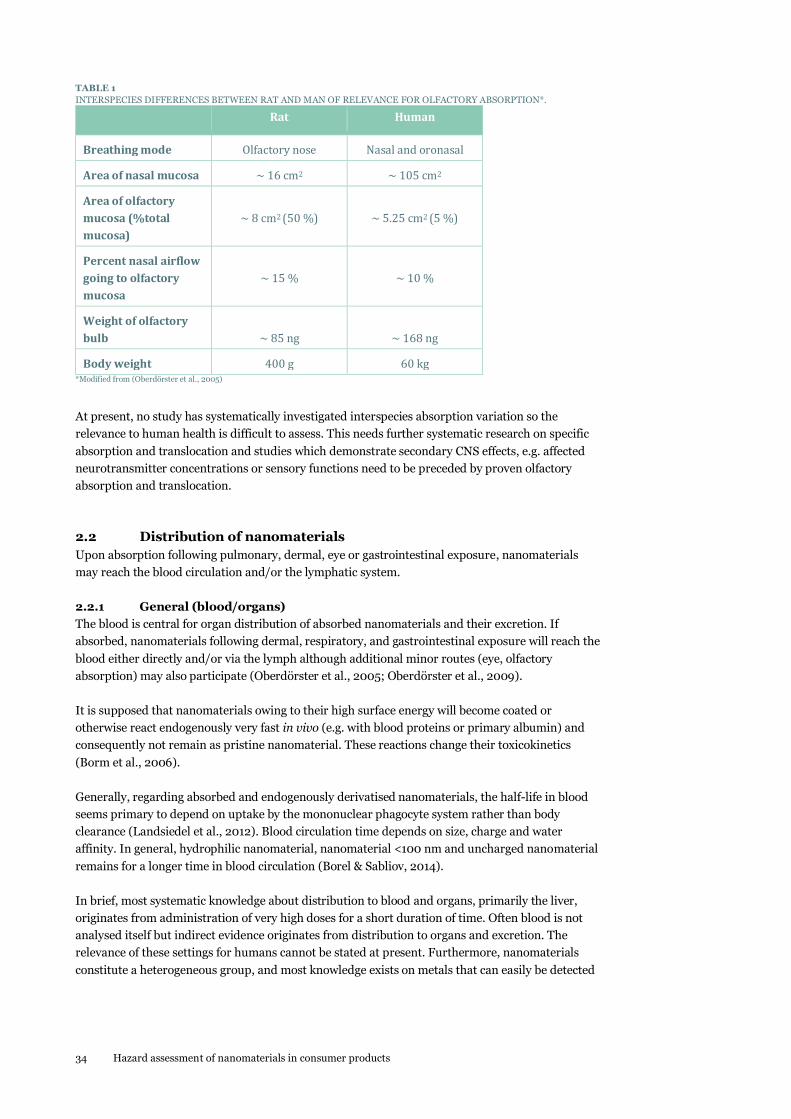
34 Hazard assessment of nanomaterials in consumer products
TABLE 1
INTERSPECIES DIFFERENCES BETWEEN RAT AND MAN OF RELEVANCE FOR OLFACTORY ABSORPTION*.
Rat Human
Breathing mode Olfactory nose Nasal and oronasal
Area of nasal mucosa ~ 16 cm2 ~ 105 cm2
Area of olfactory
mucosa (%total
mucosa)
~ 8 cm2 (50 %)
~ 5.25 cm2 (5 %)
Percent nasal airflow
going to olfactory
mucosa
~ 15 %
~ 10 %
Weight of olfactory
bulb
~ 85 ng
~ 168 ng
Body weight 400 g 60 kg *Modified from (Oberdörster et al., 2005)
At present, no study has systematically investigated interspecies absorption variation so the
relevance to human health is difficult to assess. This needs further systematic research on specific
absorption and translocation and studies which demonstrate secondary CNS effects, e.g. affected
neurotransmitter concentrations or sensory functions need to be preceded by proven olfactory
absorption and translocation.
2.2 Distribution of nanomaterials
Upon absorption following pulmonary, dermal, eye or gastrointestinal exposure, nanomaterials
may reach the blood circulation and/or the lymphatic system.
2.2.1 General (blood/organs)
The blood is central for organ distribution of absorbed nanomaterials and their excretion. If
absorbed, nanomaterials following dermal, respiratory, and gastrointestinal exposure will reach the
blood either directly and/or via the lymph although additional minor routes (eye, olfactory
absorption) may also participate (Oberdörster et al., 2005; Oberdörster et al., 2009).
It is supposed that nanomaterials owing to their high surface energy will become coated or
otherwise react endogenously very fast in vivo (e.g. with blood proteins or primary albumin) and
consequently not remain as pristine nanomaterial. These reactions change their toxicokinetics
(Borm et al., 2006).
Generally, regarding absorbed and endogenously derivatised nanomaterials, the half-life in blood
seems primary to depend on uptake by the mononuclear phagocyte system rather than body
clearance (Landsiedel et al., 2012). Blood circulation time depends on size, charge and water
affinity. In general, hydrophilic nanomaterial, nanomaterial <100 nm and uncharged nanomaterial
remains for a longer time in blood circulation (Borel & Sabliov, 2014).
In brief, most systematic knowledge about distribution to blood and organs, primarily the liver,
originates from administration of very high doses for a short duration of time. Often blood is not
analysed itself but indirect evidence originates from distribution to organs and excretion. The
relevance of these settings for humans cannot be stated at present. Furthermore, nanomaterials
constitute a heterogeneous group, and most knowledge exists on metals that can easily be detected

Hazard assessment of nanomaterials in consumer products 35
and quantified as elements, whereas knowledge about their nanoform is very limited. At present,
extrapolation to other nanomaterials for hazard assessment is not possible.
2.2.2 CNS
Access of nanomaterials to the CNS may take place via olfactory absorption or after oral, dermal
and inhalation exposure via the blood. However, an important anatomical and functional barrier
protects the brain from entry of chemicals present in the blood, namely the BBB. Generally,
crossing the BBB is restricted to molecules which are lipophilic, actively transported or small
soluble molecules < 500 Da (Hagens et al., 2007).
However, owing to their special characteristics, nanomaterials have been hypothesised to be able to
cross the BBB. Thereby, some nanomaterials, including Ir, Zn, Ag, Au, carbon particles, MnO2,
TiO2, Al2O3, and ZnO may distribute to the brain and induce toxic effects to the CNS (Boyes et al.,
2012; Hu & Gao, 2010; Sharma & Sharma, 2010; Simkó & Mattsson, 2010; Yang et al., 2010;
Oberdörster et al., 2009). No interspecies studies are conducted, thus the relevance to humans
cannot be stated.
2.2.3 Fetus
The placenta constitutes the nutritional interface between mother and fetus. As such it does not
represent a complete barrier between the maternal and fetal compartments. Studies of the passage
of ENP across the placenta are still few in number, but rodent studies describe transplacental
transport of nano-sized particles, with rates ranging from almost negligible to high (several percent
of the administered dose). At present, placental transfer has been studied and rendered possible for
particles of Au, polystyrene, nano-sized TiO2, and CNT (Huang et al., 2014; Hougaard et al., 2011a;
Menezes et al., 2011). Exposure to nanoparticles of silica and Ag or quantum dots (with a core of
cadmium) led to increased metallic content in fetal tissues in rats. Whether this was caused by
particulate or ionic transfer is unclear (Melnik et al., 2013; Buerki-Thurnherr et al., 2012; Menezes
et al., 2011). Studies in the human placenta model confirm that placental transfer and uptake may
also take place in humans (Hougaard et al., 2011a). Additional transfer of particles from mother to
offspring could potentially take place during lactation (Melnik et al., 2013).
The capability for transplacental transfer depends highly on the physico-chemical properties of the
particles. This is well illustrated in a study of 13 nm Au nanoparticles administered intravenously to
pregnant mice. Early after establishment of pregnancy, close to 1 % of the maternally administered
dose was found in the fetus and 1 % in the immediate surrounding (extraembryonic) tissues. Later,
when the placental barrier function was just established, fetal uptake was down to 0.1 %. Of note,
extraembryonic tissues now held 10 % of the maternal dose, approximately the same percentage as
was recovered from maternal liver. The pattern was similar for two types of biocompatible Au
particles, whereas accumulation was drastically reduced for the negatively charged particle (Yang et
al., 2012).
The finding has several other important implications. Timing of exposure during pregnancy is of
importance. Transfer may be higher early in pregnancy and decrease in the immediate period after
establishment of the placental barrier. In humans, transfer may increase late in pregnancy, where
also transfer of large antibody molecules takes place (Hougaard et al., 2011a). Furthermore,
particles may be taken up in extraembryonic tissues. Several studies find nanoparticles to be
internalised in placental cells at the maternal-fetal interface, consistent with the placenta acting as a
sequestration organ (Hougaard et al., 2011a; Menezes et al., 2011; Wick et al., 2010).
Size presents an important factor in placental transfer. In the human placenta model, close to 30 %
of 50 nm and 80 nm polystyrene beads transferred from maternal to fetal circulation within hours
of exposure. For 240 nm and 500 nm particles, 9 % and 1 % crossed the placenta, respectively (Wick

36 Hazard assessment of nanomaterials in consumer products
et al., 2010). Finally, transfer of particles across the placenta has been proposed to increase under
some conditions, for example inflammation (Hougaard et al., 2011a).
It does therefore not seem a question whether nano-sized particles are able to reach the fetus, but
rather to which degree it takes place and which characteristics govern particle transfer across the
placenta. Overall, nanoparticle type, size and surface composition as well as stage of embryonic
development determine the extent to which particles transfer from mother to fetus.
2.3 Metabolism
Only a limited number of studies have investigated the potential metabolism of nanomaterials.
Where such information exists, most of the studies show that the nanomaterials are not
metabolised (Landsiedel et al., 2012).
It is supposed that nanomaterials due to high surface energy will be coated or otherwise non-
enzymatically quickly react with e.g. proteins, agglomerate or agglutinate in vivo and consequently
not remain as pristine and discrete particles if absorbed.
The liver is a target organ for many nanomaterials in laboratory animals. Traditionally, first-pass
metabolism is considered to play an important role for detoxifying or activating xenobiotics in the
liver. However, the relevance of this phenomenon for nanomaterials is not known. At present it is
very limited what is known about the metabolism of nanomaterials and the underlying processes
(Borel & Sabliov, 2014; Card et al., 2011). Some general principles include:
Inorganic nanomaterials are very stable (e.g. TiO2, Au, Pt, silica) and are impossible to
metabolise endogenously by enzymes (Allen et al., 2008). Generally, metabolism does not
seem likely for inorganic nanomaterials as such, whereas any organic group attached to the
surface could be modified (Hagens et al., 2007). Breakdown of soluble nanomaterials such as
Ag or ZnO nanomaterials caused by low pH or chemical non-enzymatic oxidation/modification
are not regarded as metabolism.
Organic nanomaterials, especially those with branched side chains or hydrophilic groups,
could be metabolised e.g.by human oxidative enzymes (Hagens et al., 2007). However, these
nanomaterials are outside the scope of this report.
It has been reported that single-walled CNT can be degraded in vitro by horseradish
peroxidase (Allen et al., 2008). It is not known whether other peroxidases are able to degrade
these nanomaterials in vivo.
The database search did not reveal further relevant information on the metabolism of relevant
nanomaterial - free or bound in a matrix - and its potential consequences. In conclusion, available
data are too limited to be extrapolated to other nanomaterials in humans. A case-by-case strategy
should be applied for hazard assessment.
2.4 Excretion/accumulation
2.4.1 Excretion
The blood is central for the distribution and excretion of absorbed nanomaterials which may be
removed by renal excretion and/or in feces via bile excreted from the liver. A minor potential
excretion route may be via breast milk. However, the extent is not known (Landsiedel et al., 2012;
Oberdörster et al., 2009; Oberdörster et al., 2005).

Hazard assessment of nanomaterials in consumer products 37
Some nanomaterials are excreted directly from the port of entry. For example, the majority of orally
dosed nanomaterials are almost completely excreted via the feces without or with minimal
absorption into the body. Rats orally exposed to 14C-labelled C-60 fullerenes eliminated 97 % of the
dose in the feces within 48 hours (Yamago et al., 1995).
As reviewed by Landsiedel et al. (Landsiedel et al., 2012), two functionalised CNT, one single walled
carbon nanotubes (SWCNT) and one MWCNT are examples of nanomaterials for which an
efficiently excretion via the kidneys are seen in rats dosed by intravenous injection. Hepatic
excretion has been observed in rats exposed to polystyrene nanoparticles. Of the administered dose
4 % was excreted via the bile within the first 24 hours.
Characteristics of importance for kidney and liver excretion are size, shape, surface charge, attached
functional groups, endogenous coating, aspect ratio and internal agglomeration and/or aggregation
(Borel & Sabliov, 2014; Landsiedel et al., 2012).
Generally, kidneys excrete nanomaterial < 5 nm whereas renal clearance seems to be minimal for
nanomaterial > 5 nm. Uncharged and negatively charged particle are eliminated from the kidneys,
whereas positively charged particles may accumulate in the kidneys. Only hydrophilic particles are
eliminated via urine.
Orally dosed nanomaterial 2-200 nm in size are excreted from the liver via bile whereas particles <
500 nm in size are are not taken up via the gastrointestinal tract (GI-tract) and are excreted directly
by feces. The relevance of charge for fecal excretion is not clear.
Because of the very little translocation of nanomaterials following exposure via the main exposure
ways (lungs, skin and gastrointestinal tract) most excretion studies inclusive the above mentioned
have been performed in rodents dosed by intravenous injection. Intravenous exposure is not the
way a consumer will be exposed but this exposure way is appropriate for mechanistic studies and
simulates a worst-case scenario in which 100 % of the administered dose is absorbed and reaches
the circulation. More knowledge on excretion following oral, dermal and inhalation exposure is
urgently needed.
2.4.2 Accumulation
It seems that nanomaterials in the sizes 20-100/200 nm can remain in the blood for a longer time
and in that way add to the organ distribution (Borel & Sabliov, 2014; Wang et al., 2013b).
Studies have shown that nanomaterial in systemic circulation can accumulate in the body rather
than being excreted. The liver has been shown to accumulate more than 90 % of the translocated
nanomaterials (compared to the other organs) (Kermanizadeh et al., 2014). For example, no
excretion from rodents exposed by intravenous injection of different kinds of quantum dots could
be detected during the follow-up time of the experiment (between 10 and 28 days). Another
example is a study showing protracted elimination of 40 nm Au nanoparticles from mouse liver
following intravenous injection. Au particles were accumulated in Kupffer cells in the liver one day
after exposure. The Au content of liver from exposed mice was only decreased slightly after 6
months (Sadauskas et al., 2009a). The size of nanomaterials is important for the proportion of
nanomaterials that reaches the liver. A mouse intratracheal instillation study showed that 2 nm Au
particles translocated to the systemic circulation and was detected in the liver to a greater extent
than 40 nm and 100 nm sized Au particles (Sadauskas et al., 2009b). The smallest Au particles (2
nm) seemed to be removed not only by endocytosis by macrophages, but also to be excreted via the
urine (Sadauskas et al., 2007).
In general, distribution to organs besides liver and kidneys includes spleen, heart, muscle, placenta,
bone marrow, CNS and the lungs and most other organs if analysed (Wang et al., 2013a;
Oberdörster et al., 2009; Oberdörster et al., 2005). The liver is the major distribution organ
followed by the spleen, the lymph nodes and the bone marrow. These organs may constitute the

38 Hazard assessment of nanomaterials in consumer products
prime target organs for accumulation and for toxicity. However, most knowledge about distribution
and excretion originates from studies applying intravenous injections and studies applying high-
level exposure for a short time. No relevant study has been identified on long-term
bioaccumulation.
In conclusion, at present consistent knowledge about organ accumulation cannot be concluded
from available data. Systematic long-term, low-dose studies of bioaccumulation and persistence in
the body are needed to assist hazard assessment. Furthermore, nanomaterials constitute a
heterogeneous group and most knowledge exits on metals as they can easily be detected and
quantified as elements, whereas knowledge about their nanoforms is very limited. At present,
extrapolation from ADME and organ accumulation of bulk material and other nanomaterials is not
possible.
2.5 Summary
It is difficult to make general conclusions on the biokinetics on nanomaterials due to the very
diverse physico-chemical properties. The present knowledge on toxicokinetics of nanomaterials is
summarised below.
Absorption: For the consumer, the lungs, the gastrointestinal tract and the skin are considered to be
the primary absorption routes of nanomaterials. In addition, uptake may occur through the eyes
and the olfactory system. Pulmonary exposure to nanomaterials is the exposure route of highest
concern. Inhaled nanomaterials deposit in the alveolar region of the lungs from where they are
removed very slowly and consequently particles may stay for years. A small amount of the particles
may translocate from the lungs to the systemic circulation. Most studies on dermal absorption have
shown no to very moderate absorption of nanomaterials following application on healthy skin.
Similarly, most gastrointestinal studies have shown that many nanomaterials are almost completely
excreted by faeces although a few studies report high uptake of specific nanomaterials.
Distribution: The blood is central for organ distribution of absorbed nanomaterials and their
excretion. If absorbed, nanomaterials following dermal, respiratory and gastrointestinal exposure
will reach the blood either directly and/or via the lymph although additional minor routes (eye,
olfactory absorption) may also participate.
Metabolism: In general, nanomaterials are not metabolised.
Excretion/accumulation: Once in systemic circulation, small nanoparticles (less than 6 nm) may be
excreted in urine via renal excretion. Larger particles and other nanomaterials accumulate in
various tissues including spleen, lymph nodes and bone marrow, and liver accumulation seems to
be most dominant. The larger nanoparticles may be removed into feces via bile excreted from the
liver although the rate is very low. A minor potential excretion route may be via breast milk.
However, the extent is not known.

Hazard assessment of nanomaterials in consumer products 39
3. Adverse effects of pristine nanomaterials
3.1 Respiratory system
Pulmonary effects associated with ambient particle exposure include asthma (D'Amato et al., 2005),
chronic obstructive pulmonary disease and lung cancer (Cohen & Pope, III, 1995; Vineis &
Husgafvel-Pursiainen, 2005). However, so far most of the information on the pulmonary effects of
nanomaterials has been derived from studies with rodents.
The processes and mechanisms behind particle-induced effects in the lungs have recently been
presented by (Donaldson & Poland, 2012) and are summarised below and a few additional
references have been included where relevant.
Cellular stress
Uptake of nanomaterials in pulmonary cells (epithelial cells and macrophages) may induce cellular
stress and this determines if the nanomaterial has the potential for causing inflammation. Several
potential pathways and mechanisms for particle-induced cellular stress have been proposed: 1) The
main hypothesis for the adverse effects of nanomaterials is oxidative stress. Oxidative stress is
defined as an imbalance between oxidants and antioxidants. Nanomaterials may generate reactive
oxygen species (ROS) either directly by physicochemical properties, by soluble compounds or
transition metals, altered function of mitochondria or cellular calcium homeostasis, or indirectly
through inflammatory processes (Li et al., 2008), 2) Following uptake of positively charged nano-
materials, the positively-charged surface may interact with and destabilise the lysosomal
membrane, 3) Lysosomes may get destabilised if an acid-soluble nanomaterial dissolves within the
lysosome resulting in release of ions which may destabilise the lysosome, and 4) Frustrated
phagocytosiscytosis may be induced by high aspect ratio nanomaterials (HARN) because the
macrophages are not capable of complete enclosure of the long fibers. This results in destabilisation
of the phagolysosomal process leading to oxidative stress.
Inflammation
As already described in Paragraph 2.1.1, the primary mechanism for particle clearance in the alveoli
is macrophage-mediated phagocytosis of particles. As long as the capacity of the macrophages to
engulf the particles is not exceeded and the particles do not induce stress in the macrophages, this
process only has minor impact on macrophages and the particle-loaded macrophages are cleared by
the mucociliary escalator. However, if the macrophages are stressed they induce an inflammatory
response. The inflammatory response is characterised by macrophage secretion of cytokines,
chemokines and other signaling molecules mediating recruitment of granulocytes (especially
neutrophils but also eosinophils) and monocytes from the blood circulation into the lung lumen.
Exposure studies have shown that nanoparticles cause more inflammation in the lungs of rodents
than exposure to the same mass concentration of fine particles. There is much evidence that the
inflammatory response induced by low-toxicity low-solubility particles correlates well with the total
surface area of the pulmonary deposited particles (Saber et al., 2012b; Jacobsen et al., 2009; Tran et
al., 2000). For HARN, inflammation is induced by frustrated phagocytosis.

40 Hazard assessment of nanomaterials in consumer products
Fibrosis
Fibrosis is characterised by the accumulation of a matrix of collagen and fibronectin, proliferation
of fibroblasts and transformation of myofibroblasts. In general, persistent inflammation is
suggested to result in fibrosis.
Genotoxicity and cancer
Nanomaterials may induce genotoxicity by primary or secondary mechanisms (further described in
Paragraph 3.9).
3.2 Cardiovascular system
Human exposure to particles from the ambient air has been associated with a number of
cardiovascular conditions. These include myocardial infarction, hypertension, atherosclerosis, heart
rate variability, thrombosis, and coronary heart disease (Nelin et al., 2012). Concern has been raised
that engineered nanomaterials may have similar effects on the cardiovascular system (Saber et al.,
2014). The mechanisms behind particle-induced effects on the cardiovascular system are still not
well understood. However, there are three major theories (Schulz et al., 2005): 1) Particles
translocating from the lungs to the circulation may have a direct effect on the blood vessels and the
blood cells, and, 2) Particles may exert an indirect effect due to particle-induced pulmonary
inflammation which leads to release of signaling molecules into the bloodstream, thereby inducing
systemic inflammation, and 3) Particle-induced effects on the autonomous nervous system may
result in heart rate variability. Recently it was proposed that inhalation of (nano)particles induce
pulmonary inflammation and acute phase response and that the pulmonary acute phase response
via systemic circulation promotes atherosclerosis and cardiovascular disease (Saber et al., 2014).
Particle translocation
As already described in Paragraph 2.1.1., the degree of translocation of particles from lungs to
circulation differs considerably between studies. An example is that one study of humans exposed to 99Tc-labelled 20 nm carbon nanoparticles showed translocation from lungs to blood circulation and
accumulation of particles in the liver, while another study of the same particles showed no
translocation (Landsiedel et al., 2012 and references therein). Translocated particles reaching the
blood circulation may interact and cause adverse effects in the blood vessels. The effect of exposure
to particles on vasomotor function has been investigated by exposing vessel segments ex vivo
(reviewed by Møller et al., 2011). Such ex vivo studies enable assessment of the direct particle effect
without the effects of the accompanying inflammation. However, these ex vivo studies assess the
effects of a much larger particle concentration than present in the in vivo situation, where only a
very limited number of particles translocate to the systemic circulation and where the circulation
time seems to be relatively short. Therefore cautions should be taken before extrapolating the
results to the in vivo situation.
Systemic inflammation
A chronically elevated acute phase response is one of the most important known risk factors for
cardiovascular disease. Epidemiological studies have shown a link between air pollution and the
contents of the acute phase protein C-reactive protein (CRP) in the blood (Ridker et al., 2000). An
elevated level of an acute phase protein called serum Amyloid A (SAA) is also causally linked to the
development of atherosclerosis in mice (Dong et al., 2011). Recently, it has been shown that
exposure to a number of very diverse nanomaterials (nano-TiO2, Nano-CB, MWCNT) and other
particles result in acute phase response in the lungs of mice (Saber et al., 2013). Other studies have
shown that the acute phase response is causally related to increased risk of atherosclerosis and
cardiovascular diseases (reviewed by Saber et al., 2014). This suggests that the acute phase response
in the lungs may be a possible mechanistic explanation for particle-induced cardiovascular disease.
The extent of the particle-induced acute phase response was shown to depend on the total surface
area of pulmonary deposited particles (Saber et al., 2014).

Hazard assessment of nanomaterials in consumer products 41
Autonomic nervous system
The third theory concerns the adverse effects of particles on the autonomous nerve system, which
can either be a direct particle effect or an inflammation-mediated effect on the autonomous nervous
system.
The hypotheses described above, whereby the systemic effects of inhaled nanomaterials are caused
by translocation of particles, pulmonary inflammation and respiratory reflexes, are not mutually
excluding but assumed to interact. For instance, lung inflammation has been shown to increase the
extrapulmonary translocation of particles (Chen et al., 2006).
3.3 Gastrointestinal tract
Recent reviews have indicated that the knowledge on the toxicity of orally administered
nanomaterials in the gastrointestinal tract is limited (Bergin & Witzmann, 2013; Jepson, 2012; Card
et al., 2011). As described previously in Paragraph 2.1.2, the uptake of nanomaterials from the
gastrointestinal tract differs a lot between different types of nanomaterials. If absorbed from the
gastrointestinal tract, nanomaterials can reach the systemic circulation and accumulate in various
tissues or interact with blood components. The review by Card et al identified 30 studies on the
safety of oral exposure to food-related nanomaterials (Card et al., 2011). Of these, only few were
considered reliable because of insufficient characterisation of the tested nanomaterials in most of
the studies. Only two in vivo studies were regarded as having sufficiently described the studied
nanomaterials. One of these studies tested the oxidative damage in rats following a single oral
gavage to C60 fullerenes and SWCNT ((Folkmann et al., 2009) as cited by (Card et al., 2011)). Both
C60 and SWCNT exposure increased the hepatic and pulmonary levels of 8-oxo-7,8-dihydro-2-
deoxyguanosine, while no effects were seen in the colon. It was concluded by the authors that the
systemic effects were caused by direct genotoxicity. The other study was a 15-day repeated dose
dietary study of different types of nano-sized FePO4 and FeSO4. No increase in liver damage,
inflammatory changes or any other adverse effects were detected in the iron-supplemented rats
((Rohner et al., 2007) as cited by (Card et al., 2011)). Based on only these two in vivo studies and a
number of in vitro studies the authors concluded that the data set is too limited “to derive any
overall conclusions regarding the toxicity of nanomaterials for intended use in food and food-
related products” (Card et al., 2011). However, the authors conclude that “the current review does
provide sufficient evidence to dismiss or refute some purported generalizations. For example, the
toxicity of the nanoformulation of a certain ingredient or material was not consistently increased
as compared to the non-nanoformulation. In some cases, there were no differences in the
biological endpoints measured between the nanoformulation and non-nanoformulations and, in
the case of nanoselenium, evidence for reduced toxicity of the nanoformulation as compared to
other forms of selenium was found in several studies. Secondly, several studies demonstrated
enhanced beneficial effects of orally administered nanomaterials, with no evidence of adverse
effects. Clearly it is possible to develop engineered nanomaterials that present no safety concerns
due to oral exposure at specific dose levels” (Card et al., 2011).
Since the review by Card et al., a review on the toxicity of Ag nanoparticles has been published by
Hadrup & Lam. Ag induces a blue-grey discoloration in the skin (argyria). The review concludes
that the effects caused by Ag nanoparticles are mediated via Ag ions released from the particle
surface (Hadrup & Lam, 2014). The toxicity of nano-Ag is further discussed in Chapter 6.4.
To summarise, more studies on oral toxicity of well-characterised nanomaterials are needed to
perform a clear risk assessment of oral exposure to nanomaterials.

42 Hazard assessment of nanomaterials in consumer products
3.4 CNS
The CNS consists of the brain and the spinal cord. At present there is no internationally accepted
definition of chemically induced neurotoxicity but it is generally agreed that CNS-neurotoxicity is
any adverse effect on biochemistry, structure and/or function of the CNS during development or at
maturity induced by a chemical or physical agent. Adversity is any decreased ability to function fully
or to function based to compensatory mechanisms.
Some nanomaterials, including Ir, Zn, Ag, Au, carbon particles, MnO2, TiO2 and ZnO seem to
distribute to the brain and may induce effects on CNS (Boyes et al., 2012; Hu & Gao, 2010; Sharma
& Sharma, 2010; Simkó & Mattsson, 2010; Oberdörster et al., 2009; Yang et al., 2010) as based on
elemental analyses and thus not necessary as nanomaterials.
Human epidemiological studies, controlled animal studies and mechanistic in vitro studies have
shown that exposure to air pollution can lead to neurotoxicity (Costa et al., 2014).
Especially, much relevant knowledge on effects of nanomaterials on the brain originates from
animal studies.
Rats were dosed orally by gavage with 2.25, 4.5 or 9 mg/kg bw/day with 14 nm nano-Ag. Dosing
2.25 and 4.5 mg/kg bw/day for 14 days reduced brain dopamine (DA) neurotransmitter
concentration, whereas noradrenaline (NA) and 5-hydroxy-tryptamine (5-HT) neurotransmitter
concentrations were not affected. Dosing 2.25 and 4.5 nano-Ag mg/kg bw/day for 28 days increased
the DA concentration. Dosing 9.0 mg nano-Ag/kg bw/day for 28 increased DA and 5-HT brain
concentrations (Hadrup et al., 2012c).
Nano-ZnO, when administered intraperitoneal in doses of 4 mg/kg bw biweekly (20-80 nm) for 8
weeks to rats and in doses of 5.6 mg/kg bw every other day (8 doses in total) (20-80 nm) to mice,
have been shown to affect spatial learning and memory functions (Xie et al., 2012; Han et al., 2011).
Wang demonstrated increased brain NA and 5-HT brain neurotransmitter concentrations in mice
10 days after intranasal installation of nano-TiO2 (25-155 nm) in a dose of 50 mg/kg bw (25-125
nm) whereas the brain DA concentration was decreased (Wang et al., 2007b). Mice were instilled
250 µg CB (14 nm) (CB, Printex 90) intranasally. CB increased the extracellular concentrations in
bulbus olfactorius of glutamate and glycine neurotransmitters, whereas taurine and gamma-
aminobutyric acid concentrations were not affected (Win-Shwe et al., 2008).
ZnO is soluble whereas both nano-TiO2 and CB are insoluble. Zn may reach the brain as Zn2+
whereas TiO2 and CB both seem to reach the brain as particles.
The presence of the administered nanomaterials in nanoform in the brain has not been proven but
only evidenced by elemental analyses, thus the mechanisms underlying nanomaterials crossing of
the intact BBB remain elusive and there are still open questions as to the long-term consequences of
nanomaterials CNS-accumulation and fate (Simkó & Mattsson, 2010; Oberdörster et al., 2009).
Any possible functional consequences of the above changes in brain neurotransmitter
concentrations as induced by Ag, TiO2 and CB are not known whereas the behavioral changes as
induced by ZnO are functional changes that could be a manifestation of effects on neurotransmitter
concentrations that were not measured.
CNS-effects induced in rats and mice after olfactory exposure to ZnO, CB and TiO2 are analysed in a
number of studies. A recent key study (Gao et al., 2013) investigates the effects of nano-ZnO (30
nm) when instilled intra-nasally as a single dose in rats. Magnetic Resonance Imaging (MRI)
scanning was performed 1, 2, 3 and 7 days after instillation. The scans revealed that ZnO caused

Hazard assessment of nanomaterials in consumer products 43
significant damage to the olfactory epithelium including edema, inflammation and disruption of
epithelial structures, and mitochondrial destruction. These effects were accompanied by disturbed
sniffing behavior. At the end of the study (7 days post instillation) TEM revealed no indication of
cellular uptake of nano-ZnO as such. There is a slightly acidic milieu in the nasal mucosa (Mei et al.,
2008) so the nano- ZnO might be dissolved and taken up as Zn2+.
In a second study rats were exposed 1 day for 6 hours to 2.1 x 10⁶ particles/cm3 of nano- ZnO (38
nm) for preparation of olfactory synaptosomes (isolated presynaptic nerve endings) or 3 days for 4
hrs to 2.0, 3.4 and 6.6 x 10⁶ particles/cm3 nano-ZnO (12-14 nm) for TEM. The demonstrated
increased olfactory synaptosomal Zn concentration proved olfactory-brain absorption and
translocation and the TEM showed numerous black spots (of not investigated composition) in
olfactory bulbs and brains of the exposed animals (Kao et al., 2012).
Furthermore, nano-ZnO was reported to induce degeneration of olfactory sensory neurons (Takeda
et al., 1997; Burd, 1993). Short-term inhalation exposure of rats to nano-ZnO at a concentration of
0.5 mg/m3, 6hrs/day for 5 days induced nasal necrosis (Landsiedel et al., 2012). Pico molar
concentrations of nano-Zn have been reported to strongly enhance the odorant responses of
olfactory sensory neurons (Aschner, 2009). This indicates that Zn nanoparticles interact with
olfactory receptor neurons (Vodyanoy, 2010).
Mice were instilled 250 µg CB (14 nm) (Printex 90) intra-nasally. Microdialysis in the olfactory bulb
revealed increased extracellular concentrations of the CNS neurotransmitters glutamate and glycine
whereas the taurine and gamma-aminobutyric acid concentrations were not affected (Win-Shwe et
al., 2008).
Ten days after intranasal installation of TiO2 in mice, affected brain neurotransmitter concen-
trations were demonstrated as increased NA and 5-HTconcentrations and decreased DA concen-
tration (Wang et al., 2007b).
Overall, at present it is not possible to predict the toxicological consequences of the above findings,
if any. Normally, functional and structural findings are considered more relevant than biochemical
changes in the CNS. Most often brain biochemistry is investigated because it is more economic with
respect to time and costs than studies of functional and structural changes.
Based on ex vivo experiments with perfusion of human placenta with fluorescence labelled
polystyrene nanomaterials (50, 80, 240, 500 nm), fluorescence was demonstrated to pass the
human placenta barrier (Wick et al., 2010). Other nanomaterials (TiO2, 70nm, 10-100nm; SiO2, 35
nm) have been shown to accumulate as elements in fetal organs, including the brain of rats and
mice and induce effects on CNS (Gao et al., 2011; Yamashita et al., 2011; Hougaard et al., 2010).
Therefore, a recent US-EPA review raises concern that nanomaterials may induce developmental
neurotoxicity (DNT) following prenatal or early postnatal exposure of mothers and pup,
respectively. The review concludes “The evidence for nanomaterials DNT to date is not strong
enough to reach any conclusions about potential risk; however, it indicates that a closer look is
warranted” (Powers et al., 2013). Thus, this is still an important issue that should be further
studied.
3.5 Other organs
Effects of nanomaterials on organs distant from the route of exposure (e.g. lung, gastrointestinal
tract, skin) may be caused either by direct effects of translocated particles or by systemic
inflammation. Different kinds of nanomaterials have been shown to be able to translocate from site
of exposure (e.g. gastronintestinal tract, lungs) to organs (e.g. liver, kidney). The liver has been
shown to accumulate more than 90 % of the translocated nanomaterials (compared to the other
organs) (Kermanizadeh et al., 2014). The toxic effects of nanomaterials on the liver have been

44 Hazard assessment of nanomaterials in consumer products
summarised in a recent review (Kermanizadeh et al., 2014). Intravenous injection of nanomaterials
into rats and mice have resulted in different adverse effects in the liver such as changes in the
hepatic gene expression of genes related to detoxification, lipid metabolism and the cell cycle in rats
following injection of 20 nm Au particles ((Balasubramanian et al., 2010) as cited by
(Kermanizadeh et al., 2014)) and changes in the hepatic gene expression of genes related to
apoptosis, cell cycle, inflammation and metabolic processes following intravenous exposure of Bagg
Albino (BALB/c ) mice to 4 and 100 nm polyethyleneglycol (PEG) Au coated nanomaterials ((Cho et
al., 2009) as cited by (Kermanizadeh et al., 2014)). Kermanizadeh et al. mention a number of
factors of importance for the outcome of liver-related effects in in vivo experiments: 1) the route of
exposure is very important for the fraction of dosed nanomaterials that accumulates in the liver.
Intravenous injection results in accumulation of a much larger fraction of nanomaterials in the liver
compared to other exposure routes (e.g. lung, gastrointestinal tract), 2) the size of nanomaterials is
important for the proportion of nanomaterials that reaches the liver. For example, a mouse
intratracheal instillation study showed that 2 nm Au particles translocated to the systemic
circulation and was detected in the liver to a greater extent than 40 and 100 nm sized Au particles
(Sadauskas et al., 2009b). The smallest Au particles (2 nanometer) seemed to be removed not only
by endocytosis by macrophages but also to be excreted via the urine (Sadauskas et al., 2007), and 3)
The coating of nanomaterials by proteins may also affect the liver toxicity. The coating depends on
the route of exposure.
3.6 Skin
As discussed in Paragraph 2.1.3 there is little evidence of dermal absorption of nanoparticles
through healthy skin as well as through skin damaged by UV radiation or psoriasis. Consequently,
there is also little evidence of dermal toxicity. Discrepancies in the literature are likely to be related
to differences in techniques and methods employed, laboratory conditions and absence of
standardised evaluation protocols (Crosera et al., 2009).
In a review article on nanoparticle dermal absorption and toxicity, Crosera et al. refer to a number
of studies reporting cytotoxic effects on dermal cells exposed to different nanomaterials (Crosera et
al., 2009). No details are available on the individual studies. Examples of observed effects are
summarised in Table 2 based on information from the review article.
Based on the experimental findings reported by Crosera et al. (Crosera et al., 2009), the authors
conclude that the results are contradictory and no specific conclusions regarding dermal toxicity are
drawn. Overall it is concluded that more information in needed in order to understand under which
conditions dermal absorption may occur and what adverse effects can be expected.
Conclusion
Overall there is little evidence of dermal toxicity following typical application of nanomaterials.
However, if nanoparticles are able to penetrate the skin and enter the bloodstream, they may exert a
number of adverse effects. As demonstrated in numerous in vitro studies, these effects include
generation of reactive oxygen species, oxidative stress, protein, DNA and membrane injury,
mitochondrial damage and inflammation resulting in e.g. tissue infiltration, fibrosis, and
granuloma formation. Other proposed effects of nanoparticles entering the systemic circulation
include adverse effects on cells of e.g. the immune systems and passage into the CNS (Choksi et al.,
2010).
3.7 Eyes
The eyes may potentially be exposed to nanomaterials during manufacturing, use and disposal of
the nanomaterials (Ema et al., 2013a). This may give rise to irritation due to surface contact or
toxicity if absorbed into the eye.

Hazard assessment of nanomaterials in consumer products 45
TABLE 2
EXAMPLES OF EFFECTS OF DIFFERENT NANOPARTICLES ON DERMAL CELLS*
Compound Type of cells Outcome
Iron oxide (10 nm)
Human dermal fibroblast
· Disruption to cell cytoskeleton · Reduced proliferation.
SWCNT
Human epidermal keratinocytes
· Increased oxidative stress · Inhibited cell proliferation · Increased expression of stress responsive genes · Cellular toxicity with formation of free radicals · Accumulation of peroxidative products · Antioxidant depletion · Loss of cell viability · Ultrastructural and morphological changes · Dose-dependent irritation response with increase in IL-8 and decrease in cell viability
MWCNT
Human epidermal keratinocytes
· Irritation response · Induction of the release of proinflammatory cytokine · Alteration of protein expression (metabolism, cell signaling, stress, cytoskeletal elements, vesicular trafficking)
Ag Human epidermal keratinocytes
· Inhibited proliferation · Affected cell morphology
Cobalt chrome alloy (30 nm)
Human dermal fibroblast
· DNA damage, aneuploidy and cytotoxicity · Disintegration within the cells with the creation of electron dense deposits enriched in cobalt
TiO2 Human epidermal keratinocytes Human dermal fibroblasts Primary human melanocytes Human immortalized sebaceous gland cells
· Significant and cell-type dependent effects on cellular functions, such as viability, proliferation, apoptosis and differentiation · Inflammation · Cytotoxicity related to phase composition
Quantum dots Human epidermal keratinocytes
· Increased cytokine production and quantum dot uptake · Irritation and decreased cell viability · Release of IL-6 and IL-8
Fullerenes Human dermal fibroblast
· Oxidative damage to cell membranes and cell death · Disruption of normal cellular function through lipid peroxidation
*SUMMARISED FROM (CROSERA ET AL., 2009)
There are a few studies on eye irritation of nanomaterials. In one study performed according to the
Organisation for Economic Co-operation and Development (OECD) test guideline 405, 100 mg C-
60-fullerene and CNT were applied to rabbit eye showing a reversible but minimal potential for
irritation (Ema et al., 2013a; Ema et al., 2011). Two different products of SWCNT and one of two
MWCNT were not eye irritants whereas one other MWCNT product was a very weak irritant (Ema
et al., 2011). Weak eye irritation was confirmed for MWCNT by others (Kishore et al., 2009). Ag
nanomaterials 10 nm did not induce irritation in rabbits when studied according to OECD test
guideline 405 (Kim et al., 2013). In a study, ultrafine TiO2 particles induced reversible ocular
conjunctival redness in rabbits (Warheit et al., 2007). In conclusion, C(60) fullerene, SWCNT,
MWCNT, Ag nanomaterials and ultrafine TiO2 seem not to induce adverse irritation of the eye.

46 Hazard assessment of nanomaterials in consumer products
Concerning toxicity inside the eye, one study in rabbits investigates the toxicity of intravitreal
injected (i.e. into the eye humor) 67 or 670 µmole Au nanomaterials (<220 nm). By light
microscopy, one month post injection, neither 67 nor 670 µmole Au nanomaterials showed any
toxicity in terms of ocular inflammation, or cellular atrophy and/or disorganisation in retina or in
the optic nerve by this very special administration route (Bakri et al., 2008). This shows that if
absorbed from the environment, nano-Au <220 nm is not likely to induce any of these effects inthe
eye.
In conclusion, present knowledge about eye absorption and toxicity into the eye is too limited to be
stated and evaluated in general for hazard assessment.
3.8 Developmental and reproductive system
No epidemiological studies have been published in this area of nanotoxicology. Overall, data from
experimental studies suggest that ENP of various types possess the potential to influence male
fertility as well as fetal development, with very little knowledge with regards to female fertility.
Mechanistically, ENP may potentially affect reproduction and development due to toxicological
properties related to specific chemical constituents of the particles (e.g., Cd in quantum dots) or
properties related to the particles per se. The rationale from the latter comes from the high potential
of particles to induce oxidative stress and inflammation in biological tissues. This may have several
implications in both maternal and fetal tissues. Specifically for inhalation exposure, particles may
deposit in the airways and lungs, where oxidative stress and inflammation may arise in response.
Neither particles nor the inflammatory condition need to be confined to the lungs. Particles may
translocate and inflammatory mediators may be released to the bloodstream and transported to
organs of importance for reproduction, pregnancy and fetal development, such as neuro-endocrine
circuits, the placenta, the fetus, testicles and ovaries (reviewed by Hougaard et al., 2011a; Iavicoli et
al., 2013). Particulates may confer their oxidative and inflammatory action directly in the tissues,
and inflammatory mediators may have consequences in several tissues that in turn may interfere
with reproduction and pregnancy. Some CNT have also been proposed to interfere directly with
cellular and extracellular constituents and thereby alter vital cellular processes, a mechanism that
may be especially relevant in the early phases of embryo development (Shvedova et al., 2012).
Maternal gestational exposure to nanomaterials has been associated with effects in the offspring
that include changes in function of the CNS, immune system, male reproductive system as well as
differential expression of genes (Hougaard & Campagnolo, 2012; Hougaard et al., 2011a). These
findings are somewhat supported by epidemiological and animal studies of ultrafine particles (e.g.,
in ambient air and diesel exhaust) (Ema et al., 2013b).
Most research within this field must be categorised as hypothesis generating. Published reports
present a great diversity of study designs, with respect to kind and size of ENP, route of exposure,
model systems and species, dose levels and endpoints. Furthermore, the methodology does often
not always reflect state-of-the-art, and even if easily available, most mammalian studies do not
provide data on gestational and lactational endpoints (e.g. maternal weight gain, gestation length,
litter size etc.) (Hougaard & Campagnolo, 2012).
Developmental toxicity
Developmental toxicity embraces any effect interfering with normal development during pregnancy
as well as after birth. As exposure route probably plays a major role for induction of adverse effects,
findings are grouped by route of exposure.
Airway exposure during pregnancy (by inhalation, instillation or intranasal insufflation)
Human exposure to particles from ambient air has been associated with low birth weight, preterm
birth and being born small for gestational age (Stieb et al., 2012; Shah & Balkhair, 2011). Rodent

Hazard assessment of nanomaterials in consumer products 47
studies of gestational exposure to diesel exhaust and particles in rodents have revealed changes in
offspring growth, sexual development, male fertility, behavior, susceptibility to develop allergy and
induction of mutations in the DNA of the male fetuses that were inherited by male offspring in the
next generation (Ema et al., 2013b).
So far nano-sized particles of Cd and TiO2, CB and CNT have been studied after airway exposure.
Inhalation of cadmium oxide nanoparticles was overtly toxic to the uterine contents, probably due
to Cd affecting the placenta (Blum et al., 2012). For CB and TiO2, maternal gestational airway
exposure to any of the particles did not seem to interfere with traditional pregnancy and lactational
measures, even if airway exposure was associated with overt lung inflammation in the mother
(Jackson et al., 2012a; Hougaard et al., 2010). Gene expression in the offspring was, however, found
to differ significantly between control and exposed offspring, with female offspring being apparently
more sensitive than males (Jackson et al., 2013; Jackson et al., 2012b). CB also induced strand
breaks in liver from mothers and offspring when assessed after birth (Jackson et al., 2012a), and
has been found to adversely interfere with fertility in the male offspring (Kyjovska et al., 2013;
Yoshida et al., 2010). Maternal intranasal insufflation of both TiO2 and CB altered offspring
phenotype indicative of increased propensity to develop allergy (Fedulov et al., 2008). Exposure to
both particle types caused behavioral alterations in the offspring (Jackson et al., 2011; Hougaard et
al., 2010). A single dose of MWCNT (67 µg/kg) instilled to female mice the day before cohabitation
with male was not associated with changes in gestational and developmental measures.
Malformations have however been observed after a single intratracheal instillation of a very high
dose of MWCNT (>4 mg/kg) concomitant with significant increases in leucocyte counts in maternal
peripheral blood. At 3 mg/kg, neither malformations nor maternal leukocyte counts was
significantly changed (Hougaard et al., 2013; Fujitani et al., 2012; Ema et al., 2014).
Intravenous exposure during pregnancy
SWCNT, silica and TiO2 nanoparticles interfered adversely with mouse embryonic development
(fetal death, malformations) and placental vascularisation when administered intravenously to the
mother early in gestation (Pietroiusti et al., 2011).
Subcutaneous exposure during pregnancy
Subcutaneous injection of TiO2 to pregnant mice have been associated with significant alterations in
offspring brain; in gene expression during lactation (only assessed in male offspring), in DA levels
in some brain areas and in increased numbers of caspase-3-positive cells in the olfactory bulb (the
only investigated structure). Furthermore, TiO2 exposure interfered adversely with fertility in the
male offspring (reviewed by Hougaard & Campagnolo, 2012; Powers et al., 2013; Ema et al., 2014).
Gavage exposure
Gavage administration of multi walled carbon nanotubes (MWCNT) to pregnant rats was not
associated with treatment-related differences in pregnancy measures including body and placental
weights, but SWCNT appear embryolethal and teratogenic in mice when given by oral gavage.
Gavage exposure of pregnant or lactating mice to TiO2 interfered with function of the CNS (Gao et
al., 2011). Ag nanoparticles were without effects on classical pregnancy and birth parameters at dose
levels of up to 1,000 mg Ag/day in some but not all studies (Hong et al., 2014; Philbrook et al.,
2011). Ag has, however, shown toxic to fetal development after exposure in ionic form
(malformations and very high postnatal lethality in rodent studies) (Shavlovski et al., 1995).
Fertility
Fertility includes the reproductive processes up to and including implantation of the fertilised
ovum.
Whether administration is by inhalation or by the intravenous route, particles seem to distribute to
organs with relevance for male and female fertility, i.e. testes, the uterus and ovaries (although the
amount may vary with route of exposure) (Hougaard & Campagnolo, 2012).

48 Hazard assessment of nanomaterials in consumer products
Male fertility
A few epidemiological studies indicate that heavy road traffic might affect reproductive parameters
in men. This is corroborated by findings of decreased semen quality and hormonal changes in male
rodents exposed to diesel exhaust and particles (reviewed by Ema et al., 2013b).
The sensitivity of male fertility to nanomaterial exposure has been investigated in several rodent
studies. Most tested particles were associated with adverse effects on spermatogenesis, although a
few are without adverse effects. Both exposure through the airways and by subcutaneous injection
affected male reproductive parameters, e.g. sperm counts and male reproductive hormones
(reviewed by Lan & Yang, 2012; Ema et al., 2014).
Female fertility
Female fertility has been studied very little. Some in vitro studies indicate that nanomaterials can
be toxic to ovarian cell (reviewed by Iavicoli et al., 2013). Ex vivo studies of, e.g., pre-implantation
mouse embryos have been performed of Ag and polystyrene nanoparticles and indicate that Ag
particles are toxic during this stage of development whereas polystyrene nanoparticles are not, even
if the particles were observed to enter and distribute inside the embryo (summarised in Hougaard &
Campagnolo, 2012). Lung exposure of female mice to MWCNT the day before cohabitation with a
male introduced a 5 day delay in time to delivery of the first litter (Hougaard et al., 2013).
Additional evidence comes from a study in mice, where female mice were housed in heavy traffic
pollution. The female reproductive cycle was disturbed and becoming pregnant also took longer for
mouse couples breeding under polluted conditions (Veras et al., 2009).
3.9 Genotoxicity and cancer
A number of reviews on nanomaterial-induced genotoxicity have been published (e.g.
(Magdolenova et al., 2014; Kumar & Dhawan, 2013; Møller et al., 2013; Oesch & Landsiedel, 2012;
Xie et al., 2011; Singh et al., 2009)). Despite the magnitude of literature within this area, it is
difficult to draw general conclusions due to study limitations such as lack of standardised test
methods, incomplete particle characterisation and the diversity in the used test systems. So far,
most genotoxicity studies have been performed in vitro. A recent review by Magdolena et al. reports
that out of 112 identified genotoxicity studies only 22 are in vivo studies while the rest are
performed in vitro (Magdolenova et al., 2014).
The mechanisms behind nanomaterial-induced genotoxicity are still not well understood. It is well
agreed that these mechanisms can be divided into primary and secondary genotoxicity. Primary
genotoxicity can be further subdivided into 1) Direct primary genotoxicity which is the result of
direct interaction of the nanomaterial with the DNA, and 2) Indirect primary genotoxicity which is
the result of e.g. nanomaterial-induced reactive oxygen species, or of the release of toxic ions from
the nanomaterial. Secondary genotoxicity is caused by nanomaterial-induced inflammation
resulting in release of reactive oxygen species from activated phagocytes (Magdolenova et al., 2014).
A number of physicochemical properties (size, shape, surface properties, composition, solubility,
aggregation/agglomeration, nanomaterial uptake, presence of mutagens and transition metals
affiliated with the nanomaterials etc.) have been suggested to be important for toxicity. However, it
is not known which of these properties that is most important for genotoxicity (Magdolenova et al.,
2014). For insoluble so-called inert particles such as TiO2 and CB, the specific surface area of the
particles has been shown to be correlated to the inflammatory response (please refer to Paragraph
3.1 for more information). The fact that MWCNT and asbestos fibres of same shape and size have
been shown to induce similar genotoxic effects in rodents is an example of the effect of particle
shape for genotoxity (Poland et al., 2008). The importance of physicochemical properties for
toxicity is further discussed in Chapter 5.

Hazard assessment of nanomaterials in consumer products 49
If DNA damage is not repaired or wrongly repaired, it may lead to mutations. Cancer is the result of
an accumulation of mutations in cells. Therefore, nanomaterials capable of inducing genotoxicity
may also have the capacity to induce cancer.
It is well documented that rodents pulmonary exposed to a variety of nanomaterials (nano-TiO2,
nano-CB, SWCNT, MWCNT etc.) develop pulmonary inflammation which may result in secondary
genotoxicity and thereby also cancer. Studies in rodents have demonstrated the carcinogenic
potential of a number of nanomaterials. Instillation and inhalation studies with rodents have shown
that insoluble particles such as TiO2 and CB induce lung cancer (IARC, 2010). Another example of
the carcinogenic effect of nanomaterials is the induction of mesothelioma in animals following
intraperitoneal and intrascrotal administration of CNT (The Nordic Expert Group for Criteria
Documentation of Health Risks from Chemicals, 2013). CB and TiO2 (as groups with no size-
specifications) are both categorised by IARC as possibly carcinogenic to humans (Group 2B) (IARC,
2010).
It is not possible to give an overall conclusion on the genotoxic and carcinogenic potential of
nanomaterials as a group because of the diversity within the group of nanomaterials. However,
based on the recent reviews it can be concluded that the physicochemical characterisation of the
nanomaterials is very important for the prediction of the genotoxic potential (Kumar & Dhawan,
2013). Some of the nanomaterials are considered potential carcinogens. For example, nano-TiO2 is
considered a potential carcinogen by NIOSH. This is based on the pattern of inflammation and a
chronic animal inhalation study showing increase in adenocarcinomas in animals exposed to nano-
TiO2 (NIOSH, 2011). NIOSH concludes “that TiO2 is not a direct-acting carcinogen, but acts
through a secondary genotoxicity mechanism that is not specific to TiO2 but primarily related to
particle size and surface area.”
3.10 Immunotoxicity
Nanoparticles may interact with the immune system and result in immunosuppression or
immunostimulation. Properties such as size, charge, hydrophobicity, solubility, and presence of
targeting moieties and not least surface area determine the interaction with biological tissues and
components of the immune system, and thereby the immunotoxicity. Based on available literature,
nanoparticles do not seem to introduce new immunotoxic responses specific to nanomaterials and
traditional testing regimes are applied to investigate nanoparticle immunotoxicity. It has, however,
been mentioned that in vitro assays may need modifications because nanoparticles may interphere
with the reagents and detection methods and thereby produce unreliable results (Dobrovolskaia et
al., 2009).
Different responses are reported in the literature depending on the characteristics of the nano-
particles. Examples include elicitation of autoimmunity and involvement in allergic sensitisation. It
is, however, mentioned that it is unlikely that nanoparticles can act as haptenes and induce specific
imunoglobulin E production, and it is suggested that the mechanism more likely is based on
nanoparticles acting as adjuvants e.g. inducing specific patterns of cytokines and antibodies
(Gioacchino et al., 2011, abstract only).
Several nanoparticles have, based on animal studies, been shown to induce pro-inflammatory
effects in the lung with changes in cytokine profile. Di Gioacchino et al. concludes that "all
considered, the available data suggest that through the elicitation of an oxidative stress
mechanism, engineered NPs may contribute to pro-inflammatory disease processes in the lung,
particularly allergy" (Di et al., 2011).

50 Hazard assessment of nanomaterials in consumer products
4. Adverse effects of nanomaterials when part of a matrix
The use of nanomaterials in different kinds of materials is increasing due to product advantages
compared to the non-nano-containing conventional products. While there is increasing knowledge
on the hazard of free nanomaterials, only a limited number of studies have focused on the hazard of
nanocomposites.
4.1 Composites (solid)
Paints and lacquers are examples of a product group where nanomaterials are used in relatively
large quantities to provide advantages such as self-cleaning properties or greater scratch resistance.
A few studies have focused on the hazard of sanding dust from nanomaterial-containing paints
compared with conventional products without nanomaterials.
In a Danish project, NanoKem, the purpose was to examine how substitution of larger particles in
paints, lacquers and fillers with nanoparticles of the same chemical affects risk of exposure and
adverse health effects. The adverse health effects of sanding dust from a number of otherwise
identical paints with and without addition of nanomaterials were tested. The tested products were 8
different nanomaterials and 13 dust samples obtained by sanding different painted boards (painted
with 5 conventional products without nanoparticles and 8 versions with nanoparticles).
The products were selected in collaboration with the Danish Coatings and Adhesives Association
and therefore had industrial relevance. Sanding of some of the nanomaterial-containing paints led
to increased formation of nano-sized particles compared to the reference paint, but there was no
consistent pattern in which paints gave rise to increased formation of nano-sized sanding particles,
nor the amount (Koponen et al., 2011). Sanding dust from paints and lacquers with and without
nanomaterials were tested in mice 24 hours after a single intratracheal instillation of 54 µg test
material (Saber et al., 2012c). Sanding dust from nanoparticle-containing paints or lacquers did not
result in statistically significant increased inflammation or DNA damage as compared to dust from
conventional products even though some of the added nanomaterials induced inflammatory
responses in mice when dosed as pristine nanomaterial (nano-TiO2, photocat-TiO2, Axilate, Kaolin)
and some induced DNA damage (nano-TiO2, fine-TiO2) (Saber et al., 2012b). However, when
comparing the different paint and lacquer matrices, the genotoxic response differed between the
paint and lacquer types. Dusts from the two lacquers (one with nano-SiO2 and one without) and the
outdoor acrylic based reference paint resulted in statistically significantly increased level of DNA
damage, while dust from PVA-based paint, filler, and binders did not result in increased level of
DNA damage compared to vehicle exposed mice.
Based on the initial screening, one nanomaterial, nano-TiO2, and sanding dusts from the
corresponding paints with (Indoor-nano-TiO2) and without nano-TiO2 (Indoor-R) were tested for
dose-response relationship at different times because nano-TiO2 was inflammogenic and genotoxic

Hazard assessment of nanomaterials in consumer products 51
(Saber et al., 2012a). The tested nano-paint contained 10 % nano-TiO2. To be able to compare the
same amount of nano-TiO2 in paint, two different dose ranges for the pure nano-TiO2 (18, 54 and
162 µg) and the sanding dusts (54, 162 and 486 µg) were chosen. The 18 µg nano-TiO2 dose
approximately corresponded to the nano-TiO2 content in 162 µg of sanding dust and similarly for
the 54 µg dose of nano-TiO2 and 486 µg dose of sanding dust (indoor-nano-TiO2). There was no
additive effect of adding nano-TiO2 to the paint compared to the reference paint for any of the
measured toxicological endpoints (Saber et al., 2012a).
The same sanding dusts from paints containing nanomaterials and the added nanomaterials as
tested in the screening experiment were also tested by measuring cell surface expressions of
vascular cell adhesion molecule-1 (VCAM-1) and intracellular adhesion molecule-1 (ICAM-1) in
primary human umbilical vein endothelial cells (HUVEC). The results were in agreement with the
in vivo results. The sanding dusts with and without nanomaterials had similar toxicity and on a
mass basis the nanomaterials had a larger effect than the sanding dusts (Mikkelsen et al., 2013).
The same overall conclusion was reached in a recent study on the toxicity of nanoparticles (TiO2, Ag
and SiO2) embedded in a paint matrix compared to control paints without nanoparticles and the
pristine nanoparticles (Smulders et al., 2014). Pulmonary inflammation and systemic effects were
evaluated two and 28 days after the last of five oropharyngeal aspirations of 20 µg of particles or
milled paint exposed to UV-A as an ageing process (day 0, 7, 14, 21 and 28, total dose 100 µg). The
pristine nanomaterials induced some inflammation while little to no adverse effects were seen in
mice exposed to nanomaterials incorporated in an aged paint matrix. Of importance for the
interpretation of the study is that the content of nanomaterials in the paints as measured by
Inductively Coupled Plasma Mass Spectrometry (ICP-MS) was reduced significantly after milling
and ageing.
To the best of our knowledge, the above mentioned publications are the only ones testing the toxic
effects of sanding dusts from nanoparticle doped paints and lacquers. However, Wohlleben et al.
have published two other studies on dust obtained by sanding other types of nanocomposites than
paints and lacquers: 1) A study in rats testing toxic properties of sanding dusts from cement and
plastic with and without CNT (Wohlleben et al., 2011), and 2) an in vitro study using ‘Precision Cut
Lung Slices’ exposed to sanding dust from thermoplastic polyurethane with and without CNT
(Wohlleben et al., 2013).et al. 2013).
In the in vivo study toxicological endpoints were evaluated 3 days and 3 weeks after instillation of
0.36 mg of sanding dust from the composite cement and plastic materials and 0.09 mg CNT
(Nanocyl NC7000) in rats (Wohlleben et al., 2011). The following endpoints were analysed:
Pulmonary histology, inflammation (differential cell counts on broncho alveolar lavage (BAL) cells
and protein concentrations in bronchoalveolar lavage fluid (BALF)), genotoxicity (Comet assay in
lung tissue and micronuclei in bone marrow) and blood were analysed for hematology and acute
phase proteins. The physicochemical characterisation showed similar shape and size of the sanding
dusts with and without CNT. Overall, there were no differences between the toxicity of sanding dust
from the composites with and without CNT. The low content of CNT in the products (2 wt % in
cement and less than 5 wt % in polyoxymethylene plastic) did not allow a study design to determine
if the composite-embedded CNT resulted in a reduced toxicity compared to the free CNT at the
same CNT mass.
The cytotoxicity of sanding dust from elastic CNT-polyurethane nanocomposite was evaluated in
comparison with sanding dust from the corresponding product without CNT by use of ‘Precison Cut
Lung Slices’ (Wohlleben et al., 2013). This in vitro method assesses lung functions under cell
culture conditions. Cytotoxicity was evaluated after 24 hours by measuring membrane leakage of
lactate dehydrogenase and by measuring mitochondrial activity. No cytotoxicity was detected at the
tested doses. CNT was tested at 1 mg/ml while the sanding dusts were tested up to 20 mg/ml. As no

52 Hazard assessment of nanomaterials in consumer products
cytotoxicity was detected in this set-up, a possible difference between sanding dust with and
without CNT could not be evaluated.
To summarise, the very limited number of published studies on the toxicological effects of sanding
dusts from nanocomposites are in good agreement with each other. No additional toxicity have been
detected for any of the nanocomposites compared to the corresponding products without
nanomaterials. More studies are needed to make conclusions within this area.
4.2 Sprays
The attention towards possible adverse health effects of “nano” sprays aroused when a number of
cases of pulmonary health problems were reported among German consumers using different kinds
of “nano” sprays (“Magic Nano Glass & Ceramic” spray and “Magic Nano Bath” spray) (Hahn et al.,
2008; Hahn, 2007). However, analysis of the products documented that solid nano-sized particles
were not generated during use and therefore the adverse effects, which were also demonstrated by
rat inhalation studies (Pauluhn et al., 2008), were not caused by nanomaterials. Instead, the name
“nano” referred to the thickness of the coating generated on the surface upon spraying.
In the present report we were asked by the Danish EPA only to cover spray products containing
solid nanomaterials and therefore the above and similar studies are not further discussed. The
literature search identified only a few studies on the toxicity following inhalation of liquid aerosols
of nanocomposites.
In two of these studies, commercial (solid) nanomaterial-containing sprays were examined
(McKinney et al., 2012; Roberts et al., 2013). Both studied the pulmonary and cardiovascular effects
of antimicrobial spray products in rats.
One of the examined sprays was a spray can product marketed as containing “nano” TiO2 particles
and was intended to be used as a surface antimicrobial product, such as a bathroom sanitiser
(McKinney et al., 2012). Further information on the content was not available from the
manufacturer. Simulated consumer use of the spray resulted in 3.4 mg/m3 75 nm mean diameter
TiO2 particles and the particle concentration was 1.2 x 10⁵ particles/cm3 (Chen et al., 2010). The rats
were exposed by inhalation under the following conditions: 2.62 mg/m3 for 2 hours (low dose), 1.72
mg/m3 4hours/day for 2 days (medium dose) and 3.79 mg/m3 4hours/day for 4 days (high dose),
respectively. This resulted in three doses; 314 mg/m3 min (low dose), 826 mg/m3 min (medium
dose) and 3,638 mg/m3 min (high dose), respectively. The alveolar deposition per rat was estimated
to 4 (low dose), 10 (medium dose) and 43 µg (high dose). Twenty-four hours after end of exposure,
pulmonary (breathing rate, specific airway resistance, inflammation and lung damage) and
cardiovascular endpoints (the responsiveness of the tail artery to constrictor or dilatory agents)
were assessed. Only the high dose resulted in significant effects. These included pulmonary
inflammation, increases in breathing rate and lung cell damage. The specific airway resistance and
the cardiovascular endpoints (systemic vascular responsiveness: vasoconstriction and redilation of
ventral tail arteries) were unaffected even at the high dose. The study design did not include a
comparison of the spray product with nano-sized TiO2 powder. However, the authors compared the
response with one of their previously performed rat instillation studies in which rats were instilled
with 260 µg TiO2 and responded with an 11-fold increase in neutrophils in BAL 1 day after
instillation (Sager et al., 2008). In the present study, the neutrophil influx was doubled at the high
dose (43 µg). Extrapolation from the 43 µg to the 260 µg dose shows that a 12-fold increase in
neutrophils would have been expected in the present study. This indicates a similar response of
TiO2 as powder and when TiO2 is part of the tested spray product.
A similar study was performed to characterise the hazard of an antifungal spray containing nano-
sized Ag particles (MesoSilver, Purest Colloids, Inc.). Rats were exposed for 5 hours by inhalation to

Hazard assessment of nanomaterials in consumer products 53
100 µg/m3 MesoSilver (low dose). This was compared to the effects in rats exposed for the same
period of time to 1,000 µg/m3 Ag spray from National Institute of Standards and Technology, US
(NIST) or a spray consisting of sterile, deionised water (control mice). The alveolar deposition per
rat was estimated to 0 (control spray), 1.4 (MesoSilver) and 14 µg (NIST Ag), respectively. The
choice of the low dose (100 µg/m3) was selected to be the same as the threshold limit value time-
weighted average (TLW-TWA) set by the American Conference of Governmental Industrial
Hygienists (ACGIH) for particulate Ag. For comparison, the Danish OEL for particulate (and
soluble) Ag is 1/10 of the limit set by ACGIH, namely 0.01 mg/m3 (~10 µg/m3). The manufacturer of
the MesoSilver spray indicated the total concentration of Ag to be 20 mg/L in deionised water and
the primary nanoparticle size to be 0.65 nm. The content of Ag particles and free Ag ions was given
to be 75 % and 25 %, respectively (Roberts et al., 2013). One and 7 days after the end of exposure
the same endpoints for inflammatory and cardiovascular effects as in the above study by McKinney
et al was measured (McKinney et al., 2012). In addition, measurement of alveolar macrophage
activity and heart rate and blood pressure in response to isoproterenol and noradrenaline,
respectively, was included in the present study. Only a few pulmonary and cardiovascular changes
were reported. One day after exposure to the commercial spray product MesoSilver (low dose), rats
responded with elevated heart rate in response to isoproterenol, and one day after exposure to the
NIST silver (high dose) standard, the rats responded with an modest increase in the number of
blood monocytes (1.7 fold) and decreased dilation of tail artery after acethylcholine stimuli (Roberts
et al., 2013). The authors discuss that the difference in potency regarding the moderate
cardiovascular changes observed in rats exposed to the commercial product at low dose, which was
not seen in rats exposed to the high dose NIST silver sample, could be the higher content of Ag ions
in the commercial product. Another plausible reason for this slight change could also be adverse
effects of unknown contents of the commercial spray product.
A third study has been performed in which the toxicity of an acrylic ester-based polymer dispersion
containing nanomaterials was compared to a reference product without nanomaterials (Ma-Hock et
al., 2012). In contrast to the two previously referred studies, the products tested in this study were
not spray products but dispersions that were aerosolised in the test system. Despite this, the study
has been included because of the very limited number of studies in this field and because the study
contributes to the overall picture of the toxicological impact of inhalation of nanomaterials in liquid
products. The two products, which were tested in rats exposed by inhalation, were identical except
for the content of nanomaterials (approximately 11 % (w/w) polymer particles in the “nano”
version). Rats were exposed by nose-only exposure for 6 hours/day for 5 consecutive days at target
concentrations 3 and 10 mg/m3. Compared to the reference product without nanomaterials, the
toxicity of the nanomaterial-containing version of the products was not changed. When evaluating
the present study it should be noted that none of the products resulted in any adverse effects at the
tested concentrations (3 and 10 mg/m3) and time-points for evaluation after exposure (3 and 23
days). Thus, the NOAELs for both dispersions were >10 mg/m3 and were independent on the
content of nanomaterials.
To summarise, a very limited number of studies on the toxicological effects of sprays containing
nanomaterials have been published. A rat inhalation study of a commercial spray containing “nano”
TiO2 particles indicated similar toxicity of nano-TiO2 when part of the product and when nano-TiO2
was tested alone. Testing of a commercial Ag spray product in rats resulted in moderate cardio-
vascular effects that were not observed in rats exposed to a standard Ag reference product. The
third study evaluated in the above paragraph showed no increased toxicity of a polymer dispersion
compared to the non-nano product. More studies are needed to make conclusions within this area.
4.3 Liquids
The most commonly investigated nanomaterials in liquid matrices are TiO2 and ZnO used in
cosmetics. The SCCS has adopted opinions on both substances in the nano form in 2013 and 2012,

54 Hazard assessment of nanomaterials in consumer products
respectively. The overall conclusion in the two opinions is that the use of TiO2 and ZnO nano-
materials with specified characteristics as indicated in the opinions, at a concentration up to 25 % as
a UV-filter in sunscreens, can be considered to not pose any risk of adverse effects in humans after
application. This does, however, not apply to other applications that might lead to inhalation
exposure of the nanoparticles (such as sprayable products) (SCCS, 2014d; SCCS, 2012).
Most of the documentation is based on studies with the pristine nanomaterial, as only few relevant
studies investigating the effects of dermal application of sunscreen products are available. However,
none of these studies provide evidence of dermal absorption or other effects on the skin.
A few studies have investigated penetration of the nanomaterials in sunscreen products. Sadrieh et
al. studied dermal penetration of TiO2 particles applied in a sunscreen product to the skin from
weanling Yorkshire pigs. When exposed to UV-B radiation (sunburn simulation), the study
demonstrated that UV-B-sunburned skin slightly had enhanced in vitro or in vivo penetration of
rutile TiO2 (uncoated mixture of anatase and rutile and dimethicone/methicone copolymer-coated
rutile TiO2) present in the sunscreen formulations into the stratum corneum. The penetration was,
however, considered minimal, and there was no evidence that nano-sized or submicronised TiO2
penetrated the intact epidermis to any significant extent and there was no evidence of systemic
absorption (Sadrieh et al., 2010).
Based on a review of a number of skin penetration studies investigating penetration of solid
nanoparticles in cosmetics, Nohynek et al conclude that there is little evidence suggesting that
slightly compromised skin in general has greater susceptibility to skin penetration by small
particles. It is however concluded that some pathological skin conditions may affect skin
penetration of topically applied substances (Nohynek & Dufour, 2012).
Gulson et al have investigated dermal uptake of ZnO from sunscreen applied to human skin in vivo
over five days and found that small amounts of elemental Zn could be detected in blood and urine.
This is taken as an indication of possible passage through the skin, although at a very low level
(Gulson et al., 2012; Gulson et al., 2010). However, in the SCCS opinion on ZnO in the nano form it
is concluded that measured elemental Zn is not a result of dermal penetration of nanoparticles but
rather a result of Zn-ions released from the ZnO nanoparticles (SCCS, 2012).
4.4 Food
The application of nanomaterials in food, beverages and food contact materials in the EU is still
rare (Wilson Center, 2014) and has been hampered by concern about safety, ethical, political and
regulatory issues which all are caused by lack of knowledge (Borel & Sabliov, 2014).
In the EU, three types of nanomaterial are authorised to be used in food packaging materials:
Titanium nitride was evaluated by EFSA in 2008 and has been approved as an additive for use
in some food contact plastics since 1 May 2011. Use is limited to PET (polyethylene
terephthalate) bottles and refrigerators in a concentration up to 20 mg/kg. The Agency is not
aware of any commercial use of this additive. There is no migration from this material into
food (European Commission, 2014).
CB can be used in rubber, silicones and printing inks for e.g. meat (Additive, primary particles
10-300 nm which are aggregated and may form agglomerates, 2.5 % w/w) (European
Commission, 2014).
Silicone dioxide can be used in printing inks, paper, rubbers and silicones (Antislip-agent,
primary particles 1-100 nm which are aggregated and may form agglomerates) (European
Commission, 2014).

Hazard assessment of nanomaterials in consumer products 55
Four nanomaterials not authorised in the EU can be used in food contact materials in the US
according to the US Food and Drug Administration. They all have the status of ‘Generally
recognised as safe’ implying that they can be applied in the US in concentrations limited to fulfill
technical demands (Wagner, 2013):
Al. Applications: Filler in polymers, scratch- and abrasion-resistance in coatings, improvement
of barrier properties and UV-filter,
Ag. Applications: Antimicrobial, antibiotic and antistatic agent. Ag-nanomaterials are used in
food packaging materials,
Nanoclay (bentonite). Applications: Improvement of barrier properties) and
ZnO. Applications: UV-filter antimicrobial.
The use of these products in EU and specifically in Denmark is not known, but it is assumed to be
very limited.
Gastrointestinal exposure may occur either directly by intentional intake of food or beverages
containing nanomaterials, or indirectly by e.g. leakage from dental implants or from nanomaterials
from food packaging. In addition, gastrointestinal exposure may occur following translocation of
inhaled particles through the mucociliary escalator which are subsequently swallowed. Oral
absorption of nanomaterial from nanomaterial-containing medicine is also a potential exposure
scenario. However, in the present report we were asked by the Danish-EPA not to cover
nanomedicine and therefore such studies are not discussed.
A very limited number of studies on the adverse effects of nanomaterials in a food matrix or a food
related item such as food packaging have been identified.
Toxicity of nanomaterials when part of food
Chewing gum is an example of a food item for which there is an extensive use of nano-TiO2 as an
additive. This is, however, not approved in EU. A Chinese study was designed to characterise and
perform a preliminary assessment of TiO2 as an additive in chewing gum (Chen et al., 2013b). The
toxicity of nano-TiO2 isolated from 6 different brands of commercially available chewing gum was
evaluated by exposure of human gastric epithelial cells and human epithelial colorectal
adenocarcinoma cells. Viability, lactic acid dehydrogenase release and ROS generation was
evaluated after 24 hours exposure to 0, 10, 25, 50, 100 and 200 µg/ml nano-TiO2. Neither viability
nor membrane leakage of lactic acid dehydrogenase was affected at the tested doses, while cell
viability was slightly reduced in P25 TiO2 exposed cells included as positive control. ROS generation
for the isolated TiO2 was similar for all brands and P25 TiO2. Estimation of the amount of TiO2 in
gum before and after chewing indicated that approximately 95 % of the chewing gum content of
nano-TiO2 was swallowed during chewing. The fraction of TiO2 particles below 200 nm in the
chewing gum was more than 93 % and 18-44 % of the TiO2 particles were below 100 nm in size. The
hydrodynamic size distribution of TiO2 in one of the chewing gum brands was determined in a
simulated digestive system consisting of three different compartments and showed that the number
percentages of nano-TiO2 was lowered to 34, 13 and 22 % in 1) artificial fluid, 2) gastric fluid and 3)
intestinal fluid, respectively. Under the given experimental conditions this in vitro study does not
indicate high toxicity. However, further studies and especially in vivo studies are needed to evaluate
the potential toxicity associated with the chewing of nano-TiO2 containing chewing gum.
In vivo studies in laboratory animals dosed with nanomaterials via the diet
A thorough review evaluates the oral safety of food-related nanomaterials that have potential use
from in vitro and in vivo studies in laboratory. Only the in vivo studies are considered here (Card et
al., 2011).

56 Hazard assessment of nanomaterials in consumer products
No human studies were identified. In laboratory animals, 21 studies were identified on oral
administration (17 by gavage, 4 by diet) (Card et al., 2011).
Rohner et al. dosed rats with ferric phosphate (used for iron fortification) ad libitum in their diet at
concentrations at 10 and 20 mg/kg diet (10.7, 30.5, or 64.2 nm) for 15 days. Histological
examinations and measurement of thiobarbituric acid reactive substances as indication of oxidative
stress showed no toxicity (Rohner et al., 2007).
Jia et al. dosed female and male rats with Se-nanomaterials (20-60 nm) at 4 different
concentrations in the diet for 13 weeks corresponding to doses of 0.19/ 0.12, 0.33/0.22, 0.44/0.31,
and 0.50/0.42 mg Se/kg bw/day (female/male) . The authors concluded a NOAEL at 0.33 and 0.22
mg Se/kg bw/day for female and male rats, respectively (Jia et al., 2005).
Chan et al. dosed mice with nano-sized Black Soybeans (52 % <100 nm, 42 % 100-200 nm) in the
diet at a concentration of 10 % for 12 weeks. The Black Soybeans were grounded to nano-size by a
company in Taiwan (details not specified). The study indicated enhanced immune system
responsiveness (Chan et al., 2009).
Pigs were orally dosed with 0.5 % montmorillonite nanocomposite (MNC) in the diet for 83 days.
This corresponded to an oral intake of 11 mg MNC/animal/day. The size was not specified (Xu et al.,
2004).
No adverse toxic effects were reported in any of the above studies. None of the studies characterised
the nanomaterials in the diet or in any organ (Card et al., 2011).
In conclusion, at present there is no clear overview of food items and contact materials actually
containing nanomaterials in EU, but present and especially future consumer exposure to
nanomaterials via food seems likely. However, initiatives regarding analytical tools need to be
developed for verification of data from industry, for product control, for labeling information and to
validate database information. Such knowledge is, bearing in mind the European Commission’s
recommended definition from 2011 of nanomaterials, mandatory (Danish EPA, 2013c). Therefore,
intensive method development and validation are in progress, some of which are funded by the EU
(Linsinger et al., 2013; Loeschner et al., 2013a; Loeschner et al., 2013b). Here, a recent study
investigates and evaluates the liberation of spiked nanomaterials from a food matrix (chicken meat)
and their characterisation afterwards by asymmetric field flow fractionation in combination with
on-line optical detection and mass spectrometry (Loeschner et al., 2013b).
Assessment of exposure is hampered by gap of knowledge on all important aspects including
detection of nanomaterials in food, consumption, exposure, toxicokinetics, gastrointestinal
absorption, distribution, metabolism, absorption and all endpoints of toxicity. Free, insoluble
nanomaterials including agglomerates are considered of highest concern to the consumers
(Bouwmeester et al., 2009).
It was overall concluded that there are not sufficient reliable data for relevant assessment of the
safety of oral exposure to food-related nanomaterials (Card et al., 2011). Furthermore, in a complex
matrix such as food, more knowledge is needed about the interaction between nanomaterials and
various food components, the interplay between nanomaterials and the microbial flora in the gut
and ADME of the nanomaterial form.
Potential carry-over from meat and liver
Three studies have been carried out in pigs and chicken administered nano-Ag through their diet or
in drinking water. In the first study, pigs administered 20 or 40 ppm nano-Ag in their feed for 5
weeks showed no Ag retention in muscle and only minimal retention in the liver; 0.435 and 0.837

Hazard assessment of nanomaterials in consumer products 57
µg/g wet tissue, respectively (Fondevila et al., 2009). In the second study, poultry administered 25
ppm nano-Ag in drinking water had very low concentrations in muscle and liver (Ahmadi &
Kordestany, 2011). In the third study, chicken were administered up to 15 ppm nano-Ag in their
feed up to 42 days. Ag was distributed to various organs including eatable tissues. The highest
concentrations were demonstrated in breast, femur muscles and the liver; 13.5, 14.2 and 6.8 ppm,
respectively (Ahmadi & Kordestany, 2011). In neither of these studies were the size of the
nanomaterials specified, and it was not demonstrated that the nanomaterials were on nano-form in
the diet, drinking water or in the tissue. However, Ag should not to be present in the nanoform in
the meat and after eating it. The studies suggest that animals fed with nanomaterial-containing food
represent a potential for carry-over if the meat is used for consumption. Furthermore, these are
academic studies, and it is not likely that such meat or liver are on the market in EU.
Study in humans via Ag nanomaterial solution
In a very recent study, healthy humans were orally dosed with 10 ppm Ag nanomaterials (5-10 nm)
in an experimental aqueous solution for 3, 7 and 14-days or to 32 ppm (32.8 nm) for 14 days
corresponding to 100 and 480 µg/person/day. Persons underwent thorough metabolic analyses (all
common clinical biochemical analyses in plasma), complete blood counts, urine analyses, and chest
and abdominal MRI. The authors concluded that no important changes on any of these parameters
at any time or dose were detected (Munger et al., 2014).
Nanocomposites used as dental fillings
Nanocomposites have the potential, when used as dental fillings, to restore affected teeth following
dental caries. There is a long lasting exposure because the nanocomposite will be in contact with the
oral tissue for many years. The cytotoxicity and genotoxicity of an extract from a dental nano-
composite containing 35 wt % nanosilica filler was evaluated in vitro by exposure to human lung
fibroblast cells (Medical Research Council cell strain 5 (MRC-5 cells)) (Musa et al., 2013). The cells
were exposed to cell medium containing an extract from the nanocomposite. The extract was
prepared in two steps: 1) 24 hours incubation of a suspension of 0.2 g/mL granulated cured
nanocomposite in cell media at 37⁰C and 2) filtration of the suspension through a 0.45 µm filter.
The cytotoxicity was evaluated by the monocyte mediated cytotoxicity assay (MTT assay) following
72 hours exposure to various concentrations of nanocomposite extract. The genotoxicity was
determined at doses resulting in less than 50 % cell death. Genotoxicity was assessed by the comet
assay and chromosome aberration tests (6, 24 and 48 hours exposure) with or without addition of a
metabolic activation system. No genotoxic effects were detected at the used test conditions. The
value of the study is limited in this context due to shortcomings in relation to the assessment of the
toxicity of nanomaterial-containing composites. No characterisation of the nanocomposite extract
was performed. Therefore it cannot be assessed if any of the nano-content of the suspension was
liberated to the cell media. In addition, the choice of a lung fibroblast cell line is not an optimal
choice for the assessment of oral toxicity.
Summary
Three types of nanomaterial (TiN, CB, SiO2) are authorised by EU to be used in food packaging
materials. In addition, four nanomaterials (Al, Ag, nanoclay, ZnO) may be used in food contact
materials in the US. All such products may be available in Denmark. Their production and use is not
known but is assumed to be limited. However, future consumer exposure seems likely.
Studies suggest that farming animals fed with nanomaterial-containing food may represent a
potential for carry-over if the meat is used for consumption. However, this was no longer in the
nanoform.
To gain knowledge, analytical means need to be developed for verification of data from industry,
product control, labeling information, to validate database information and to quantify and verify
nanomaterials in diet, drinking water and organs from toxicological studies.

58 Hazard assessment of nanomaterials in consumer products
In laboratory animals, four studies dosing nanomaterials via diet were identified. No adverse toxic
effects were reported. It is stressed that none of the studies characterised the nanomaterials in the
diet, drinking water or in any organ owing to lack of appropriate methods.
In a study, healthy humans were orally dosed via drinking fluid with nano-Ag for 14 days. No
adverse effects were detected.
In a complex matrix such as food, more knowledge is needed about the interaction between
nanomaterials and various food components, the interplay between nanomaterials and the
microbial flora in the gut, ADME and toxicity of the nanomaterial form.
At present, free, insoluble nanomaterials including agglomerates are considered of highest concern
to the consumers.
Any hazard assessment of nanomaterials in food is hampered by a deep gap of knowledge on all
important aspects including detection and quantification of nanomaterials, consumption,
toxicokinetics, and by lack of sufficient and consistent data on toxicity.
At present, any evaluation needs to be on a case-by-case basis and to be performed by skilled
experts.

Hazard assessment of nanomaterials in consumer products 59
5. Physico-chemical factors of importance for toxicity
A number of different physico-chemical properties have been shown to be important for nano-
material-induced toxicity. This chapter summarises the present knowledge based on examples
within this area. For more information on specific types of nanomaterials, please refer to Chapter 6.
5.1 Size
Regarding pulmonary exposure, the size of the nanomaterial is an important determinant for lung
deposition (further described in Paragraph 2.1.1). In brief, nanoparticles with aerosolised or
aggregate/agglomerate size in the size range from 10-100 nm will deposit in the alveoli to a greater
degree than larger particles. The mucociliary system removes particles deposited in the upper
airways very efficiently. However, the mucociliary system is not present in the alveoli resulting in
much longer clearance time (half-time of particles in the alveolar region is months in rats and years
in humans) (Paragraph 2.1.1 and (Braakhuis et al., 2014)). Size is also an important factor regarding
translocation. In general, when comparing the translocation of a particle of the same chemical
composition but with different sizes, the translocation has been shown to be higher for smaller
particles compared to the larger. For example, 2 nm Au particles translocated, following
intratracheal instillation in mice, from the lungs to the liver, while the 40 and 100 nm sized Au
particles did not translocate (Sadauskas et al., 2009b).
5.2 Agglomeration/aggregation
Aerosolised nanomaterials aggregate or agglomerate into larger sized particles over time and
therefore aerosol exposure will often consist of aggregates or agglomerates rather than of single
nanomaterials. Agglomeration of nanomaterials is caused by adhesion of particles to each other by
weak forces. These larger agglomorates can rather easily be disrupted. Aggregates of nanomaterials
are connected with covalent or metallic bonds which are difficult to disrupt. It is not known if
agglomerates are broken up to single particles in the compartments of the body. However, several
studies have shown that single particles form agglomerates in biological fluids (further described in
Paragraph 5.10 Behavior in biological media). The toxicity of aggregates/agglomerates of
nanomaterials may be different than the toxicity of the individual particles (Landsiedel et al., 2012).
As described in Paragraph 2.1.1, the deposition pattern of particles in the different parts of the
respiratory tract is strongly dependent on the size of the aerosolised particle agglomerate/aggregate
and this affects toxicity. On the other hand, pulmonary inflammation, in terms of neutrophil influx
and pulmonary acute phase response, are proportional to BET surface area that is the total surface
area of the primary particles irrespectively of the aggregation state.
5.3 Specific surface area
The smaller the size of the particles, the larger are their specific surface area. It is well established
that the inflammatory response of low toxicity-low solubility particles is proportional to the total
surface area of the instilled primary particles rather than the mass (e.g. by (Saber et al., 2012b;

60 Hazard assessment of nanomaterials in consumer products
Stoeger et al., 2006; Jacobsen et al., 2009). Chronic inflammation has been associated with
respiratory and cardiovascular diseases, as previously described in Chapter 3.
5.4 Shape
The shape of nanomaterials has been shown to affect toxicity. For example, fibre-shaped
nanomaterials are more toxic compared to spherical-shaped nanomaterials of the same chemical
composition (Braakhuis et al., 2014). Fibres are cleared much slower from the lungs compared to
spherical particles, and short fibres have been shown to be cleared faster than longer fibres. Fibres
longer than 15-20 µm cannot be phagocytised by macrophages and induce frustrated phagocytosis
(Braakhuis et al., 2014; Donaldson et al., 2010).
5.5 Crystallinity
SiO2 is an example of a substance for which the crystal phase is important for toxicity. SiO2 is
available in multiple crystalline and amorphous phases. While inhalation of crystalline SiO2
(quartz) can lead to the development of silicosis, bronchitis and lung cancer, these adverse health
effects have not been observed following exposure to amorphous SiO2 (IARC Monographs on the
evaluation of carcinogenic risks to humans, 1997). Another example is TiO2 which exists in two
main crystal phases, rutile and anatase, and where anatase TiO2 has been shown to be more toxic
than the rutile form (Johnston et al., 2009).
5.6 Dissolution rate
Some nanomaterials are insoluble in the biological environment (e.g. lung lining fluid) while others
may dissolve at the portal of entry or after being phagocytised leading to formation of ions. The
insoluble particles may lead to chronic inflammation in the lungs. Inflammation causes release of
ROS which can result in DNA damage and lead to the development of cancer. An example of a
soluble and toxic nanomaterial is ZnO. When testing the solubility of nano-ZnO in different media,
it was shown that 90 % of the nano-ZnO was dissolved within a day when incubated with artificial
lysosomal fluid (pH = 4.5) while nano-ZnO did not dissolve in artificial intestinal fluid (pH =7.4)
(Cho et al., 2011). Nano-Ag particles are another example of a soluble nanomaterial. In contrast to
nano-ZnO, nano-Ag particles are rather non-toxic (Hadrup & Lam, 2014). As illustrated in the
example given above, pH differences may affect dissolution rate and therefore result in different
exposure to ions in different compartments of the body. Overall, the dissolution rate of nano-
materials depends on particle size, coating, stability, manufacturing process and biological
environment (Braakhuis et al., 2014).
5.7 Chemical composition
The chemical composition of the nanomaterial is a major determinant of the toxicity. For example,
nano-ZnO is a highly toxic nanomaterial following pulmonary deposition. The high toxicity of nano-
ZnO has been suggested to be a combination of the large alveolar deposition of nano-ZnO and the
following release of toxic Zn ions in the lyzosomes (Cho et al., 2011). Nanomaterials often contain
more than one chemical. The particles may be surface modified to provide the nanomaterial with
certain properties (as described in Paragraph 5.8) or nanomaterials may contain impurities
introduced during the production or in the environment. As an example, CNT and nano-sized CeO2
have been shown to absorp certain toxic chemicals (Feng et al., 2012; Fang et al., 2008; Yan et al.,
2008). Moreover, CNT often contain different metal oxides in various amounts. The metal oxides
are impurities from the production process (Danish EPA, 2015a). Therefore, the toxicity of a certain
nanomaterial is a combination of the toxicity of the nanomaterial per see and the toxicity of the
absorped contaminants.

Hazard assessment of nanomaterials in consumer products 61
5.8 Surface modifications
The surface of nanomaterials can be extensively modified by surface functionalisation, a process by
which surface moieties are attached to the surface. Surface modifications may reduce or increase
the toxicity of nanomaterials. As an example, surface modifications of SAS have been show to
modify toxicity. A recent study investigated the toxicological effects of one uncoated SAS and four
surface-modified SAS (surface modifications: polyacrylate, PEG, phosphate amino). Rats were
exposed by inhalation for 5 consecutive days with 14- or 21-day post-exposure observation.
Uncoated SAS induced moderate pulmonary inflammation but no systemic effects. Because no
adverse effects were observed in rats exposed to SAS coated with PEG, phosphate or amino, the
results suggest that some types of surface modification masks the toxicity of SAS. However, in
contrast to the uncoated SAS the acrylated SAS induced effects in the spleen while no pulmonary
effects were seen. This indicates that surface coating with acrylate increases the systemic but not the
pulmonary toxicity (Landsiedel et al., 2014). In another recent study, the toxicity of COOH-
modified SAS was compared to uncoated SAS following intraperitoneally injection in mice The
inflammatory response was evaluated by measuring cytokines in the peritoneal cavity lavage fluid
and found to be significantly reduced in mice exposed to the surface modified SAS compared to the
uncoated SAS (Morishige et al., 2012).
5.9 Surface charge
A review by Braakhuis et al. concluded that positively charged nanomaterials are taken up by cells
more easily than neutral and negatively charged nanomaterials which resulted in a higher degree of
inflammation and cell death (Braakhuis et al., 2014). The surface charge can be measured by the
zeta-potential, which is the electric potential created between the charged groups associated with
the surface of a particle and the suspension medium.
5.10 Behavior in biological media
Nanomaterials behavior in biological media has been reviewed recently (Landsiedel et al., 2012). As
soon as a nanomaterial is taken up by an organism its surface usually becomes coated by proteins.
This spontaneous coating by proteins is called the protein corona. The composition of the corona
depends on the exposure route. If the nanomaterials are inhaled, the corona will be composed of
proteins present in the lung lining fluid, while nanomaterials entering the blood stream will become
coated with plasma proteins. Certain types of absorbed proteins such as e.g. albumin can counteract
agglomeration of nanomaterials. This protection against agglomeration is exploited in studies in
which a well dispersed particle suspension is needed (e.g. when preparing particle suspensions for
intratracheal instillation).
To summarise, the hazard of a specific nanomaterial is not a result of a single physico-chemical
property but is rather the result of a combination of the properties.

62 Hazard assessment of nanomaterials in consumer products
6. Hazard evaluation of a selection of nanomaterials
6.1 Carbon black (CB)
6.1.1 Introduction
The purpose of this chapter on hazard assessment of CB is to serve as background documentation
for the risk assessment of mascara containing CB. Thus, the main focus will be the hazards
associated with dermal and eye exposure to CB because those are the most relevant exposure routes
associated with consumer use of mascara. The assessment will primarily be based on the recent
opinion on CB adopted by the SCCS (SCCS, 2014c). The aim of this SCCS opinion on CB was
specifically to decide if CB is safe for use as a colorant with a concentration up to 10 % in cosmetic
products and is therefore considered highly relevant for the present purpose. The text is partly
condensed from this SCCS opinion. To some extent, key references of importance for the chosen
risk scenario are referred based on the description in the SCCS evaluation.
CB is a low-solubility particle which is primarily manufactured from hydrocarbon sources although
CB used as food colorant is manufactured from vegetable sources. CBs produced from hydrocarbon
sources are nanomaterials used as reinforcement in rubber and as black pigment in plastics, paints,
coatings and inks (IARC, 2010). Furthermore, CB is approved for use as a cosmetic colorant (e.g.
mascaras and eyeliners) (SCCS, 2014c). The yearly production of CB was approximately 10 million
tonnes in 2005. CB consists of spherical primary particles (10-500 nm in diameter) that form
aggregates (50-600 nm in diameter) and agglomerates (many micrometers in diameter). The
content of extractable adsorbents is less than 1 % (IARC, 2010). CB has been used as a model
particle within the area of particle toxicology. Therefore, there is a rather large database on the
toxicity of CB.
Vegetable carbon is approved as a food additive (E 153) in Europe and consists of finely divided
carbon manufactured by steam activation of carbonised raw material of vegetable origin (EFSA
Panel on Food Additives and Nutrient Sources added to food (ANS), 2012). EFSA concluded in their
evaluation that the currently marketed vegetable carbon products do not contain nanoparticles.
Because of this and the chosen risk scenario of mascara containing CB, vegetable carbon will not be
further dealt with in the present report.
6.1.2 Biokinetics
The text is partly condensed from the SCCS (SCCS, 2014c). To some extent, the text also refers to
the more detailed description of general particle attributes in the chapter on biokinetics in the
present report (Chapter 2).
Absorption
The pulmonary retention of inhaled CB particles has been studied in experimental animals (rats,
mice and hamsters). CB is a typical insoluble particle and the deposition, clearance and
translocation of CB is therefore already described in the general chapter on pulmonary absorption
(Paragraph 2.1.1). CB particles have been found to translocate from the lungs to other organs (e.g.
liver, spleen and brain). It has been shown in experimental animal studies that CB can act as a

Hazard assessment of nanomaterials in consumer products 63
carrier of adsorbed material such as polyaromatic hydrocarbons. This results in a slower clearance
from the lungs compared to the clearance following exposure to the material alone (SCCS, 2014c).
After oral administration uptake and distribution from the gut to gut-associated
lymphoid tissue has been described (SCCS, 2014c).
The SCCS concludes that: ”…the available data show that there is no indication of CB particles
(>20 nm) being absorbed through the intact skin” (SCCS, 2014c). This is based on the results from
three in vitro studies using human skin. In one study, absorption of CB, from a typical eyeliner
formulation containing 6 % 20-30 nm CB applied on the surface of human skin in vitro and left for
24 hours, was evaluated. No absorption of CB was detected by TEM ((Hallegot & Grégoire, 2011) as
cited by (SCCS, 2014c)). However, as the SCCS noted that no accepted guideline was used. Another
limitation of the study is that only a single skin sample was used. In two other studies by the same
group ((Johnson, 2013a; Johnson, 2013b) as cited by (SCCS, 2014c)), the in vitro absorption of CB
through dermatomed human skin was analysed by TEM following 24 hours exposure to a 12 % CB
formulation. In the first study tape stripping was performed ((Johnson, 2013a) as cited by (SCCS,
2014c)) while the skin surface was only washed-off while no tape stripping was performed in the
second study ((Johnson, 2013b) as cited by (SCCS, 2014c)). No CB particles were detected in any of
the tape strips beyond the second tape strip (T2 level). The authors concluded that “…the absence of
CB particles in the lower tape strips, epidermis and dermis indicates that CB does not penetrate
into or beyond the Stratum Corneum following application of the cosmetic formulation.”
((Johnson, 2013a; Johnson, 2013b) as cited by (SCCS, 2014c)). SSCS notes that the available studies
on skin absorption of CB have several shortcomings, such as: 1) TEM imaging was not considered to
be sufficiently quantitative, 2) The smallest particles tested were 20 nm and therefore the available
data do not give any information regarding possible uptake of particles smaller than 20 nm and 3)
The CB samples are not characterised with regard to purity and the amounts of aggregates in the
formulation before and after exposure (SCCS, 2014c).
The SCCS did not report any studies on absorption of CB into the eyes (SCCS, 2014c).
Distribution
The SCCS concludes that inhaled CB may be retained in the lung for a considerable time period.
However, translocation from the lungs to the circulation may result in accumulation in other organs
such as liver, spleen and brain. The SCCS concludes that translocation of CB nanomaterial through
the skin following dermal application is unlikely. The SCCS concludes that some reports indicate
distribution of CB particles from the gut to lymphoid tissue following oral administration (SCCS,
2014c). If particles are intravenous injected in mice, particles accumulates in the liver
(Kermanizadeh et al., 2014).
Metabolism
The SCCS does not report any studies on metabolism of CB (SCCS, 2014c). In general, no
information indicates any metabolism of insoluble particles including CB.
Excretion/accumulation
The SCCS does not report any studies on the excretion/metabolism of CB (SCCS, 2014c). If particles
reach the circulation in mice, particles accumulate in the liver (Kermanizadeh et al., 2014).
6.1.3 Adverse effects of CB
Respiratory system
Several studies have reported that nano-sized CB induced pulmonary inflammation in mice and rats
following exposure by inhalation (e.g. (Jacobsen et al., 2009; Saber et al., 2005; Driscoll et al.,
1996)) and intratracheal instillation (Bourdon et al., 2012; Gilmour et al., 2004). As reported in
Paragraph 1.5, it is generally acknowledged that the specific surface area of insoluble particles

64 Hazard assessment of nanomaterials in consumer products
including CB is proportional to the induced inflammation. As reported in Paragraph XXX on
Genotoxicity and cancer, CB is categorised as possibly carcinogenic to humans by the WHOs IARC
(IARC, 2010). Systemic effects following inhalation of CB has been reported and will be dealt with
in the relevant paragraphs below. Systemic effects may be caused by particles translocating from the
lung into the circulation or be caused by systemic inflammation/acute phase response. Subchronic
studies were summarised by the SCCS and showed that rats, mice and hamsters responded similarly
following inhalation of CB at 1 and 7 mg/m3. The NOAEL for inhalation of CB nanomaterials for all
three species was 1 mg/m3. In contrast, inhalation of 7 mg/m3 and 50 mg/m3 CB induced
pathological lung effects in mice and rats that were irreversible in the recovery period for up to 11
months after exposure (SCCS, 2014c).
Cardiovascular system
Effects on the cardiovascular system are not addressed specifically by the SCCS. However, the SCCS
concludes that absorption of CB nanomaterials through intact skin is unlikely (SCCS, 2014c) and
therefore we conclude that the risk of cardiovascular effects following dermal exposure is
considered to be negligible. In contrast by pulmonary exposure, CB has been shown to generate
vasomotor dysfunction and to exacerbate the progression of atherosclerosis (Møller et al., 2011).
CB inhalation/instillation has recently been shown to induce a pulmonary acute phase in mice
(Saber et al., 2013) and has been suggested to be the causal link between particle inhalation and
cardiovascular disease (Saber et al., 2014).
Gastrointestinal tract
SCCS concludes that, the maximal non-lethal dose of CB after single administration by the oral
route in rats is higher than 10 000 mg/kg bw, and CB is therefore considered to have no acute
toxicity potential by the oral route. Repeated dose toxicity of 20-30 nm CB by oral gavage was tested
in a 13 week study in rats following the OECD guidelines. Rats were dosed by gavage (0, 100, 300,
and 1,000 mg/kg/day) for 90 consecutive days. The NOAEL of CB in the evaluated study was 1,000
mg/kg/day (SCCS, 2014c).
CNS
CB-induced effects on CNS are not addressed by the SCCS and are not supposed to be of any
concern following dermal/eye exposure (SCCS, 2014c). General information on particle-induced
CNS effects is addressed in Paragraph 3.4.
Other organs
Hepatic DNA damage was observed in mice intratracheally instilled with CB (Bourdon et al., 2012).
Skin
Application of CB on intact and abraded skin from rabbits under occlusion for up to 24 hours did
not result in any cutaneous signs of oedema or erythema. Neither did the testing of up to 10 % CB in
sunflower oil result in any irritation when tested in an in vitro model of reconstructed human
epidermis (SCCS, 2014c).
The SCCS concludes that no carcinogenicity was observed following dermal exposure to CB (SCCS,
2014c). More details in Paragraph on genotoxicity and cancer.
Eyes
The SCCS refers a study on eye irritation: “The acute ocular irritation potential of undiluted CB
(furnace blacks Printex G: Degussa AG, 1977d; Spezialschwarz 4: Degussa AG, 1977e; Printex-
140: Degussa AG, 1978c) was evaluated following a single instillation to rabbit eyes. No irritant
effects were found in any of the animals at any observation time. The study authors concluded
“that under the conditions used in those studies, CB was considered to be non-irritating to rabbit
eyes“” (SCCS, 2014c).

Hazard assessment of nanomaterials in consumer products 65
Developmental and reproductive system
The SCCS identified five studies, four with mice and one with rats, on the reproductive toxicity of
CB. The SCCS concludes “…that oral and dermal exposure to carbon black is of little concern in
relation to reproductive toxicity, however, inhalation exposure should be avoided”(SCCS, 2014c).
Genotoxicity and cancer
CB has been categorised as possibly carcinogenic to humans (Group 2B). This is based on sufficient
evidence for the carcinogenicity of CB in experimental animals and inadequate evidence in humans
for the carcinogenicity of CB (IARC, 2010). The carcinogenicity of CB was tested in three skin
painting studies from the 1950s. The SCCS evaluates that the carcinogenicity of CB by topical
application cannot be evaluated based on these old studies because important information is
lacking such as the doses and type of CB used. In addition, the evaluation is hampered by the fact
that 1 % benzene was used as a vehicle for some extracts (SCCS, 2014c). The SCCS concludes “…that
carbon black can induce malignant tumors in female rats after inhalation exposure or
intratracheal instillations. The potency of carbon black particles with a diameter of 14 nm was
higher than the potency of carbon black particle with diameter of 95 nm. Thus, the evidence
presented indicates that smaller nanoparticles have a higher potency of causing tumors in lung
than relatively larger nanoparticles. There is no empirical support for a dose threshold from the
animal carcinogenicity studies” (SCCS, 2014c).
With regard to genotoxicity the SCCS concludes: “Carbon black nano particles have been shown to
induce single strand breaks both in cell-free studies as well as in mammalian cells. In addition,
carbon black has been shown to induce mutations in an alveolar epithelial cell line. The genotoxic
effects of nano carbon black in vitro are probably at least in part caused by primary genotoxicity.
Conceptually, primary genotoxicity might operate via various mechanisms, such as the actions of
ROS (e.g., as generated from reactive particle surfaces), or DNA–adduct formation by reactive
metabolites of particle associated organic compounds (e.g., polycyclic aromatic hydrocarbons)”
(SCCS, 2014c).
6.1.4 Adverse effect of carbon black when part of a matrix
Only two studies (one in vivo and one in vitro) on the toxic effect of CB when part of a solid matrix
were identified. Both studies compared the toxicity of the same sample of sanding dust from paint
with and without nano-sized CB in. The in vivo study tested pulmonary inflammation and DNA
damage in mice following pulmonary deposition. The in vitro study tested the expression of
markers of importance for cardiovascular disease. None of the studies detected any additional
effects for the nanopaint. Neither the SCCS opinion nor our own literature search
reported/identified any studies on adverse effects of CB when part of solid matrix following
dermal/eye application.
In details, in the in vivo study, sanding dust from indoor acrylic paint with and without 2.5 % 95 nm
CB particles was tested by intratracheal instillation of 54 µg in mice. Pulmonary inflammation,
oxidative stress and DNA damage was assessed after 24 hours (Saber et al., 2012c). The same CB as
used in the sanding dust study was tested in another study with an identical set-up (Saber et al.,
2012b). Neither the CB alone nor the sanding dusts with and without CB induced any significant
effects. In addition to the indoor acrylic paint with and without CB, the testing included sanding
dust from 11 other different paints (in total 8 with and 5 without nanomaterials). For none of the
sanding dusts additional toxicity was detected for the nanoparticle-containing dusts compared with
the dusts obtained by sanding traditional products without nanoparticles. However, the
paint/lacquer types differed in toxicity. Thus, sanding dust from the tested lacquer was found to be
genotoxic. However, the genotoxic effect did not depend on the addition of nanomaterial in the
lacquer. The study is further described in Paragraph 4.1.

66 Hazard assessment of nanomaterials in consumer products
The same sanding dusts and CB as described above were tested in vitro by exposure of primary
human umbilical vein endothelial cells by measuring cell surface expression of vascular cell
adhesion and intracellular adhesion molecules (Mikkelsen et al., 2013). The results were similar to
the in vivo studies in that no additional toxicity was detected for the nanomaterial containing dusts
compared to the dusts from the conventional paints. The study is further described in Paragraph
4.1.
6.1.5 Physico-chemical properties of importance for toxicity
For insoluble particles including CB specific particle characteristics such as size, specific surface
area and carcinogenic compounds adhered to the particle core and ROS generating capability have
been suggested to be important for particle-induced toxicity. For example chemically identical CB
particles induced different systemic and pulmonary responses depending upon the particle size
(Gilmour et al., 2004). There is large evidence that that the specific surface area of insoluble
particles in general is proportional to the induced inflammation (Jacobsen et al., 2009; Donaldson
et al., 2002; Tran et al., 2000). In a recent study, inflammation measured as neutrophil influx in
mice instilled with five different types of particles, of which two were CBs, correlated strongly with
the specific surface area (Saber et al., 2012b). Due to the large specific surface areas of CB particles,
various chemicals and metals may be adsorbed to the carbon core. Polyaromatic hydrocarbons are
some of the most commonly identified chemicals in CB extract (IARC, 2010). The presence of
carcinogenic polyaromatic hydrocarbons may affect the toxicity of the particles. Particles may lead
to the formation of ROS by different mechanisms. ROS may react with DNA and cause DNA
damage.
6.1.6 Summary
This hazard assessment of CB in a matrix is primarily based on the recent SCCS opinion on CB
(SCCS, 2014c). The aim of this SCCS opinion on CB was specifically to decide if CB is safe for use as
a colorant with a concentration up to 10 % in cosmetic products and is therefore considered highly
relevant for the present purpose, namely to serve as an input for the hazard part of the risk
assessment of CB in mascara. The focus in the present assessment was on hazard related to skin and
eye exposure because these routes of exposure are considered to be the most relevant in relation to
consumer use of mascara. Three studies on skin absorption of cosmetic formulations containing CB
(all 20-30 nm in size) were evaluated by the SCCS and did not indicate any skin absorption. As
emphasised by the SCCS, the conclusion on no risk of adverse effects of up to 10 % as CB as a
colorant in cosmetic products is only valid when the skin is intact and when the CB particles are 20
nm or larger. No studies on eye absorption of CB were evaluated by the SCCS. Therefore, hazard
associated with eye absorption cannot be evaluated even though it is highly relevant for the hazard
assessment of consumer’s use of mascara containing CB. The risk of eye irritation of CB cannot be
excluded (SCCS, 2014c). We agree with the final concluding remarks of the SCCS opinion stressing
that the skin absorption studies have only been done for CB sizes above 20 nm and that it is
therefore not possible to conclude on cosmetic products containing smaller sized CB.
The conclusions by the SCCS, IARC and EFSA on CB are inserted below.
The Scientific Committee on Consumer safety (SCCS)
On December 2013, the SCCS adopted a draft opinion on CB on nanoform. March 2014 a revised
and final opinion was published following a commenting period (SCCS, 2014c). On the basis of the
available evidence, the SCCS concluded: “...that the use of carbon black CI 77266 in its nano-
structured form with a size of 20 nm or larger at a concentration up to 10 % as a colorant in
cosmetic products, is considered to not pose any risk of adverse effects in humans after
application on healthy, intact skin. However, on the basis of the evidence provided, an eye
irritation potential of carbon black cannot be excluded. This opinion does not apply to
applications that might lead to inhalation exposure to carbon black nanoparticles, where the
preparation might lead to inhalable particles”. Furthermore, the SCCS states that the purity of CB

Hazard assessment of nanomaterials in consumer products 67
nanomaterials used in cosmetic products should be above 97 % and that the impurity profile of CB
should be comparable with those nanomaterials tested for toxicity in the submission and should
also comply with the US Food and Drug Administration specifications with respect to CB produced
by furnace method.
In their safety evaluation the SCCS concludes:
“The calculation of margin of safety (MoS) is not relevant for this assessment given the very low,
if any, dermal penetration of nano-carbon black when applied on skin, and in consideration of the
low toxicity (NOAEL for oral administration of carbon black to rats as 1000 mg/kg bw/d)
observed. The NOAEL for inhalation of carbon black nanomaterials for rats, mice and hamsters is
reported to be 1 mg/m3. In view of the potential for persistent lung inflammation, similar to other
nanomaterials evaluated so far, applications that might lead to inhalation exposure of the
consumer to carbon black nanoparticles (such as powders or sprayable products) are not
recommended” (SCCS, 2014c).
This hazard assessment of CB does not deal with vegetable carbon because the vegetable carbon
particles on the market today are not nano-sized (EFSA Panel on Food Additives and Nutrient
Sources added to food (ANS), 2012). However, for completeness the recent assessment of vegetable
carbon by EFSA is inserted below.
The European Food Safety Authority (EFSA)
EFSA recently considered the available toxicological data on vegetable carbon (E153) (EFSA Panel
on Food Additives and Nutrient Sources added to food (ANS), 2012). EFSA concluded that the
toxicological data base was too limited to establish an Acceptable Daily Intake for vegetable carbon.
Based on the reported use levels, vegetable carbon containing less than 1.0 μg/kg of residual
carcinogenic polyaromatic hydrocarbons expressed as benzo[a]pyrene was concluded not to be of
safety concern. This conclusion was also stated to be based on the long historical use of CB for
medicinal purposes, the inertness of vegetable carbon and for essentially no absorption following
oral intake.
The International Agency for Research on Cancer (IARC)
IARC has categorised CB as possibly carcinogenic to humans (Group 2B). This is based on sufficient
evidence for the carcinogenicity of CB in experimental animals and inadequate evidence in humans
for the carcinogenicity of CB (IARC, 2010).

68 Hazard assessment of nanomaterials in consumer products
6.1.7 Input to risk assessment
Table 3 summarises key hazard data to be used for the risk assessment of CB.
TABLE 3
SUMMARY TABLE FOR CB
Su
bs
tan
ce
Ex
po
su
re
r
ou
tes
Cr
itic
al
eff
ec
t
N(L
)OA
EL
/ N
(L)O
AE
C
DN
EL
/
As
se
ss
me
nt
fac
tor
s
(AF
)
So
ur
ce
Pristine Inhalation NOAEL for pulmonary inflammation: 1 mg/m³ (rats, mice, hamsters)
Irritation (respiratory tract): 2 mg/m³ (long term exposure4)
(SCCS, 2014c)
Oral NOAEL (rats): 1000 mg/kg bw/day
(SCCS, 2014c)
Dermal
Eye
Matrix bound Inhalation
Oral
Dermal No absorption, no risk1
Eye No data2
Occupational exposure limit
1. DK 3.5 mg/m³ (bulk form)3
Comments / uncertainties
1 No risk of adverse effects following dermal application of up to 10 % CB (above 20 nm) as colorant in cosmetic products (SCCS, 2014c). No knowledge on uptake from damaged skin or uptake of particles smaller than 20 nm. 2 No studies on eye absorption were evaluated by the SCCS and therefore no conclusion on hazard following eye exposure can be taken (SCCS, 2014c). 3 (Danish Working Environment Authority, 2007) 4 (European Chemicals Agency (ECHA), 2014)
Conclusion No risk of adverse effects following dermal application of up to 10 % CB (above 20 nm) as colorant in cosmetic products. No knowledge on uptake from damaged skin or uptake of particles smaller than 20 nm (SCCS, 2014c).
6.2 Carbon nanotubes (CNT)
6.2.1 Introduction
CNT are incorporated into different kinds of matrices such as plastics and cement to increase
strength or reduce weight. Concern has been raised that CNT due to their physical similarity to
asbestos may induce similar adverse effects in humans. CNT constitute a very broad group of
chemicals varying with regard to wall number (single- and multiwalled), length and composition.
The purpose of this chapter on hazard assessment of CNT is to serve as background documentation
for the risk assessment of use (wear and tear) and sanding of a golf club containing CNT. The
assessment will also address the potential hazards associated with the impact of UV-exposure that
may result in increased liberation of free CNT over time. Thus, the main focus will be the hazards
associated with 1) exposure via the dermal route, because this is the most likely exposure from the

Hazard assessment of nanomaterials in consumer products 69
intended consumer use of a golf club, and 2) pulmonary exposure to sanding dust from a CNT
containing golf club, because this the most relevant exposure route associated with this scenario,
respectively. Part of this paragraph is literally identical to the summary part on human toxicology of
CNT from the just published report on carbon nanotubes from the Danish EPA (Danish EPA,
2015a). No reviews have been identified regarding the hazard of CNT when part of a matrix.
Therefore, the hazard assessment of CNT when part of a matrix is based on original scientific
publications.
6.2.2 Biokinetics
This paragraph is literally identical to the summary part on human toxicology of CNT from the just
published report on carbon nanotubes from the Danish EPA (Danish EPA, 2015a).
Absorption and distribution
Regarding absorption from the gastrointestinal tract, the Danish EPA report concludes: “For
assessment of oral uptake, a recent well-performed study with a low detection limit shows no
ingested CNT beyond the GI tract. Thus, there is no evidence to suggest that CNT are taken up
from the GI tract“ (Danish EPA, 2015a). The same conclusion is reached by another recent review
report from the Danish EPA specifically focusing on the systemic absorption of nanomaterials by
oral exposure: “None of the in vivo studies identified in the open literature for the purpose of this
review measured absorption of CNTs after oral exposure of laboratory animals in terms of blood
and tissue levels. Thus, the reviewed in vivo studies neither prove nor rule out any absorption of
CNTs from the gastro-intestinal tract following oral administration. However, the lack of toxicity
observed in animals treated with oral doses of CNT compared with the toxicity seen in animals
after i.v. or i.p. administration indicated that either no or a very low absorption occurred after
oral exposure “ (Danish EPA, 2015a). Thus, both these recent reviews come up with the same
conclusion that there is no evidence that CNT are absorbed from the gastrointestinal tract.
Regarding absorption from the lungs, the Danish EPA report concludes: “After lung exposure,
pulmonary dosed CNT on the other hand is slowly cleared from the lungs after being phagocytised
by macrophages. Phagocytosis seems to occur relatively fast. The macrophages also transport the
CNT into the subplural regions of the lungs, which is a prerequisite if CNT are to cause
mesotheliomas. Half-lives of up to 300 days have been reported. There is evidence that CNT
translocate from the lungs via the blood vessels to secondary organs. Thus, 1 % of inhaled CNTs
were found in the tracheobronchial lymph nodes in murine lungs after 1 day and 7 % after 336
days, whereas 0.01 % and 0.04 % were localised in extra-pulmonary tissues after 1 day and after
336 days, respectively. After lung exposure, pulmonary dosed CNT on the other hand is slowly
cleared from the lungs after being phagocytosed by macrophages. Phagocytosis seems to occur
relatively fast. The macrophages also transport the CNT into the subplural regions of the lungs,
which is a prerequisite if CNT are to cause mesotheliomas. Half-lives of up to 300 days have been
reported “ (Danish EPA, 2015a).
No studies on translocation of CNT to the blood from the skin were identified in a review on CNT
(Johnston et al., 2010b). The recent EPA report on dermal absorption of nanomaterials came to the
same conclusion: ”...all of these [studies on dermal application of CNT] have focused on the toxicity
of CNT towards the skin and not on penetration” and “Therefore, no definitive conclusions as to
the penetration efficiency of CNTs can be drawn” (Danish EPA, 2013b). Neither did our literature
search identify any studies addressing the absorption of CNT through the skin. It can therefore be
concluded that so far there are no studies available to evaluate if skin absorption of CNT is possible.
However, as described in Paragraph 2.1.3, in general the absorption of insoluble nanomaterials has
been shown to be very low.
Metabolism
No data so far indicates that CNT are metabolised.

70 Hazard assessment of nanomaterials in consumer products
Excretion
Regarding excretion/accumulation the Danish EPA report concludes: “There is evidence that CNT
translocate from the lungs via the blood vessels to secondary organs. Thus, 1 % of inhaled CNTs
were found in the tracheobronchial lymph nodes in murine lungs after 1 day and 7 % after 336
days, whereas 0.01 % and 0.04 % were localised in extra-pulmonary tissues after 1 day and after
336 days, respectively. Once CNT reach the blood vessels, there is amble evidence that they will
accumulate in Kupffer cells in the liver with a very low rate of elimination. Several different kinds
of CNT have been observed in liver cells up to 1 year after exposure. In a quantitative study, 0.03
% of the inhaled MWCNT localised to the liver after 336 days. In another quantitative study, 0.75
% of radioactively labelled MWCNT dosed by aspiration localised to the liver and 0.2 % localised
to the spleen after 1 year“ (Danish EPA, 2015a).
6.2.3 Adverse effects of carbon nanotubes
This chapter is almost literally identical to the summary and conclusion part on human toxicology
of CNT from the just published report on carbon nanotubes (Danish EPA, 2015a). However, a few
adjustments and additions have been made to follow the outline of the present report.
Respiratory system
Based on OECD guideline, subchronic (90 days) inhalation studies, a No Observed Adverse Effect
Concentration (NOAEC) (Pauluhn, 2010) and a Lowest Observed Adverse Effect Concentration
(LOAEC) (Ma-Hock et al., 2009) of 0.1 mg/m3 MWCNT were identified for pulmonary
inflammation. Based on these studies, Aschberger et al propose 0.25 µg/m3 as the chronic human
INEL for inhalation for the general public, using overall assessment factors of 100 (details specified
in the (Aschberger et al., 2010)).
Regarding respiratory effects the Danish EPA report concludes: “Numerous studies have
investigated the pulmonary toxicity of CNT and found negative toxicological effects at reasonable
doses. We have put emphasis on the two recently published long-term inhalation studies following
OECD guidelines. In both studies, rats were exposed to two different commercially available
MWCNT and subclinical symptoms of inflammation was observed at a concentration of 0.1
mg/m3, which was the lowest tested dose and therefore yielding a Lowest Observed Effect Level.
Both studies used aggregated or agglomerated MWCNT rather than single CNT fibres. It is not
known whether inhalation of MWCNT that do not form aggregates will have effects at lower
concentrations” (Danish EPA, 2015a).
Cardiovascular system
Regarding CNT-induced cardiovascular effects the Danish EPA report concludes: “There is evidence
that pulmonary exposure to SWCNT in combination with a high-fat diet leads to plaque
progression, and there is evidence that CNT present in the blood in high concentrations will
promote platelet aggregation. Since the publication of the Danish EPA report on CNTs it has been
shown that pulmonary exposure to CNTs result in an induction of the acute phase response (Saber
et al., 2014). This is interesting because acute phase proteins in blood are predictors of risk of
cardiovascular disease in prospective epidemiological studies” (Danish EPA, 2015a).
Gastrointestinal tract
Regarding oral uptake of CNT the Danish EPA report concludes: “For assessment of oral uptake,
there is very little literature, but no ingested CNT has been detected beyond the GI tract. Thus,
there is no evidence to suggest that CNT are taken up from the GI tract” (Danish EPA, 2015a).

Hazard assessment of nanomaterials in consumer products 71
Skin
Based on a dermal exposure study, a NOAEL for inflammation of 40 µg of free CNT/mouse and a
LOAEL of 80 µg free CNT/mouse for 5 days was identified (Murray et al., 2009). The study was not
a guideline study but was used by Aschberger et al due to the lack of better studies. This limitation
was accounted for by applying an extra assessment factor. Based on this study, Ascberger et al
propose the following INEL’s: INELacute 4.7 mg/person and INELchronic 0.78 mg/person (details
specified in (Aschberger et al., 2010)).
Regarding CNT-induced dermal effects the Danish EPA report concludes: “Dermal inflammation
was observed after exposure to unpurified SWCNT, but not after exposure to purified SWCNT and
commercially available MWCNT. This might be caused by the high levels of impurities in the
unpurified SWCNT rather than the CNT” (Danish EPA, 2015a).
Eyes
Eye toxicity was not addressed in the report on CNT by the Danish EPA (Danish EPA, 2015a) or in
the other recent reviews addressed for this report (NIOSH, 2013; Johnston et al., 2010b). Our
literature search identified a study summarising a few studies on CNT-induced eye toxicity (Ema et
al., 2011). Acute eye irritation of two SWCNT and two MWCNT was tested in rabbits. One of the
MWCNT was a very weak acute irritant to the eyes one hour after eye application, while the other
tested CNT were nontoxic under the test conditions. The effects were reversible since no effects
were observed 24 hours after eye application.
Developmental and reproductive system
Regarding CNT induced developmental and reproductive effects the Danish EPA report concludes:
“In reprotoxicological tests, CNTs were found not to accumulate in testis cells after IP9 injection of
CNTs, and injection of CNTs had no effects on male fertility. The significance of the found results is
highly questionable, since there is little evidence that CNT will enter circulation at all. However,
the results indicate that even if CNT would enter the body, there are no indications of direct effects
of CNT on male fertility. However, reprotoxicological effects have been found for other NMs10 and
ascribed to be most likely caused by indirect effects after pulmonary CNT exposure” (Danish EPA,
2015a; Hougaard et al., 2013).
Genotoxicity and cancer
Regarding CNT-induced genotoxicity and cancer the Danish EPA report concludes: “Instillation of
SWCNT induced single strand breaks in DNA in BAL cells after 24 hours. Oral dosing of the same
SWCNT induced single strand breaks in DNA in liver cells. Mitsui MWCNT-7 MWCNT has caused
mesotheliomas in rat and in a susceptible mouse model in a dose-dependent manner. Mitsui
MWCNT-7 MWCNT are long MWCNT with a low Fe content. In contrast, SWCNT and a shorter
MWCNT did not cause mesothelioma in rats. Thus, this evidence suggests that long and straight
MWCNT like Mitsui MWCNT-7 may be carcinogenic” (Danish EPA, 2015a).
Immunotoxicity
Regarding CNT-induced immunotoxicity the Danish EPA report concludes: “Suppressed T-cell
response was found by two independent groups using inhalation or instillation, respectively, of
two different MWCNT. Moreover, increased expression of TGF- which initiates the immune-
supression response was found both after inhalation of MWCNT and SWCNT” (Danish EPA,
2015a).
9 Intraperitoneal 10 Nanomaterials

72 Hazard assessment of nanomaterials in consumer products
6.2.4 Adverse effect of CNT when part of a matrix
The use of CNT in different kinds of materials is increasing due to product advantages compared to
conventional products. While there is increasing knowledge on the hazard of free CNT, only a
limited number of studies have focused on the hazard of CNT when part of a matrix. Wohlleben et
al. have published two studies on dust obtained by sanding different kinds of nanocomposites: 1) a
study in rats testing toxic properties of sanding dusts from cement and plastic with and without
carbon nanotubes (Wohlleben et al., 2011) and 2) an in vitro study using ‘Precision Cut Lung Slices’
exposed to sanding dust from thermoplastic polyurethane with and without CNT (Wohlleben et al.,
2013).
In the in vivo study, toxicological endpoints were evaluated 3 days and 3 weeks after instillation of
0.36 mg of sanding dust from the composite cement and plastic materials and 0.09 mg CNT
(Nanocyl NC7000) (Wohlleben et al., 2011). The following endpoints were analysed: pulmonary
histology, inflammation (differential cell counts on BAL cells and protein concentrations in BALF)
and genotoxicity (Comet assay in lung tissue and micronuclei in bone marrow), and blood were
analysed for hematology and acute phase proteins. The physicochemical characterisation showed
similar shape and size of the sanding dusts with and without CNT. Overall, there were no
differences between the toxicity of sanding dust from the composites with and without CNT. The
low content of CNT in the products (2 wt % in cement and less than 5 wt % in plastic)
(polyoxymethylene)) did not allow for a study design to determine if the composite-embedded CNT
resulted in a reduced toxicity compared to the free CNT at the same CNT mass.
The cytotoxicity of sanding dust from elastic CNT-polyurethane nanocomposite was evaluated in
vitro in comparison with sanding dust from the corresponding product without CNT by use of
‘Precison Cut Lung Slices” (Wohlleben et al., 2013). No cytotoxicity was detected at the tested doses
(CNT at 1000 µg/ml; sanding dusts up to 20 mg/ml), hence a possible difference between sanding
dust with and without CNT could not be evaluated.
To summarise, the two published studies on the toxicological effects of sanding dusts from
nanocomposites containing CNT are in good agreement with each other: No additional toxicity was
detected for the nanocomposites compared to the corresponding products without nanomaterials.
More studies are needed to make conclusions within this area.
6.2.5 Physico-chemical properties of importance for toxicity
CNT constitute a very broad group of chemicals varying with regard to wall number (single- or
multi walled), length and composition (e.g. metal content). The physico-chemical attributes are
suspected to affect the toxicity. A recent review concluded that the most important drivers of the
toxicity of CNT were: 1) CNT length, 2) metal content, 3) tendency to aggregate/agglomerate and 4)
surface chemistry (Johnston et al., 2010b). The relationship between the physico-chemical
characteristics of CNT and toxicity is summarised by (Johnston et al., 2010b): ” There has been a
focus on the properties of CNTs that might account for toxicity, with SWCNTs often being shown
to be more toxic than MWCNTs, although this is difficult to confirm due to other parameters that
differ between CNT samples such as length. In fact longer length (> 15 mm) has been
demonstrated to result in greater pathogenicity in some in vivo models and frustrated
phagocytosis in vitro. Increased functionalization of the surface chemistry and reduced metallic
impurities have both been associated with a relative decrease in toxicity, while the consequences
of aggregation/agglomeration are highly dependent upon the model used. While physico-
chemical characterization of CNT samples is clearly important for such studies, evaluating the
attributes of CNTs that are responsible for driving CNT toxicity is also complicated by the
experimental design. While some studies indicate an ability of CNT to elicit oxidative stress and
inflammation, which ultimately culminate in cytotoxicity in vitro or disease in vivo, more work is
required to establish whether this is applicable for all routes of exposure and target organs,”
(Johnston et al., 2010b).

Hazard assessment of nanomaterials in consumer products 73
6.2.6 Summary
The purpose of this chapter on hazard assessment of CNT is to serve as background documentation
for the risk assessment of use (wear and tear) and sanding of a golf club containing CNT. With
regard to the intended use of a golf club, dermal exposure is considered to be the only relevant
exposure route. No studies were identified on the dermal toxicity of CNT incorporated in a solid
matrix. If the CNT-containing gulf club is sanded, sanding dust-containing CNT and potentially free
CNT may be liberated and exert toxicity primarily by the pulmonary and dermal route. Two studies
were identified on the toxicity of sanding dusts from different kinds of CNT composites (Wohlleben
et al., 2013; Wohlleben et al., 2011). None of the studies showed increased toxicity of sanding dust
from the CNT materials compared to the conventional products without CNT. However, it has not
yet been tested if sanding dust from UV-exposed materials has a different toxicity profile that may
be due to an increased liberation of free CNT.
Based on the OECD guideline, subchronic (90 days) inhalation studies, a No Observed Adverse
Effect Concentration (NOAEC) (Pauluhn, 2010) and a Lowest Observed Adverse Effect
Concentration (LOAEC) (Ma-Hock et al., 2009) of 0.1 mg/m3 MWCNT were identified for
pulmonary inflammation. Based on these studies, Aschberger et al propose 0.25 µg/m3 as the
chronic human INEL for inhalation for the general public, using overall assessment factors of 100
(details specified in the (Aschberger et al., 2010)).
Based on a dermal exposure study, a NOAEL for inflammation of 40 µg of free CNT/mouse and a
LOAEL of 80 µg free CNT/mouse for 5 days was identified (Murray et al., 2009). The study was not
a guideline study but was used by Aschberger et al due to the lack of better studies. This limitation
was accounted for by applying an extra assessment factor. Based on this study, Ascberger et al
propose the following INEL’s :NELacute 4.7 mg/person and INELchronic 0.78 mg/person (details
specified in (Aschberger et al., 2010)).
Because no relevant oral exposure scenarios of CNT are known, no INEL was proposed in the risk
assessment of CNT by Aschberger et al. (Aschberger et al., 2010).
As reviewed in the Danish EPA report on CNT, occupational exposure limits for CNT exposure has
been suggested by a number of scientists and is summarised as follows (Danish EPA, 2015a):
“Based on scientifically derived occupational exposure limits and results from reviews, the
proposed inhalation exposure limits are one to three orders of magnitude lower than the currently
regulatory enforced exposure limits for CNT. The highest exposure limit was derived by
Kobayashi et al who suggested that an exposure of 210 µg/m3 was acceptable for working
8 hours/day 5 days a week. Pauluhn has suggested an occupational exposure limit of 50 µg/m3
based on his own OECD guideline inhalation experiment. In 2010, NIOSH proposed a
recommended exposure limit (REL) of 7 µg/m3 of CNT or carbon nanofibers in air as an eight-
hour, time-weighted average, respirable mass concentration
(www.cdc.gov/niosh/docket/review/docket161A.html). Just recently, NIOSH revised this REL
exposure limit to be 1 µg carbon/m3 in April, 2013 (http://www.cdc.gov/niosh/docs/2013-145).
Previously, Aschberger proposed a comparable OEL (1 µg/m3) and an exposure limit of
0.25 µg/m3 for the general public (including consumers) (Aschberger et al., 2010). The earliest
exposure limits were based on a smaller data set, and also reflect results from tests on different
CNT. It is unclear whether the differences are fully related to different procedures for
establishment of exposure limits or differences in materials. From a precautionary principle, we
favour the lowest derived exposure limits due to the long pulmonary retention times of CNT and
the fact that none of the risk assessments are based on the most critical endpoint, mesothelial
cancer” (Danish EPA, 2015a).
We agree with the conclusions from the recent Danish EPA report on CNT.

74 Hazard assessment of nanomaterials in consumer products
6.2.7 Input to risk assessment
Table 4 summarises key hazard data to be used for the risk assessment of CNT.
TABLE 4
SUMMARY TABLE FOR CNT
Su
bs
tan
ce
Ex
po
su
re
r
ou
tes
Cr
itic
al
eff
ec
t
N(L
)OA
EL
/
N(L
)OA
EC
DN
EL
/
As
se
sm
en
t fa
cto
rs
(A
F)
So
ur
ce
Pristine Inhalation Pulmonary inflammation
0.1 mg/m³ (rats)
INEL: 0.25 µg/m³ / 100
NOAEL: (Pauluhn, 2010) (Ma-Hock et al., 2009) INEL: (Aschberger et al., 2010)
Oral
Dermal Inflammation NOAEL: 40 µg/mouse LOAEL: 80 µg/mouse
INEL (acute): 4.7 mg/person / INEL (chronic): 0.78 mg/person
(Aschberger et al., 2010)
Eye
Matrix bound
Inhalation
Oral
Dermal
Eye
Occupational exposure limit
1. To our knowledge, no regulatory or legal binding OELs for CNT exist.
2. NIOSH has suggested 1 µg/m³ (NIOSH, 2013)
Comments / uncertainties
The authors stress that the term INELs is used instead of DNELs because the authors do not want to give the impression that these values could be used for regulatory risk assessment: The INELs were derived from studies on certain types of CNT and the evaluated endpoint was inflammation and not long-term effects such as carcinogenicity.
Conclusion NIOSH has suggested 1 µg CNT/m³as OEL. The toxicity of CNT-containing sanding dust was not increased compared to sanding dust from the conventional product in a mouse study.
6.3 Amorphous silica (nano-SAS)
6.3.1 Introduction
The purpose of this chapter on hazard assessment of nano-SAS is to serve as background
documentation for the risk assessment of the following consumer products containing nano-SAS: 1)
food items and food containers, 2) face powder and 3) “easy to clean” surface impregnation. Thus,
the focus will be the hazards associated with exposure by the gastrointestinal route (relevant for the
food items and food container scenarios) and exposure by the dermal and the inhalation route
(relevant for the face powder and the “easy to clean” surface impregnation product).
There are three main types of silica: 1) crystalline silica, 2) naturally occurring amorphous silica,
and 3) SAS. SAS is the primary form used in consumer products and for that reason, only a hazard
assessment of SAS will be performed in this report. SASs can be further categorised into two groups
based on the manufacturing method: 1) manufacturing by the thermal route (pyrogenic silica), and

Hazard assessment of nanomaterials in consumer products 75
2) manufacturing by the wet route (precipitated silica or silica gel) (European Centre for
Ecotoxicology and Toxicology of Chemicals (ECETOC), 2006). SAS is a group of materials
consisting of a mixture of primary nanoparticles, nano- or micrometer-sized aggregates and
micrometer-sized agglomerates (Fruijtier-Pôlloth, 2012). SASs are hydrophilic and are sometimes
surface-modified to increase hydrophobicity (Fruijtier-Pôlloth, 2012).
Industrial production of SAS was initiated in the 1950s and the annual world production exceeded 1
million ton in 2006 (European Centre for Ecotoxicology and Toxicology of Chemicals (ECETOC),
2006). SAS has been used for decades in a number of different consumer products such as as food
additive (E 551) without limitations regarding particle size (Michel et al., 2013) and cosmetics, as
filler in the rubber industry or as anticaking agent for drug preparations (Michel et al., 2013;
Dekkers et al., 2011). Recently, also newer engineered nano-scaled SAS have been put into
production (Jensen & Saber, 2014).
It has recently been debated in the scientific literature whether or not the available hazard data for
the traditional commercial SAS products can be extrapolated to be valid for the new engineered SAS
(Bergin & Witzmann, 2013; Dekkers et al., 2013; Bosch et al., 2012). The main difference between
the traditional and the new engineered SAS is that the former usually consists of a mixture of larger
aggregates/agglomerates and only of a limited amount of primary nano-scaled particles while the
latter potentially can be produced as well-dispersed homogeneous nano-scaled particles. In
addition, the engineered SAS may be surface modified and this may also affect the toxicity. The
argument for using the original hazard data is that primary particles of SAS have been in the nano-
scale already in the past, and even though the primary particles aggregate/agglomerate it is likely
that nano-scaled particles have been part of SAS types tested in the older studies (Michel et al.,
2013; Bosch et al., 2012). The counterargument has been presented by Dekkers et al., who argues
that the amount of nano-scaled SAS in the studies of bulk SAS is unknown and that the toxicity
profile may be different for the intentionally nano-scaled SAS (Dekkers et al., 2013; Dekkers et al.,
2011).
Because of this uncertainty regarding the hazard of engineered nano-scaled SAS, this assessment
tries to summarise both the conclusions of the hazard assessment of the “traditional” SAS presented
in the ECETOC report (European Centre for Ecotoxicology and Toxicology of Chemicals (ECETOC),
2006) and from Fruijtier-Pölloth et al. (Fruijtier-Pôlloth, 2012) and the available information from
recent reviews on the newer single dispersed nano-scaled SAS (Dekkers et al., 2013; Dekkers et al.,
2011). To some extent a few key references of importance for the chosen risk scenarios are also
referred.
6.3.2 Biokinetics
Absorption
ECETOC concludes that there is limited accumulation of SAS in body tissues after ingestion and
that SAS is rapidly eliminated (European Centre for Ecotoxicology and Toxicology of Chemicals
(ECETOC), 2006).
Based on the limited literature concerning absorption of nano-SiO2 from the gastrointestinal tract,
it is concluded by Dekkers et al that nano-SiO2 can be assumed to be absorbed from the gastro-
intestinal tract. No studies were available to quantify the absorption of SAS or nano-SAS from the
gastrointestinal tract (Dekkers et al., 2013). A recent study indicates translocation from the
gastrointestinal tract and accumulation of a 7 nm sized SAS following 84 days of oral exposure to
SAS mixed with food (van der Zande et al., 2014). The study is further described in Paragraph 1.4.
ECETOC reports a study in which rats were intratracheally instilled with 2 mg pyrogenic SAS,
where 82 % and 18 % of the dose was retained in the lungs after 6 hours and 2 days, respectively.
After 2 days the half-life was approximately 11 days ((Ernst et al., 2002) as cited by (European

76 Hazard assessment of nanomaterials in consumer products
Centre for Ecotoxicology and Toxicology of Chemicals (ECETOC), 2006)). ECETOC concludes that
SAS is rapidly eliminated from the lungs after end of exposure.
ECETOC does not report any studies on dermal absorption of SAS. However, two studies on
intradermal injection of pyrogenic SAS showed that SAS was removed quickly from the site of
injection, as after both 2 months ((Klosterkötter, 1969) as cited by (European Centre for
Ecotoxicology and Toxicology of Chemicals (ECETOC), 2006)) and 6 weeks ((Degussa, 1964) as
cited by (European Centre for Ecotoxicology and Toxicology of Chemicals (ECETOC), 2006)) more
than 95 % of the dose was eliminated.
Distribution
Silica nanoparticles (synthesised by precipitation) administered systemically has been shown to
distribute to the liver and kidney (summarised by van der Zande et al., 2014). This is similar to what
have been shown for other particles.
Metabolism
No data on the metabolism of SAS was identified by ECETOC. It is concluded by ECETOC that “SAS
is generally considered to undergo no metabolism except perhaps some conjugation” ECETOC
(European Centre for Ecotoxicology and Toxicology of Chemicals (ECETOC), 2006).
Excretion/accumulation
ECETOC concludes that SAS is soluble in physiological media and soluble chemical compounds are
formed. These are excreted via the urine without modification after intestinal resorption (European
Centre for Ecotoxicology and Toxicology of Chemicals (ECETOC), 2006).
6.3.3 Adverse effects of SAS
The overall conclusion by ECETOC is that “SAS is essentially non-toxic in humans via the oral,
dermal/ocular and inhalation routes of exposure and no data exist on systemic effects in humans”
(European Centre for Ecotoxicology and Toxicology of Chemicals (ECETOC), 2006). The
background for these conclusions from ECETOC supplemented with knowledge from more recent
papers on toxicity of engineered nano-SAS is summarised below.
Respiratory system
In epidemiological studies, occupational exposure to SAS did not result in adverse pulmonary
effects (European Centre for Ecotoxicology and Toxicology of Chemicals (ECETOC), 2006). A large
number of repeated dose, subchronic and chronic inhalation studies in rodents with SAS in
concentrations from 0.5 mg/m3 to 150 mg/m3 have been evaluated by ECETOC: The LOAELs and
NOAELs obtained from studies with rodents were typically 1-50 mg/m3 and 0.5-10 mg/m3,
respectively. These differences were by ECETOC evaluated as particle size-dependent, i.e. in general
the NOAEL/LOAEL decreased by particle size (European Centre for Ecotoxicology and Toxicology
of Chemicals (ECETOC), 2006).
The acute pulmonary toxicity of 14 and 213 nm sized colloidal silica particles following intratracheal
instillation was evaluated in mice ((Kaewamatawong et al., 2006; Kaewamatawong et al., 2005)
both as cited by (Fruijtier-Pôlloth, 2012)). The smaller particles induced more lung inflammation
and tissue damage than the larger particles.
Rats exposed for one- or three-days to de novo synthesised, aerosolised amorphous silica
nanoparticles (1.8 or 86 mg/m3 corresponding to 3.7 x 10⁷ or 1.8 x 10⁸ particles/m3) did not result
in pulmonary inflammation, genotoxic- or adverse lung histopathological effects ((Sayes et al.,
2010) as cited by (Fruijtier-Pôlloth, 2012)).

Hazard assessment of nanomaterials in consumer products 77
A recent study investigated the toxicological effects of one uncoated nano-SAS (precipitated
amorphous silica) and four surface-modifications thereof (surface modifications: polyacylate, PEG,
phosphate, amino). Rats were exposed by inhalation for 5 consecutive days with 14- or 21-day post-
exposure observation (Landsiedel et al., 2014). The NOAEC for uncoated nano-SAS (15 nm) was
assessed to be 2.5 mg/m3 based on pulmonary inflammation, while the NOAEC for pulmonary
inflammation was assessed to be at least 50 mg/m3 for the PEG, phosphate and amino-coated nano-
SAS (all 15 nm). The NOAEC for the acrylate coated nano-SAS (20 nm) differed regarding the local
and systemic effects as the NOAEC for pulmonary effects was at least 50 mg/m3 while the NOAEC
for systemic effects was 0.5 mg/m3.
In a sub-chronic study, rats were exposed to three different types of nano-SAS (precipitated, gel and
pyrogenic) by inhalation for 6 hours a day on 5 consecutive days (1, 5 or 25 mg/m3) and pulmonary
effects were evaluated 1 day, 1 or 3 months after last exposure (Arts et al., 2007). The effects were
compared with the effects in rats exposed to 25 mg/m3 crystalline silica. Pulmonary inflammation
(measured as influx of neutrophils) was induced both in rats exposed to SAS and crystalline silica.
At day 1, the inflammatory response was greater in rats exposed to 25 mg/m3 SAS than in rats
exposed to crystalline silica. However, after 1 and 3 months the neutrophil influx in rats exposed to
SAS was almost reversed while the neutrophil influx in rats exposed to crystalline silica was
increased. For all types of SAS the NOAEL was evaluated as 1 mg/m3.
To summarise, the NOAEL of different types of SAS seems to depend on particle size and surface
modifications.
Cardiovascular system
As already referred in the introduction to Paragraph 1.3, ECETOC evaluated that no data exists on
systemic effects in humans (European Centre for Ecotoxicology and Toxicology of Chemicals
(ECETOC), 2006). However, our own literature search identified 3 recent studies on the
cardiovascular effects of SAS (1 in vivo and 2 in vitro).
The cardiovascular toxicity of different sizes (30, 60, 90 and 600 nm) and different doses (2, 5 and
10 mg/kg bw) of amorphous silica particles was investigated after intratracheal instillation in
Wistar rats. Silica was detected in the heart which suggested translocation of silica from the lungs to
the circulation. The translocation was dependent on the size of silica. The serum concentration of
the acute phase protein CRP and several cytokines was increased in rats exposed to 10 mg/kg bw for
three of the particles. The authors concluded that the cardiovascular toxicity of silica nanoparticles
was related to particle size and dose. Oxidative stress could be involved in inflammatory reaction
and endothelial dysfunction, all of which could aggravate cardiovascular toxicology. In addition,
endothelial nitric oxide/nitric oxide synthase system disorder caused by nanoparticles could be one
of the mechanisms for endothelial dysfunction (Du et al., 2013).
Two recent in vitro studies have shown that SAS may affect platelet aggregation (Corbalan et al.,
2012) and the vasodilator function of the aortic vessels (Akbar et al., 2011).
Gastrointestinal tract
In general, reviews on the toxicity of SAS suggest safety for consumers when exposed to SAS by food
intake (Fruijtier-Pôlloth, 2012; European Centre for Ecotoxicology and Toxicology of Chemicals
(ECETOC), 2006). Based on liver toxicity in rats, LOAEL has been determined to 1,500 mg/kg
bw/day (Dekkers et al., 2011). These evaluations are based on rather old studies from the 1980s and
may not, as previously discussed, represent the hazard of the new engineered nano-SAS. No in vivo
studies on the toxicity of ingested nano-scaled SAS were identified by the recent review on nano-
particle toxicity by the oral route (Bergin & Witzmann, 2013).

78 Hazard assessment of nanomaterials in consumer products
However, recently concern has been expressed by Dekkers et al. (Dekkers et al., 2013) because silica
nanoparticles have been detected in human consumption products and because nano-sized silica
particles are still present in the intestinal content of an in vitro digestion model which simulates the
conditions of the human gastrointestinal tract (Peters et al., 2012).
CNS
No data were found concerning neurotoxicity in humans following exposure to SAS (European
Centre for Ecotoxicology and Toxicology of Chemicals (ECETOC), 2006). No studies reporting
effects or no-effects on CNS are mentioned in the recent review by Fruijtier-Pölloth (Fruijtier-
Pôlloth, 2012).
Other organs
As summarised by van der Zande et al, several recent studies demonstrate particle-, size, and dose-
related liver toxicity following intravenous or intraperitoneal exposure to silica nanoparticles.
Fibrosis was observed in the liver from rats following oral exposure to one type of SAS (NM-202)
while the other tested SASs did not. These effects were described as much lower in both severity and
incidence as observed in studies in which silica have been systemically administered (van der Zande
et al., 2014).
Skin
ECETOC concludes that SAS is neither a skin irritant nor a sensitiser. Repeated skin exposure does
not result in any significant toxicity. However, dryness and cracking may be the result of repeated
skin exposure (European Centre for Ecotoxicology and Toxicology of Chemicals (ECETOC), 2006).
A literature search of more recent studies on the toxicity of nano-SAS following dermal application
identified a single study (Nabeshi et al., 2011): The transdermal penetration and biodistribution was
evaluated by TEM after application of 70 nm sized SAS particles on the skin on the inner side of
both ears of mice. After 28 days SAS particles were detected in the skin, the regional lymph nodes,
the liver, the cerebral cortex and the hippocampus. The study suggests that SAS particles may
translocate through the skin and be systemically distributed. However, the results from this study
needs to be confirmed by other studies before a final conclusion on skin uptake can be made.
Eyes
ECETOC concludes that SAS is essentially non-toxic in humans via the ocular route of exposure
(European Centre for Ecotoxicology and Toxicology of Chemicals (ECETOC), 2006). Our own
literature search did not identify any recent studies on eye effects of SAS or nano-SAS.
Developmental and reproductive system
ECETOC concludes that no developmental toxicity was induced by SAS tested in several species
(European Centre for Ecotoxicology and Toxicology of Chemicals (ECETOC), 2006).
Genotoxicity and cancer
In 1997 IARC reviewed the carcinogenicity of SAS and concluded that amorphous silica is not
classifiable to its carcinogenicity to humans (Group 3) (IARC Monographs on the evaluation of
carcinogenic risks to humans, 1997). ECETOC concluded in 2006 that the reviewed literature
indicates that SAS is non genotoxic in in vivo assays (European Centre for Ecotoxicology and
Toxicology of Chemicals (ECETOC), 2006).
These conclusions are in line with the recent review by Fruijtier-Pölloth which is also based on
earlier reports including IARC and ECETOC but is updated with the more recent relevant literature.
Fruijtier-Pölloth concludes that there is no evidence for SAS-induced mutagenicity or SAS-induced
cancer in animals or humans (European Centre for Ecotoxicology and Toxicology of Chemicals
(ECETOC), 2006).

Hazard assessment of nanomaterials in consumer products 79
Immuntoxicity
As reviewed by ECETOC, contact allergy has never been observed in workers at SAS production
plants despite that SAS production plants have existed for more than 50 years (European Centre for
Ecotoxicology and Toxicology of Chemicals (ECETOC), 2006). In a recent study, rats orally exposed
to two different types of SAS (NM-202 (100, 500 or 1,000 mg/kg bw/day) and a commercially
available food grade SAS (100, 1000 or 2,500 mg/kg bw/day)) did not result in any immunotoxic
effects after 28- and 84 days of exposure (van der Zande et al., 2014).
6.3.4 Adverse effect of SAS when part of a matrix
Only a few studies on the toxic effect of nano-SAS when part of a matrix were identified.
Two studies compared the toxicity of the same sample of sanding dust from paint/lacquer with and
without different types of nano-sized SAS in an in vivo and an in vitro study, respectively (reviewed
by Jensen & Saber, 2014). The in vivo study tested pulmonary inflammation and DNA damage in
mice following pulmonary deposition. The in vitro study tested the expression of markers of
importance for cardiovascular disease. None of the studies detected any additive effects for the
nanoparticle containing paint/lacquer. The studies are further described below.
The toxicological effects of three different nano-SAS particles, sanding dusts from reference paints
and lacquer with and without the same nano-SAS particles were tested in mice by intratracheal
instillation. One of the testet nano-SAS products (Axilat) induced inflammation in mice 24 hours
after intratracheal instillation of 54 µg/mouse. The toxicity was masked when Axilat was part of a
binder matrix. These data indicate that the toxicity of sanding dust of these paints and lacquer types
depends more on the paint matrix than the added fillers and nanomaterials (Jensen & Saber, 2014;
Saber et al., 2012c). The studies are further described in Paragraph 4.1.
The same sanding dusts and nano-SASs as described above were tested in vitro by exposure of
primary human umbilical vein endothelial cells by measuring cell surface expression of vascular cell
adhesion molecules and intracellular adhesion molecules (Mikkelsen et al., 2013). The results were
in agreement withthe in vivo studies as no additional toxicity was detected for the nanomaterial-
containing dusts compared to the dusts from the conventional paints. The study is further described
in Paragraph 4.1.
Nanocomposites have the potential for use as dental fillings to restore affected teeth following
dental caries. There is a long lasting exposure because the nanocomposite will be in contact with the
oral tissue for many years. The cytotoxicity and genotoxicity of an extract from a dental nano-
composite containing 35 wt % nano-SiO2 filler was evaluated in vitro by exposure of MRC-5 cells
(Musa et al., 2013). The cells were exposed to cell medium containing an extract from the nano-
composite. The extract was prepared in two steps: 1) 24 hours incubation of a suspension of 0.2
g/mL granulated cured nanocomposite in cell media at 37⁰C and 2) filtration of the suspension
through a 0.45 µm filter. The cytotoxicity was evaluated by the MTT assay following 72 hours
exposure to various concentrations of nanocomposite extract. The genotoxicity was determined at
doses resulting in less than 50 % cell death. Genotoxicity was assessed by the comet assay and
chromosome aberration tests (6, 24 and 48 hours exposure) with or without addition of a metabolic
activation system. No genotoxic effects were detected at the used test conditions. The value of the
study is limited in this context due to shortcomings in relation to the assessment of the toxicity of
nanomaterial-containing composites.No characterisation of the nanocomposite extract was
performed: Therefore it cannot be assessed if any of the nano-content of the suspension was
liberated to the cell media. In addition, the choice of a lung fibroblast cell line is not an optimal
choice for the assessment of oral toxicity.
The effects of two different types of hydrophilic, pyrogenic nano-SAS (SAS1: 7 nm and SAS2: NM-
202, 10-25 nm) mixed with a food matrix were tested in rats by oral exposure (van der Zande et al.,

80 Hazard assessment of nanomaterials in consumer products
2014). Nano-SAS was mixed with standard feed and chocolate milk to make the combination more
palatable and rats were orally exposed to 100, 1,000 or 2,500 mg/kg bw/d of SAS1 and 100, 500 or
1,000 mg of SAS2 (NM-202) for 28 days, or to the highest dose of SAS1 or SAS2 for 84 days. None
of the exposure to the SASs resulted in detectable tissue accumulation after 28 days after exposure.
However, 84 days of exposure to the smallest of the SAS types (SAS1) resulted in detectable
accumulation of silica in the spleen. Histopathology showed increased incidence of liver fibrosis in
rats exposed to SAS2 for 84 days. The SASs were characterised both in the feed matrix and in the
intestinal content by an in vitro digestion model. The nano-size fraction of the total SAS content
was 40 w/w % in the SAS1 feed mixture while the SAS2 feed mixture contained 100 w/w % SAS in
nano-size. In the intestinal content, more than 50 % of the SAS1 and 80 % of the SAS2 was present
as 5-200 nm sized particles. An examination of the intestinal content by the in vitro digestion
model indicated that the two highest tested doses resulted in more gelation than the low dose.
Lower gelation is suggested to result in higher bioaccesability. On that background, the authors
concluded that it would be relevant to test the long-term effects with lower and more realistic
consumer exposure levels.
Two of the identified studies on the toxicity of SAS when part of a matrix was studied on the effects
of adding nano-SAS to paints and lacquers. The addition of nano-SAS did not result in any
increased adverse effect and the effects were more dependent on the paint/lacquer matrix than the
added nano-SAS particles (further described above in Paragraph 2.2). However, this has only been
tested for a few types of paints/lacquers and the same result may not be true for other types of
matrices that may be less durable (mechanically and/or water-soluble) or for aged matrices
following years of UV-exposure and humidity. In the latter case, free particles may be released over
time as it has been shown for other types of paints containing nano-Ag and nano-TiO2 (Kaegi et al.,
2010; Kaegi et al., 2008).
6.3.5 Physico-chemical properties of importance for toxicity
If free SAS particles are liberated from the matrix, several physicochemical properties have been
shown to affect the toxicity.
The physicochemical properties, in particularly solubility and particle size, affect the toxicity of SAS.
SASs have a rather low solubility in water (1.9-2.5 mmol/L~114-150 mg/L (European Centre for
Ecotoxicology and Toxicology of Chemicals (ECETOC), 2006). Surface treatments result in lower
water solubility because the hydrophobicity increases (Dekkers et al., 2013). Dekkers et al. raise the
concern that for nano-SAS used in many studies on kinetics and toxicity, the water solubility is not
given (Dekkers et al., 2013). The importance of size has been demonstrated in a study in which mice
were intratracheally instilled with 14 and 213 nm sized silica. The inflammatory response was
greater in mice exposed to the smaller particles compared to the larger particles ((Kaewamatawong
et al., 2006; Kaewamatawong et al., 2005) as cited by (Fruijtier-Pôlloth, 2012)). The hepatic toxicity
was also dependent on particle size in mice intravenously injected with 70 nm, 300 nm or 800 nm
sized silica particles. The 70 nm sized silica induced liver injury while the larger particles did not
((Nishimori et al., 2009) as cited by (Fruijtier-Pôlloth, 2012)). In addition to particle size and
solubility, also surface modifications of SAS have been shown to modify the toxicity A recent study
investigated the toxicological effects of one uncoated and four surface-modifications thereof
(surface modifications: polyacylate, PEG, phosphate amino). Rats were exposed by inhalation for 5
consecutive days with 14- or 21-day post-exposure observation (Landsiedel et al., 2014). Uncoated
SAS induced moderate pulmonary inflammation but no systemic effects. Because no adverse effects
were observed in rats exposed to SAS coated with PEG, phosphate or amino, the results suggest that
the some types of surface modification masks the toxicity of SAS. However, in contrast to the
uncoated SAS, the acrylated SAS induced effects in the spleen while no pulmonary effects were
seen. This indicates that surface coating of acrylate increases the systemic but not the pulmonary
toxicity. In another recent study, the toxicity of COOH-modified SAS was compared to uncoated
SAS following intraperitoneal injection in mice (Morishige et al., 2012). The inflammatory response

Hazard assessment of nanomaterials in consumer products 81
was evaluated by measuring cytokines in the peritoneal cavity lavage fluid and found to be
significantly reduced in mice exposed to the surface-modified SAS compared to the uncoated SAS.
In summary, the above studies on the effects of surface modification of SAS suggest that some types
of surface modification may reduce the toxicity of SAS while other modifications may increase the
toxicity. SAS forms aggregates and agglomarates, and a recent study suggest that larger aggregates
and agglomerates of SAS can break up into smaller aggregates in conditions similar to the
gastrointestinal fluid (van der Zande et al., 2014).
To summarise, the toxicity of SAS is dependent on the physicochemical properties (e.g. size, surface
modifications, solubility etc). It is therefore a general problem that many studies on the toxicity of
SAS only include limited characterisation of SAS and the amount of nano-sized silica in the SAS
products (Dekkers et al., 2013). These modified SAS particles may therefore have a different
toxicological profile than the corresponding uncoated SAS product and will need to be assessed on a
case-by-case basis (Fruijtier-Pôlloth, 2012).
6.3.6 Summary
This hazard assessment of SAS as part of a matrix is primarily based on the recent reviews by
Dekkers et al (Dekkers et al., 2013; Dekkers et al., 2011), a review of the hazard of SAS (Fruijtier-
Pôlloth, 2012),, an IARC monograph (IARC Monographs on the evaluation of carcinogenic risks to
humans, 1997) and the ECETOC report on SAS (European Centre for Ecotoxicology and Toxicology
of Chemicals (ECETOC), 2006). The purpose of the present paragraph is to serve as an input for the
hazard part of the risk assessment of SAS in 1) food items and food containers, 2) face powder and
3) “easy to clean” impregnation. Thus, the focus will be the hazards associated with exposure by the
gastrointestinal route (relevant for the food items and food container scenarios) and exposure by
the dermal and the inhalation route (relevant for the face powder and the “easy to clean”
impregnation product).
The critical effect following oral exposure is assessed as the hepatic effect. The NOAEL has been
suggested to be 1,500 mg/kg bw/day (Dekkers et al., 2011).
The critical effect following pulmonary exposure is pulmonary inflammation. Based on the
evaluation by ECETOC, the LOAELs and NOAELs were typically 1-50 mg/m3 and 0.5-10 mg/m3,
respectively (European Centre for Ecotoxicology and Toxicology of Chemicals (ECETOC), 2006).
These differences were by ECETOC evaluated as, i.e. in general the NOAEL/LOAEL decreased by
particle size. Our literature search identified several recent studies showing that the
NOAEL/LOAEL were affected by size and surface modification, highlighting that the physico-
chemical properties have to be taken into account.
No studies were identified by ECETOC on the dermal or oral absorption of SAS (European Centre
for Ecotoxicology and Toxicology of Chemicals (ECETOC), 2006). For that reason no
NOAEL/LOAEL has been suggested. However, the overall conclusion by ECETOC is that “SAS is
essentially non-toxic in humans via the oral, dermal/ocular and inhalation routes of exposure and
no data exist on systemic effects in humans”. We identified a single recent study showing dermal
uptake of 70 nm sized SAS (Nabeshi et al., 2011). After 28 days, SAS particles were detected in the
skin, the regional lymph nodes, the liver, the cerebral cortex and the hippocampus. The study
suggests that SAS particles may translocate through the skin and be systemically distributed.
However, the results from this study needs to be confirmed by other studies before a final
conclusion on skin uptake can be made.
The conclusions by ECETOC and EFSA on SiO2 are inserted below.

82 Hazard assessment of nanomaterials in consumer products
ECETOC, 2006
“This report has been produced as part of the ECETOC Joint Assessment of Commodity Chemicals
(JACC) programme. It presents a critical evaluation of the physico-chemical properties,
toxicology, ecotoxicology and environmental fate and impact of (non-crystalline) synthetic
amorphous silica (SAS, plural: SASs). SASs are white, fluffy powders or milky-white dispersions
of these powders (usually in water). SASs are hydrophilic, but can be made hydrophobic by
surface treatment. SASs are produced by the wet route (precipitated silica, silica gel) or the
thermal route (pyrogenic silica). SASs are used in various industrial applications (e.g. thickening
of elastomers) and in consumer products (e.g. cosmetics and pharmaceuticals). Crystalline and/or
amorphous silicas are ubiquitous on the earth in soils and sediments, and in living organisms (e.g.
diatoms), but only the dissolved form is bioavailable. On a global scale, the level of man-made SAS
represents up to 2.4 % of the dissolved silica naturally present in the aquatic environment. The
rate of SAS released into the environment during the product life cycle is negligible in comparison
with the natural flux of silica in the environment. Based on available data, SAS is not toxic to
environmental organisms (apart from physical desiccation). In conclusion, SAS presents a low
risk for adverse effects to the environment.
When experimental animals inhale SAS dust, it dissolves in the lung fluid and is rapidly
eliminated. If swallowed, the vast majority of SAS is excreted in the faeces and there is little
accumulation in the body. Following absorption across the gut, SAS is eliminated via urine
without modification in animals and humans. SAS is not expected to be broken down
(metabolised) in mammals. Both the mammalian and environmental toxicology of SASs are
significantly influenced by the physical and chemical properties, particularly those of solubility
and particle size. SAS has no acute intrinsic toxicity by inhalation. Adverse effects, including
suffocation, that have been reported were caused by the presence of high numbers of respirable
particles generated to meet the required test atmosphere. These results are not representative of
exposure to commercial SASs and should not be used for human risk assessment. Though repeated
exposure of the skin may cause dryness and cracking, SAS is not a skin or eye irritant, and it is not
a sensitiser.
Repeated-dose and chronic toxicity studies confirm the absence of toxicity when SAS is swallowed
or upon skin contact. Long-term inhalation of SAS caused some adverse effects in animals
(increases in lung inflammation, cell injury and lung collagen content), all of which subsided after
exposure. Neither inhalation nor oral administration caused neoplasms (tumours). SAS is not
mutagenic in vitro. No genotoxicity was detected in in vivo assays. SAS does not impair
development of the foetus. Fertility was not specifically studied, but the reproductive organs in
long-term studies were not affected.
In humans, SAS is essentially non-toxic by mouth, skin or eyes, and by inhalation. Epidemiology
studies show little evidence of adverse health effects due to SAS. Repeated exposure (without
personal protection) may cause mechanical irritation of the eye and drying/cracking of the skin”
(European Centre for Ecotoxicology and Toxicology of Chemicals (ECETOC), 2006).
EFSA (2009)
“The present opinion deals only with the safety of calcium silicate, silicon dioxide/silicic acid gel,
as sources of silicon (Si) and with the bioavailability of silicon from these sources. The Panel notes
that one petitioner also applied for the use of calcium silicate as source of Calcium silicate and
silicon dioxide/silicic acid gel added to food supplements The EFSA Journal (2009) 1132, 18-24
calcium. The safety of silicon and calcium itself, in terms of amounts that may be consumed and
the consideration of silicon as a nutrient, are outside the remit of this Panel.
The Panel notes the low solubility of calcium silicate in hydrochloric acid and its practical
insolubility in water, but in the absence of specific data, the Panel cannot conclude on the

Hazard assessment of nanomaterials in consumer products 83
bioavailability of either calcium or silicon from the source. No data have been submitted on the
bioavailability of silicon from either silicon dioxide or silicic acid gel. However, several studies
have shown that silicon present in a similar form was readily available from foods and in many
cases showed absorption similar to that of silicon from liquids. Furthermore, given the conversion
of silicon dioxide/silicic acid to orthosilicic acid upon hydration and the bioavailability of silicon
from orthosilicic acid, the Panel considers that silicon from silicon dioxide/ silicic acid gel is
bioavailable.
The Panel concludes that, in view of the Safe Upper Level for silicon of 700 mg silicon/day
established by the EVM11 for supplemental use and of 2500 mg calcium/day for adults established
by the SCF12, and given the exposure to calcium and to silicon resulting from the proposed uses of
calcium silicate as a source of respectively silicon and calcium in food supplements, the use of
calcium silicate in food supplements at the proposed use levels is of no safety concern, provided
that it complies with the specifications for its use as a food additive. The Panel also concludes that
the use of silicon dioxide up to 1500 mg SiO2/day (equal to 700 mg of silicon/day) and of silicic
acid gel to supply up to 200 mg silicon/day added to food supplements is of no safety concern”
(EFSA Panel on Food Additives and Nutrient Sources added to food (ANS), 2009).
11 Expert group on Vitamins and Minerals 12 Scientific Committee on Food

84 Hazard assessment of nanomaterials in consumer products
6.3.7 Input to risk assessment
Table 5 summarises key hazard data to be used for the risk assessment of nano-SAS.
TABLE 5
SUMMARY TABLE FOR NANO-SAS
Su
bs
tan
ce
Ex
po
su
re
r
ou
tes
Cr
itic
al
eff
ec
t
N(L
)OA
EL
/
N(L
)OA
EC
DN
EL
/
As
se
sm
en
t fa
cto
rs
(A
F)
So
ur
ce
Pristine Inhalation Pulmonary inflammation
LOAELsrodent1: 1-50 mg/m³ NOAELsrodent1: 0.5-10 mg/m³
4 mg/m³ 4 (European Centre for Ecotoxicology and Toxicology of Chemicals (ECETOC), 2006)
Oral Hepatic effects Liver fibrosis
LOAELrat2 : 1,500 mg/kg bw/day LOAELrat3: 1,000 mg/kg bw/day
(Dekkers et al., 2011) (van der Zande et al., 2014)
Dermal Neither a skin irritant nor a sentitiser Dryness and cracking may be the result of repeated exposure
(European Centre for Ecotoxicology and Toxicology of Chemicals (ECETOC), 2006)
Eye Non-toxic in humans via the ocular route of exposure
(European Centre for Ecotoxicology and Toxicology of Chemicals (ECETOC), 2006)
Matrix bound
Inhalation
Oral
Dermal
Eye
Occupational exposure limit
1. DK 2.0 mg/m³(bulk form) 5
Comments / uncertainties
1 In general, the NOAEL/LOAEL decreased by particle size (European Centre for Ecotoxicology and Toxicology of Chemicals (ECETOC), 2006) 2 This evaluation is based on rather old studies from the 1980s and may not represent the hazard of the new engineered nano-SAS (Dekkers et al., 2011) 3 Only one of two tested nano-SAS in the study by (van der Zande et al., 2014) reached statistical significance. Only one dose was tested and the response was evaluated after 84 days of exposure 4 Workers, hazard via the inhalation route (bulk form) (European Chemicals Agency (ECHA), 2014) 5 (Danish Working Environment Authority, 2007)
Conclusion No studies have been identified concerning the adverse effects of silica when part of the chosen consumer products for the risk assessment scenarios. Therefore no NOAELs or LOAELs for silica as part these matrices can be given. A description of hazard related to a few other consumer products is found in Paragraph 6.3.4.

Hazard assessment of nanomaterials in consumer products 85
6.4 Nano silver (nano-Ag)
6.4.1 Introduction
The main purpose of this chapter on hazard assessment of Ag in its nanoform (nano-Ag) is to serve
as background documentation for the risk assessment when used in food supplements, paints for
spraying, nano-filtering, disinfectant pump and propellant sprays, textiles and wound dressing.
There exist recent relevant reviews (Scientific Committee on Emerging and Newly Identified Health
Risks (SCENIHR), 2014a; Hadrup & Lam, 2014; Johnston et al., 2010a) and a report on nano-Ag
(Danish EPA, 2011). In addition, some relevant, recent research, papers as identified in the open
scientific literature, are included.
Several investigations have demonstrated continuous release of Ag ions from the surface of nano-Ag
particles in solution (Danish EPA, 2011). In biological systems Ag becomes oxidized to the
monovalent cation, Ag+. Ag+ reacts with important biologically very abundant ions such as Cl-, S--,
and Se to form complexes with very low solubility. In addition, Ag+ forms complexes with proteins.
Thereby the chemical and toxicological available Ag+ concentration in biological fluids and organs is
very low. Ag has no known essential function in man. The daily human intake has been estimated to
bo 0.007-0.5 µg/kg bw/day as the sum from all routes of exposure (Hadrup & Lam, 2014).
Ionic Ag has been used for centuries as an antimicrobial agent. Today, nano-Ag is widely applied in
different kinds of products including food packaging materials (the EU migration limit of Ag into
food is 0.05 mg/kg food(Scientific Committee on Emerging and Newly Identified Health Risks
(SCENIHR), 2014a)), food supplements (only to be used if allowed by EU), textiles, paints,
electronics, household products, cosmetics, medical devices including wound dressings, water
disinfectant and sprays for rooms and cloths. The oral and dermal routes of exposure are regarded
as being the most relevant for man although inhalation following use in sprays may occur (Scientific
Committee on Emerging and Newly Identified Health Risks (SCENIHR), 2014a; Hadrup & Lam,
2014).
In order to describe the potential toxicity of nano-Ag, it is considered relevant to take into account
both data on nano-Ag and conventional forms (metal, salts, colloids) of Ag.
At present, human risk assessment of Ag is most often based on epidemiological studies showing
development of argyria in humans. Here an oral NOAEL of 5 µg/kg bw/day can be calculated from a
lifetime oral NOAEL of 10 g Ag/life by assuming a person of 70 kg is living 75 years (WHO, 2003).
Argyria was the critical effect. SCENIHR has concluded that Ag and nano-Ag may have low toxic
potential following ingestion, dermal exposure and inhalation (Scientific Committee on Emerging
and Newly Identified Health Risks (SCENIHR), 2014a).
6.4.2 Biokinetics
Absorption
Oral exposure: Animal studies strongly suggest that nano-Ag was dissolved by release of ionic Ag
that afterwards was deposited as insoluble salts in tissues and organelles (Hadrup & Lam, 2014;
Danish EPA, 2013c).
After oral exposure to nano-Ag, Ag absorbed in the gastro-intestinal tract can be detected in the
blood from where it is further distributed to organs (Danish EPA, 2013c). Several examples of
argyria show oral absorption in man. In a rat study it was suggested that 1-4 % of the oral dose of
nano-Ag is taken up systemically whereas the absorption is about 18 % in man after exposure to
ionic Ag (Hadrup & Lam, 2014).

86 Hazard assessment of nanomaterials in consumer products
Inhalation exposure: After inhalation exposure of laboratory animals, Ag has been detected
primarily in lungs but also in secondary organs such as liver, spleen, kidney, heart, olfactory bulb,
brain and kidneys (Smulders et al., 2014; Danish EPA, 2011). This might be direct uptake via the
lungs (Smulders et al., 2014) or due to uptake via the gastrointestinal-tract after clearance from the
lungs (Scientific Committee on Emerging and Newly Identified Health Risks (SCENIHR), 2014a).
In humans, absorption of Ag following pulmonary administration of nano-Ag has been shown in the
blood but the organ distribution was not investigated (Scientific Committee on Emerging and Newly
Identified Health Risks (SCENIHR), 2014a).
Dermal exposure: Dermal absorption to serum through damaged skin has been reported in humans
applying wound dressings containing nano-Ag. No toxic effects were reported due to nano-Ag. Less
than <0.1 % of the dose was estimated to be absorbed (Danish EPA, 2011; Moiemen et al., 2011;
Vlachou et al., 2007).The absorption through intact and deceased human skin, if any, is uncertain,
perhaps due to strong bindings to proteins and cell surface structures in the skin, wound and
plasma (Walker & Parsons, 2014).
None of the available studies clarified whether oral, inhalation or dermal absorption is as nano-Ag,
Ag ions or a combination. If nano-Ag is absorbed, nanomaterial may continuously release Ag ions
into the tissue and may therefore be more toxic than non-nanoforms (Danish EPA, 2011).
In conclusion, Ag from nano-Ag can especially be absorbed via oral exposure and after inhalation
exposure. Minimal dermal absorption can also take place through damaged human skin.
Distribution
Ag seems to be distributed to all of the organs investigated (Hadrup & Lam, 2014). The main organs
for distribution are spleen, liver, kidney and sometimes the testes (Scientific Committee on
Emerging and Newly Identified Health Risks (SCENIHR), 2014a) whereas a lesser distribution is
reported to other organs including the brain and skin epidermis (Hadrup & Lam, 2014; Danish
EPA, 2011). Ag from 8 nm nano-Ag seems to be able to pass the placental barrier (Lee et al., 2012).
None of the available studies clarify whether distribution following oral, inhalation and dermal
absorption occurs as nano-Ag-, Ag ions or a combination thereof (Danish EPA, 2011). It should be
stressed that in one rat study, dosing nano-Ag (14 nm) and soluble Ag-acetate to rats for 28 days,
nano-scale structures of Ag also containing chloride, sulfur and selenium were shown indicating
formation of heavy-soluble Ag complexes (Loeschner et al., 2011).
Metabolism
Ag as an element cannot be metabolised by endogenous enzymes and there is no overall data
suggesting how Ag is further transformed once in the body (Danish EPA, 2011).
Excretion
Excretion of Ag takes place by feces via the bile and by the urine. Most orally administered Ag from
Ag nitrate (90-99.6 %) is excreted via feces in mice, rats, dogs and monkeys indicating low levels of
absorption. Urinary excretion is very low. In humans, retention of orally administered Ag was
determined to be 18 % in a woman already suffering from argyria (Hadrup & Lam, 2014).
6.4.3 Adverse effects of nano-Ag
Animal studies
Acute studies:
The acute toxicity of Ag is low. In rats an LD50 (lethal dose, 50 %) was found to be 280 mg/kg bw
and in rabbits it was found to be 800 mg/kg bw (Tamimi et al., 1998).Orally administered nano-Ag
Hazard assessment of nanomaterials in consumer products 87
was not toxic to guinea pigs in oral single doses up to 5,000 mg/kg bw/day (Maneewattanapinyo et
al., 2011).
Repeated-dose toxicity studies
Subacute studies:
In a 28-day OECD guideline repeated oral dose toxicity study on nano-Ag in mice, dose-dependent
liver toxicity was reported at relative high doses. No oral NOAEL could be established owing to
limited histopathological information (Danish EPA, 2011).
In a key series of comprehensive toxicological studies (Hadrup et al., 2012b; Loeschner et al., 2011)
rats were dosed orally by gavage for 28 days with well-characterised and stable nano-Ag (14 ± 2 nm
for 90 %) in doses of 2.25, 4.5 or 9 mg/kg bw/day. The organ distribution of Ag in decreasing order
was: small intestine (bound to the surface), stomach, kidney as observed in glomeruli and proximal
tubules, renal papilla and liver as observed around the central vein and portal tract. Ag was also
detected in lung, muscle, brain and plasma in lower concentrations. There was low 24-hour urinary
excretion (0.005 % of daily dose) but a much higher excretion via feces (63 % of daily dose).
Spherical, strongly aggregated granules were found in lysosomes of macrophages localised in
lamina propria and in individual granula in the basal lamina of the epithelium at about the same
size as the nanomaterial, i.e. approximately 12 nm or smaller. In the lysosomes the granula were
composed of Ag, Se and S after dosing with nano-Ag (Loeschner et al., 2011). The authors conclude
that the toxicity of nano-Ag seems to be very low.
One study applying the doses as specified above placed focus on the brain. Animals were not
affected by dosing (general appearance, behavior) but doses at 2.25 and 4.5 mg/kg bw/day for 14
days reduced the brain’s DA neurotransmitter concentration (no dose-relation), whereas NA and 5-
HT neurotransmitter concentrations were not affected. The doses of 2.25 and 4.5 nano-Ag mg/kg
bw/day for 28 days increased DA concentration (not dose related). Dosing 9.0 mg/kg increased the
5-HT concentration in the brain (Hadrup et al., 2012c).
In a second study applying the doses as specified above there were no toxicological findings in
animals dosed with nano-Ag (clinical appearance, feed intake, body weight or pathological
changes). Dosing changed haematological parameters (haematocrit increased at the dose of 9
mg/kg bw/day; mean corpuscular haemoglobin decreased at all three doses, not dose-related; and
the mean cell hemoglobin concentration decreased at the doses of 4.5 and 9 mg/kg bw/day, not
dose-related). Clinical biochemical parameters and relative organ weight (in adrenals, brain, heart,
kidney, liver, lymphnode mesentarialis, ovaries, spleen, testes and thymus) were not affected. This
study suggests a NOAEL >9 mg/kg bw/day (highest tested dose) for 28 days to 14 nm nano-Ag
(Hadrup et al., 2012b).
In a third study, metabolomic differences in the composition of urine were found in females, not in
males. In females nano-Ag increased the excretion of uric acid and allantoin (its metabolite)
(Hadrup et al., 2012a).
Subchronic studies:
In a 90 day OECD guideline study with daily oral doses of 0, 30, 125 and 500 mg/kg bw/day of 56
nm nano-Ag the liver was found to be the target organ. Dose-related effects on activity of liver
enzymes and cholesterol was reported at and above 125 mg/kg bw/day. This may reflect liver
toxicity which was also indicated by histopathological findings (bile duct hyperplasia, fibrosis and
pigmentation) and dose-dependent accumulation of Ag in the liver. A NOAEL of 30 mg/kg bw/day
and a LOAEL of 125 mg/kg bw/day were established based on the effects on the liver (Kim et al.,
2010). The relevance of these findings and at which exposure levels this might be relevant to
humans needs to be further investigated.

88 Hazard assessment of nanomaterials in consumer products
Long-term studies:
No relevant study exist on the induction of long-term effects was found.
Respiratory system
Two 28 day studies on inhalation of nano-Ag in rats showed no consistent toxic effect whereas a 90
day rat inhalation study on 18-19 nm nano-Ag showed accumulation in lungs and liver accompanied
by inflammatory responses in the lungs and altered lung function indicating a LOAEC at the lowest
dose corresponding to 49 µg/m3. Ag was also found in liver, olfactory bulb, brain and kidneys
(Danish EPA, 2011).
A 28 day study in mice ((Smulders et al., 2014), for details please refer to Paragraph 4.1)
investigated the organ distribution and toxicity of pristine nano-Ag (average size 25-28 nm) in mice
exposed to nano-Ag by oropharyngeal aspiration at days 0, 7, 14, 21 and 28. The doses
corresponded to 20 µg/dose or 100 µg in total. Some mice were sacrificed at day 30 or after a
recovery period at day 56. At day 30, nano-Ag induced increased neutrophils in BAL and a two-fold
increase in keratinocyte chemoattractant and interleukin-1β (IL-1β) concentrations in lung tissue.
Disposition of Ag, as an element measured by ICP-MS, was found in lung, liver, spleen and kidney.
At day 56 after the recovery period from the last dosing at day 28, no effects were identified showing
that the acute effects were reversible.
In a 90 day inhalation study of 18 nm nano-Ag in rats at concentrations of 0.7, 1.4 and 2.9 x 10⁶
particles/cm3 for 6 hours/day indicated dose-dependent inflammatory responses in the lungs for all
doses. However, these findings were not statistically significant (Sung et al., 2008).
In another study with 18-19 nm nano-Ag (Sung et al., 2009) male and female rats were dosed as
follows: control (fresh-air), low dose (0.6 x 10⁶ particle/cm3, 49 µg/m3), middle dose (1.4 x 10⁶
particle/cm3, 133 µg/m3) and high dose (3.0 x 10⁶ particle/cm3, 515 µg/m3). The animals were
exposed 6 h/day, 5 days/week for 13 weeks in whole-body inhalation chambers. In addition to
mortality and clinical observations, body weight, food consumption and pulmonary function tests
were recorded weekly. At the end of the study, the rats were subjected to a full necropsy, blood
samples were collected for hematology and clinical chemistry tests, and the organ weights were
measured. Bile-duct hyperplasia in the liver increased dose dependently in both the male and
female rats. Histopathological examinations indicated dose-dependent increases in lesions related
to Ag nanoparticle exposure, including mixed inflammatory cell infiltrate, chronic alveolar
inflammation and small granulomatous lesions. Target organs for Ag nanoparticles were considered
to be the lungs and liver in both the male and female rats. No adverse effect was noted at the dose
level of 1.4 x 10⁶ particle/cm3 (133 µg/m3) and overall a NOAEL of 100 µg/m3 was suggested by the
authors (Sung et al., 2009).
However, when measuring the lung function, a decrease in lung fuction was noted at all dose-levels
(Sung et al., 2008) suggesting that the low dose of 49 µg/m3 should be considered as an LOAEL for
subchronic inhalation exposure to nano-Ag (Christensen et al., 2010).
The available studies, applying respiratory exposure, demonstrate translocation of Ag to liver,
spleen and brain. However, it is not possible to state if Ag is particulate or ionic due to lack of
appropriate techniques (Johnston et al., 2010a).
Cardiovascular system
Ionic Ag in drinking water (doses not specified) was reported to induce reversible heart hypertrophy
in rats and birds and different effects on the hematocrit value whereas nano-Ag increased the
hematocrit value. However, no study has shown any nano-Ag induced histopathological changes in
the heart (Hadrup & Lam, 2014).

Hazard assessment of nanomaterials in consumer products 89
Gastrointestinal tract
In two laboratory animal oral studies of acute toxicity the studies have shown evidence of
gastrointestinal-inflammation due to excessive doses of nano-Ag. However, the relevance of these
findings is dubious (Danish EPA, 2011).
Dosing nano-Ag (4.5 and 9 mg/kg bw/day) has resulted in high deposition of Ag in the
gastrointestinal tract and three investigations have reported pathological findings including
damages in the epithelium and intestinal glands, increased goblet cell that have released their
mucus granules and abnormal ileum pigmentation due to excessive doses. A NOAEL could not be
set (Hadrup & Lam, 2014).
At present, it is not possible to state the relevance, if any, of effects on the gastrointestinal tract
induced by these excessive doses of nano-Ag.
CNS
It still remains controversial if Ag crosses the BBB (Hadrup & Lam, 2014). However, 14 nm nano-Ag
orally administered for 28 days affected brain NA, DA and 5-HT neurotransmitter concentrations in
rat brain, but no behavioral effects were registered (Hadrup et al., 2012c). These effects cannot be
taken as evidence for Ag crossing the BBB since they may be indirect effects secondary to effects
induced elsewhere in the body. The relevance of these effects needs to be further investigated.
A case study reported induction of myoclonic status epilepticus in man after oral ingestion of
excessive amounts of colloidal Ag (Hadrup & Lam, 2014).
Other organs
Ionic Ag and nano-Ag have shown effects on liver enzyme activities in plasma, increased serum
cholesterol and increased urinary excretion of uric acid and its metabolite allantoin. No
hepatotoxicity due to ionic Ag or nano-Ag was observed 28 days of dosing (Hadrup & Lam, 2014).
Immunotoxicity
Nano-Ag and ionic Ag induced decreased weight of thymus after 28 days of administration (Hadrup
& Lam, 2014). Nano-Ag has also been shown to increase plasma and lung concentrations of
different interleukins in mice (Hadrup & Lam, 2014; Smulders et al., 2014).
Nano-Ag (15 nm) was orally administered to mice at a dose of 2.5 mg. Three days post exposure
evidence of inflammation was shown including changes in four genes used as markers for
inflammation. The concentration was not investigated in any organ (Johnston et al., 2010a). The
toxicological outcome of these findings needs to be evaluated.
Skin
No information on repeated dermal application was identified (Danish EPA, 2011).
Ag uptake from wound dressing over burned skin has resulted in significant serum concentrations
whereas any uptake over intact skin cannot be quantified (Scientific Committee on Emerging and
Newly Identified Health Risks (SCENIHR), 2014a).
In a study on nano-Ag, dermal irritation and corrosion test in rabbits were negative (Kim et al.,
2013).
Eyes
No consistent adverse effects have been documented.
In a study on nano-Ag, an acute eye irritation test in rabbits was negative (Kim et al., 2013).
Developmental and reproductive system
A recent study conducted according to OECD test guideline 422, where 8 nm citrate capped nano-
Ag was administered by oral gavage, did not reveal any treatment-related toxic maternal effects or
effects on development or reproduction. Two other studies in mice and two studies in rats

90 Hazard assessment of nanomaterials in consumer products
confirmed these negative results (Scientific Committee on Emerging and Newly Identified Health
Risks (SCENIHR), 2014a).
Genotoxicity.
In vitro, controversial results on genotoxicity have been reported by SCENIHR. Genotoxicity could
not be confirmed by the few existing in vivo studies. It was concluded that further studies are
necessary for a final conclusion on genotoxicity to be drawn (Scientific Committee on Emerging and
Newly Identified Health Risks (SCENIHR), 2014a).
In a recent review it is concluded that Ag has only limited genotoxic effects, if any at all (Hadrup &
Lam, 2014).
Irritation, corrosion and sensitisation
No studies have been identified on the irritational effects to eyes, lungs or skin of nano-Ag and
nano-Ag is not expected to be corrosive. No studies have been identified on the sensitising effects of
nano-Ag, and based on no reported effects when used in wound dressings, nano-Ag is not expected
to be a dermal sensitiser (Danish EPA, 2011).
Studies in humans
In an expert assessment of Ag, WHO (WHO, 2003) indicated that, as shown in several cases, the
only known clinical picture of chronic Ag intoxication is that of argyria, following chronic exposure
to Ag including colloidal Ag and manifested as permanent discoloration of skin and eyes (Wadhera
& Fung, 2005; Kim et al., 2009; Chang et al., 2006). Other toxic effects in humans have only been
observed after very high concentrations (Danish EPA, 2011).
For many years AgNO3 1 %, has been used prophylactic in newborns against gonocococcal
ophthalmia neonatorum (Scientific Committee on Emerging and Newly Identified Health Risks
(SCENIHR), 2014a).
In a recent study, healthy humans were orally dosed with 10 ppm nano-Ag (5-10 nm) for 3, 7 and
14-days or to 32 ppm nano-Ag (32.8 nm) for 14 days corresponding to 100 and 480 µg/person/day,
respectively. Persons underwent metabolic studies, blood cell counts, urine analyses, sputum
induction, and chest and abdomen MRI. The authors concluded that no important changes on any
of these parameters at any time or dose were detected (Munger et al., 2014). This is in accordance
with WHO (WHO, 2003).
Underlying mechanisms
The toxicity of nano-Ag appears to be mediated by the induction of oxidative stress (ROS induction)
that might stimulate inflammation and genotoxic events, apoptosis and necrosis in different organs
(Danish EPA, 2011).
Intestinal bacteria flora and resistance
Due to its bacteriostatic effect, nano-Ag can induce changes in the composition of the bacterial
flora, and there is an, until now, ongoing inconclusive debate on the development of resistance
induced by nano-Ag (Scientific Committee on Emerging and Newly Identified Health Risks
(SCENIHR), 2014a). Moreover, one (Gunawan et al., 2013) of two recent studies that were not
considered/available to SCENIHR showed that Bacillus sp adapt to nano-Ag cytotoxicity under
controlled experimental conditions in vitro. In the other study, 28 days oral dosing of rats with
nano-Ag did not show any in vivo changes in the caecal bacterial flora or any induction of resistance
because the amount of ceacal firmicutes bacteria or bacteroidetes bacteria and the expression of
three Ag resistance genes (silRS, silP, silCBA) were not affected (Hadrup et al., 2012b). Being an in
vivo study, the second study is considered more relevant than the in vitro study. However, these
two studies do not change the relevance of the opinion of SCENIHR that no consistent

Hazard assessment of nanomaterials in consumer products 91
documentation is available at this moment and that this represents a serious gap of knowledge
(Scientific Committee on Emerging and Newly Identified Health Risks (SCENIHR), 2014a).
6.4.4 Adverse effect of nano-Ag when part of a matrix
Only a few available studies address adverse effect of nano-Ag when part of composites.
Food
Three studies have been carried out in pigs and chicken administered nano-Ag through their diet or
drinking water. Pigs were administered 20 or 40 ppm nano-Ag in their feed for 5 weeks. They
showed no Ag retention in muscle and only minimal retention in the liver; 0.435 and 0.837 µg/g
wet tissue, respectively, thus indicating dose-response relationship (Fondevila et al., 2009). Poultry
administered 25 ppm nano-Ag in their drinking water had very low concentrations in muscle and
liver (Ahmadi & Kordestany, 2011). When chickens were administered up to 15 ppm nano-Ag in
their feed for up to 42 days, Ag was distributed to various organs including eatable tissues. The
highest concentrations were demonstrated in breast, femur muscles and the liver; 13.5, 14.2, and
6.8 ppm, respectively (Ahmadi & Kordestany, 2011). In none of these studies was the nanomaterial
size specified, and it was not demonstrated that the nanomaterials were on nano-form in the diet,
drinking water or tissue.
The studies suggest that animals fed with nanomaterial-containing food and drinking water
represent a potential for carry-over if the meat is used for consumption. However, these are
academic studies, and it is not likely that such meat or liver are on the market in EU.
In order to better predict the possible oral absorption of nano-Ag, more knowledge is needed about
the interaction between nano-Ag and various co-digested food items or types of food, and food
supplements (Danish EPA, 2013c).
Wound dressings
In wound dressings, Ag+-ions are liberated from different Ag-formulations and constitute the active
principle. Released Ag rapidly interacts with proteins that may be absorbed and translocated
systematically. A recent review considers risk of clinical application of Ag-containing wound
dressings (nano-form not specified in any) on damaged human skin for weeks. It is concluded that
the risk of absorbed/protein bound Ag+ to induce short- or long-term local or systematic toxicity is
“considered to be low to negligible” (Walker & Parsons, 2014).
SCENIHR has recently prepared a preliminary opinion “Guidance on the Determination of
Potential Health Effects of Nanomaterials Used in Medical Devices” to be implemented. It
recommends a four- phased approach based on potential release and characteristic of
nanomaterials (Scientific Committee on Emerging and Newly Identified Health Risks (SCENIHR),
2014b).
Paints
A 28 day comparative study in mice by Smulders et al. investigated the organ distribution and
toxicity of prestine nano-Ag (average size 25-28 nm) and paint powder containing nano-Ag as
obtained after painting, removing the 24 hours dried paint using a spartula and milling to end up
with a powder containing larger (>10 µm) and smaller particles (<1µm). Particles were likewise
obtained from the same paint without nano-Ag. The mice were exposed to these Ag-particles and
the control particles by oropharyngeal aspiration of 25 µL suspensions at days 0, 7, 14, 21 and 28.
This corresponded to 20 µg/dose or 100 µg in total. Some mice were sacrificed at day 30 or after a
recovery period at day 56. At day 30, prestine nano-Ag induced increased neutrophils in BAL and a
two-fold increase in IL-1β and keratinocyte chemoattractant concentrations in lung tissue.
Disposition of Ag, as an element, was measured by ICP-MS, and was found in lung, liver, spleen and
kidney. At day 56 after the recovery period from last dosing at day 28, no effects of prestine nano-
Ag were identified, showing that the acute effects were reversible. There was no significant organ

92 Hazard assessment of nanomaterials in consumer products
distribution or toxic effect of the tested paint powder at day 30, therefore the effects after recovery
was not tested at day 56. The authors concluded “that even though direct exposure to nano-Ag
induced some toxic effects, once they were incorporated in a matrix little to no adverse
toxicological effects were identified”(Smulders et al., 2014).
Spray: As described in details in Paragraph 4.2, a recent study has been performed to characterise
the hazard of exposure to Ag-containing aerosols as generated during use of spray products
(Roberts et al., 2013).
In brief, rats were single-exposed for 5 hours by inhalation to 100 µg Ag/m3 MesoSilver (low dose),
1,000 µg Ag/m3 from a spray from NIST (high dose) or to a spray consisting sterile, deionized water
(control). The mean aerodynamic diameters of the aerosol were 33 nm (low dose) and 39 nm (high
dose). No nano-Ag diameter was specified in the aerosols, but nano-sized particles were
demonstrated in the exposure chambers after drying of the aerosols.
The alveolar deposition of Ag per rat was estimated to 0 (control spray), 1.4 (MesoSilver) and 14 µg
(NIST silver), respectively. Only a few pulmonary and cardiovascular changes were registered: One
day after the exposure to the spray product from MesoSilver (low dose), rats responded with
elevated heart rate when stimulated with isoproterenol, and one day after exposure to the NIST
silver (higher dose), the rats responded with a modest increase in the number of blood monocytes
(1.7 fold) and decreased dilation of tail artery following stimulation with acethylcholine.
The authors conclude that “short-term inhalation of nano-Ag did not produce apparent marked
acute toxicity in this animal model” (Roberts et al., 2013).
It can be concluded, that no adverse effects have been identified when nano-Ag is part of food,
wound dressing, or paints. Modest effects were observed in rats exposed to a spray product
containing nano-Ag.
6.4.5 Physico-chemical properties of importance for toxicity
There exists no consistent information on the importance of form, shape or surface chemistry.
However, binding in compounds with low solubility, aggregation and especially agglomeration seem
to reduce toxicity (Scientific Committee on Emerging and Newly Identified Health Risks
(SCENIHR), 2014a).
Elementary Ag is rapidly oxidised to Ag+ in biological fluids, especially at low pH, i.e. in the stomach
and in lysosomes following uptake in cells. Therefore, Ag nanoparticles are dissolved after oral or
pulmonary uptake, and the Ag+ are distributed throughout the body. The concentration of free Ag+
in biologic fluids is low due to formation of low-solubility complexes with Cl-, selenium and sulfide.
Due to the oxidation of nano-Ag, the toxicity of nano-Ag will be very similar to the toxicity of Ag+.
6.4.6 Summary and conclusions
Key properties
Nano-Ag dissolves in solution and releases Ag+ that seems to bind to proteins in serum and organs.
The released Ag+ could be responsible for the described biological/toxicological effects (Hadrup &
Lam, 2014).
Critical effects
Critical effects are based on oral exposure and comprise development of argyria in humans, liver
toxicity in rats and increased serum concentration of transforming growth factor beta (TGF-β) and
increased lung cytokine concentrations in mice, as specified below.
At present, human risk assessment of Ag is most often based on accumulation of silver and the
development of argyria as seen in epidemiological studies and human case studies. Taking into
account the pharmakokinetic aspects of oral silver exposure and accumulation, an oral NOAEL of

Hazard assessment of nanomaterials in consumer products 93
10 g Ag for life-time exposure was estimated by WHO. This corresponds to an oral NOAEL of 5
µg/kg bw/day for every-day exposure during lifetime (WHO, 2003).
Recently, in a rat 90-day oral study of nano-Ag, a NOAEL of 30 mg/kg bw/day for nano-Ag was set
based on indications of liver toxicity (Scientific Committee on Emerging and Newly Identified
Health Risks (SCENIHR), 2014a).
Increased serum concentration of TGF-β and lung cytokine concentrations was shown in orally
dosed mice (Smulders et al., 2014; Park et al., 2010) and a NOAEL of 0.25 mg/kg bw/day was set
(Park et al., 2010).
No study applying dermal exposure identifies any relevant critical effects. When part of a complex
matrix (food, wound dressing, paints), no adverse effects of nano-Ag could be identified.
Thresholds
Oral exposure: Based on the development of argyria in humans, an oral NOAEL of 5 µg Ag/kg
bw/day can be set. This value was calculated on a life dose of 10 g during 75 years for a person of 70
kg bw. Underlying data originated from epidemiological studies and case studies. This NOAEL has
been adopted by EFSA when assessing the safety in humans of silver zenolite A used for food
contact materials (EFSA Panel on Food Contact Materials, 2011). The US EPA has published an oral
reference dose of 5 µg Ag/kg bw/day in relation to life-time exposure to Ag (U.S.Environmental
Protection Agency, 1996).
Recently, in a rat 90-day oral study a NOAEL of 30 mg/kg bw/day for nano-Ag was set based on
indications of liver toxicity (Scientific Committee on Emerging and Newly Identified Health Risks
(SCENIHR), 2014a). However, it has to be further elucidated whether the liver toxicity as found in
experimental animal studies is to be considered as a further critical effect and at which exposure
levels this would be relevant in humans.
In the general population, a daily human intake of Ag of 0.007-0.5 µg/kg bw/day has been
estimated, which is sufficiently below the NOAEL values.
Inhalation exposure: In connection with a REACH registration of silver DNELs for inhalation
exposure of 0.1 mg Ag/m3 for workers and 0.04 mg Ag/m3 for the general population have been set
by the REACH registrant (European Chemicals Agency (ECHA), 2014). The Danish Working
Environment Authority has set an occupational limit value of 0.01 mg/m3 for silver (Danish
Working Environment Authority, 2007).
From subchronic inhalation exposure to rats a LOAEL of 49 µg/m3 (90 days exposure 6 h/day, 5
days/week) can be derived based on impairment of lung function at this dose level (Sung et al.,
2008; Christensen et al., 2010).
Dermal exposure: No data in relation to dermal exposure allow for identification of a specific
N(L)OAEL value.
Summary and conclusion
Nano-Ag is oxidised to Ag+ in biological fluids. Released Ag+ quickly reacts with highly abundant
anions such as e.g. Cl- (present at 0.9 % wt/v of biological fluids) to form almost insoluble AgCl. Ag
has also been shown to be bound to proteins and in the previously mentioned low-solubility
complexes with chloride, sulfur and selenium. Such binding of Ag+ results in low availability of Ag+
in biological fluids and thus minimises toxicity. Hazard assessment of nano-Ag based on Ag+
concentration resulting from total solubilisation of Ag to Ag+ seems to represent a wost-case
scenario.

94 Hazard assessment of nanomaterials in consumer products
The above studies in laboratory animals and the epidemiological studies in humans show low
toxicity and no consistent adverse effects have been reported. In its overall conclusion, SCENIHR
concluded that the toxicity of Ag in its different forms is considered to be low following ingestion,
dermal exposure and inhalation (Scientific Committee on Emerging and Newly Identified Health
Risks (SCENIHR), 2014a).

Hazard assessment of nanomaterials in consumer products 95
6.4.7 Input to risk assessment
Table 6 summarises key hazard data to be used for the risk assessment of Ag.
TABLE 6
SUMMARY TABLE FOR Ag
Su
bs
tan
ce
Ex
po
su
re
r
ou
tes
Cr
itic
al
eff
ec
t
N (
L)O
AE
L /
N
(L)O
AE
C
DN
EL
/
As
se
ss
me
nt
fac
tor
s (
AF
) S
ou
rc
e
Pristine Inhalation
Lesions in the lungs Impairment of lung function
NOAEL 100 µg/m3 LOAEL 49 µg/m3
Sung et al., 2009 Sung et al., 2008; Christensen et al., 2010
Oral Argyria in humans
NOAEL: 5 µg/kg bw/day1,2,3
(Scientific Committee on Emerging and Newly Identified Health Risks (SCENIHR), 2014a; U.S.Environmental Protection Agency, 1996; EFSA Panel on Food Contact Materials, 2011; WHO, 2003)
Dermal Low toxicity if any1
(Scientific Committee on Emerging and Newly Identified Health Risks (SCENIHR), 2014a)
Eye No relevant study Matrix bound
Inhalation
Low acute toxicity when part of spray
(Roberts et al., 2013)
Oral No adverse effect3,4
(EFSA Panel on Food Contact Materials, 2011; WHO, 2003; U.S.Environmental Protection Agency, 1996)
Dermal Less than 0.1 % of dose was absorbed in humans when applied as wound dressings. No toxic effects have been reported
(Walker & Parsons, 2014; Moiemen et al., 2011; Vlachou et al., 2007)
Eye No relevant study Occupa-tional exposure limit
DK: OEL: 0.01 mg/m³ (bulk form) (Danish Working Environment Authority, 2011)
REACH registration: DNEL for workers 0.1 mg/m³(bulk form) DNEL for general population 0.04 mg/m3 (bulk form)
Set by the REACH registrant (European Chemicals Agency (ECHA), 2014)
Comments / uncertain-ties
1 Studies in laboratory animals and the epidemiological studies in humans show low toxicity and no consistent adverse effects. In its overall conclusion SCENIHR concluded that the toxicity of Ag in its different forms is considered to be low following ingestion, dermal exposure, and inhalation (Scientific Committee on Emerging and Newly Identified Health Risks (SCENIHR), 2014a). 2 As the sum of human life-time from all exposure routes 3 There is documentation of uptake of Ag after oral exposure, however the form, including the nano-form, is not documented 4 A concentration in food supplements corresponding to an oral uptake of up to 5 µg Ag/kg bw/day is considered without adverse effects in humans. This should apply to other matrices
Conclusion The concentration of free Ag+ will be very low in serum and organs due to immediate binding in insoluble compounds. Overall, the toxicity of Ag in its different forms is considered to be low following ingestion, dermal exposure and inhalation. Any hazard assessment on nano-Ag based on the free Ag+ concentration assumed or resulting from total solubilisation of the dose of nano-Ag will represent a worst-case scenario.

96 Hazard assessment of nanomaterials in consumer products
6.5 Nano titanium dioxide (nano-TiO2)
6.5.1 Introduction
The purpose of the chapter on hazard assessment of nano-sized TiO2 is to serve as background
documentation for the risk assessment of TiO2 used in chewing gum, sunscreen, sunscreen lipstick,
paint and cement and in relation to sanding of a surface painted with nano-TiO2-containing paint.
The different applications of TiO2 involve exposure by the oral, dermal, eye and pulmonary route
and focus will therefore be on the hazards associated with these exposure routes.
TiO2 occurs in nature as rutile, anatase or brookite crystals and in two high-pressure forms, a
monoclinic baddeleyite-like form and an orthorhombic α-PbO2-like form. The rutile form is the
most common in nature. In commercial products, rutile and anatase TiO2 are the preferred forms as
brookite is more difficult to obtain as a pure phase. The difference in the anatase and the rutile
forms lie in the arrangement of the titanium and oxygen atoms in the unit cell. Both the rutile and
anatase forms exhibit photocatalytic reactivity, with the anatase form being the most reactive.
TiO2 is widely used as a pigment to increase whiteness or opacity in industrial and consumer
products such as paints, coatings, adhesives, paper and paperboard, plastics and rubber, printing
inks, coated fabrics and textiles, catalyst systems, ceramics, floor coverings, roofing materials,
cosmetics and pharmaceutical products, food colorants etc. In the nano-form, TiO2 has proven
useful as a UV-filter in e.g. sunscreen products, because it, unlike the larger macro form, scatters
very little visible light and thereby appears transparent on the skin rather than opaque white.
Rutile TiO2 and anatase TiO2 are assigned with CAS Registry Numbers 1317–70–0 (EC No.: 215-
280-1) and 1317–80–2 (EC No.: 1317-80-2), respectively. In addition, the CAS Registry number
13463–67–7 (EC No.: 236-675-5) is used as a general term for TiO2 (both anatase and rutile). TiO2
(CAS Registry No.: 13463–67–7) is registered under REACH13 (Registration, Evaluation,
Authorisation and Restriction of Chemicals), and the majority of registrations derive from a lead
dossier of a REACH registration joint submission.
The SCCS has evaluated the safety of TiO2 in the nano form when used as a UV-filter in cosmetics
with focus on both rutile and anatase forms (SCCS, 2014d). Several other reviews are available as
well as a vast amount of literature describing the toxicity of nano-TiO2 in the pure form and as part
of a matrix. Due to the widespread use in cosmetics and sunscreen products, much of the literature
base has considered toxicity from exposure via the dermal route. In the following, focus will be on
information generated from in vivo studies, where relevant.
A recent survey from the Danish EPA on "Occurrence and effects of nano-sized anatase titanium
dioxide in consumer products" (Danish EPA, 2015b) also summarises the toxicity, exposure and
risk of nano- TiO2 with focus on the anatase form based on both reviews and background papers.
Information from this survey will also be included in the following.
6.5.2 Biokinetics
Absorption
A recent report from the Danish EPA provides an overview of systemic absorption of nanoparticles
by oral exposure (Danish EPA, 2013c). Four in vivo studies on the absorption of TiO2 nanoparticles
have been identified, three studies in rats and one in mice.
13 Regulation (EC) No 1907/2006 of the European Parliament and of the Council of 18 December 2006 concerning the
Registration, Evaluation, Authorisation and Restriction of Chemicals (REACH), establishing a European Chemicals Agency,
amending Directive 1999/45/EC and repealing Council Regulation (EEC) No 793/93 and Commission Regulation (EC)
No 1488/94 as well as Council Directive 76/769/EEC and Commission Directives 91/155/EEC, 93/67/EEC, 93/105/EC and
2000/21/EC

Hazard assessment of nanomaterials in consumer products 97
Results of an investigation of the distribution of TiO2 nanoparticles (anatase crystals with hydroxyl
groups on the surface) following administration of doses up to 200 mg/kg bw by gavage in young
and adult rats indicated a very low absorption from the gastro intestinal tract. No translocation of
the nanoparticles, located in the mucosa of the stomach and in the small intestine, into systemic
circulation was observed (Wang et al., 2013b). Geraets et al. confirmed these observations in a study
investigating tissue distribution and blood kinetics following oral and intravenous administration of
one single or five repeated doses of TiO2 nanoparticles with a mean particle size in the range of 38 -
267 nm (primary particle size 6 – 90 nm). The results demonstrated a very limited bioavailablility
after oral exposure, mainly distribution to the liver, spleen and lung, and slow tissue elimination.
The latter may indicate a potential accumulation following long-term frequent exposure. Only
minor differences in kinetic profile were observed between the different nanoparticles (Geraets et
al., 2014).
In a 28-day study in male Wistar rats receiving a low dose (1 mg/kg bw/day) and a high dose (100
mg/kg bw/day) of water suspensions of anatase, rutile or micro-sized TiO2 particles (the food
additive E 171) by gavage, the absorption and distribution of TiO2 nanoparticles was investigated.
The content of titanium was measured in the liver, and the results indicated that TiO2 nanoparticles
are more readily absorbed than micro-sized particles (E 171), with the rutile form being the best
absorbed. There was no major difference in measured levels of titanium in tissues between micron-
sized particles at the low dose and at the high dose, whereas the titanium concentration in the liver
increased significantly in rats receiving the high dose of rutile TiO2 nanopaticles intragastrically.
Levels in the liver were measured at 0.500±0.036 mg/kg following doses of 1 mg/kg and 0.370
±0.267 mg/kg following the high dose of 100 mg/kg (Onishchenko et al., 2012).
Another study investigated the distribution of TiO2 nanoparticles (25 and 80 nm, crystal form not
specified) and fine TiO2 (155 nm) particles in CD-1 mice administered a single large dose of 5 mg/kg
bw by gavage. The results indicated that TiO2 nanoparticles were absorbed from the gastrointestinal
tract after oral exposure to extremely high doses and distributed to liver, spleen, lung and kidneys
(Wang et al., 2007a).
The uptake of rutile TiO2 particles (nominal size 500 nm) was investigated in female rats receiving
doses of 12.5 mg/kg bw by oral gavage for 10 days. Histological examination demonstrated that 500
nm TiO2 particles were absorbed and translocated to systemic organs; mainly the liver and to a
lesser extent the spleen. Absorption of 6.5 % of the total dose of TiO2 particles in the 500 nm size
range administered orally over 10 days was calculated based on the results. The study results
indicated that a minor part of the rutile form of TiO2 with a nominal size of 500 nm was absorbed
and translocated to mainly the liver and the spleen after oral exposure (Jani et al., 1994).
Agglomeration of TiO2 was seen in the suspensions administered to the animals in most studies and
that raises the question about which size distribution and form the experimental animals were
actually exposed to. The overall conclusions regarding the influence of physical and chemical
properties on the absorption of nano-sized TiO2 are that agglomeration decreases the absorption of
the anatase form and that the rutile form is better absorbed than the anatase form following oral
intake/administration (Danish EPA, 2013c).
Brun et al. demonstrated in vivo and ex vivo that agglomerates of TiO2 nanoparticles cross both the
regular ileum epithelium lining and through Peyer’s patches. Transepithelial passage was, however,
shown to be low (Brun et al., 2014).
Another recent report from The Danish EPA provides a comprehensive overview of dermal
absorption of nanomaterials (Danish EPA, 2013b).
According to (Danish EPA, 2013b), TiO2 nanoparticles are generally reported to penetrate no
further than the stratum corneum. However, deeper penetration into the basal cell layer and even

98 Hazard assessment of nanomaterials in consumer products
dermis has been reported. Actual penetration is, however, often reported as being a very small
fraction or infrequent.
Results from a study with skin from weanling Yorkshire pigs exposed to UV-B radiation (sunburn
simulation) demonstrated that UV-B-sunburned skin slightly enhanced the in vitro or in vivo
penetration of rutile TiO2 present in the sunscreen formulations into the stratum corneum. The
penetration was, however, considered minimal and there was no evidence of any significant
penetration of the intact epidermis or evidence of systemic absorption (Sadrieh et al., 2010).
No conclusive evidence to indicate penetration of TiO2 nanoparticles through the skin to viable cells
of the epidermis has been presented. The SCCS opinion highlights that a number of studies have
shown that nano-sized TiO2 particles can penetrate into the outer layers of the stratum corneum,
and enter hair follicles and sweat glands, and therefore recommends not to use TiO2 with high
photocatalytic activity in sunscreen formulations in order to avoid deposition of photocatalytic
active TiO2 in these areas and possible generation of ROS (SCCS, 2014d).
Reports from the Danish EPA (Danish EPA, 2013b) and the SCCS (SCCS, 2014d) emphasise that
limited studies are available studying the influence of damaged and flexed skin on the
penetration/absorption of nanomaterials.
Few studies regarding pulmonary absorption and translocation are available. Li et al. studied
intratracheal instillation of 3 nm TiO2 in mice. The results showed that instilled nano-sized TiO2
could induce lung damage and change the permeability of alveolar-capillary barrier, and also
indicated that TiO2 nanoparticles may pass through the BBB and induce brain injury through
oxidative stress response (Li et al., 2010).
Hougaard et al. studied inhalation exposure of time-mated mice (C57BL/6BomTac) exposed 1
hour/day to 40 mg/m3 aerosolised powder (1.7·106 n/cm3; peak-size: 97 nm) on gestation days 8-18
and effects on maternal lung inflammation, gestational and litter parameters, offspring
neurofunction and fertility. The study results did not demonstrate translocation of inhaled
nanoparticles to offspring liver tissue where the titanium content was found to be below the
detection limit. However, the detection limit was relatively high (Hougaard et al., 2010; Hougaard
et al., 2011b).
In a review of current toxicological data on TiO2 nanoparticles, Shi et al. conclude that TiO2
nanoparticles can translocate, although at a low rate, from the lung into the circulatory system to
systemic tissues and from the nasal cavity into sensory nerves to the nervous system. Available
evidence does not allow distinguishing between the anatase and rutile forms with or without coating
(Shi et al., 2013).
Distribution
In the SCCS opinion on TiO2 in the nano form it is concluded that the limited available evidence
suggest that if TiO2 nanoparticles become systemically available, they may accumulate mainly in the
liver with a very slow clearance (SCCS, 2014d).
Chen et al. examined particle distribution in mice administered doses between 0 and 2,593 mg/kg
bw of 80 nm and 100 nm anatase TiO2 by intraperitoneal injection in a two week acute toxicity
study. The results demonstrated that 1, 2, 7 and 14 days post-exposure, accumulation of TiO2
nanoparticles (80 nm, 100 nm, anatase) was highest in the spleen, followed by the liver, the kidneys
and the lungs (Chen et al., 2009).
In the lungs most particles in the size range of 1–5 nm distribute throughout the nasopharyngeal,
tracheobronchial and alveolar regions. Particles of 1 nm and 20 nm are mostly distributed in the

Hazard assessment of nanomaterials in consumer products 99
nasopharyngeal region and alveolar regions, respectively. Particles of 0.5-10 μm remain on the
epithelial surface of the airways and alveoli (Shi et al., 2013).
Husein et al. investigated gene expression, protein synthesis and particle retention in mouse lungs
following intratracheal instillation of rutile, nano-sized TiO2 in female C57BL/6-mice at doses of 18,
54 and 162 μg/mouse. Mice were sampled 1, 3 and 28 days post-exposure. Results demonstrated
dose-dependent deposition and sustained retention of nano-TiO2 over 28 days following exposure
for both the lowest and the highest dose (Husain et al., 2013).
Hougaard et al. (Hougaard et al., 2010; Hougaard et al., 2011b) found that following eleven days of
inhalation of TiO2 nanoparticles in mice, high amounts (21 %) of the titanium deposited in the lungs
still retained in the lungs 26-27 days following the last exposure. Animals were exposed 1 hour/day
at 40 mg TiO2/m3 corresponding to half the 8-hour time weighted average OEL according to Danish
Regulations (Danish Working Environment Authority, 2007).
Metabolism and excretion
No specific literature was identified regarding metabolism and excretion of nano-sized TiO2.
Clearance of nano-sized TiO2 distributed to the liver does however seem to be slow.
6.5.3 Adverse effects of nano-TiO2
Several reviews are available including information on the toxicity of TiO2 and the nano form. The
summary of adverse effects of nano-TiO2 presented in this Paragraph is based on available reviews
which have also provided background for the survey on anatase TiO2 (Danish EPA, 2015b) together
with information from selected, more recent studies.
Acute toxicity
Oral toxicity studies in rats with anatase/rutile mixtures of TiO2 (85 % anatase and 15 % rutile)
coated with trimethoxy-caprylylsilane or trimethoxy-n-octyl-silane show low acute oral toxicity with
LD50 values above 2,150 mg/kg (single dose study) (SCCS, 2014d). Results from other studies
suggest that different ages may require different biomarkers for identifying and monitoring
oral toxicity of nanoparticles (Wang et al., 2013b).
An LD50 value of > 2,000 mg/kg bw from an older study on dermal toxicity of ultrafine TiO2 have
been reported by the applicant in the SCCS opinion (SCCS, 2014d). Based on the results from
absorption/penetration studies with TiO2 nanoparticles, no systemic toxicity is expected when
nanoparticles are applied to healthy or UV-damaged skin.
In vivo inhalation studies in mice and rats have shown that there is clear evidence that inhalation of
TiO2 nanoparticles is more toxic than inhalation of micro-sized TiO2, and that the effect is dose-
dependent (Grassian & Adamcakova-Dodd, 2007).
Subchronic, repated dose toxicity
Based on a 60 days oral gavage study in mice exposed to anatase TiO2 nanomaterials (primary
particle size 5 nm), the SCCS concludes that a LOAEL of 5 mg/kg bw/day may be derived based on
impaired neurofunction and behaviour at all dose levels (SCCS, 2014d). Results from a 30 days oral
(gavage) study in mice exposed to anatase TiO2 nanomaterials with a primary particle size of 5 nm,
a NOAEL of 62.5 mg/kg bw/day was suggested, based on body weight reduction, increased
coefficients of the liver, kidney, spleen and thymus and serious damage to liver function at doses ≥
125 mg/kg bw/day (SCCS, 2014d).
Sub-chronic inhalation studies have demonstrated inflammatory responses, epithelial hypertrophy
and hyperplasia in the lungs at high exposure doses. The response appears to be influenced by
particle size and crystal form of the nanoparticle resulting in differences in pulmonary clearance

100 Hazard assessment of nanomaterials in consumer products
and inflammatory response, and rats seems to be more sensitive than mice and hamsters, possibly
due to lung overload leading to progression of histopathological lesions (Bermudez et al., 2004;
Bermudez et al., 2002).
Respiratory system
Effects on the respiratory system has been the focus of several studies investigating the toxicity of
nano-TiO2 following inhalation, intratracheal instillation and intranasal (oro-pharyngial) exposure.
TiO2 nanoparticles administered through the lung can produce inflammatory responses in the form
of infiltration of inflammatory cells and interstitial thickening. Effects may be of a transient nature.
Particle size/surface area and exposure dose of TiO2 nanoparticles have an important impact on the
pulmonary toxicity. Liu et al. demonstrated that intratracheal instillation of 5 and 21 nm TiO2
particles in rat lungs at a dose level of 50.0 mg/kg TiO2 can induce pulmonary lesions whereas 50
nm TiO2 could not. When exposure dose was 0.5 mg/kg, lesions in lungs treated with 50 nm TiO2
primary particles were significantly more severe than those treated with 5 and 21 nm TiO2 primary
particles. The authors concluded "that low doses of small-size nanoparticles could be transported
to other organs through the circulation which, to a certain extent, may reduce the burden to lung
tissue. Due to their large size, 50 nm TiO2 particles cannot enter the circulation through
pulmonary alveoli, but they can deposit in the alveolar wall, which promotes damage to lung
tissue". Pulmonary toxicity caused by 5 nm TiO2 particles was found to be more severe than toxicity
caused by 21 and 50 nm TiO2 particles. Primary particles of 5 nm nanoparticles may suppress the
phagocytotic ability of alveolar macrophages if the exposure dose is ⩾50 mg/kg, (Liu et al., 2009).
NIOSH has referred to a study by Tran et al. (Health and Safety Executive (HSE) Great Britain,
1999) who estimated NOAELs for fine-sized TiO2 particles based on the relationship between the
particle surface area dose, overloading of lung clearance and neutrophilic inflammation in male
Wistar rats exposed by whole body inhalation (7 hours/day, 5 days/week) to either 25 mg/m3 for 7.5
months (209 calendar days) or to 50 mg/m3 for 4 months (118 calendar days). Findings
demonstrated that retardation of alveolar macrophage-mediated clearance, particle transfer to the
lung-associated lymph nodes and influx of polymorphonuclear leukocytes were related to the lung
burden as particle surface area dose. NIOSH refers that "a mean airborne concentration of 3
mg/m3 fine-sized TiO2 was estimated as the NOAEL, which was defined as a 95 % probability that
the lung responses would be below those predicted using the “no overload level” for the average
animal"(NIOSH, 2011).
Cardiovascular system
Studies are available linking exposure to nano TiO2 with cardiovascular disease. Chen et al.
examined the inhalation toxicology of nano-TiO2 in an atherosclerosis susceptible animal model
(ApoE knockout mice; ApoE-/- mice). ApoE-/- mice received tracheal instillation of anatase nano-
TiO2 particles with a diameter ranging from 5 to 10 nm at doses of 100 microgram, 50 microgram
and 10 microgram and phosphate buffered saline (PBS) solution per week respectively, totally for
six weeks. Indicators of inflammation such as endothelial dysfunction and lipid metabolism in
serum were measured, and plaque formation on the aorta was determined. After six weeks of
treatment, there was significant difference between the high dose group and PBS control group in
terms of CRP, nitric oxide, endothelial nitric oxide synthases, total cholesterol and high density
lipoprotein cholesterol in serum. The results showed that tracheal instillation of nano-TiO2 particles
induced considerable systemic inflammation, endothelial dysfunction and lipid metabolism
dysfunction, contributing to the progression of atherosclerosis (Chen et al., 2013a). Modestly
increased plaque progression was also found in ApoE-/- mice following intra-tracheal instillation of
nano-TiO2 (Mikkelsen et al., 2011).

Hazard assessment of nanomaterials in consumer products 101
In the 3-week oral toxicity study conducted by Wang et al., heart injury was observed in young rats
as well as slight liver and kidney injury in adult rat was observed following oral exposure at doses of
0, 10, 50, 200 mg/kg bw/day for 30 days (Wang et al., 2013b).
Pulmonary exposure to nano-TiO2 was shown to induce a pulmonary acute phase response in a
time- and dose-dependent manner (Saber et al., 2013; Halappanavar et al., 2011), leading to
systemic circulation of acute phase proteins. Acute phase proteins are risk factors for cardiovascular
disease (Saber et al., 2014).
Gastrointestinal tract
In the study by Brun et al., it was demonstrated that the agglomerates of TiO2 also induce
epithelium impairment and persist in gut cells where they can possibly induce chronic damage.
Transepithelial passage is, however, low (Brun et al., 2014).
CNS
In a 30-days study involving nasal instillation of 500 µg/mouse every other day for a total of 30
days, it was shown that both rutile and anatase TiO2 nanoparticles bypass the BBB and translocate
via the olfactory nerve to the brain, where they accumulate within the cerebral cortex, thalamus and
hippocampus (main target). Exposure to what must be considered a very high dose resulted in
morphological alterations and loss of neurones in the hippocampus, induction of oxidative stress
and initiation of inflammation (Danish EPA, 2011) referring (Wang et al., 2008).
Other organs
In the 3-week oral toxicity study conducted by Wang et al., liver and heart injury in young rats as
well as slight liver and kidney injury in adult rats was observed following oral exposure at doses of
0, 10, 50 and 200 mg/kg bw/day for 30 days (Wang et al., 2013b).
Skin
Based on two unpublished studies involving 85 % anatase and 15 % rutile TiO2, coated with
trimethoxy-caprylylsilane or trimethoxy-n-octyl-silane, the SCCS have concluded that TiO2
nanomaterials appear to be either mild or non-irritant to skin. There is no information available to
further characterise the test substances. The primary irritation index was estimated to be zero and
0.3, and the materials were regarded as non-irritant on rabbit skin (SCCS, 2014d).
Eyes
The SCCS have concluded that nano-sized TiO2 can be regarded as having a low eye irritation
potential. This conclusion is based on two unpublished studies involving 85 % anatase and 15 %
rutile TiO2, coated with trimethoxy-caprylylsilane or trimethoxy-n-octyl-silane. There was no
information to further characterise the test substances (SCCS, 2014d).
Developmental and reproductive system
In the SCCS opinion it is stated that no conclusive evidence is available regarding reproductive and
developmental toxicity of nano-TiO2 particles (SCCS, 2014d). Shi et al. concluded that although
experimental evidence shows that absorbed TiO2 particles may be able to move across the placenta
into fetal tissue, it has not yet been established whether human exposure to TiO2 particles causes
reproductive and developmental toxicities (Shi et al., 2013). Available studies on exposure of
pregnant mice showed no evidence of particle translocation, no effect on classical reproductive
parameters, no effect on DNA strand break levels in liver in offspring, but found changes in hepatic
gene expression in newborn offspring (Jackson et al., 2013; Hougaard et al., 2010). No effects on
DNA stability (assessed as microsattelite instability) were detected in the germline cells of F1
females exposed in utero to nano-TiO2 from gestation day 8-18 relative to control females (Boisen et
al., 2012). In contrast, maternal airway exposure to nano-TiO₂ tended to reduce sperm counts in the

102 Hazard assessment of nanomaterials in consumer products
F1 generation male offspring, although the effect was not statistically significant (Kyjovska et al.,
2013).
Genotoxicity and cancer
In a study in mice, Trouiller et al. investigated the genotoxicity, oxidative DNA damage and
inflammation of nano-sized TiO2. A mixture of 75 % anatase and 25 % rutile TiO2 with a particle size
of 21 nm was administered to male mice for 5 days in the drinking water with doses corresponding
to 0, 50, 100, 250 and 500 mg/kg bw. Pregnant dams were dosed with 500 mg/kg bw for 10 days at
gestation days 8.5 to 18.5 post-coitum in drinking water. DNA single strand breaks were measured
in male mice by the comet assay at the highest dose tested (500 mg/kg bw). Furthermore,
nanoparticles were shown to induce detectable clastogenicity in mice peripheral blood. DNA double
strand breaks were measured by λ-H2AX immunostaining assay in bone marrow, showing an
increase in double strand breaks in a dose-dependent manner. Oxidative DNA damage (8-hydroxy-
2' -deoxyguanosine) was measured in the liver at the highest dose tested and a pro-inflammatory
response, measured as changes in cytokine expression, was seen in peripheral blood. The study
showed that the mixture of anatase and rutile nano-sized TiO2 administrated orally is systemically
distributed to different tissues such as blood, bone marrow and liver, where it can induce
genotoxicity at relatively high exposure levels. The inflammatory response and oxidative damage in
liver indicate that the mechanism behind the observed genotoxicity may be due to a secondary
response following inflammation and oxidative stress (Trouiller et al., 2009). The same study also
demonstrated that maternal exposure to 500 mg/kg TiO2 nanoparticles during gestation resulted in
significantly elevated frequencies of DNA deletions in the offspring.
Several in vitro studies are also available with both negative and positive results. The Australian
National Industrial Chemicals Notification and Assessment Scheme (NICNAS) concludes in a
review from 2014 that the effects observed in both in vitro and in vivo studies are considered
secondary to an inflammatory response and/or oxidative stress with no direct correlation to
physicochemical properties of TiO2 nanoparticles (Australian NICNAS, 2014).
IARC has concluded that there is inadequate evidence in humans for the carcinogenicity of TiO2 but
sufficient evidence in experimental animals. Titanium was categorised as a group 2B carcinogen
(possibly carcinogenic to humans) (IARC, 2010). NIOSH has concluded that "titanium dioxide is
not a direct-acting carcinogen, but acts through a secondary genotoxicity mechanism that is not
specific to titanium dioxide but primarily related to particle size and surface area". It is expected
that carcinogenicity occurs following pulmonary particle overload and thus has a threshold, and
that the effects are considered to be caused by the particle exposure rather than the specific
chemical substance (NIOSH, 2011).
As summaried by Shi et al., "the mechanisms of metal-induced carcinogenesis are not well
understood. Both genetic and non-genetic factors elicited by TiO2 nanopaticles in cells may
predispose to carcinogenicity". Shi et al. also suggest that ROS formation, induction of
inflammation and alterations in cell signal transduction induced by TiO2 nanoparticles may play an
important role in the etiology of their carcinogenesis (Shi et al., 2013).
Immunotoxicity
In a study by Fu et al., an evaluation of the systemic immune effects of nano-TiO2 was investigated
in Sprague Dawley rats treated by intratracheal instillation with nano-TiO2 at doses of 0.5, 4 and 32
mg/kg bw, with micro-TiO2 at 32 mg/kg bw and with 0.9 % NaCl, respectively. The study was
conducted to improve the overall understanding of the immune response associated with inhalable
nano-TiO2. The animals were exposed twice a week for four consecutive weeks. In summary, it was
demonstrated that nano-TiO2 induced pathological changes in the spleen and cervical and axillary
lymph nodes including slight congestion and brown particulate accumulation. Furthermore,
increased proliferation of spleen-derived T cells and B cells following mitogen stimulation and

Hazard assessment of nanomaterials in consumer products 103
enhanced natural killer cell activity was observed by repeated instillation of nano-TiO2. The number
of B cells was also increased in the blood. The results suggested that nano-TiO2 exposure may
modify the systemic immune response (Fu et al., 2014).
6.5.4 Adverse effect of TiO2 when part of a matrix
Food
TiO2 is a common additive in many food products including chewing gum. It is registered as a food
additive in the EU as E 171. Results of a study by Chen et al. found that over 93 % of TiO2 in gum is
nano-TiO2, and that the nano-TiO2 was easily liberated from the chewing gum and could be
swallowed by a person who chews gum. They also conclude based on preliminary cytotoxicity assays
that the gum nano-TiO2 particles in sugar-coated chewing gum are relatively safe for gastro-
intestinal cells within 24 hours even at a concentration of 200 μg/mL (Chen et al., 2013b). Weir et
al. have estimated that a child in general consumes 2-4 times as much TiO2 per kg bw as an adult.
Children consume relatively more sweet products, which have a high content of TiO2, than adults
(Weir et al., 2012).
Paints
Saber et al. investigated dose-response relations of inflammation and DNA damage in mice exposed
to a single intratracheal instillation of 18, 54 and 162 μg of Nano-TiO2 or 54, 162 and 486 μg of
sanding dust from paint containing Nano-TiO2 and paint without Nano-TiO2. There was was no
effect of adding Nano-TiO2 to the paint compared to the reference paint for any of the measured
toxicological endpoints. Endpoints included presence of inflammatory cells in lung fluids
(inflammatory response), mRNA expression of TGF-β (fibrotic response) and DNA strand breaks in
broncheoalveolar lavage cells and liver tissue by the Comet assay as a sensitive assay for
genotoxicity (DNA damage). Nano-TiO2 particles were shown to be inflammogenic and to induce
hepatic DNA-damage at the highest dose, but did not induce DNA damage in lung lining cells.
Increased pulmonary inflammation was observed in mice exposed to sanding dusts from paints
with and without TiO2 independent of TiO2-content. No DNA damage was observed in BAL cells or
liver tissue (Saber et al., 2012a). In a screening study, Saber et al. concluded that addition of
nanoparticles to paint or lacquers did not increase the potential of sanding dust for causing
inflammation, oxidative stress or DNA damage in mice exposed by intratracheal instillation to a
single dose of 54 μg of sanding dust from boards painted with or without nanoparticles. The results
suggested that the paint/lacquer matrix is more important as determinant of DNA damage than the
nanomaterial (Saber et al., 2012c).
Smulders et al. investigated exposure to pristine TiO2 nanoparticles and powder from aged paints
containing TiO2 particles in male BALB/c OlaHsd mice through oropharyngeal aspiration once a
week for five weeks. A limited but significant increase of neutrophils in BAL fluid was observed in
mice exposed to pristine TiO2 nanoparticles at day 30. No increase was observed in mice exposed to
powder from aged paints. Overall it was concluded that nano-TiO2 particles incorporated in a
complex paint matrix had little or no adverse toxicological effects on the exposed mice (Smulders et
al., 2014).
Cosmetic/Sunscreen
Sadrieh et al. demonstrated that UV-B-sunburned skin from weanling Yorkshire pigs slightly
enhanced the in vitro or in vivo penetration of TiO2 (uncoated mixture of anatase and rutile and
dimethicone/methicone copolymer-coated rutile TiO2) present in sunscreen formulations into the
stratum corneum. The penetration was considered minimal, and there was no evidence that nano-
sized or submicronised TiO2 penetrated the intact epidermis to any significant extent and no
evidence of systemic absorption (Sadrieh et al., 2010).
104 Hazard assessment of nanomaterials in consumer products
The overall conclusion from the SCCS based on the available evidence for TiO2 in the nano-form is,
that the use of TiO2 nanomaterials at a concentration up to 25 % as a UV-filter in sunscreens, can be
considered not to pose any risk of adverse effects in humans after application on healthy, intact or
sunburnt skin. The required characteristics include: i) a purity of ≥99.5 %, ii) a median primary
particle size based on the number-based size distribution of 30 to 100 nm or larger, iii) TiO2
particles that are composed of mainly the rutile form, or rutile with up to 15 % anatase, iv)
photostability in the final formulation, and v) a coating with a coating material considered safe and
which is stable in the final formulation and during use. The conclusion from the SCCS does however
not apply to applications that might lead to inhalation exposure to TiO2 nanoparticles (such as
powders or sprayable products) (SCCS, 2014d).
Dust from sanding of cement
No studies have been identified investigating inhalation exposure to sanding dust from cement
containing nano-TiO2.
Few in vivo studies are available studying inhalation exposure to different spray products.
McKinney et al. studied rats exposed to an antimicrobial spray (whole body) at 314 mg/m3 min (low
dose), 826 mg/m3 min (medium dose) and 3,638 mg/m3 min (high dose) of TiO2 under the
following conditions: 2.62 mg/m3 for 2 hours, 1.72 mg/m3 4 hours/day for 2 days and 3.79 mg/m3 4
hours/day for 4 days, respectively. Pulmonary and cardiovascular effects were monitored 24 hours
post-exposure. The authors found no significant pulmonary or cardiovascular changes at low and
middle dose levels. Significant increases in breathing rate, pulmonary inflammation and lung cell
injury were observed at the high dose level. Based on the results it was concluded that occasional
consumer exposure to this spray product should not be a hazard whereas extended exposure of
workers routinely applying this product to surfaces should be avoided (McKinney et al., 2012).
NIOSH has, in the background document for the recommendation of a threshold of 3.4 mg/m3 for
fine TiO2 and 0.3 mg/m3 for ultrafine TiO2 (< 100 nm) for up to 10 hours/day during a 40-hour
work week based on the an evaluation of the carcinogenic potential of TiO2, also evaluated the
potential for coatings to modify the toxicity of TiO2, as many industrial processes apply coatings to
TiO2 particles. Based on the available scientific literature, NIOSH conclude that TiO2 toxicity has
been shown to increase after coating with various substances (NIOSH, 2011).
6.5.5 Physico-chemical properties of importance for toxicity
Nano-TiO2 is insoluble in water (SCCS, 2014d). For pulmonary exposure, the size of the aerosolised
particle-aggregate will determine the pulmonary deposition pattern, and thereby indirectly the rate
of clearance, since clearance from the alveolar region is much slower than clearance from the upper
airways. The total surface area of the primary particles will predict the inflammatory and acute
phase response.
The chemical compositions of industrially relevant TiO2 particles, which will appear in consumer
products, have been shown to vary considerably although this is seldom declared by the suppliers
(Saber et al., 2012b).
The size of the primary particle and of aggregates and surface chemisty are also important
determinant of oral uptake.
6.5.6 Summary
Key properties
TiO2 has low solubility in water. Absorption through skin to viable cells of the epidermis has not
been demonstrated and absorption from the gastrointestinal tract after oral exposure has only been
observed at very high doses. TiO2 nanoparticles have been shown to translocate at a low rate from

Hazard assessment of nanomaterials in consumer products 105
the lung into the circulatory system to systemic tissue and from the nasal cavity into sensory nerves
to the nervous system.
Mixtures of anatase/rutile TiO2 with and without coating show low acute oral toxicity with LD50
values above 2,150 mg/kg (single dose tested). No in vivo tests investigating dermal toxicity have
been identified and systemic toxicity is not expected based on results from absorption/penetration
studies showing that nano-TiO2 is not likely to pass the stratum corneum.
Acute and subchronic inhalation studies have demonstrated a pulmonary inflammation response
(neutrophil and macrophage infiltration) at occupationally relevant doses.
Available studies indicate either mild or non-irritating effects to skin and eyes and nano-TiO2 is
considered to have low or no sensitising potential.
Liver, kidney and heart injury has been observed following oral exposure of rats. TiO2 nanoparticles
can bypass the BBB and translocate via the olfactory nerve to the brain. Effects on the nervous
system have been observed following nasal instillation in mice.
Nano-TiO2 has demonstrated the potential to cause DNA damage, although documentation is not
considered conclusive. NIOSH has concluded that TiO2 is not a direct acting carcinogen, but acts
through a secondary genotoxicity mechanism which is related to particle size and surface area more
than the specific material.
No conclusive evidence is available regarding reproductive and developmental toxicity of nano-TiO2
particles. Available studies on exposure of pregnant mice showed no evidence of particle
translocation, no effect on classical reproductive parameters but found changes in hepatic gene
expression in newborn offspring and marginal effects on sperm counts.
Lung tumours are observed in rats exposed to a mixture of nano-sized anatase and rutile TiO2 but
are considered a result of particle overload and of little relevance for man. So far the mechanisms
behind a possible carcinogenic potential are not fully understood. Several factors such as ROS
formation may play a role. IARC has categorised TiO2 as a Group 2B carcinogen (possibly
carcinogenic to humans) (IARC, 2010).
A study evaluating the systemic immune effects following intratracheal instillation of nano-TiO2 in
rats demonstrated that nano-TiO2 may induce a systemic immune response.
Critical effects
Based on the available information on lung toxicity following inhalation, inflammation appears to
be the most critical effect in relation to long-term exposure to nano-TiO2. Inflammation is
determined by the total surface area of the deposited particles.
Key studies
A NOAEL of 62.5 mg/kg bw/day was established based on a 30 days oral (gavage) study in mice
exposed to anatase TiO2 nanomaterials with a primary particle size of 5 nm.
A LOAEL of 5 mg/kg bw/day was derived based on impaired neurofunction and behaviour based on
a 60 days oral gavage study in mice exposed to anatase TiO2 nanomaterials with primary particle
size 5 nm.
Thresholds
The OEL (8-hour average) for TiO2 in all forms (calculated as Ti) is 6 mg/m3 in Denmark,
corresponding to 10 mg/m3 TiO2.

106 Hazard assessment of nanomaterials in consumer products
NIOSH has recommended a threshold of 3.4 mg/m3 for fine TiO2 and 0.3 mg/m3 for ultrafine TiO2
(< 100 nm) for up to 10 hours/day during a 40-hour work week (NIOSH, 2011).
6.5.7 Input to risk assessment
Table 7 summarises key hazard data to be used for the risk assessment of TiO2.
TABLE 7
SUMMARY TABLE FOR TiO2
Su
bs
tan
ce
Ex
po
su
re
r
ou
tes
Cr
itic
al
eff
ec
t
N(L
)OA
EL
/
N(L
)OA
EC
DN
EL
/
As
se
ss
me
nt
fac
tor
s (
AF
) S
ou
rc
e
Pristine Inhalation Pulmonary inflammation Secondary genotoxic mechanism involving chronic inflammation and cell proliferation
0.004 mg/m³ (ultrafine TiO2)1 0.3 mg/m³ 2
- (NIOSH, 2011) (NIOSH, 2011)
Oral Impaired neurofunction and behaviour
5 mg/kg bw/day
(SCCS, 2014d)
Dermal No indication of absorption
- (SCCS, 2014d)
Eye Low eye irritation potential No information on absorption
- (SCCS, 2012)
Matrix bound
Inhalation3 Nanoparticles in matrix (paint) had little or no adverse effect on mice
(Saber et al., 2012c)
Oral Use level confectionary: 0.068 %
(EFSA Panel on Food additives, 2005)
Dermal Safe up to 25 % in cosmetic with maximum 5 % anatase TiO2
(SCCS, 2014d)
Eye No data
Occupational exposure limit
1. DK: 6 mg/m³ (as Ti) corresponding to 10 mg/m³ TiO2 (bulk form) (Danish Working Environment Authority, 2007)
2. US: NIOSH REL: 0.3 mg/m³ (nano-TiO2) (NIOSH, 2011)
Comments / uncertainties
1 Occupational exposure concentrations designed to prevent pulmonary inflammation and thus prevent the development of secondary toxicity 2 Occupational exposure that would allow reduction of the risk of lung tumors to a 1/1,000 lifetime excess risk level 3 (SCCS, 2014b) recommends not to use nano-TiO2 in sprayable products due to demonstrated carcinogenic activity
(NIOSH, 2011)
Conclusions Nano-TiO2 has low acute toxicity from the oral route No penetration to viable epidermis is demonstrated Inhalation toxicity and inflammatory effects are the critical effects Inflammatory response may promote genotoxicity and possible carcinogenicity in experimental animals
(NIOSH, 2011; IARC, 2010)

Hazard assessment of nanomaterials in consumer products 107
6.6 Nano zink oxide (nano-ZnO)
6.6.1 Introduction
The main purpose of this chapter on hazard assessment of ZnO in nanoform (nano-ZnO) is to serve
as background documentation for the risk assessment of sun screen pump sprays containing nano-
ZnO. Thus, the main focus will be the hazards associated with dermal exposure because this is the
most relevant exposure route associated with consumer use of sun screen pump spray containing
nano-ZnO. However, oral and pulmonary exposure may occur but to a lesser extent. The assessment
will primarily be based on the opinion on ZnO adopted by the SCCS (SCCS, 2012), an addendum for
this opinion adopted by the SCCS (SCCS, 2014a) and an opinion by the SCCS specifying the
meaning of the term “sprayable applications/products” (SCCS, 2014b). The aim of the first SCCS
opinion on ZnO was specifically to decide if ZnO in nanoform is safe for use as an UV-filter with a
concentration up to 25 % in cosmetic products and is therefore considered highly relevant for the
present purpose. The text is partly condensed from these three SCCS opinions (SCCS, 2012; SCCS,
2014a; SCCS, 2014b). To some extent, key references of importance for the chosen risk scenario are
referred.
The SCCS assessment applies to nano-ZnO with similar characteristics of the nano-ZnO submitted
by manufacturers to the dossier (for detailed information, please refer to Paragraph 6.6.6).
A number of studies have demonstrated that nano-ZnO releases Zn2+ in aqueous solution (SCCS,
2012; Massalski, 1990) including artificial body fluids. Thus, in order to describe the potential
toxicity of nano-ZnO both data on nano-ZnO and conventional forms (metal, salts and colloids) can
be applied.
As an essential trace element and part of human food, at present and during evolution, Zn is
regarded to be of low toxicity to man in relation to oral toxicity (Baek et al., 2012). Furthermore, the
amount of absorbed Zn is likely to be insignificant compared to the large amount of Zn already
present in the body (SCCS, 2012).
6.6.2 Biokinetics
This text is partly condensed from opions on ZnO from the Scientific Commity on Consumer Safety
(SCCS, 2012).
Absorption/distribution
Regarding dermal absorption, the SCCS concludes: “None of the studies or projects yielded any
evidence that nano-sized ZnO particles are able to cross the skin barrier in intact or compromised
skin. The literature data and the data which were provided for this submission suggest that there
is only minimal absorption and resulting systemic availability of zinc from application of ZnO
nanoparticles containing sunscreens on the skin. Whether this zinc is available as zinc oxide
nanoparticles or zinc ions has not been determined. These studies include numerous in vitro
(using human, porcine and nude mice skin) and in vivo human volunteer studies. In studies that
analysed particles, no penetration beyond the stratum corneum was seen. In studies that analysed
Zn, small amounts were detected in deeper skin layers and receptor fluid/blood. Zn could only be
detected in one out of seven of the in vitro studies evaluated, and was detected in the receptor
liquid by elemental analysis with ICP-MS indicating some passage (maximally 0.03 % of the
applied dose) of the skin barrier. It was shown that some Zn may pass the skin barrier in a human
volunteer study in which a small fraction of the blood Zn-pool was demonstrated to originate
from a dermally applied sunscreen preparation. No major differences were seen between
coated/uncoated ZnO nanomaterials, and no significant differences were observed between
damaged skin versus normal healthy skin” (SCCS, 2012).

108 Hazard assessment of nanomaterials in consumer products
In view of the discussion above, it is assumed that penetration of the skin, if any, is caused by Zn
ions released from ZnO nanoparticles. Therefore the solubility of ZnO is one of the critical
parameters that should be considered in the characterisation of ZnO used for sunscreen
formulations.
Regarding oral absorption/distribution, the SCCS concludes: “After oral exposure there is some
uptake of Zn in the systemic circulation. ZnO nanoparticles of approximately 50 nm in size (TEM
evaluation) were compared to ZnO microparticles showing at least one diameter >100 nm (TEM
evaluation). After oral and intraperitoneal administration for both ZnO nanoparticles and
microparticles, Zn could be observed in serum indicating uptake from the GI–tract, either as
particulate materials or as dissolved Zn ions. For ZnO nanoparticles the systemic availability was
somewhat higher compared to that of ZnO microparticles as indicated by Zn measurements by
ICP-MS. Zn showed a higher distribution in the liver, spleen and lung after treatment with ZnO
nanoparticles compared to treatment with ZnO microparticles” (SCCS, 2012).
In the safety evaluation of a sunscreen containing 25% ZnO, the SCCS evaluation used an oral
absorption rate for ZnO of 20% (based on read-across) and a dermal skin penetration rate of 0.03%
(based on in vitro skin permeation data for nano-ZnO using human skin samples) (SCCS, 2012).
In a report from the Danish EPA it is overall concluded that the oral absorption rate of Zn from
nano-ZnO is dose-dependent (5-17 % at the low dose and 28-33 % at the high dose), and to a lesser
extent also size dependent (Danish EPA, 2013c). Zn absorption is slightly higher from the small
particles compared to the larger ones, which could be due to a higher dissolution rate of smaller
particles compared to their larger counterparts (Danish EPA, 2013c).
Regarding pulmonary absorption, the SCCS concludes: “After inhalation exposure elevated zinc
levels were detected in various organs, most likely due to zinc ions dissolved from the ZnO
particles” (SCCS, 2012).
Metabolism
Being an element Zn cannot be metabolised by endogenous enzymes.
Excretion
When orally administered as nano-ZnO, Zn was mainly excreted via the feces and only minimally
via urine (Danish EPA, 2013c). This was confirmed by Baek et al. showing that 0.32-1.47 % of a dose
of nano-ZnO was excreted via urine, while most was excreted via feces. The percentage excreted via
urine decreased with increasing dose, while percentage excretion via feces increased in a dose-
related manner (probably due to limited absorption) (Baek et al., 2012). The 70 nm nanomaterial
seems to be cleared via urine and feces a bit slower than to 20 nm nanomaterials, but not
statistically significant.
In a 13-week oral study the excretion with feces was dose-related. There was a dose-response
related increase in Zn concentration in urine (Cho et al., 2013).
6.6.3 Adverse effects of nano-ZnO
Respiratory system
SCCS conclude that “Upon inhalation of ZnO nanoparticles, serious local effects in the lung were
observed. Even if this may be due to the solubilized Zn ions, the effects are a direct result of the
exposure to the ZnO nanoparticles. Therefore, the SCCS is of the opinion that, on the basis of
available information, the use of ZnO nanoparticles in spray products that could lead to exposure
of the consumer’s lungs to nano ZnO by inhalation cannot be considered safe”, and furthermore
state: “In view of the lung inflammation induced by ZnO particles after inhalation, the use of ZnO

Hazard assessment of nanomaterials in consumer products 109
in cosmetic products that may result in exposure of the consumer’s lungs by inhalation is of
concern” (SCCS, 2014b).
The SCCS conclusion was based on a few relevant in vivo inhalation toxicity studies identified by
the SCCS:
A 5-day study applying head-nose inhalation exposure of triethoxycaprylsiloxane coated nano-ZnO
in rats (0, 0.5, 2.5 or 12.5 mg/m3), including a 14-day recovery period, resulted in local
concentration-related inflammation at all doses in the lungs as indicated by changed parameters in
BALF and histological examinations. No systemic effects were shown and no NOAEL was
established (SCCS, 2012).
A recent repeated-dose 90-days OECD guideline 413 inhalation study on nano-ZnO including a 28-
days recovey period in male rats exists. Rats were nose-only exposed for 0, 0.3, 1.5 or 4.5 mg/m3 6
hours/day, 5 days/week. The particle size was not specified. No persistent toxicity was found.
Transient (recovered within the 28-day recovery period) local effects on the respiratory tract were
only observed in the highest dosed group. The authors concluded a NOAEL of 1.5 mg/m3 based on
observations in BAL and lung histopathology but, in contrast to the SCCS, for this study concluded a
NOAEL of 0.3 mg/m3 based on activation of lung macrophages and lung draining lymph nodes
(SCCS, 2014a).
No signs of clinical symptoms were found in humans following inhalation of nano-ZnO for 2
hours/day for three days by mouth piece at a concentration of 500 µg/m3 (SCCS, 2012).
Our own literature search identified a recent study on the pulmonary toxicity of nano-ZnO. In a
recent subacute and subchronic inhalation toxicity study mice were whole body exposed to 3.6±0.5
(subacute) or 3.3±0.6 (subchronic) mg/m3 of nano-ZnO (average size 40 nm) 4 hours/day for 2 or
13 weeks, respectively (Adamcakova-Dodd et al., 2014). Subacute exposure resulted in minimal
pulmonary inflammation (elevated IL-12) and macrophage inflammatory protein 1α in BAL) that
was normalised after 3 weeks. No other cytokines/chemokines were affected in BAL. There was no
effect after 13 weeks of exposure. There was no cytotoxicity or histopathological changes. In the 13-
weeks study, the total dose was 10.9 mg/kg bw corresponding to a daily dose of 121 µg/kg bw/day.
No adverse effects were reported and the authors concluded that the subchronic inhalation toxicity
of nano-ZnO was low.
Cardiovascular system
Effects on the cardiovascular effects of nano-ZnO are not addressed specifically in the SCCS opinion
from 2012 (SCCS, 2012). However in general, cardiovascular effects due to dermal exposure of
nano-Zn are considered to be of limited risk for the consumer. A recent study investigated the
toxicity to the heart of rats orally exposed for 600 and 1,000 mg/kg bw/day of nano-ZnO (50 nm)
for 5 days by oral exposure. This is the first and only identified study on heart toxicity. It showed
dose-related decreased weight of heart and increased concentrations of several cardiac injury
markers as well as the cardiac Ca2+ concentration (Baky et al., 2013). Further studies are necessary
to determine the importance of this findning.
Gatrointestinal tract
Regarding oral toxicity, the SCCS concludes the following: “In one exploratory study in mice,
systemic availability of Zn was indicated after a single oral exposure. However, no differences
were observed between ZnO administered as nanoscale or microscale particles. It is likely that
absorbed Zn in the GI-tract was in the dissolved ionic form. In view of the data provided, oral
exposure of nano-ZnO via applications of nano-ZnO as a cosmetic ingredient in sunscreens should

110 Hazard assessment of nanomaterials in consumer products
be considered to be of a similar risk to micron-sized ZnO as previously evaluated in the RAR14.
The NOAEL for oral intake of ZnO is 50 mg/bw day15 (Reference 44, sub III). The oral exposure to
ZnO nanoparticles as cosmetic ingredient in sunscreens is limited to accidental ingestion of small
fractions of lip products and sun protection products and can be considered to be low” (SCCS,
2012).
In a recent 13-week subchronic study by Seok et al., where rats were administered nano-ZnO in
doses of 67.1, 134.2, 268.4 or 536.8 mg/kg bw/day. There was no effect on food or water
consumption or on organ weight for any sex. Body weight gain was lower in male rats. The highest
dose female and male rats at the 536.8 mg/kg bw/day dose showed significant effects on anemia-
related hematological parameters and development of moderate pancreatitis with focal lymphocyte
infiltration and mild acinar apoptosis. There were no pathological changes in any other organs
including the liver and the brain of either sex. The authors conclude a NOAEL for nano-ZnO in rats
corresponding to 268.4 mg/kg bw/day in rats (Seok et al., 2013).
CNS
The SCCS opinion does not address nano-ZnO-induced effects (SCCS, 2012). Our literature search
did not identify any studies on CNS effects of dermal application of nano-ZnO, but our literature
search identified a single study on CNS effects following oral exposure: Mice were administered
nano-ZnO (size <100 nm) in a dose of 500 mg/kg bw/day for 21 days. The results indicated that
statistically significant oxidative stress was induced in the brain. The DA and NA neurotransmitter
concentrations were increased in the brain cerebral cortex (Shrivastava et al., 2014). In addition, we
have identified a few studies on different endpoints for CNS effects following other exposure routes
of little importance for the present risk scenario (intraperitoneal exposure (Han et al., 2011),
intranasal (Gao et al., 2013) or pulmonary exposure (Kao et al., 2012). It is difficult to interpret the
consistency and toxicological relevance of these isolated findings from non-guideline studies and
therefore it is uncertain if or how these data can be used in a risk assessment context.
Immunotoxicity
No studies on immunotoxicity were presented in the SCCS opinion (SCCS, 2012).
Other organs
A few intravenous toxicity studies of nano-ZnO are discussed by the SCCS. These studies show that,
if absorbed and thereby reaching the systemic circulation, nano-ZnO has the potential to induce
hepatic toxicity. The SCCS concludes the following: “In general it can be concluded that based on
the observations on serum liver enzyme levels and histopathology, the systemic availability of
either ZnO nanoparticles or Zn ions has the potential to induce liver toxicity” (SCCS, 2012).
Skin
The SCCS opinion concludes on skin irritation: “A skin irritation study (not according to a
guideline) was performed in which male Guinea pigs were exposed to 25 % and 40 % of 20 nm
ZnO dispersed in ethanol. No effects were observed at any time during the administration and
observation periods in the 25 % test substance group. Slight erythema was observed in one of
three animals in the 40 % test substance group on day 3 of administration” (SCCS, 2012).
As further discussed in Paragraph 6.6.2 regarding absorption/distribution, the SCCS opinion
concludes the following on dermal absorption: “From the available information, there is no
indication for penetration of ZnO nanoparticles through the skin. In one study it was shown that
Zn from ZnO nanoparticles in a tested sunscreen formulation made a minor contribution to the
blood Zn pool of human volunteers. This shows that some Zn was absorbed from the sunscreen,
14 Risk assessment report 15 SCCS, 2012 concludes an oral NOAEL of “50 mg/bw day”. Correctly is should have been 50 mg/day (corresponding to 0.833
mg/kg bw/day).

Hazard assessment of nanomaterials in consumer products 111
although it was not known whether this was absorbed in nanoparticulate form or as solubilized
Zn ions. Considering the dissolution of ZnO, it is most likely that the zinc was absorbed in ionic
form. The overall weight of evidence therefore suggests that a very small proportion of Zn ions
released from the ZnO nanoparticles may be available for systemic exposure when applied
dermally” (SCCS, 2012).
SCCS noted that: ”The sensitization assay was not performed according to a recognized OECD
guideline. Furthermore, a concurrent positive control with a well known weak sensitizer was not
included in this assay, so there is no certainty as to whether the test system used was able to
identify weak sensitizers. However, for eight contact sensitizers, similar responses were found in
the GPMT (Guinea pig maximisation test) performed according to the OECD Guideline 406, and
in a shortened test. The validity of this (or any) test for demonstrating sensitization potency of
nanomaterials has not yet been demonstrated. The inclusion of a positive particle control might
overcome this problem. However, no positive particle control has been identified thus far” (SCCS,
2012).
Eyes
The SCCS refers a single study on eye irritation from a ZnO nanomaterial: “The ZnO nanomaterial,
both as a neat dispersion and 25 % solution as used in sunscreen, was slightly and transiently
irritating to the eyes when tested in rabbits.
Developmental and reproductive system
SCCS concluded that “given the data available, it is concluded that zinc oxide is of no concern for
reproductive toxicity”. In a recent OECD Guideline 421-like study, nano-ZnO <100 nm at a dose of
500 mg/kg bw/day was given to female rats from 2 weeks before mating and until postnatal day 4
(Jo et al., 2013). Males were also exposed 2 weeks before mating. There was no effect on male and
female fertility or the number of implants but a reduced number of newborn and live pubs induced
by increased fetal resorption. There was decreased pup bodyweight. Zn was distributed to the pups.
Genotoxcity and cancer
Regarding genotoxcicity the SCCS concludes: “Based on the available database and additional in-
depth evaluation of the studies, and in view of the uncertainties over whether or not nanoparticles
reached the target cells/DNA in the tests, there is no conclusive evidence to conclude whether or
not micro-or nano-sized ZnO particles pose a mutagenic/genotoxic, photo-toxic or photo-
mutagenic/genotoxic risk to humans. However, where ZnO nanoparticles are applied on the skin
in a sunscreen formulation, there is sufficient evidence to conclude that due to the very low if any
systemic exposure, the risk to the consumer is negligible. The evidence from in vitro and in vivo
studies presented in this dossier (reference to SCCS), and other studies on different metal/metal
oxide nanoparticles (e.g. titanium dioxide – Nanoderm Project1) shows that penetration of nano
or larger particles is generally limited to the upper few layers of the stratum corneum and there is
no significant dermal penetration of the particles to systemic circulation. Whilst this leaves the
possibility for nanoparticle mediated local effects, it diminishes the possibility for any harmful
effects at the systemic level” (SCCS, 2012).
Regarding carcinogenicity the SCCS concludes: “Specific data on carcinogenicity studies of ZnO
nanomaterials are not available. In view of the occurring dissolution of the ZnO nanoparticles it
can be assumed that the carcinogenic risk is similar to the conventionally manufactured ZnO
preparations. According to the EU Risk assessment report (Reference 44, submission III), there is
no clear experimental or epidemiological evidence for a direct carcinogenic action of zinc or its
compounds” (SCCS, 2012).

112 Hazard assessment of nanomaterials in consumer products
6.6.4 Adverse effect of nano-ZnO when part of a matrix
Two in vivo studies in humans have been conducted to study the uptake of nano-ZnO when nano-
ZnO containing sunscreen was applied on human volunteers using real-life conditions of use.
In the first study, sunscreen containing 20 % (wt/wt) of nano-ZnO (19 nm) or larger sized ZnO (120
nm) was applied (Gulson et al., 2010). The stable 68Zn-isotope was spiked to distinguish uptake
from endogenous Zn. Sunscreen application was performed over 5 days. Blood and urine samples
were analysed by ICP-MS. The authors concluded that small amount of Zn, less than 0.001 % of the
applied dose, passed through the skin as detected as elemental Zn. Whether it was as Zn-ions or
particulate Zn could not be specified by the applied analysis. There was a gender difference between
the absorption of Zn as blood and urine levels of 68Zn were higher in women treated with nano-
sunscreen compared to men.
In the second study, applying the same experimental procedures, 30 nm nano-ZnO particles were
used (Gulson et al., 2012). This study also showed very low levels of dermal absorption (<0.01 %) of
the applied dose. Whether the 10-fold higher dermal absorption of the 30 nm particles can be
explained by different particle size, experimental or individual variation is not known. Taken
together, the reported levels of absorption were very low both when considered as absorption and
when compared to endogenous Zn levels. No adverse effects were reported in any of the two studies.
The SCCS assumes that the skin penetration “if any, is caused by Zn-ions released from the ZnO
nanoparticles” and that there is no evidence for dermal absorption of nano-ZnO particles (SCCS,
2012).
It is stressed by the SCCS that use of nano-ZnO containing cosmetics that may result in pulmonary
exposure is of concern:“In view of the lung inflammation induced by ZnO particles after
inhalation, the use of ZnO in cosmetic products that may result in exposure of the consumer’s
lungs by inhalation is of concern” (SCCS, 2014b).
No toxicological studies on oral or pulmonary exposure to nano-ZnO when part of any
product/matrix have been identified.
6.6.5 Physico-chemical properties of importance for toxicity
Nano-ZnO dissolves in solution including artificial gastrointestinal fluid and lung fluids and
releases Zn2+ that could be responsible for the potential biological and toxicological effects.
The SCCS evaluates the physico-chemical characteristics that are important for the hazard of nano-
ZnO (SCCS, 2014a).
“• When compared in terms of solubility, micro-sized ZnO has been shown to be less soluble (in
water), and equally soluble (in tissue culture medium) compared to nano-ZnO.
• Experimental evidence shows that both nano and non-nano particulate forms of ZnO are not
absorbed through the skin. Also on a theoretical ground, larger sized (nonnano) particles of ZnO
are less likely to be absorbed through the skin than the nanoforms.
• As far as evaluated in the toxicity testing, micro-sized ZnO has been shown to induce either
similar toxic effects (in terms of general toxicity, lung toxicity after inhalation, uptake from
gastrointestinal-tract, serum liver enzyme presence) or lower toxic effects (in terms of
genotoxicity, liver histopathology) when compared to nano-sized ZnO. No toxic effects were
observed at similar doses in a human volunteer inhalation study for both nano-sized ZnO (40 nm)
and fine ZnO (291 nm).

Hazard assessment of nanomaterials in consumer products 113
Thus the calculation of MoS16 for nano-sized ZnO for use as a cosmetic ingredient in sunscreen
formulations can also be used for the non-nano form of ZnO.
The SCCS would like to point out that a re-evaluation may be needed in the case of use of other
specific coatings or specific absorption enhancers in the formulation, which can promote the
dermal penetration of ZnO particles (nano or non-nano), or indications of dermal absorption
after long-term use of nano ZnO containing formulations.”
The SCCS concludes that the SCCS opinion is valid for nano-ZnO nanomaterials with the following
characteristics (SCCS, 2014a):
1. ZnO nanoparticles of purity ≥96%, with wurtzite crystalline structure and physical appearance
as clusters that are rod-like, star-like and/or isometric shapes, with impurities consisting only of
carbon dioxide and water, whilst any other impurities are less than 1% in total.
2. ZnO nanoparticles with a median diameter (D50: 50% of the number below this diameter) of
the particle number size distribution above 30 nm, and the D1 (1% below this size) above 20nm.
3. ZnO nanoparticles that are either uncoated or coated with triethoxycaprylylsilane,
dimethicone, dimethoxydiphenylsilanetriethoxycaprylylsilane cross-polymer, or octyl triethoxy
silane. Other cosmetic ingredients can be used as coatings as long as they are demonstrated to the
SCCS to be safe and do not affect the particle properties related to behaviour and/or effects,
compared to the nanomaterials covered in the current opinion.
4. ZnO nanoparticles that have a comparable solubility to that reported in the dossier, i.e. below
50 mg/L (approximately the maximum solubility of the ZnO nanomaterials for which data are
provided in the dossier).
Smaller nano-ZnO have been shown to dissolve/dissociate faster and release Zn2+ in biological
fluids faster than larger ZnO particles. This may be a consequence of the larger specific surface area
of nanomaterials (Danish EPA, 2011).
6.6.6 Summary and conclusions
Nano-ZnO dissolves in biological fluids including artificial gastrointestinal fluid and lung fluid to
form Zn2+ that seems to be distributed systematically to organs as Zn2+. This hazard assessment of
nano-ZnO in a matrix is primarily based on the recent SCCS opinion on ZnO (SCCS, 2012) and two
addendums related to this opinion (SCCS, 2014a; SCCS, 2014b).The aims of these SCCS opinions on
ZnO was specifically to decide if ZnO in nanoform is safe for use as an UV-filter with a
concentration up to 25 % in cosmetic products and is therefore considered highly relevant for the
present purpose, namely to serve as input for for the hazard part of the risk assessment of nano-
ZnO in a pump spray sunscreen.
The SCCS has the the following overall conclusion:
“In summary, it is concluded on the basis of available evidence that the use of ZnO nanoparticles
with the characteristics as indicated below, at a concentration up to 25% as a UV-filter in
sunscreens, can be considered not to pose a risk of adverse effects in humans after dermal
application. This does not apply to other applications that might lead to inhalation exposure to
ZnO nanoparticles (such as sprayable products). Also, this assessment only applies to ZnO
nanoparticles that are included in this dossier, or are similar materials that have the following
characteristics” (SCCS, 2012) (for more on the characteristics, please refer to Papragraph 6.6.5).
16 Margin of Safety

114 Hazard assessment of nanomaterials in consumer products
The SCCS concludes based on the following background information:
•” There is no evidence for the absorption of ZnO nanoparticles through skin and via the oral
route. Even if there was any dermal and/or oral absorption of ZnO nanoparticles, continuous
dissolution of zinc ions would lead to complete solubilization of the particles in the biological
environment. In the MoS calculation, the calculation of the exposure to ZnO nanoparticles
assuming Zn2+ uptake results in acceptable MoSs for both the oral and dermal routes.
• Nano ZnO-containing cosmetic formulations are likely to contain a small proportion of
solubilized zinc, a further small proportion of which may be absorbed through skin and other
routes. The rate and amount of the absorbed zinc is, however, likely to be insignificantly small
compared to the large zinc pool already present in the body.
• Although the current evidence in relation to potenial genotoxicity of ZnO is not conclusive, the
use of nano ZnO in cosmetic products should not pose a risk to the consumer in the absence of a
significant systemic exposure.
• Based on the parameters described in the dossier, the different particle sizes, surface
modifications, and crystalline structures and morphologies investigated do not significantly alter
the uptake, bioavailability and overall safety profile.
• The different typical formulations as described in this submission also do not change the overall
safety profile of the tested ZnO nanoparticles” (SCCS, 2012).
The key studies on toxicity following exposure by different exposure routes are summarised below:
Inhalation exposure has shown effects in the lungs and a NOAEL of 0.3 mg/m3 has been established
(SCCS, 2014a). In 2014, the SCCS concluded that “in view of the lung inflammation induced by
ZnO particles after inhalation, the use of ZnO in cosmetic products that may result in exposure of
the consumer’s lungs by inhalation is of concern” (SCCS, 2014b). In the 90-days inhalation key
study described in Paragraph 6.6.3 regarding the respiratory system, SCCS concluded a NOAEL of
0.3 mg/m3 based on activation of lung macrophages and lung draining lymph nodes (SCCS, 2014a).
Toxic effects have been shown in liver of mice and pancreas of rats orally exposed to nano-ZnO.
Generally the oral toxicity is considered to be low in laboratory animals. The NOAEL of 50 mg/day
has been set in a 90-days study in humans (SCCS, 2012). The relevance and applicability of this
NOAEL can be questioned as the real no effect level may be considerable higher. Without specifying
the reference, the SCCS in 2012 refers “An NOAEL of 50 mg/day” corresponding to 0.83 mg
Zn2+/day was derived from a 10-week oral study with human volunteers. The applicability of this
NOAEL can be questioned as the real no effect level may be considerable higher (SCCS, 2012).
The dermal absorption, if any, is very low and when used in sunscreens up to a concentration of 25
% that are applied dermally as creams, the SCCS concluded that this does not pose any risk (SCCS,
2012). We agree with the final concluding remarks of the SCCS opinion stressing that inhalation
exposure to ZnO nanoparticles by using e.g. sprayable products is of concern.

Hazard assessment of nanomaterials in consumer products 115
6.6.7 Input to risk assessment
Table 8 summarises key hazard data to be used for the risk assessment of ZnO.
TABLE 8
INPUT PARAMETERS FOR ZnO
Su
bs
tan
ce
Ex
po
su
re
r
ou
tes
Cr
itic
al
eff
ec
t
N(L
)OA
EL
/
N(L
)OA
EC
DN
EL
/
As
se
ss
me
nt
fac
tor
s (
AF
) S
ou
rc
e
Pristine Inhalation Effects in mice lungs Activation in rats of lung macrophages and lung draining lymph nodes
NOAEL: 0.3 mg/m³
(Adamcakova-Dodd et al., 2014) (SCCS, 2014a)
Oral Rat: Pancreatitis Humans: Adverse effects not reported
NOAEL: 268.4 mg/kg bw/day NOAEL: 50 mg/day or 0.833 mg/kg bw/day2 LOAEL: not given2
(Seok et al., 2013) (SCCS, 2012)
Dermal Not specified3 (SCCS, 2012)
Eye No data
Matrix bound Inhalation Not specified Nano-ZnO in sprays cannot be considered safe
(SCCS, 2012)
Oral No indication of absorption as nano-ZnO
(SCCS, 2012)
Dermal Safe up to 25 % in sunscreen creams3
(SCCS, 2012)
Eye No data
Occupational exposure limit
DK: OEL: 4 mg ZnO/m³ for inhalable particles and fumes (Danish Working Environment Authority, 2007)
REACH registration : DNEL: 2.5 mg ZnO/m³ for inhalable particles for the general population as set by the REACH registrant
(European Chemicals Agency (ECHA), 2014)
Comments / uncertainties
1 Based on a repeated dose, 90-days inhalation study in rats. Particle size was not specified 2 The evaluation was based on human Zn supplement studies where no adverse effect traditionally is wanted. NOAEL was estimated for a 60-kg person. No LOAEL was reported or propably even could be set. The real no effect level may be considerably higher. The relevance of this NOAEL is questioned 3 A dermal absorption rate of 0.03 % for nano-Zn is used in the safety evaluation
Conclusion See above for the pristine nanomaterial and when bound in matrix

116 Hazard assessment of nanomaterials in consumer products
6.7 Nano zirconium dioxide (nano-ZrO2)
6.7.1 Introduction
Zirconium (Zr) does not exist as a free metal in nature. Zirconium dioxide (ZrO2), which is also
referred to as zirconium oxide or zirconia, is an inorganic metal oxide that is mainly used in ceramic
materials. ZrO2 succeeds zirconium as the compound of the element Zr that most frequently occurs
in nature. Zr is a heavy metal of which 0.016 % is found in the earth crust and which, thus, occurs
more frequently than the elements Cl and Cu. Its great hardness, low reactivity and high melting
point have made it the oldest mineral that can be found on the earth (Data and knowledge on
nanomaterials - DaNa 2.0, 2011).
Zr does not occur massively but is bound in minerals, mainly in zirconate (ZrO2-SiO2, ZrSiO4) and
baddelyite (ZrO2), and most of the material used is chemically extracted from these two minerals. In
nature, ZrO2 occurs in the mineral form as baddelyite, a modification in monoclinic crystal lattices
(which is often found as weathered grit in gravel). ZrO2 resulting from baddelyite, which is also
known as zirconia, is a course oxide that presents a monoclinic crystal structure at room
temperature. The monoclinic structure of pure ziconia is stable up to 1,170°C. Between this
temperature and 2,370°C tetragonal zirconia is formed, while cubic zirconia is formed at
temperatures above 2,370°C. After processing, and depending on the cooling process, the tetragonal
phase becomes monoclinic at about 970°C. Due to polymorphism, pure zirconia cannot be used at
elevated temperatures due to a large volume change (3-5 %) which occurs during cooling to the
monoclinic phase. This change is sufficient to exceed the elastic and fracture limits, resulting in
cracks and flaws in ceramics (Volpato et al., 2011).
Different oxides, such as yttrium oxide (Y2O3), calcium oxide (CaO) or magnesium oxide (MgO), can
be added to ZrO2 to stabilise it, allowing the tetragonal form to exist at room temperature after
sintering. The addition of stabilisers is used in the production of e.g. dental ceramics with
properties such as high flexural strength and fracture toughness, high hardness, excellent chemical
and thermal resistance and good conductivity ions (Volpato et al., 2011).
ZrO2 often contains high amounts of natural radioactive impurities of long half-life, such as thorium
(Th) and uranium (U).
ZrO2 is widely used in the ceramics industry. ZrO2, at both nano- and micro-scale, is also a widely
used metal oxide used for ceramic implants and in densitry. Implants based on ZrO2 ceramics
(yttrium-stabilised tetragonal polycrystals) were introduced into dental implantology as an
alternative to titanium implants because of the tooth-like colour, the mechanical strength and the
biocompatibility (Depprich et al., 2008). Biocompatibility does, however, depend on the
purification of the radioactive contents (Volpato et al., 2011). Yttrium-stabilised tetragonal
polycrystals (Y-TZP) are generally used when there is a need for a material which is exceptionally
strong, wear-resistant, chemically inert and which has a high fracture toughness. Combined with a
more elastic behavior with a bending strength and high hardness this makes Y-TZP ceramics useful
for dental implants as well as aerospace applications. Y-TZP components can be produced with
extremely fine surface finishes. This comes primarily from the sub-micron crystal size that is always
associated with this family of stabilised zirconias (Superior Technical Ceramics, 2013).
The preparation of Y-TZP zirconia ceramic has been described in a dissertation including a
literature review of ZrO2in densitry and fracture resistance. The preparation begins with the
incorporation of Y2O3 (yttria). Yttria as stabilising component is critical for the material quality in
the final product. The incorporation is done by the so-called sol-gel process in which the mixing at
the atomic level occurs. Starting from the zirconium sand, zirconium silicate (ZrSiO4) is obtained by
chemical release of ZrOCl2, which together with YCl3 is used as starting material for the sol-gel
process. The sol-gel process is the same as hydrolysis. After the dispersion of Y2O3, a ceramic nano-

Hazard assessment of nanomaterials in consumer products 117
powder with an average primary grain diameter of less than 100 nanometers is produced by multi-
stage washing, drying and calcination processes in conjunction with a final milling step (Katz,
2007).
Other types of zirconia used in densitry include 1) ceramics based on zirconia combined with a
matrix of alumina (Al2O3), forming a structure known as ‘zirconia toughened alumina’ (alumina
reinforced with zirconia grains), and 2) ‘magnesia partially stabilised zirconia’ (Mg-PSZ) where the
microstructure of Mg-PSZ consists of an array of cubic zirconia partially stabilised by 8 to 10 mol %
of magnesium oxide (Volpato et al., 2011).
Only limited information referring to the hazards of the ZrO2 and in particular the nano-form has
been identified and no specific reviews of the compound were identified.
ZrO2 is registered under REACH with CAS No. 1314-23-4 in the tonnage band 10,000 – 100,000
tonnes per annum, as a reaction mass of CeO2 and ZrO2 (1,000-10,000 tonnes per annum) and as a
mass of SiO2 and ZrO2 (1-10 tonnes per annum). Information on manufacture and use of ZrO2
include manufacture of ceramics. In the following, reference will be made to the information on the
macro chemical where no information is available for the nano-form of ZrO2, which, according to
the four key particle size distribution studies, has a mean diameter (D50 percentile17) of 3.347 –
107.387 µm (European Chemicals Agency (ECHA), 2014).
The purpose of the chapter on hazard assessment of nano-sized ZrO2 is to serve as background
documentation for the risk assessment of ZrO2 used in dental fillings (implants). This particular use
involves exposures of the consumer related to the application process (with spatula), to sanding and
polishing, and to contact with the material migrating from the dental filling to the oral mucosa and
saliva. Focus will on be the hazards associated with exposure by the inhalation route and exposure
by the gastrointestinal route. In addition, exposure to the eye will be covered. Inhalation may in
reality be less relevant, as sanding and polishing usually takes place with suction and application of
water. It is also mentioned in relation to Y-TZP that sandblasting of this material should be
eliminated to maintain the surface integrity and prevent transformation from the tetragonal to the
monoclinic form (Volpato et al., 2011).
6.7.2 Biokinetics
Absorption
No information on absorption and distribution of nano-ZrO2 has been identified.
Oral: A qualitative assessment of the toxicokinetic behaviour is presented in the REACH
registration dossier based on physico-chemical properties as well as on toxicological data available
for both macro-ZrO2 and other Zr compounds. According to this assessment, and due to the
extremely low solubility of ZrO2, significant oral absorption via passive absorption is not expected.
It is, however, noted that it may be possible for small particles to be taken up by pinocytosis18.
Based on this assumption, and in the absence of reliable experimental data, a worst-case oral
absorption factor of 10 % is proposed in the dossier (European Chemicals Agency (ECHA), 2014).
Inhalation: In the registration dossier it is noted that the particle size distribution of macro-ZrO2 is
dependent on the production process of the material as well as on the anticipated use, and that
particle size distributions vary widely with D50 values roughly between 3 and 100 µm. It is
therefore concluded that at least some ZrO2 materials contain particles that can reach the alveolar
region of the respiratory tract (50 % of the particles with an aerodynamic diameter of 4 µm are
17 50 % percentile
18 The uptake of fluid and dissolved substances by a cell by invagination of the cell membrane followed by formation of vesicles
within the cell.

118 Hazard assessment of nanomaterials in consumer products
assumed to belong to the respirable fraction, i.e., the fraction that reaches the alveoli). Because of
the extremely low water solubility, particles depositing in the alveolar region would mainly be
engulfed by alveolar macrophages. The macrophages will then either translocate particles to the
ciliated airways or carry particles into the pulmonary interstitium and lymphoid tissues. Particles
which settle in the tracheo-bronchial region would mainly be cleared from the lungs by the
mucociliary mechanism and swallowed. A small amount may, however, be taken up by phagocytosis
and transported to the blood via the lymphatic system. Based on this, and in the absence of reliable
experimental data, a worst-case inhalation absorption factor of 10 % is proposed in the dossier
(European Chemicals Agency (ECHA), 2014).
Dermal: Because of the very low water solubility there is little potential for dermal absorption and
also no indication of adverse effects. Based on this, and in the absence of reliable experimental data,
a worst case dermal absorption factor of 10 % is proposed. It is noted in the registration dossier,
that the actual absorption factors for zirconium dioxide will be much lower and there is reference to
data on zirconium dichloride oxide in mouse and rat showing oral absorption to be at levels of 0.01
to 0.05 % of the administered dose (European Chemicals Agency (ECHA), 2014).
It is mentioned that this 'water soluble' zirconium compound (zirconium dichloride) could be
regarded as a reference for ZrO2 as it will instantaneously be converted to ZrO2 in aqueous solutions
at physiologically relevant pH levels. However, in the absence of reliable experimental data, the
registrant suggested that the worst-case absorption factor of 10 % would not be lowered (European
Chemicals Agency (ECHA), 2014).
Distribution
No information on distribution of nano-ZrO2 has been identified.
No relevant data are presented for the macro chemical or other Zr-compounds in the registration
dossier.
Metabolism
No information on metabolism of nano-ZrO2 has been identified.
No relevant data are presented for the macro chemical or other Zr-compounds in the registration
dossier.
Excretion
No information on excretion of nano-ZrO2 has been identified.
In the REACH registration dossier it is concluded that, since no effects were observed in rats after
oral exposure to (single) high doses of ZrO2 and absorption via the gastrointestinal tract is expected
to be extremely limited, elimination can be expected to occur mainly via the faeces (European
Chemicals Agency (ECHA), 2014).
6.7.3 Adverse effects of nano- ZrO2
ZrO2 is generally described as a substance of low toxicity.
In a comparative study, the cytotoxicity of 24 manufactured nanoparticles of similar equivalent
spherical diameter and various elemental compositions was investigated in human alveolar
epithelial and macrophage cell lines; the A549 cell line, representative of alveolar type II cells and
the THP-1 cell line (Phorbol Myristate Acetate-differentiated monocytes to macrophages). These
two cell types were chosen because they are potential targets of nanomaterials in vivo after
inhalation. In the test titania, alumina, ceria, Ag, Ni and zirconia-based nanomaterials showed low

Hazard assessment of nanomaterials in consumer products 119
to moderate toxicity. No correlation between cytotoxicity and equivalent spherical diameter or
specific surface area was found (Lanone et al., 2009).
In vitro toxicity, antioxidant potential and bioactivity of nano- and micro-ZrO2 and -TiO2 particles
was evaluated using NIH 3T3 rodent embryonic fibroblasts (3T3), di(phenyl)-(2,4,6-trinitrophenyl)
iminoazanium and simulated body fluid, respectively. The cell line viability % indicated that both
ZrO2 and TiO2 nanoparticles were less toxic than the micro-particles up to 200 mg/ml. Higher
antioxidant activity was seen in the nano form compared to the micron counterparts. The simulated
body fluid study revealed that nanoparticles possessed higher bioactivity as they contained higher
percentage of calcium deposition compared with the microparticles. Overall, it was concluded that
cytotoxicity alone was not a sufficient evaluation method for toxicity (Karunakaran et al., 2013).
Brunner et al. studied the effect of solubility on cytotoxicity of oxide nanoparticles including ZrO2 in
human mesothelioma and 3T3 cell lines. The study was developed as a pre-screening method to
provide a relative measure for cytotoxicity of nanomaterials. Cytotoxicity of insoluble nanoparticles,
including ZrO,2 was relatively low up to 30 ppm. A rough comparison of the investigated
nanomaterials showed the following order of cytotoxicity in human mesothelioma cells after 3 days
of treatment: Fe2O3 ≈ asbestos > ZnO > CeO2 ≈ ZrO2 ≈ TiO2 ≈ Ca3(PO4)2. This remained consistent
for 6 days of treatment. For 3T3 cells, reduced proliferation, expressed as lower DNA content,
followed the order: ZnO > asbestos ≈ ZrO2 > Ca3(PO4)2 ≈ Fe2O3 ≈ CeO2 ≈ TiO2 and showed an
unexpected strong response of zirconia after 3 days of exposure. The overall cell culture activity
(MTT assay) was drastically reduced for ZnO and asbestos while ZrO2, Ca3(PO4)2, Fe2O3 and CeO2
were not much affected. After 6 days both cytotoxicity parameters in 3T3 cells recovered for zirconia
while cultures exposed to Zn and asbestos were irreversibly affected (Brunner et al., 2006).
In the REACH registration dossier, a reliable acute oral toxicity study according to OECD guideline
423 and according to Good Laboratory Practice is presented. Six female rats were administered
2,000 mg/kg bw CC10 zirconium oxide by gavage. No clinical signs, effects on body weight or other
treatment-related effects were observed.
Respiratory system
Acute pulmonary effects of ZrO2 with primary particle size 40 nm (particle size range 10-130 nm)
were investigated in male Wistar rats. Dust aerosols were generated from ZrO2 with a target
concentration of 0.5, 2.5 and 10 mg/m3. Groups of 14 animals were head-nose exposed to the dust
aerosols for 6 hours a day for five consecutive days. The respiratory tract was evaluated by light
microscopy in groups of six animals, either immediately after the last exposure or 3 weeks
thereafter (study days 5 and 26), as well as the content of the test material in the lung and in the
mediastinal lymph nodes. BALF and histopathological sections of the entire respiratory tract were
examined. BAL was performed in satellite animals (five animals per group and time point) 3 days
after the exposure and 3 weeks thereafter (study days 8 and 29) (Landsiedel et al., 2010a).
Landsiedel et al. elaborates on the results of what is believed to be the same study as just mentioned
(Landsiedel et al., 2009). Several biochemical and cytological parameters as well as a large panel of
cytokines/chemokines were measured in the in BALF. Pulmonary deposition and clearance and test
material translocation into extra-pulmonary organs were also assessed. It was concluded that nano-
ZrO2 did not induce any treatment-related effects in cytological, protein, enzyme, cytokine or
chemokine levels in the BALF or in cytokine levels in the lung tissue, even though a comprehensive
panel of 68 cell mediators was assessed both in the BALF and lung tissue. Likewise, the
hematological parameters and acute phase protein levels in the blood remained unchanged. There
were no histopathological changes of the respiratory tract, and cell proliferation rates and apoptotic
reactions in lung cells were comparable to those from the control groups. Thus, ZrO2 did not show
any adverse effects at the highest tested aerosol concentration of 10 mg/m3 and a NOAEC of 10
mg/m3 was established based on the study results. ZrO2 surface-coated with either 3,6,9-

120 Hazard assessment of nanomaterials in consumer products
trioxadecanoic acid (TODA) or acrylate was also tested without observation of any adverse effects
(Landsiedel et al., 2014).
Klein et al. refers to two reports investigating the effects of zirconium compounds on the lung health
of workers. In one report from 1981, 32 manual finishers of zirconium metal were exposed to 5.75–
14.7 mg/m3 of dust (25 % zirconium). No exposure-related symptoms were observed. Another
report on ZrO2-exposed workers showed that even under long-term exposure conditions of up to 20
years and peak concentrations up to 30 mg/m3, zirconium exposure elicited neither abnormal chest
radiographs, for example granuloma formation, nor did it impair lung function parameters such as
Forced Expiratory Volume1/Forced Vital Capacity (FEV1/FVC) (Klein et al., 2012).
Supplier information regarding health and safety from two different suppliers of nano-ZrO2
(American Elements® and Sigma Aldrich®) about nano-ZrO2 powder indicates that the substance
should be classified as a respiratory irritant. No studies have been identified to support this
conclusion.
Cardiovascular system
No studies resulting in systemic toxicity of nano- or macro-ZrO2 have been identified.
Gastrointestinal tract
No studies resulting in systemic toxicity of nano- or macro-ZrO2 have been identified.
CNS
No studies resulting in CNS toxicity of nano- or macro-ZrO2 have been identified.
Other organs
No studies resulting in systemic toxicity of nano- or macro-ZrO2 have been identified.
Skin
No studies investigating the effects of skin exposure have been identified.
Zirconium is one of four granuloma-forming chemicals (beryllium, zirconium, silica and talc)
(James, 2000). Earlier uses of zirconia have included cosmetic (dermatological) applications such
as deodorants. However, due to reports of zirconia as the cause of deodorant axillary granulomas in
sensitised individuals this use was stopped (James, 2000; Ganrot, 1986).
Supplier information regarding health and safety from the two different suppliers of nano-ZrO2
powder indicates that the substance should be classified as a skin irritant. However, no studies have
been identified to support this conclusion.
Study results for the macro-substance reported in the REACH registration dossier show that macro-
ZrO2 was not considered irritating based on a dermal irritation/corrosion study according to OECD
guideline 404 and Good Laboratory Practice (European Chemicals Agency (ECHA), 2014).
Eyes
No studies resulting in eye irritation of nano- or macro-ZrO2 have been identified.
Supplier information regarding health and safety from the two different suppliers of nano-ZrO2
powder indicates that the substance should be classified as an eye irritant. However, no studies have
been identified to support this conclusion.

Hazard assessment of nanomaterials in consumer products 121
Developmental and reproductive system
No studies resulting in developmental and reproductive effects of nano- or macro-ZrO2 have been
identified.
Genotoxicity and cancer
No studies resulting in genotoxic or carcinogenic effects of nano- or macro-ZrO2 have been
identified.
Immunotoxicity
As mentioned in relation to effects on the skin, zirconium compounds have been shown to be
involved in production of axillary granulomas when used in deodorants, as it was the case for both
zirconium lactate and zirconium aluminium glysate. This effect is sometimes considered an allergic
reaction (Health Council of the Netherlands, 2002). The Health Council of the Netherlands further
refers to an abstract describing immunostimulating effects reported for zirconium oxide after single
or repeated injections (every other day for 2, 4 or 6 months) into the thorax cavity or the
peritoneum of mice (Health Council of the Netherlands, 2002).
No further information on ZrO2 (nano or macro) immunotoxicity has been identified.
6.7.4 Adverse effect of ZrO2 when part of a matrix
Composites
Adverse effects related to ZrO2 nanoparticles derived from ceramic materials have also been
investigated in a few studies.
In a study by Lucarelli et al., the interaction of nanoparticles derived from ceramic objects and
handicrafts with the human body was investigated with emphasis on the defense mechanisms. ZrO2
nanoparticles were examined for the ability to modulate the innate defense functions of human
macrophages (U937 myelomonocytic cell line) and epithelial cells (liver-derived cell line HepG2),
which are two cell types involved in the response to mineral dust. ZrO2 did not induce necrosis of
the cultured cells. The proliferation rate and cell survival was also unaffected up to 400 µg
nanoparticles/106 cells for up to 96 hours. The production of cytokines involved in inflammatory
response was also investigated and ZrO2 nanoparticles selectively increased IL-1β levels without
effect on tumor necrosis factor-α levels, whereas TiO2 nanoparticles did not show any significant
effect and SiO2 dramatically induced production and release of both cytokines (Lucarelli et al.,
2004).
Covacci et al. studied the in vitro mutagenic and oncogenic effects of a new Y-TZP. This ceramic was
sintered from high purity powders. For comparison, ceramics made from unpurified zirconia
powder were also tested. Fibroblasts irradiated by a linear accelerator were used as positive control.
The results obtained showed that Y-TZP ceramic did not elicit either mutagenic or transforming
effect on C3H/10T(1/2) (10T(1/2)) cells and it was concluded that ceramic from high purity
powders can be considered suitable for biomedical applications regarding the effects of its content
of radioactive impurities (Covacci et al., 1999).
Olmedo et al. performed a long-term evaluation of the distribution, destination and potential risk of
TiO2 and ZrO2 micro-particles that might result from the corrosion process of biomaterials used in
implants. Wistar rats were injected intraperitoneal with an equal dose of either TiO2 or ZrO2
suspension. The following end-points were evaluated at 3, 6 and 18 months: (a) the presence of
particles in blood cells and liver and lung tissue, (b) Ti and Zr deposit quantitation, (c) oxidant–
antioxidant balance in tissues and (d) O2- generation in alveolar macrophages. Results from the
study were presented as follows: "Ti and Zr particles were detected in blood mononuclear cells and
in organ parenchyma. At equal doses and times post-administration, Ti content in organs was
consistently higher than Zr content. Ti elicited a significant increase in O2- generation in the lung
compared to Zr. The consumption of antioxidant enzymes was greater in the Ti than in the Zr

122 Hazard assessment of nanomaterials in consumer products
group. The present study shows that the biokinetics of TiO2 and ZrO2 depends on particle size,
shape, and/or crystal structure" (Olmedo et al., 2011).
Depprich et al. introduced 24 screw-type zirconia implants (yttrium-stabilised tetragonal
polycrystals) with roughened surfaces produced by acid etching and 24 implants of commercially
pure titanium with acid etched surfaces, all of similar shape and surface structure, into the tibia of
12 Göttinger minipigs. Block biopsies were harvested 1 week, 4 weeks or 12 weeks (four animals
each) after surgery. Scanning electron microscopy analysis was performed at the bone implant
interface. Results showed remarkable bone attachment already after 1 week which increased further
to intimate bone contact after 4 weeks, observed on both zirconia and titanium implant surfaces.
After 12 weeks, osseointegration without interposition of an interfacial layer was detected. At the
ultrastructural level, there was no obvious difference between the osseointegration of zirconia
implants with modified ablative surfaces and titanium implants with a similar surface topography
(Depprich et al., 2008).
6.7.5 Physico-chemical properties of importance for toxicity
Nano-ZrO2 is an insoluble white powder. The solubility has been found reported in the range of 4-
190 ppm. Water solubility of the macro-ZrO2, reported in the REACH registration dossier, is < 55
µg/L.
Form
Nano- ZrO2 is available in a monoclinic (<1170°C), tetragonal (1170-2370°C) or cubic (>2370°C)
form.
Shape
Particle morphology is generally described as irregular/rounded and spherical. Particle size range is
10-130 nm (Landsiedel et al., 2010b).
Metal content
Metal content in ZrO2 is approximately 74 %. Impurities may be present, e.g. Al.
Tendency to aggregate/agglomerate
No specific information has been identified. In relation to a particular toxicity study, agglomerate
size of uncoated ZrO2 as test material was reported at 1,000-5,000 nm (scanning electron
microscopy) (Landsiedel et al., 2014).
Surface chemistry
In the same study as mentioned above, surface chemistry was reported as follows (Landsiedel et al.,
2014):
Zr: 24 %; O: 53 %; C: 19 % (C-C, C-O, O-C=O); N: 3 %, Al: 1 %
6.7.6 Summary
ZrO2 is generally described as a substance of low toxicity. There are very few in vivo studies
available investigating the toxicity of ZrO2.
ZrO2 has extremely low solubility in water and absorption is expected to be low from all exposure
routes. When deposited in the alveolar region, particles are expected to be engulfed by alveolar
macrophages and only a small amount to end up in the blood via the lymphatic system.
No specific adverse effects have been identified for the substance in the more recent literature. In
older literature immunostimulating effects are reported following injections in the thorax cavity and

Hazard assessment of nanomaterials in consumer products 123
the peritoneum of mice. In addition, an ability to cause axilliary granulomas when applied in
deoderants is described.
The OEL (8-hour average) for zirconium compounds (calculated as Zr) is 5 mg/m3 in Denmark
(Danish Working Environment Authority, 2007). OSHA in the US has applied the same value.
6.7.7 Input to risk assessment
Table 9 summarises key hazard data to be used for the risk assessment of ZrO2.
TABLE 9
SUMMARY TABLE FOR ZrO2
Su
bs
tan
ce
Ex
po
su
re
r
ou
tes
Cr
itic
al
eff
ec
t
N(L
)OA
EL
/
N(L
)OA
EC
DN
EL
/
As
se
ss
me
nt
fac
tor
s (
AF
)
So
ur
ce
Pristine Dental filling
Inhalation NA 10 mg/m³
Oral No data
Dermal No data
Eye No data
Matrix bound
Inhalation No data
Oral No data
Dermal No data
Eye No data
Occupational exposure limit
1. DK: 5 mg/m³ (as Zr) corresponding to 6.75 mg/m³ ZrO2 (bulk form)
2. OSHA (US): 5 mg/m³ (as Zr)
Comments / uncertainties
1 No effects observed at highest dose
Conclusion Low toxicity, all exposure routes Some evidence of immunotoxic potential Cytotoxicity of the nano-form seems to be comparable with nano-TiO2

124 Hazard assessment of nanomaterials in consumer products
7. Bridging between hazard and risk assessment
The aim of this chapter is to serve as bridging between the hazard as presented in this report and
the risk assessment to be carried out as the next step in the main project. Focus will be on the 20
exposure scenarios chosen to be addressed specifically in this project as well as general
considerations for assessing risks associated with consumer product containing nanomaterials. The
chapter will also outline methodological considerations and uncertainties associated with bridging
between hazards and risks.
Examples of the occurrence and exposure of nanomaterials in consumer products is published in a
separate report from the main project (“Exposure assessment of nanomaterials in consumer
products”). In brief, consumer products may contain nanomaterials in liquid suspension (e.g.
cosmetics and paints) or products where nanomaterials are incorporated in a solid matrix (e.g.
cured paint and sports equipment). Therefore, in most cases consumers are only exposed to free
nanomaterials to a limited extent. However, free nanomaterials may be released from solid matrices
containing nanomaterials during the use phase, such as for example when sanding a nanomaterial-
containing product. Release may be increased from spray products or by degradation of the matrix
caused by wear and tear.
Hazard assessment for consumers when using nanomaterial-containing products is challenging
because very little is known regarding adverse effects of nanomaterials when part of a matrix. A
limited number of animal studies on how toxicity is affected when nanomaterials are part of a
matrix have been published. Most of these have focused on the effects following pulmonary
exposure of nanomaterial-containing spray products (three studies) or dusts obtained by sanding of
nanomaterial-containing paints, cement or thermoplastic (three studies). Other studies have
focused on the uptake of ZnO from sunscreen. In some of the studies described in this report, the
toxicity of the nanomaterial-containing material was compared to the conventional product without
nanomaterial. None of these studies showed increased toxicity of the nanomaterial-containing
product compared to the conventional product. However, the studies only assessed acute toxicity up
to 1 month after the last exposure. No DNELs for nanomaterials when part of matrix was identified
in our literature search. This problem is in principle the same for the assessment of "traditional"
chemicals in matrices. However, they may be more pronounced for nanomaterials where the
physical entity and characteristics may be especially important and affect the properties of the
material.
Due to lack of information on hazards of nanomaterials when part of a consumer product, the
hazard data for the free/pristine nanomaterials may be used to predict the hazard in a worst-case
scenario where it is assumed that the pristine nanomaterial is released from the product or that the
consumer is in direct contact with the nanomaterial in the product. DNELs for materials such as for
example CB, TiO2 and Ag, can be found on the ECHA dissemination site as extracted from REACH
registrations by importers and manufacturers. However, these DNELs are usually representative for
the macro-form of the material rather than for the nano-sized particles. Furthermore, it has to be
noted that derivation of the DNEL-values is the responsibility of the registrants and is the outcome
of the registrant’s interpretation of the data and use of assessment factors. Thus, these DNEL-values
cannot be regarded as generally accepted values. For some of the materials, DNELs are given both

Hazard assessment of nanomaterials in consumer products 125
for consumer and occupational exposure. In general, the assessment factors recommended to be
used for deriving consumer DNELs are a factor 2 larger than the assessment factors used for
deriving occupational DNELs. Thus, the consumer DNELs are generally lower than occupational
DNELs (European Chemicals Agency (ECHA), 2012). In the ECHA guidance on information
requirements and chemical safety assessment and the Appendix R8-15 on recommendations for
nanomaterials it is mentioned in relation to evaluation of the quality of the whole database used to
calculate the DNEL that application of an extra assessment factor to account for deficiencies within
the data set can be particularly relevant for nanomaterials.
As described above, consumers are only exposed to free nanomaterials to a limited extent. This is in
contrast to workers who potentially are at higher risk of exposure to free nanomaterials. Therefore,
research has primarily focused on the challenges regarding worker safety. OELs are traditionally
based on the toxicity of the macro-material. Therefore, concern has been raised whether the existing
OELs are sufficient when it comes to the nanoform. A recent workshop report summarises the
challenges regarding setting OELs for engineered nanomaterials (Gordon et al., 2014). Although
consumers (see above) are not assumed to be exposed to free nanomaterials to the same extent as
workers, the requirements for sufficient hazard data are relevant in relation to setting DNELs for
consumers as well. The report lists the following conditions as important barriers for the
establishment of health-based exposure limits for nanomaterials:
1. There is a lack of hazard data. This is particularly the case for long-term animal inhalation
experiments and epidemiological studies. The continuous introduction of new nanomaterials
(inclusive variations of “first generation” nanomaterials such as TiO2 and CB) with an
enormous diversity of physico-chemical attributes makes it unlikely that appropriate hazard
data can be generated for each compound.
2. There is a limited understanding of how the physico-chemical properties affect the toxicity of a
nanomaterial and it is still not possible to predict the toxicity based on the physico-chemical
properties alone.
3. There is an uncertainty regarding the most appropriate dose and exposure metric.
Traditionally existing OELs are mass-based (mg/m3) with the exception of asbestos which is
given as the number of fibres in the air (fibres/m3). Animal studies have shown correlations
between hazard of nanomaterials and a number of different dose metrics (surface area,
number, volume etc). More research is required to clarify if other dose-metrics than mass
should be considered when setting exposure limits.
4. There is still a lack of standardised and validated methods for measuring air concentrations of
nanomaterials.
Thus, there are several barriers for setting occupational exposure levels for nanomaterials, and to
our knowledge there are presently no legally binding exposure limits for nanomaterials. However,
some initiatives have been taken. For example, NIOSH has recommended that the airborne
exposure limits of TiO2 should be size-dependent. NIOSH propose an exposure limit of 2.4 mg/m3
for fine-TiO2 (0.1 – 4 µm) and 0.3 mg/m3 for ultrafine-TiO2 (< 0.1 µm), as time-weighted average
concentrations for up to 10 hours/day during a 40-hours work week (NIOSH, 2011). NIOSH has
also proposed a recommended exposure limit for CNT at 1 µg/m3 (NIOSH, 2013). A draft criteria
document for the Scientific Committee on Occupational Exposure Limits (SCOEL) on CNT was
submitted in 2013. In addition, some suggestions for exposure limits for nanomaterials have been
proposed by manufacturers and researchers.
Hazard assessment of nanomaterials should be done for each form of a nanomaterial with specific
characteristics separately unless it is evidenced that different nanomaterial may be grouped.
However, for both economical and time reasons it is not possible to test all specific forms of
nanomaterials before use. Therefore, a lot of effort is put into intelligent testing strategies and
grouping of nanomaterials in order for in the future to be able to predict the adverse effects based
on the physico-chemical properties (Stone et al., 2014; Finnish Institute of Occupational Health,

126 Hazard assessment of nanomaterials in consumer products
2013). One of the research strategies emphasised that knowledge of the biological mechanism of
action would enable grouping and ranking of nanomaterials and allow design of high through-put
testing and computer-based hazard testing of nanomaterials (Quantitative nanoStructure-Activity
Relationship) (Stone et al., 2014). However, nano(Q)SAR is still in early stages of development and
its implementation is far from reality (Tantra et al., 2014).
In Chapter 6 we performed a hazard assessment of seven nanomaterials (CB, CNT, SiO2, Ag, TiO2,
ZnO and ZrO2). This aim of this hazard assessment is to serve as input for the risk assessment of 20
scenarios. For each material, specific uncertainties related to the hazard assessment and selection of
NOAEL values and derivation of DNELs are highlighted where relevant in the summay tables
serving as input for the risk assessment.
The hazard assessments are, to the extent possible, based on data for the nanomaterials although
information is limited for some of the substances. However, data for the non-nano form of the
substances have been used where data for the nanomaterials were not available. This is also the case
in relation to OELs and identified DNEL values referred to in the assessments. Detailed information
about the studies behind the DNEL values identified from the REACH registration dossiers is
generally not provided with the information available on the ECHA homepage.
The quality of the available hazard data of the nanomaterials vary and often the tested materials are
not sufficiently characterised to allow robust evaluation of the mode of action and comparison of
the results for different materials. This is also linked to the need for further development of
validated test systems and the fact that nanomaterials vary endlessly.
The DNEL values identified in the summary tables are as mentioned primarily derived for the non-
nano form of the substances and most of them for the purpose of occupational exposure using
assessment factors developed for the non-nano form of the substances. The applicability of these
values will be evaluated and discussed against all available data in relation to the risk assessment to
be carried out as the next step in the main project. If not considered appropriate, new DNELs may
be derived, e.g. by introducing an extra assessment factor as suggested in the ECHA guidance for
nanomaterials. However, this may vastly underestimate the real hazard. An example could be
extrapolating CNT hazard from CB hazard. If the available DNELs are not considered appropriate
for the risk assessment, even when introducing an extra assessment factor, an attempt will be made
to suggest alternative specific DNEL values. As a consequence of data limitation, some endpoints
relevant for the exposure scenarios may have to be assesses based on other routes of exposure.
Overall, the main uncertainties related to hazard assessment and derivation of DNELs for
nanomaterials in consumers products for further risk assessment are:
Hazard data for pristine nanomaterials are applied in absence of hazard data for the
nanomaterials in matrix.
Results are not always available for the most relevant exposure routes to be addressed.
As detailed characteristics of the nanomaterials are often not available, the information used
for evaluation of nanomaterials today may be based on data generated for different forms of
the particles with varying surface area, coating or size distribution. There is little data on how
these physico-chemical parameters influence toxicity.The quality of data varies significantly
and test results with insufficient characterisation of the nanoform may be included in the
evaluations but may be less useful.
It is not known if the generally applied assessment factors are valid for nanomaterials.

Hazard assessment of nanomaterials in consumer products 127
8. Summary
Background
Under the Agreement "Better Control of Nanomaterials" (“Bedre styr på nanomaterialer”), the
Danish Environmental Protection Agency (Danish EPA) has commissioned a number of projects
aiming to investigate and generate new knowledge on the presence of nanomaterials in products on
the Danish market and to assess the possible associated risks to consumers and the environment.
This report is part of a series of four from a project which addresses consumer exposure and risk
assessment of nanomaterials in products on the Danish market.
The aim of this report is to perform a hazard assessment of nanomaterials in consumer products.
The consumer is potentially exposed to nanomaterials in their final, intended use, i.e. when the
nanomaterials are part of a matrix, and therefore, this report focuses on the hazard of nano-
materials when part of a consumer matrix. However, free nanomaterials may be liberated during
the use phase and therefore the hazard of pristine nanomaterials is also described.
The aim of the report is to refer consensus rather than isolated findings. Since the focus of the
hazard evaluation is on nanomaterials during their intended use, the referred literature concerning
pristine nanomaterials consists primarily of reviews, while all identified original studies of hazard
related to nanomaterials as part of a product are described.
The structure of the report is that we review the data relevant to assessing the hazard of nano-
materials in consumer products, i.e. biokinetics of nanomaterials (Chapter 2), adverse effects of
pristine nanomaterials (Chapter 3), adverse effects of nanomaterials when part of a matrix (Chapter
4), physico-chemical factors of importance for toxicity (Chapter 5), perform a hazard assessment of
a selection of nanomaterials (Chapter 6), discuss how to bridge between hazard and risk assessment
(Capter 7) and finally we summarise and conclude (Chapter 8).
Biokinetics
For the consumer, the lungs, gastrointestinal tract and the skin are considered to be the primary
absorption routes for nanomaterials. In addition, uptake may occur through the eyes and the
olfactory system. We did not identify any studies covering how biokinetics are modified when the
nanomaterial is part of a matrix.
Inhalation of particles results in deposition of particles in the respiratory tract (nasopharyngeal,
tracheobronchial and alveolar regions). The fate of the particles after deposition depends on a
combination of physico-chemical properties of the particles and responses both locally in the lung
and in other parts of the body. The larger particles are trapped by the mucociliary system in the
upper airways and are removed relatively fast (hours to days). In contrast, the smaller particles
deposit in the alveolar region where they can stay for years in humans. A low degree of translocation
of particles from the lung to the circulation has been described.
In 2013 the Danish EPA published in 2013 a report on systemic absorption of nanomaterials by oral
exposure which gives an overview of the existing literature (Danish EPA, 2013c). In general, the
reviewed literature indicates that the gastrointestinal absorption is low. It is concluded that
physico-chemical characteristics such as size, agglomeration and crystal structure may affect
absorption. The report concludes that the number of publications on gastrointestinal absorption is

128 Hazard assessment of nanomaterials in consumer products
increasing, but only a few of these have been well performed. In particular it is of concern that only
a few of the physico-chemical parameters that are expected to influence absorption of nanoparticles
were reported.
In 2013 the Danish EPA has published a report on dermal absorption of nanomaterials which
provides an overview and evaluation of the knowledge base regarding dermal absorption of nano-
materials (Danish EPA, 2013b). The overall conclusion in the report is that absorption of particles
in the nano-range through the skin is possible although it seems to occur to a very low degree.
However, the level of penetration, depending on chemistry and experimental conditions, may be
greater for particles in the nano-range than for larger particles.
The present knowledge about absorption of nanomaterials into the eye and the eye toxicity is too
limited to be assessed and evaluated in general for the hazard assessment.
Olfactory absorption of nanomaterials has been demonstrated in laboratory animals and may also
be relevant for humans (Elder et al., 2006). At present, consistent knowledge is too limited for
hazard assessment in general or specifically.
Only a limited number of studies have investigated the potential metabolism of nanomaterials.
Where such information exists, most of the studies show that the nanomaterials, and in particular
insoluble nanomaterials, are not metabolised (Landsiedel et al., 2012).
Upon absorption following pulmonary, dermal, eye or gastrointestinal exposure, nanomaterials
may reach the blood circulation and/or the lymphatic system. The liver is the major distribution
organ and 40 nm Au particles have been shown to accumulate in the Kupffer cells of the liver rather
than being excreted (Sadauskas et al., 2009a). The placenta constitutes the nutritional interface
between mother and fetus. Studies of the passage of engineered nanoparticles (ENP) across the
placenta are still few in number, but rodent studies describe transplacental transport of nano-sized
particles, with rates ranging from almost negligible to high (several percent of the administered
dose).
Adverse effects of pristine nanomaterials
Pulmonary exposure to nanomaterials may cause pulmonary inflammation, fibrosis, DNA damage
and cancer. Concern has been raised that pulmonary exposure may also result in adverse
cardiovascular effects as seen for human exposure to particles from the ambient air. The
mechanisms behind the cardiovascular effects are suggested to be a systemic inflammatory and
acute phase response, particle translocation or respiratory reflexes.
The knowledge on the toxicity of orally administered nanomaterials in the gastrointestinal tract is
limited and it is therefore not possible to identify any overall conclusions regarding the toxicity of
nanomaterials for intended use in food and food-related products (Card et al., 2011). However, the
toxicity of the nanoformulation of a specific ingredient or material was not always consistently
increased as compared to the non-nanoformulation.
Overall, there is little evidence of dermal toxicity following topical application of nanomaterials. In
general, the uptake through intact healthy skin is very low. However, if nanomaterials are able to
penetrate the skin and enter the bloodstream, they may exert a number of adverse effects.
Adverse effects of nanomaterials when part of a matrix
Only a very limited number of studies have focused on the hazard of nanocomposites. Most of these
have focused on the hazard following pulmonary deposition of dust obtained by sanding different
types of nanocomposites (paint, cement and thermoplastic). The published studies on the
toxicological effects of sanding dusts from nanocomposites are in good agreement with each other.

Hazard assessment of nanomaterials in consumer products 129
No additional toxicity (inflammation and genotoxicity) have been detected for any of the nano-
composites compared to the corresponding products without nanomaterials. A few studies on the
toxicological effects of sprays containing nanomaterials have been published. A rat inhalation study
of a commercial spray containing nano titanium dioxide (nano-TiO2) particles indicated similar
toxicity of nano-TiO2 when part of the product and when nano-TiO2 was tested alone. Testing of a
commercial Ag spray product in rats resulted in moderate cardiovascular effects that were not
observed in rats exposed to a standard Ag reference product. A third study showed no increased
toxicity of a polymer dispersion compared to the non-nano product. Thus, based on the very few
available studies it seems that the toxicity of nanomaterials following pulmonary exposure are
masked in solid matrices while the toxicity of nanomaterials in some cases remains in spray
products.
A few studies have investigated penetration of nanomaterials in sunscreen products. The
penetration of nanomaterial containing cosmetics through skin was considered minimal, and there
was no evidence that nano-sized or submicronised TiO2 penetrated the intact epidermis to any
significant extent or evidence of systemic absorption.
The oral safety of food-related nanomaterials that have potential use from in vitro and in vivo
studies in laboratory was reviewed by Card et al. No adverse toxic effects were revealed in any of the
very few studies with well performed physico-chemical characterisation of the nanomaterials. None
of the studies characterised the nanomaterials in the diet or in any organ (Card et al., 2011).
No Derived No Effect Levels (DNELs) on hazard of nanomaterials as part of a matrix have been
identified. More studies are needed to make conclusions within this area.
Physico-chemical factors of importance for toxicity
A number of physico-chemical properties (size, shape, surface properties, composition, solubility,
aggregation/agglomeration, nanomaterial uptake, presence of mutagens and transition metals
associated with the nanomaterials etc.) have been suggested to be important for toxicity of nano-
materials. For insoluble so-called inert particles such as TiO2 and carbon black (CB), the specific
surface area of the particles has been shown to be correlated to the inflammatory response. The fact
that multiwall carbon nanotubes (MWCNT) and asbestos fibres have been shown to induce similar
genotoxic effects in rodents is an example of the effect of particle shape for genotoxity. For soluble
nanomaterials, such as for example zinc oxide (ZnO), the liberation of Zn ions is important for the
potential toxicity. The importance of physico-chemical factors for toxicity stresses the need for
toxicological studies using well-characterised nanomaterials.
Hazard assessment of seven nanomaterials
A specific hazard assessment has been performed for seven nanomaterials with relevance for
consumer exposure to serve as input for the consumer risk assessment of 20 scenarios. The
consumer risk assessment will be published in the final report. The nanomaterials have been chosen
to represent a diverse group of nanomaterials, i.e. 1) “inert” insoluble particles such as TiO2 and CB,
2) soluble nanoparticles such as ZnO and Ag and 3) high aspect ratio particles such as carbon
nanotubes (CNT) since nanomaterials constitute a very broad group which differs with respect to
chemistry, solubility, coating etc. The main conclusions on the hazard assessment of the seven
nanomaterials are inserted below.
Carbon black (CB)
The hazard assessment of CB in a matrix is primarily based on the recent Scientific Committee on
Consumer Safety (SCCS) opinion on CB (SCCS, 2014c). The aim of this SCCS opinion on CB was
specifically to decide if CB is safe for use as a colorant with a concentration up to 10 % in cosmetic
products, and is therefore considered highly relevant for the present purpose, namely to serve as an
input for the hazard part of the risk assessment of CB in mascara. The focus in the present

130 Hazard assessment of nanomaterials in consumer products
assessment was on hazard related to skin and eye exposure because these routes of exposure are
considered to be the most relevant in relation to consumer use of mascara. Three studies on skin
absorption of cosmetic formulations containing CB (all 20-30 nm in size) were evaluated by the
SCCS and did not indicate any skin absorption. As emphasized by the SCCS, the conclusion on no
risk of adverse effects of up to 10 % as CB as a colorant in cosmetic products is only valid when the
skin is intact and the CB particles are 20 nm or larger. No studies on eye absorption of CB were
evaluated by the SCCS. Therefore, hazard associated with eye absorption cannot be evaluated even
though it is highly relevant for the hazard assessment of consumer use of mascara containing CB.
The risk of eye irritation of CB cannot be excluded (SCCS, 2014c). We agree with the final
concluding remarks of the SCCS opinion stressing that the skin absorption studies have only been
done for CB sizes above 20 nm and that it is therefore not possible to conclude on cosmetic
products containing smaller sized CB.
Carbon nanotubes (CNT)
The hazard assessment of CNT will serve as background documentation for the risk assessment of
use (wear and tear) and sanding of a golf club containing CNT. With regard to the intended use of a
golf club, dermal exposure is considered to be the only relevant exposure route. No studies were
identified on the dermal toxicity of CNT incorporated into a solid matrix. If the CNT containing golf
club is sanded, sanding dust containing CNT and potentially free CNT may be liberated and exert
toxicity primarily by the pulmonary and dermal routes. The hazard assessment of free CNT is based
on a recently published report on risk assessment of CNT by the Danish EPA (Danish EPA, 2015a).
Animal studies have shown that pulmonary exposure to CNT consistently give asbestos-like
toxicological response characterised by persistent inflammation, granulomas and fibrosis with low
no-effect levels. Chronic human indicative no-effect levels (INELs) for the general public have been
suggested to be 0.25 µg/m3 (inhalation) and 0.78 and 2.3 mg/person (dermal) for two different
scenarios (Aschberger et al., 2010). Two studies were identified on the toxicity of sanding dusts
from different types of CNT composites (Wohlleben et al., 2011; Wohlleben et al., 2013). None of the
studies showed increased toxicity of sanding dust from the CNT materials compared to the
conventional products without CNT. However, it has not yet been tested if sanding dust from
ultraviolet (UV)-exposed or otherwise weathered materials have a different toxicity profile due to a
potentially increased liberation of free CNT.
Nano-sized synthetic amorphous silica (nano-SAS)
The hazard assessment of synthetic amorphous silica (SAS) as part of a matrix is primarily based on
recent reviews by Dekkers et al (Dekkers et al., 2011; Dekkers et al., 2013), a review of the hazard of
SAS by (Fruijtier-Pôlloth, 2012), and reports on SAS by the International Agency for Research on
Cancer (IARC) (IARC Monographs on the evaluation of carcinogenic risks to humans, 1997) and by
the European Centre for Ecotoxicology and Toxicology of Chemicals (ECETOC) through the Joint
Assessment of Commodity Chemicals (JACC) program (European Centre for Ecotoxicology and
Toxicology of Chemicals (ECETOC), 2006). The purpose of a hazard assessment is to serve as an
input for the hazard part of the risk assessment of SAS in 1) food items and food containers, 2) face
powders, and 3) “easy to clean” impregnation. Thus, the focus is put on the potential hazards
associated with exposure by the gastrointestinal route (relevant for the food items and food
container scenarios) and exposure by the dermal and the inhalation routes (relevant for the face
powder and the “easy to clean” impregnation product). The critical effect following oral exposure is
assessed as the hepatic effect. The No Observed Adverse Effect Level (NOAEL) has been suggested
to be 1,500 mg/kg Body Weight (bw)/day (Dekkers et al., 2011). The critical effect following
pulmonary exposure is pulmonary inflammation. Based on the evaluation by ECETOC, the Lowest
Observed Adverse Effect Levels (LOAELs) and NOAELs were typically 1-50 mg/m3 and 0.5-10
mg/m3, respectively (European Centre for Ecotoxicology and Toxicology of Chemicals (ECETOC),
2006). These differences were by ECETOC evaluated as particle size-dependent: i.e. in general the
NOAEL/LOAEL decreased by particle size. Our literature search identified several recent studies
showing that the NOAEL/LOAEL was affected by size and surface modification, highlighting that

Hazard assessment of nanomaterials in consumer products 131
the physico-chemical properties have to be taken into account. No studies were identified by
ECETOC on the dermal or oral absorption of SAS (European Centre for Ecotoxicology and
Toxicology of Chemicals (ECETOC), 2006). For that reason no NOAEL/LOAEL has been suggested.
Thus, the overall conclusion by ECETOC is that “SAS is essentially non-toxic in humans via the
oral, dermal/ocular and inhalation routes of exposure and no data exist on systemic effects in
humans”.
Nano-sized silver (Nano-Ag)
The purpose of this hazard assessment is to serve as an input for the hazard part of the risk
assessment of Ag when used in food supplements, paints for spraying, nano-filtering, disinfectant
pump and propellant sprays, textiles and wound dressings. Thus, pulmonary, gastrointestinal and
dermal exposures are all relevant exposure routes for the chosen risk scenarios.
The hazard assessment is mainly based on recent reports and references therein on nano-Ag by The
Scientific Committee on Emerging and Newly Identified Health Risks (SCENIHR) and The Danish
EPA (Scientific Committee on Emerging and Newly Identified Health Risks (SCENIHR), 2014a;
Danish EPA, 2011) and recent reviews (Hadrup & Lam, 2014; Johnston et al., 2010a). Ag has no
known essential function in man. The daily human intake has been estimated to be 0.007-0.5 µg/kg
bw/day as the sum from all routes of exposure. Nano-Ag dissolves in solution and releases Ag+.
There is substantial evidence suggesting that the released Ag+ are responsible for toxicological
effects (Hadrup & Lam, 2014). The best described adverse effects in humans caused by long-term
exposure to Ag is a permanent bluish-grey discoloration (argyria or agyrosis) of the skin and/or
eyes (Scientific Committee on Emerging and Newly Identified Health Risks (SCENIHR), 2014a).
Human risk assessment of Ag is most often based on epidemiological studies showing development
of argyria. World Health Organisation (WHO) has set a NOAEL of 5 µg/kg bw/day as the sum of all
routes of exposure (WHO, 2003). This NOAEL has been adopted by The European Food Safety
Agency (EFSA) (EFSA Panel on Food Contact Materials, 2011). Likewise, The United States
Environmental Protection Agency (US EPA) has published an oral reference dose of 5 µg Ag/kg
bw/day in relation to a life-time exposure to Ag (U.S.Environmental Protection Agency, 1996).
Dermal absorption through damaged skin has been reported in humans applying wound dressings
containing nano-Ag. No toxic effects were reported in these test persons and less than <0.1 % of
dose was estimated to be absorbed (Vlachou et al., 2007; Danish EPA, 2011; Moiemen et al., 2011;
Walker & Parsons, 2014). The DNEL, as set by the REACH registrant, is 0.1 mg Ag/m3 and 0.04 mg
Ag/m3 in the air for workers and the general population, respectively (European Chemicals Agency
(ECHA), 2014). SCENIHR concluded that the toxicity of Ag, including nanoparticles of Ag, to
humans is generally low. Futhermore, it is concluded that more data is needed to understand 1) the
bacterial response to ionic Ag as well as Ag nanoparticles and 2) the hazard associated with the
dissemination of resistance (Scientific Committee on Emerging and Newly Identified Health Risks
(SCENIHR), 2014a). Overall, we agree in these thresholds and consider them applicable for the risk
assessment.
Nano-sized titanium dioxide (Nano-TiO2)
The hazard assessment of nano-sized TiO2 is intended to serve as background documentation for
the risk assessment of TiO2 used in chewing gum, sunscreen, sunscreen lipstick, paint and cement
and in relation to sanding a surface painted with nano-TiO2-containing paint. The different
applications of TiO2 involve exposure by the oral, dermal, eye and pulmonary route and focus is
therefore on the hazards associated with these exposure routes.
The SCCS has concluded that nano-TiO2 at concentrations up to 25 % (wt/wt) and containing
maximum 5 % anatase in sunscreens for dermal application are considered safe for the consumers.
Use of TiO2 nanoparticles in sprayable products is not recommended (SCCS, 2014d). Based on the
available information, lung toxicity following inhalation appears to be the most critical effect in
relation to long-term exposure to nano-TiO2. Deposition in the lung depends on the dose and
particles may be retained in the lung for a long time. Inflammation appears to be determined by the

132 Hazard assessment of nanomaterials in consumer products
surface area of the particles. A NOAEL of 62.5 mg/kg bw/day was established based on a 30 days
oral (gavage) study in mice exposed to anatase TiO2 nanomaterials with a primary particle size of 5
nm. A LOAEL of 5 mg/kg bw/day was derived based on impaired neurofunction and behaviour
from a 60 days oral gavage study in mice exposed to anatase TiO2 nanomaterials with primary
particle size 5 nm. The occupational exposure limit (8-hour average) for TiO2 in all forms
(calculated as Ti) is 6 mg/m3 in Denmark corresponding to 10 mg/m3 TiO2. The National Institute
for Occupational Safety and Health (NIOSH) has recommended a threshold of 2.5 mg/m3 for fine
TiO2 and 0.3 mg/m3 for ultrafine TiO2 (< 100 nm) for up to 10 hours/day during a 40-hour work
week (NIOSH, 2011).
Nano-sized zinc oxide (Nano-ZnO)
The purpose of this hazard assessment of ZnO in nanoform (nano-ZnO) is to serve as background
documentation for the risk assessment of sunscreen pump sprays containing nano-ZnO. The most
relevant exposure route associated with consumer use of nano-ZnO sunscreen pump spray is
dermal exposure. However, oral and pulmonary exposure may also occur but to a lesser extent.
Nano-ZnO dissolves in biological fluids including artificial gastrointestinal fluid and lung fluid to
form Zn2+ that seems to be distributed systematically to organs. This hazard assessment of nano-
ZnO in a matrix is primarily based on the recent SCCS opinion on ZnO (SCCS, 2012) and two
addendums related to this opinion (SCCS, 2014a; SCCS, 2014b). The aim of these SCCS opinions on
ZnO was specifically to decide if ZnO in nanoform is safe for use as an UV-filter with a
concentration up to 25 % in cosmetic products and is therefore considered highly relevant for the
present purpose. It is concluded by the SCCS that the use of the different forms of ZnO
nanoparticles as specified in the opinions at a concentration up to 25% as a UV-filter in sunscreens
can be considered not to pose a risk of adverse effects in humans after dermal application. However,
this does not apply to other applications that might lead to inhalation exposure to ZnO
nanoparticles (such as sprayable products) because pulmonary exposure induces serious pulmonary
effects. Therefore, the SCCS concludes that the use of ZnO nanoparticles in spray products that
could lead to exposure of the consumer’s lungs to nano-ZnO by inhalation cannot be considered
safe.
Nano-sized zirconia (Nano-ZrO2)
The hazard assessment of nano-sized zirconium dioxide (ZrO2) is intended to serve as background
documentation for the risk assessment of ZrO2 used in dental fillings (implants). This particular use
involves exposures of the consumer related to the application process (with spatula), to sanding and
polishing, and to contact with the material migrating from the dental filling to the oral mucosa and
saliva. Focus will be on the hazards associated with exposure by the inhalation route and exposure
by the gastrointestinal route. In addition, exposure to the eye will be covered. ZrO2 is generally
described as a substance of low toxicity. There are very few in vivo studies available investigating
the toxicity of ZrO2. ZrO2 has extremely low solubility in water and absorption is expected to be low
from all exposure routes. When deposited in the alveolar region, particles are expected to be
engulfed by alveolar macrophages and only a small amount to end up in the blood via the lymphatic
system (European Chemicals Agency (ECHA), 2014). No specific adverse effects have been
identified for the substance in the more recent literature. In older literature, immunostimulating
effects are reported following injections in the thorax cavity and the peritoneum of mice. In
addition, an ability to cause axilliary granulomas when applied in deoderants is described (Health
Council of the Netherlands, 2002). The general occupational exposure limit (8-hour average) for Zr
compounds (calculated as Zr) is 5 mg/m3 in Denmark. The Occupational Safety and Health
Administration (OSHA) in the US have applied the same value.
Bridging between hazard and risk assessment
Only a few studies on the hazard of nanomaterials as part of a matrix have been published and no
DNELs for nanomaterials when part of matrix have been identified. Although consumers are not
assumed to be exposed to free nanomaterials to the same extent as workers, the requirements for

Hazard assessment of nanomaterials in consumer products 133
sufficient hazard data are relevant in relation to setting DNELs for consumers as well. There are a
number of important barriers for the establishment of health-based exposure limits for nano-
materials: 1) There is a lack of hazard data, 2) there is a limited understanding of how the physico-
chemical properties affect the toxicity of a nanomaterial, 3) there is an uncertainty regarding the
most appropriate dose and exposure metric, and 4) there is still a lack of standardised and validated
methods for measuring air concentrations of nanomaterials. As described above there are several
barriers for setting exposure threshold limits for nanomaterials and to our knowledge presently no
legally binding specific exposure limits for nanomaterials exist. However, some initiatives have been
taken. For example, NIOSH has proposed specific Occupational Exposure Limits (OELs) for 1) all
CNT, and 2) for nano-sized TiO2 compared to larger sized TiO2.
Overall, the main uncertainties related to hazard assessment and derivation of DNELs for nano-
materials in consumers products for further risk assessment are:
Hazard data for pristine nanomaterials are applied in absence of hazard data for the nano-
materials in matrix.
Results are not always available for the most relevant exposure routes to be addressed.
As detailed characteristics of the nanomaterials are often not available, the information used
for evaluation of nanomaterials today may be based on data generated for different forms of
the particles with varying surface area, coating, and size distribution. There is little data on
how the physicochemical parameters influence toxicity.
The quality of data varies significantly and test results with insufficient characterisation of the
nanoform may be included in the evaluations but may be less useful.
It is not known if the generally applied assessment factors are valid for nanomaterials.
Conclusion
The aim of this report is to perform a hazard assessment of nanomaterials in consumer products.
The consumer is potentially exposed to nanomaterials in their final, intended use, i.e. when the
nanomaterials are part of a matrix, and therefore, this report focuses on the hazard of nano-
materials when part of a consumer matrix. Only a few studies on the hazard of nanomaterials as
part of a matrix have been published and no DNELs for nanomaterials when part of matrix have
been identified. A few studies on pulmonary exposure to sanding dust from solid matrices and spray
products containing nanomaterials have been published. No additional hazard was observed when
nanomaterials were part of a solid matrix, and the matrix was most important for the toxicity. A few
studies indicate that the hazard of nanomaterials may not always be masked when part of a liquid
matrix (e.g. spray products). Dermal application of CB containing mascara and nano-ZnO
containing sun screen resulted in no to very moderate dermal absorption. The studies on gastro-
intestinal absorption and toxicity are too few for conclusion. More research is needed to
characterise the hazard to consumers exposed to nanomaterials.
Due to the lack of information on hazard of nanomaterials when part of a consumer product, the
hazard of free nanomaterials can be used to predict the hazard in a worst-case scenario. However,
use of hazard data for free nanomaterials may also be challenging because of knowledge gaps. In
general, there is a lack of long-term toxicity studies. The continuous introduction of new nano-
materials (with new surface modifications etc) makes it impossible to test all nanomaterials.
Therefore recent research strategies have emphasised that grouping and ranking are necessary tools
to predict hazard.

134 Hazard assessment of nanomaterials in consumer products
References
1. Adamcakova-Dodd A, Stebounova LV, Kim JS, Vorrink SU, Ault AP, O'shaughnessy PT,
Grassian VH & Thorne PS. Toxicity assessment of zinc oxide nanoparticles using
sub-acute and sub-chronic murine inhalation models. Part Fibre.Toxicol.
2014;11:15.
2. Ahmadi F & Kordestany AH. Investigation on silver retention in different organs and
oxidative stress enzymes in male broiler fed diet supplemented with powder of
nanao silver. American-Eurasian Journal of Toxicological Sciences 2011;3(1):28-
35.
3. Akbar N, Mohamed T, Whitehead D & Azzawi M. Biocompatibility of amorphous silica
nanoparticles: Size and charge effect on vascular function, in vitro.
Biotechnol.Appl.Biochem. 2011;58(5):353-362.
4. Allen BL, Kichambare PD, Gou P, Vlasova II, Kapralov AA, Konduru N, Kagan VE & Star A.
Biodegradation of single-walled carbon nanotubes through enzymatic catalysis.
Nano.Lett. 2008;8(11):3899-3903.
5. Arts JHE, Muijser H, Duistermaat E, Junker K & Kuper CF. Five-day inhalation toxicity
study of three types of synthetic amorphous silicas in Wistar rats and post-
exposure evaluations for up to 3 months. Food Chem.Toxicol. 2007;45(10):1856-
1867.
6. Aschberger K, Johnston HJ, Stone V, Aitken RJ, Hankin SM, Peters SA, Tran CL &
Christensen FM. Review of carbon nanotubes toxicity and exposure--appraisal of
human health risk assessment based on open literature. Crit Rev.Toxicol.
2010;40(9):759-790.
7. Aschner M. Chapter 8 - Nanoparticles: Transport across the olfactory epithelium and
application to the assessment of brain function in health and disease. Prog.Brain
Res. 2009;180:141-152.
8. Australian NICNAS. Nano titanium dioxide technical information sheet. Nanomaterial
health hazard review: Health effects of titanium dioxide nanoparticles. 2014. The
Australian NICNAS. Accessed Oct. 14 1910.
http://www.nicnas.gov.au/communications/issues/nanomaterials-
nanotechnology/nicnas-technical-activities-in-nanomaterials/nano-titanium-
dioxide-human-health-hazard-review/nano-titanium-dioxide-technical-
information-sheet.
9. Baek M, Chung HE, Yu J, Lee JA, Kim TH, Oh JM, Lee WJ, Paek SM, Lee JK, Jeong J,
Choy JH & Choi SJ. Pharmacokinetics, tissue distribution, and excretion of zinc
oxide nanoparticles. Int.J Nanomedicine. 2012;7:3081-3097.
10. Bakri SJ, Pulido JS, Mukherjee P, Marler RJ & Mukhopadhyay D. Absence of histologic
retinal toxicity of intravitreal nanogold in a rabbit model. Retina 2008;28(1):147-
149.

Hazard assessment of nanomaterials in consumer products 135
11. Baky NA, Faddah LM, Al-Rasheed NM, Al-Rasheed NM & Fatani AJ. Induction of
inflammation, DNA damage and apoptosis in rat heart after oral exposure to zinc
oxide nanoparticles and the cardioprotective role of alpha-lipoic acid and vitamin
E. Drug Res.(Stuttg) 2013;63(5):228-236.
12. Balasubramanian SK, Jittiwat J, Manikandan J, Ong CN, Yu LE & Ong WY.
Biodistribution of gold nanoparticles and gene expression changes in the liver
and spleen after intravenous administration in rats. Biomaterials
2010;31(8):2034-2042.
13. Bergin IL & Witzmann FA. Nanoparticle toxicity by the gastrointestinal route: evidence
and knowledge gaps. Int.J.Biomed.Nanosci.Nanotechnol. 2013;3(1-2).
14. Bermudez E, Mangum JB, Asgharian B, Wong BA, Reverdy EE, Janszen DB, Hext PM,
Warheit DB & Everitt JI. Long-term pulmonary responses of three laboratory
rodent species to subchronic inhalation of pigmentary titanium dioxide particles.
Toxicol.Sci 2002;70(1):86-97.
15. Bermudez E, Mangum JB, Wong BA, Asgharian B, Hext PM, Warheit DB & Everitt JI.
Pulmonary responses of mice, rats, and hamsters to subchronic inhalation of
ultrafine titanium dioxide particles. Toxicol.Sci 2004;77(2):347-357.
16. Blum JL, Xiong JQ, Hoffman C & Zelikoff JT. Cadmium associated with inhaled cadmium
oxide nanoparticles impacts fetal and neonatal development and growth. Toxicol
Sci 2012;126(2):478-486.
17. Boisen AM, Shipley T, Jackson P, Hougaard KS, Wallin H, Yauk CL & Vogel U.
NanoTIO(2) (UV-Titan) does not induce ESTR mutations in the germline of
prenatally exposed female mice. Part Fibre.Toxicol. 2012;9:19.
18. Borel T & Sabliov CM. Nanodelivery of bioactive components for food applications: types
of delivery systems, properties, and their effect on ADME profiles and toxicity of
nanoparticles. Annu.Rev.Food Sci Technol. 2014;5:197-213.
19. Borm PJ, Robbins D, Haubold S, Kuhlbusch T, Fissan H, Donaldson K, Schins R, Stone V,
Kreyling W, Lademann J, Krutmann J, Warheit D & Oberdorster E. The potential
risks of nanomaterials: a review carried out for ECETOC. Part Fibre.Toxicol
2006;3:11.
20. Bosch A, Maier M & Morfeld P. Nanosilica? Clarifications are necessary! Nanotoxicology.
2012;6(6):611-613.
21. Bourdon JA, Saber AT, Jacobsen NR, Jensen KA, Madsen AM, Lamson JS, Wallin H,
Moller P, Loft S, Yauk CL & Vogel UB. Carbon Black Nanoparticle Instillation
Induces Sustained Inflammation and Genotoxicity in Mouse Lung and Liver. Part
Fibre.Toxicol. 2012;9:5.
22. Bouwmeester H, Dekkers S, Noordam MY, Hagens WI, Bulder AS, de HC, ten Voorde SE,
Wijnhoven SW, Marvin HJ & Sips AJ. Review of health safety aspects of
nanotechnologies in food production. Regul.Toxicol.Pharmacol. 2009;53(1):52-
62.
23. Boyes WK, Chen R, Chen C & Yokel RA. The neurotoxic potential of engineered
nanomaterials. Neurotoxicology 2012;33(4):902-910.
24. Braakhuis HM, Park MV, Gosens I, de Jong WH & Cassee FR. Physicochemical
characteristics of nanomaterials that affect pulmonary inflammation. Part
Fibre.Toxicol. 2014;11:18.

136 Hazard assessment of nanomaterials in consumer products
25. Brown JS, Zeman KL & Bennett WD. Ultrafine particle deposition and clearance in the
healthy and obstructed lung. Am.J.Respir.Crit Care Med. 2002;166(9):1240-1247.
26. Brun E, Barreau F, Veronesi G, Fayard B, Sorieul S, Chaneac C, Carapito C, Rabilloud T,
Mabondzo A, Herlin-Boime N & Carriere M. Titanium dioxide nanoparticle
impact and translocation through ex vivo, in vivo and in vitro gut epithelia. Part
Fibre.Toxicol. 2014;11:13.
27. Brunner TJ, Wick P, Manser P, Spohn P, Grass RN, Limbach LK, Bruinink A & Stark WJ.
In vitro cytotoxicity of oxide nanoparticles: comparison to asbestos, silica, and the
effect of particle solubility. Environ.Sci Technol. 2006;40(14):4374-4381.
28. Buerki-Thurnherr T, von MU & Wick P. Knocking at the door of the unborn child:
engineered nanoparticles at the human placental barrier. Swiss.Med Wkly.
2012;142:w13559.
29. Burd GD. Morphological study of the effects of intranasal zinc sulfate irrigation on the
mouse olfactory epithelium and olfactory bulb. Microsc.Res.Tech.
1993;24(3):195-213.
30. Card JW, Jonaitis TS, Tafazoli S & Magnuson BA. An appraisal of the published literature
on the safety and toxicity of food-related nanomaterials. Crit Rev.Toxicol.
2011;41(1):22-49.
31. Chan YC, Wu CC, Chan KC, Lin YG, Liao JW, Wang MF, Chang YH & Jeng KC. Nanonized
black soybean enhances immune response in senescence-accelerated mice. Int.J
Nanomedicine. 2009;4:27-35.
32. Chang AL, Khosravi V & Egbert B. A case of argyria after colloidal silver ingestion. J
Cutan.Pathol. 2006;33(12):809-811.
33. Chen BT, Afshari A, Stone S, Jackson M, Schwegler-Berry D, Frazer DG, Castranova V &
Thomas TA. Nanoparticles-containing spray can aerosol: characterization,
exposure assessment, and generator design. Inhal.Toxicol. 2010;22(13):1072-
1082.
34. Chen J, Dong X, Zhao J & Tang G. In vivo acute toxicity of titanium dioxide nanoparticles
to mice after intraperitioneal injection. J Appl.Toxicol 2009;29(4):330-337.
35. Chen J, Tan M, Nemmar A, Song W, Dong M, Zhang G & Li Y. Quantification of
extrapulmonary translocation of intratracheal-instilled particles in vivo in rats:
Effect of lipopolysaccharide. Toxicology. 2006;222(3):195-201.
36. Chen T, Hu J, Chen C, Pu J, Cui X & Jia G. Cardiovascular effects of pulmonary exposure
to titanium dioxide nanoparticles in ApoE knockout mice. J
Nanosci.Nanotechnol. 2013a;13(5):3214-3222.
37. Chen XX, Cheng B, Yang YX, Cao A, Liu JH, Du LJ, Liu Y, Zhao Y & Wang H.
Characterization and preliminary toxicity assay of nano-titanium dioxide additive
in sugar-coated chewing gum. Small 2013b;9(9-10):1765-1774.
38. Cho WS, Duffin R, Howie SE, Scotton CJ, Wallace WA, MacNee W, Bradley M, Megson IL
& Donaldson K. Progressive severe lung injury by zinc oxide nanoparticles; the
role of Zn2+ dissolution inside lysosomes. Part Fibre.Toxicol. 2011;8:27.
39. Cho WS, Kang BC, Lee JK, Jeong J, Che JH & Seok SH. Comparative absorption,
distribution, and excretion of titanium dioxide and zinc oxide nanoparticles after
repeated oral administration. Part Fibre.Toxicol. 2013;10:9.

Hazard assessment of nanomaterials in consumer products 137
40. Cho WS, Kim S, Han BS, Son WC & Jeong J. Comparison of gene expression profiles in
mice liver following intravenous injection of 4 and 100 nm-sized PEG-coated gold
nanoparticles. Toxicol.Lett. 2009;191(1):96-102.
41. Choksi AN, Poonawalla T & Wilkerson MG. Nanoparticles: a closer look at their dermal
effects. J.Drugs Dermatol. 2010;9(5):475-481.
42. Christensen FM, Johnston HJ, Stone V, Aitken RJ, Hankin S, Peters S & Aschberger K.
Nano-silver - feasibility and challenges for human health risk assessment based
on open literature. Nanotoxicology. 2010;4(3):284-295.
43. Cohen AJ & Pope CA, III. Lung cancer and air pollution. Environ.Health Perspect.
1995;103 Suppl 8:219-224.
44. Corbalan JJ, Medina C, Jacoby A, Malinski T & Radomski MW. Amorphous silica
nanoparticles aggregate human platelets: potential implications for vascular
homeostasis. Int.J Nanomedicine. 2012;7:631-639.
45. Costa LG, Cole TB, Coburn J, Chang YC, Dao K & Roque P. Neurotoxicants are in the air:
convergence of human, animal, and in vitro studies on the effects of air pollution
on the brain. Biomed.Res.Int. 2014;2014:736385.
46. Covacci V, Bruzzese N, Maccauro G, Andreassi C, Ricci GA, Piconi C, Marmo E, Burger W
& Cittadini A. In vitro evaluation of the mutagenic and carcinogenic power of high
purity zirconia ceramic. Biomaterials 1999;20(4):371-376.
47. Crosera M, Bovenzi M, Maina G, Adami G, Zanette C, Florio C & Filon LF. Nanoparticle
dermal absorption and toxicity: a review of the literature. Int.Arch.Occup.Environ
Health 2009;82(9):1043-1055.
48. D'Amato G, Liccardi G, D'Amato M & Holgate S. Environmental risk factors and allergic
bronchial asthma. Clin.Exp.Allergy 2005;35(9):1113-1124.
49. Danish EPA. Survey on basic knowledge about exposure and potential environmental and
health risks for selected nanomaterials. 1370. Danish Environmental Protection
Agency; 2011.
50. Danish EPA. Dermal absorption of nanomaterials. Copenhagen: The Danish
Environmental Protection Agency; 2013a.
51. Danish EPA. Dermal absorption of nanomaterials - update of databases. Copenhagen: The
Danish Environmental Protection Agency; 2013b.
52. Danish EPA. Systemic absorption of nanomaterials by oral exposure. Copenhagen: The
Danish Environmental Protection Agency; 2013c.
53. Danish EPA. Carbon nanotubes - Types, products, market, and provisional assessment of
the associated risks to man and the environment. Copenhagen: The Danish
Environmental Protection Agency; 2015a (manuscript in preparation).
54. Danish EPA. Occurrence and effects of nanosized anatase titanium dioxide in consumer
products. The Danish Environmental Protection Agency; 2015b (manuscript in
preparation).
55. Danish Working Environment Authority. Guideline on limit values for substances and
materials (title translated from Danish). C.0.1. Danish Working Environment
Authority; 2007.

138 Hazard assessment of nanomaterials in consumer products
56. Danish Working Environment Authority. Executive order no. 507 of 17 May 2011 on limit
values for substances and materials (title translated from Danish). 507. 2011.
57. Data and knowledge on nanomaterials - DaNa 2.0. Zirconium dioxide - Material
information. 2011. Data and knowledge on nanomaterials - DaNa2.0. Accessed
Aug. 2014. http://nanopartikel.info/en/nanoinfo/materials/zirconium-
dioxide/material-information.
58. De Lorenzo AJD. The Olfactory Neuron and the Blood-Brain Barrier. In: Ciba Foundation
Symposium - Internal Secretions of the Pancreas (Colloquia on Endocrinology).
John Wiley & Sons, Ltd.; 1970. p. 151-176.
59. Degussa. Kurzgefasster Abschlussbericht über gewerbehygienisch-experimentelle
Untersuchungen mit dem Kieselsäure-Füllstoff R972. Unpublished report.
Münster: Staatsinstitut für Staublungenforschung und Gewerbehygiene; 1964.
60. Dekkers S, Bouwmeester H, Bos PM, Peters RJ, Rietveld AG & Oomen AG. Knowledge
gaps in risk assessment of nanosilica in food: evaluation of the dissolution and
toxicity of different forms of silica. Nanotoxicology. 2013;7(4):367-377.
61. Dekkers S, Krystek P, Peters RJ, Lankveld DP, Bokkers BG, van Hoeven-Arentzen PH,
Bouwmeester H & Oomen AG. Presence and risks of nanosilica in food products.
Nanotoxicology. 2011;5(3):393-405.
62. Depprich R, Zipprich H, Ommerborn M, Naujoks C, Wiesmann HP, Kiattavorncharoen S,
Lauer HC, Meyer U, Kubler NR & Handschel J. Osseointegration of zirconia
implants compared with titanium: an in vivo study. Head Face.Med 2008;4:30.
63. Di GM, Petrarca C, Lazzarin F, Di GL, Sabbioni E, Boscolo P, Mariani-Costantini R &
Bernardini G. Immunotoxicity of nanoparticles. Int.J.Immunopathol.Pharmacol.
2011;24(1 Suppl):65S-71S.
64. Dobrovolskaia MA, Germolec DR & Weaver JL. Evaluation of nanoparticle
immunotoxicity. Nat.Nanotechnol. 2009;4(7):411-414.
65. Donaldson K, Brown D, Clouter A, Duffin R, MacNee W, Renwick L, Tran L & Stone V. The
pulmonary toxicology of ultrafine particles. J Aerosol Med 2002;15(2):213-220.
66. Donaldson K, Murphy FA, Duffin R & Poland CA. Asbestos, carbon nanotubes and the
pleural mesothelium: a review of the hypothesis regarding the role of long fibre
retention in the parietal pleura, inflammation and mesothelioma. Part
Fibre.Toxicol. 2010;7:5.
67. Donaldson K & Poland CA. Respiratory system. In: Adverse effects of engineered
nanomaterials. Exposure, toxicology, and impact on human health. San Diego:
Elsevier; 2012. p. 121-138.
68. Dong Z, Wu T, Qin W, An C, Wang Z, Zhang M, Zhang Y, Zhang C & An F. Serum amyloid
A directly accelerates the progression of atherosclerosis in apolipoprotein E-
deficient mice. Mol.Med 2011;17(11-12):1357-1364.
69. Driscoll KE, Carter JM, Howard BW, Hassenbein DG, Pepelko W, Baggs RB & Oberdorster
G. Pulmonary inflammatory, chemokine, and mutagenic responses in rats after
subchronic inhalation of carbon black. Toxicol Appl Pharmacol 1996;136(2):372-
380.

Hazard assessment of nanomaterials in consumer products 139
70. Du Z, Zhao D, Jing L, Cui G, Jin M, Li Y, Liu X, Liu Y, Du H, Guo C, Zhou X & Sun Z.
Cardiovascular toxicity of different sizes amorphous silica nanoparticles in rats
after intratracheal instillation. Cardiovasc.Toxicol. 2013;13(3):194-207.
71. EFSA Panel on Food Additives and Nutrient Sources added to food (ANS). Calcium silicate
and silicon dioxide/silicic acid gel added for nutritional purposes to food
supplements. The EFSA Journal
2009;2009(6):http://dx.doi.org/10.2903/j.efsa.2009.1132.
72. EFSA Panel on Food Additives and Nutrient Sources added to food (ANS). Scientific
Opinion on the re-evaluation of vegetable carbon (E 153) as a food additive. The
EFSA Journal 2012;2012(4):http://dx.doi.org/10.2903/j.efsa.2012.2592.
73. EFSA Panel on Food additives fpaamicwfA. Opinion of the Scientific Panel on food
additives, flavourings, processing aids and materials in contact with food (AFC)
on Titanium dioxide. The EFSA Journal
2005;2005(3):http://dx.doi.org/10.2903/j.efsa.2005.163.
74. EFSA Panel on Food Contact Materials EFaPAC. Scientific Opinion on the safety
evaluation of the substance, silver zeolite A (silver zinc sodium ammonium
alumino silicate), silver content 2 – 5 %, for use in food contact materials. The
EFSA Journal 2011;2011(2):http://dx.doi.org/10.2903/j.efsa.2011.1999.
75. Elder A, Gelein R, Silva V, Feikert T, Opanashuk L, Carter J, Potter R, Maynard A, Ito Y,
Finkelstein J & Oberdorster G. Translocation of inhaled ultrafine manganese
oxide particles to the central nervous system. Environ.Health Perspect.
2006;114(8):1172-1178.
76. Ema M, Hougaard KS, Kishimoto A & Honda K. Reproductive and developmental toxicity
of carbon-based nanomaterials: A litterature review. Nanotoxicology 2014
(resubmitted after revision).
77. Ema M, Matsuda A, Kobayashi N, Naya M & Nakanishi J. Evaluation of dermal and eye
irritation and skin sensitization due to carbon nanotubes.
Regul.Toxicol.Pharmacol. 2011;61(3):276-281.
78. Ema M, Matsuda A, Kobayashi N, Naya M & Nakanishi J. Dermal and ocular irritation and
skin sensitization studies of fullerene C60 nanoparticles. Cutan.Ocul.Toxicol.
2013a;32(2):128-134.
79. Ema M, Naya M, Horimoto M & Kato H. Developmental toxicity of diesel exhaust: a review
of studies in experimental animals. Reprod.Toxicol. 2013b;42:1-17.
80. Ernst H, Rittinghausen S, Bartsch W, Creutzenberg O, Dasenbrock C, Gorlitz BD, Hecht M,
Kairies U, Muhle H, Muller M, Heinrich U & Pott F. Pulmonary inflammation in
rats after intratracheal instillation of quartz, amorphous SiO2, carbon black, and
coal dust and the influence of poly-2-vinylpyridine-N-oxide (PVNO). Exp.Toxicol
Pathol. 2002;54(2):109-126.
81. European Centre for Ecotoxicology and Toxicology of Chemicals (ECETOC). Synthetic
amorphous Silica (CAS No. 7631-86-9). 51. Brussels: ECETOC AISBL; 2006.
82. European Chemicals Agency (ECHA). Guidance on information requirements and
chemical safety assessment. Appendix R8-15: Recommendations for
nanomaterials. European Chemicals Agency (ECHA); 2012.
83. European Chemicals Agency (ECHA). Information on Chemicals. 2014. European
Chemicals Agency (ECHA). Accessed July 2014.

140 Hazard assessment of nanomaterials in consumer products
84. European Commission. Union Guidelines on Regulations (EU) No 10/2011 on plastic
materials and articles intended to come into contact with food. Brussels:
European Commission; 2014.
85. Fang J, Shan XQ, Wen B, Lin JM, Lu XC, Liu XD & Owens G. Sorption and desorption of
phenanthrene onto iron, copper, and silicon dioxide nanoparticles. Langmuir
2008;24(19):10929-10935.
86. Fedulov AV, Leme A, Yang Z, Dahl M, Lim R, Mariani TJ & Kobzik L. Pulmonary exposure
to particles during pregnancy causes increased neonatal asthma susceptibility.
Am J Respir.Cell Mol.Biol. 2008;38(1):57-67.
87. Feng Q, Zhang Z, Ma Y, He X, Zhao Y & Chai Z. Adsorption and desorption characteristics
of arsenic onto ceria nanoparticles. Nanoscale Res.Lett. 2012;7:84.
88. Finnish Institute of Occupational HealthSavolainen K, Backman U, Brouwer D, Fadeel B,
Fernandes T, Kuhlbusch T, Landsiedel R, Lynch I & Pylkkänen L, eds. Nanosafety
in Europe 2015-2025: Towards safe and sustainable nanomaterials and
nanotechnology innovations. Helsinki: Finnish Institute of Occupational Health;
2013.
89. Folkmann JK, Risom L, Jacobsen NR, Wallin H, Loft S & Moller P. Oxidatively damaged
DNA in rats exposed by oral gavage to C60 fullerenes and single-walled carbon
nanotubes. Environ Health Perspect 2009;117(5):703-708.
90. Fondevila M, Herrer R, Casallas MC, Abecia L & Ducha JJ. Silver nanoparticles as a
potential antimicrobial additive for weaned pigs. Animal Feed Science and
Technology 2009;150(34):259-269.
91. Fröhlich E & Roblegg E. Models for oral uptake of nanoparticles in consumer products.
Toxicology 2012;291(1-3):10-17.
92. Fruijtier-Pôlloth C. The toxicological mode of action and the safety of synthetic amorphous
silica-a nanostructured material. Toxicology 2012;294(2-3):61-79.
93. Fu Y, Zhang Y, Chang X, Zhang Y, Ma S, Sui J, Yin L, Pu Y & Liang G. Systemic immune
effects of titanium dioxide nanoparticles after repeated intratracheal instillation
in rat. Int.J Mol.Sci 2014;15(4):6961-6973.
94. Fujitani T, Ohyama K, Hirose A, Nishimura T, Nakae D & Ogata A. Teratogenicity of multi-
wall carbon nanotube (MWCNT) in ICR mice. J Toxicol.Sci 2012;37(1):81-89.
95. Ganrot PO. Metabolism and possible health effects of aluminum. Environ.Health Perspect.
1986;65:363-441.
96. Gao L, Yang ST, Li S, Meng Y, Wang H & Lei H. Acute toxicity of zinc oxide nanoparticles
to the rat olfactory system after intranasal instillation. J Appl.Toxicol.
2013;33(10):1079-1088.
97. Gao X, Yin S, Tang M, Chen J, Yang Z, Zhang W, Chen L, Yang B, Li Z, Zha Y, Ruan D &
Wang M. Effects of developmental exposure to TiO2 nanoparticles on synaptic
plasticity in hippocampal dentate gyrus area: an in vivo study in anesthetized
rats. Biol.Trace Elem.Res. 2011;143(3):1616-1628.
98. Geiser M & Kreyling WG. Deposition and biokinetics of inhaled nanoparticles. Part
Fibre.Toxicol. 2010;7:2.

Hazard assessment of nanomaterials in consumer products 141
99. Geraets L, Oomen AG, Krystek P, Jacobsen NR, Wallin H, Laurentie M, Verharen HW,
Brandon EF & de Jong WH. Tissue distribution and elimination after oral and
intravenous administration of different titanium dioxide nanoparticles in rats.
Part Fibre.Toxicol. 2014;11:30.
100. Gilmour PS, Ziesenis A, Morrison ER, Vickers MA, Drost EM, Ford I, Karg E, Mossa C,
Schroeppel A, Ferron GA, Heyder J, Greaves M, MacNee W & Donaldson K.
Pulmonary and systemic effects of short-term inhalation exposure to ultrafine
carbon black particles. Toxicol.Appl.Pharmacol. 2004;195(1):35-44.
101. Gordon SC, Butala JH, Carter JM, Elder A, Gordon T, Gray G, Sayre PG, Schulte PA, Tsai
CS & West J. Workshop report: strategies for setting occupational exposure limits
for engineered nanomaterials. Regul.Toxicol.Pharmacol. 2014;68(3):305-311.
102. Grassian VH & Adamcakova-Dodd A. Inflammatory response of mice to manufactured
titanium dioxide nanoparticles: Comparison of size effects through different
exposure routes. Nanotoxicology 2007;1(3):211-226.
103. Gulson B, McCall M, Korsch M, Gomez L, Casey P, Oytam Y, Taylor A, McCulloch M,
Trotter J, Kinsley L & Greenoak G. Small amounts of zinc from zinc oxide
particles in sunscreens applied outdoors are absorbed through human skin.
Toxicol.Sci 2010;118(1):140-149.
104. Gulson B, Wong H, Korsch M, Gomez L, Casey P, McCall M, McCulloch M, Trotter J,
Stauber J & Greenoak G. Comparison of dermal absorption of zinc from different
sunscreen formulations and differing UV exposure based on stable isotope
tracing. Sci Total Environ. 2012;420:313-318.
105. Gunawan C, Teoh WY, Marquis CP & Amal R. Induced adaptation of Bacillus sp. to
antimicrobial nanosilver. Small 2013;9(21):3554-3560.
106. Hadrup N & Lam HR. Oral toxicity of silver ions, silver nanoparticles and colloidal silver--
a review. Regul.Toxicol.Pharmacol. 2014;68(1):1-7.
107. Hadrup N, Lam HR, Loeschner K, Mortensen A, Larsen EH & Frandsen H.
Nanoparticulate silver increases uric acid and allantoin excretion in rats, as
identified by metabolomics. J Appl.Toxicol. 2012a;32(11):929-933.
108. Hadrup N, Loeschner K, Bergstrom A, Wilcks A, Gao X, Vogel U, Frandsen HL, Larsen EH,
Lam HR & Mortensen A. Subacute oral toxicity investigation of nanoparticulate
and ionic silver in rats. Arch.Toxicol. 2012b;86(4):543-551.
109. Hadrup N, Loeschner K, Mortensen A, Sharma AK, Qvortrup K, Larsen EH & Lam HR.
The similar neurotoxic effects of nanoparticulate and ionic silver in vivo and in
vitro. Neurotoxicology 2012c;33(3):416-423.
110. Hagens WI, Oomen AG, de Jong WH, Cassee FR & Sips AJ. What do we (need to) know
about the kinetic properties of nanoparticles in the body?
Regul.Toxicol.Pharmacol. 2007;49(3):217-229.
111. Hahn A. Acute health impairments due to "Magic Nano" sealing sprays in Germany. EH&S
Nano News 2007;2(4):4.
112. Hahn A, Begemann K, Meyer H, Preußner K & Spielmann H. "Nano" sealing sprays health
impairments in Germany. Clin Toxicol 2008;46:351-421.
113. Hainfeld JF & Powell RD. New frontiers in gold labeling. J Histochem.Cytochem.
2000;48(4):471-480.

142 Hazard assessment of nanomaterials in consumer products
114. Halappanavar S, Jackson P, Williams A, Jensen KA, Hougaard KS, Vogel U, Yauk CL &
Wallin H. Pulmonary response to surface-coated nanotitanium dioxide particles
includes induction of acute phase response genes, inflammatory cascades, and
changes in microRNAs: A toxicogenomic study. Environ.Mol.Mutagen.
2011;52(6):425-39 (doi: 10.1002/em.20639).
115. Hallegot P & Grégoire S. Carbon black - In vitro absorbtion of carbon particles through
human skin - Visualisation of carbon particles using Transmission Electron
Microscopy. L'Oreal study No PH/OD/11.0273/CB. 2011.
116. Han D, Tian Y, Zhang T, Ren G & Yang Z. Nano-zinc oxide damages spatial cognition
capability via over-enhanced long-term potentiation in hippocampus of Wistar
rats. Int.J Nanomedicine. 2011;6:1453-1461.
117. Health and Safety Executive (HSE) Great Britain. Investigation and prediction of
pulmonary responses to dust. CRR 216. Sudbury: HSE Books; 1999.
118. Health Council of the Netherlands. Zirconium and zirconium compounds; health-based
reassessment of administrative occupational exposure limits. 2000/15OSH/059.
The Hague: Health Council of the Netherlands; 2002.
119. Hong JS, Kim S, Lee SH, Jo E, Lee B, Yoon J, Eom IC, Kim HM, Kim P, Choi K, Lee MY,
Seo YR, Kim Y, Lee Y, Choi J & Park K. Combined repeated-dose toxicity study of
silver nanoparticles with the reproduction/developmental toxicity screening test.
Nanotoxicology. 2014;8(4):349-362.
120. Hougaard KS & Campagnolo L. Reproductive toxicity of engineered nanoparticles. In:
Fadeel B, Pietroiusti A & Shvedova AA, eds. Adverse effects of engineered
nanoparticles. Elsevier; 2012. p. 225-248.
121. Hougaard KS, Fadeel B, Gulumian M, Kagan VE & Savolainen K. DEvelopmental toxicity
of engineered nanoparticles. In: Gupta RC, ed. Reproductive and developmental
toxicology. San Diego: Academic Press; Elsevier; 2011a. p. 269-290.
122. Hougaard KS, Jackson P, Jensen KA, Sloth JJ, Loschner K, Larsen EH, Birkedal RK,
Vibenholt A, Boisen AM, Wallin H & Vogel U. Effects of prenatal exposure to
surface-coated nanosized titanium dioxide (UV-Titan). A study in mice. Part
Fibre.Toxicol. 2010;7:16.
123. Hougaard KS, Jackson P, Jensen KA, Sloth JJ, Loschner K, Larsen EH, Birkedal RK,
Vibenholt A, Boisen AM, Wallin H & Vogel U. Correction: Effects of prenatal
exposure to surface-coated nanosized titanium dioxide (UV-Titan). A study in
mice. Part Fibre Toxicol 2011b;8:14.
124. Hougaard KS, Jackson P, Kyjovska ZO, Birkedal RK, De Temmerman PJ, Brunelli A,
Verleysen E, Madsen AM, Saber AT, Pojana G, Mast J, Marcomini A, Jensen KA,
Wallin H, Szarek J, Mortensen A & Vogel U. Effects of lung exposure to carbon
nanotubes on female fertility and pregnancy. A study in mice. Reprod.Toxicol
2013;41:86-97.
125. Hu YL & Gao JQ. Potential neurotoxicity of nanoparticles. Int.J Pharm. 2010;394(1-2):115-
121.
126. Huang X, Zhang F, Sun X, Choi KY, Niu G, Zhang G, Guo J, Lee S & Chen X. The genotype-
dependent influence of functionalized multiwalled carbon nanotubes on fetal
development. Biomaterials 2014;35(2):856-865.

Hazard assessment of nanomaterials in consumer products 143
127. Husain M, Saber AT, Guo C, Jacobsen NR, Jensen KA, Yauk CL, Williams A, Vogel U,
Wallin H & Halappanavar S. Pulmonary instillation of low doses of titanium
dioxide nanoparticles in mice leads to particle retention and gene expression
changes in the absence of inflammation. Toxicol Appl.Pharmacol.
2013;269(3):250-262.
128. IARC. Carbon Black, Titanium Dioxide, and Talc. IARC Working Group on the evaluation
of carcinogenic risks to humans. (93). 2010. Lyon: WHO; The International
Agency for Research on Cancer. IARC Monographs on the Evaluation of
Carcinogenic Risks to Humans.
129. IARC Monographs on the evaluation of carcinogenic risks to humans. Silica, some
silicates, coal dusts and para-aramid fibrils. IARC Monographs on the evaluation
of carcinogenic risks to humans. (68):1-506. 1997. Lyon: IARC.
130. Iavicoli I, Fontana L, Leso V & Bergamaschi A. The effects of nanomaterials as endocrine
disruptors. Int.J Mol.Sci 2013;14(8):16732-16801.
131. Illum L. Transport of drugs from the nasal cavity to the central nervous system. Eur.J
Pharm.Sci 2000;11(1):1-18.
132. Jackson P, Halappanavar S, Hougaard KS, Williams A, Madsen AM, Lamson JS, Andersen
O, Yauk C, Wallin H & Vogel U. Maternal inhalation of surface-coated nanosized
titanium dioxide (UV-Titan) in C57BL/6 mice: effects in prenatally exposed
offspring on hepatic DNA damage and gene expression. Nanotoxicology
2013;7(1):85-96.
133. Jackson P, Hougaard KS, Boisen AM, Jacobsen NR, Jensen KA, Moller P, Brunborg G,
Gutzkow KB, Andersen O, Loft S, Vogel U & Wallin H. Pulmonary exposure to
carbon black by inhalation or instillation in pregnant mice: Effects on liver DNA
strand breaks in dams and offspring. Nanotoxicology. 2012a;6(5):486-500.
134. Jackson P, Hougaard KS, Vogel U, Wu D, Casavant L, Williams A, Wade M, Yauk CL,
Wallin H & Halappanavar S. Exposure of pregnant mice to carbon black by
intratracheal instillation: toxicogenomic effects in dams and offspring. Mutat.Res.
2012b;745(1-2):73-83.
135. Jackson P, Vogel U, Wallin H & Hougaard KS. Prenatal exposure to carbon black (printex
90): effects on sexual development and neurofunction. Basic Clin
Pharmacol.Toxicol 2011;109(6):434-437.
136. Jacobsen NR, Moller P, Jensen KA, Vogel U, Ladefoged O, Loft S & Wallin H. Lung
inflammation and genotoxicity following pulmonary exposure to nanoparticles in
ApoE-/- mice. Part Fibre.Toxicol 2009;6(1):2.
137. James DG. A clinicopathological classification of granulomatous disorders. Postgrad.Med
J 2000;76(898):457-465.
138. Jani PU, McCarthy DE & Florence AT. Titanium dioxide (rutile) particle uptake from the
rat GI tract and translocation to systemic organs after oral administration. Int J
Pharm 1994;105(2):157-168.
139. Jensen KA & Saber AT. Case study scenario: Paints and lazquers with silica nanoparticles.
In: Wohlleben W, Kuhlbusch T, Lehr C-M & Schnekenburger J, eds. Safety of
Nanomaterials along their Lifecycle: Release, Exposure and Human Hazards.
Taylor & Francis; 2014. p. 1-380.

144 Hazard assessment of nanomaterials in consumer products
140. Jepson MA. Gastrointestinal Tract. In: Fadeel B, Pietroiusti A & Shvedova A, eds. Adverse
Effects of Engineered Nanomaterials
Exposure, Toxicology, and Impact on Human Health. San Diego: Elsevier; 2012.
p. 209-224.
141. Jia X, Li N & Chen J. A subchronic toxicity study of elemental Nano-Se in Sprague-Dawley
rats. Life Sci 2005;76(17):1989-2003.
142. Jo E, Seo G, Kwon JT, Lee M, Lee B, Eom I, Kim P & Choi K. Exposure to zinc oxide
nanoparticles affects reproductive development and biodistribution in offspring
rats. J Toxicol.Sci 2013;38(4):525-530.
143. Johnson IR. Carbon black - In vitro absorption of carbon black particles through
dermatomed human skin - Visualisation of carbon particles using Transmission
Electron Microscopy. Dermal Technology Laboratory Study No. JV2223. 2013a.
144. Johnson IR. Carbon black - In vitro absorption of carbon black particles through
dermatomed human skin - Visualisation of carbon particles using Transmission
Electron Microscopy. Dermal Technology Laboratory Study No. JV2235. 2013b.
145. Johnston HJ, Hutchison G, Christensen FM, Peters S, Hankin S & Stone V. A review of the
in vivo and in vitro toxicity of silver and gold particulates: particle attributes and
biological mechanisms responsible for the observed toxicity. Crit Rev.Toxicol.
2010a;40(4):328-346.
146. Johnston HJ, Hutchison GR, Christensen FM, Peters S, Hankin S, Aschberger K & Stone
V. A critical review of the biological mechanisms underlying the in vivo and in
vitro toxicity of carbon nanotubes: The contribution of physico-chemical
characteristics. Nanotoxicology. 2010b;4(2):207-246.
147. Johnston HJ, Hutchison GR, Christensen FM, Peters S, Hankin S & Stone V. Identification
of the mechanisms that drive the toxicity of TiO(2 )particulates: the contribution
of physicochemical characteristics. Part Fibre.Toxicol. 2009;6:33.
148. Kaegi R, Sinnet B, Zuleeg S, Hagendorfer H, Mueller E, Vonbank R, Boller M & Burkhardt
M. Release of silver nanoparticles from outdoor facades. Environ.Pollut.
2010;158(9):2900-2905.
149. Kaegi R, Ulrich A, Sinnet B, Vonbank R, Wichser A, Zuleeg S, Simmler H, Brunner S,
Vonmont H, Burkhardt M & Boller M. Synthetic TiO2 nanoparticle emission from
exterior facades into the aquatic environment. Environ Pollut. 2008;156(2):233-
239.
150. Kaewamatawong T, Kawamura N, Okajima M, Sawada M, Morita T & Shimada A. Acute
pulmonary toxicity caused by exposure to colloidal silica: particle size dependent
pathological changes in mice. Toxicol Pathol. 2005;33(7):743-749.
151. Kaewamatawong T, Shimada A, Okajima M, Inoue H, Morita T, Inoue K & Takano H.
Acute and subacute pulmonary toxicity of low dose of ultrafine colloidal silica
particles in mice after intratracheal instillation. Toxicol Pathol. 2006;34(7):958-
965.
152. Kao YY, Cheng TJ, Yang DM, Wang CT, Chiung YM & Liu PS. Demonstration of an
olfactory bulb-brain translocation pathway for ZnO nanoparticles in rodent cells
in vitro and in vivo. J Mol.Neurosci. 2012;48(2):464-471.

Hazard assessment of nanomaterials in consumer products 145
153. Karunakaran G, Suriyaprabha R, Manivasakan P, Yuvakkumar R, Rajendran V & Kannan
N. Screening of in vitro cytotoxicity, antioxidant potential and bioactivity of nano-
and micro-ZrO2 and -TiO2 particles. Ecotoxicol.Environ.Saf 2013;93:191-197.
154. Katz F. Literaturübersicht über Zirkoniumdioxid in der Zahnmedicin und
Bruchbelastbarkeit am Beispiel von Slot-Inlay Brückengerüsten. [Thesis]. Albert-
Ludwigs-Universität Freiburg; 2007.
155. Kermanizadeh A, Gaiser BK, Johnston H, Brown DM & Stone V. Toxicological effect of
engineered nanomaterials on the liver. Br.J Pharmacol. 2014;171(17):3980-3987.
156. Kim JS, Song KS, Sung JH, Ryu HR, Choi BG, Cho HS, Lee JK & Yu IJ. Genotoxicity, acute
oral and dermal toxicity, eye and dermal irritation and corrosion and skin
sensitisation evaluation of silver nanoparticles. Nanotoxicology. 2013;7(5):953-
960.
157. Kim Y, Suh HS, Cha HJ, Kim SH, Jeong KS & Kim DH. A case of generalized argyria after
ingestion of colloidal silver solution. Am J Ind Med 2009;52(3):246-250.
158. Kim YS, Song MY, Park JD, Song KS, Ryu HR, Chung YH, Chang HK, Lee JH, Oh KH,
Kelman BJ, Hwang IK & Yu IJ. Subchronic oral toxicity of silver nanoparticles.
Part Fibre.Toxicol. 2010;7:20.
159. Kishore AS, Surekha P & Murthy PB. Assessment of the dermal and ocular irritation
potential of multi-walled carbon nanotubes by using in vitro and in vivo methods.
Toxicol.Lett. 2009;191(2-3):268-274.
160. Klein CL, Wiench K, Wiemann M, Ma-Hock L, van RB & Landsiedel R. Hazard
identification of inhaled nanomaterials: making use of short-term inhalation
studies. Arch.Toxicol. 2012;86(7):1137-1151.
161. Klosterkötter W. Gewerbehygienisches Gutachter über die hochdisperse Kieselsäure 'HDK
V 15'. Unpublished report. Burghausen: Institut für Hygiene und Arbeitsmedizin;
Wacker Chemie; 1969.
162. Koponen IK, Jensen KA & Schneider T. Comparison of dust released from sanding
conventional and nanoparticle-doped wall and wood coatings.
J.Expo.Sci.Environ.Epidemiol. 2011;21:408-418.
163. Kreyling WG, Semmler-Behnke M, Takenaka S & Moller W. Differences in the biokinetics
of inhaled nano- versus micrometer-sized particles. Acc.Chem.Res.
2013;46(3):714-722.
164. Kumar A & Dhawan A. Genotoxic and carcinogenic potential of engineered nanoparticles:
an update. Arch.Toxicol. 2013;87(11):1883-1900.
165. Kuo TR, Lee CF, Lin SJ, Dong CY, Chen CC & Tan HY. Studies of intracorneal distribution
and cytotoxicity of quantum dots: risk assessment of eye exposure.
Chem.Res.Toxicol. 2011;24(2):253-261.
166. Kyjovska ZO, Boisen AM, Jackson P, Wallin H, Vogel U & Hougaard KS. Daily sperm
production: application in studies of prenatal exposure to nanoparticles in mice.
Reprod.Toxicol 2013;36:88-97.
167. Lan Z & Yang WX. Nanoparticles and spermatogenesis: how do nanoparticles affect
spermatogenesis and penetrate the blood-testis barrier. Nanomedicine.(Lond)
2012;7(4):579-596.

146 Hazard assessment of nanomaterials in consumer products
168. Landsiedel R, Fabian E, Ma-Hock L, van RB, Wohlleben W, Wiench K & Oesch F. Toxico-
/biokinetics of nanomaterials. Arch.Toxicol. 2012;86(7):1021-1060.
169. Landsiedel R, Ma-Hock L, Hofmann T, Wiemann M, Strauss V, Treumann S, Wohlleben
W, Groters S, Wiench K & van RB. Application of short-term inhalation studies to
assess the inhalation toxicity of nanomaterials. Part Fibre.Toxicol. 2014;11:16.
170. Landsiedel R, Ma-Hock L, Kroll A, Hahn D, Schnekenburger J, Wiench K & Wohlleben W.
Testing metal-oxide nanomaterials for human safety – Online supporting
information. 2009.
171. Landsiedel R, Ma-Hock L, Kroll A, Hahn D, Schnekenburger J, Wiench K & Wohlleben W.
Testing metal-oxide nanomaterials for human safety. Adv.Mater.
2010a;22(24):2601-2627.
172. Landsiedel R, Ma-Hock L, van RB, Schulz M, Wiench K, Champ S, Schulte S, Wohlleben W
& Oesch F. Gene toxicity studies on titanium dioxide and zinc oxide
nanomaterials used for UV-protection in cosmetic formulations. Nanotoxicology.
2010b;4:364-381.
173. Lanone S, Rogerieux F, Geys J, Dupont A, Maillot-Marechal E, Boczkowski J, Lacroix G &
Hoet P. Comparative toxicity of 24 manufactured nanoparticles in human
alveolar epithelial and macrophage cell lines. Part Fibre Toxicol 2009;6:14.
174. Lee Y, Choi J, Kim P, Choi K, Kim S, Shon W & Park K. A transfer of silver nanoparticles
from pregnant rat to offspring. Toxicol.Res. 2012;28(3):139-141.
175. Li N, Xia T & Nel AE. The role of oxidative stress in ambient particulate matter-induced
lung diseases and its implications in the toxicity of engineered nanoparticles. Free
Radic.Biol.Med 2008;44(9):1689-1699.
176. Li Y, Li J, Yin J, Li W, Kang C, Huang Q & Li Q. Systematic influence induced by 3 nm
titanium dioxide following intratracheal instillation of mice. J
Nanosci.Nanotechnol. 2010;10(12):8544-8549.
177. Linsinger TP, Chaudhry Q, Dehalu V, Delahaut P, Dudkiewicz A, Grombe R, von der KF,
Larsen EH, Legros S, Loeschner K, Peters R, Ramsch R, Roebben G, Tiede K &
Weigel S. Validation of methods for the detection and quantification of
engineered nanoparticles in food. Food Chem. 2013;138(2-3):1959-1966.
178. Liu R, Yin L, Pu Y, Liang G, Zhang J, Su Y, Xiao Z & Ye B. Pulmonary toxicity induced by
three forms of titanium dioxide nanoparticles via intra-tracheal instillation in
rats. Progress in Natural Science 2009;19(5):573-579.
179. Loeschner K, Hadrup N, Qvortrup K, Larsen A, Gao X, Vogel U, Mortensen A, Lam HR &
Larsen EH. Distribution of silver in rats following 28 days of repeated oral
exposure to silver nanoparticles or silver acetate. Part Fibre.Toxicol. 2011;8:18.
180. Loeschner K, Navratilova J, Kobler C, Molhave K, Wagner S, von der KF & Larsen EH.
Detection and characterization of silver nanoparticles in chicken meat by
asymmetric flow field flow fractionation with detection by conventional or single
particle ICP-MS. Anal.Bioanal.Chem. 2013a;405(25):8185-8195.
181. Loeschner K, Navratilova J, Legros S, Wagner S, Grombe R, Snell J, von der KF & Larsen
EH. Optimization and evaluation of asymmetric flow field-flow fractionation of
silver nanoparticles. J Chromatogr.A 2013b;1272:116-125.

Hazard assessment of nanomaterials in consumer products 147
182. Lucarelli M, Monari E, Gatti AM & Boraschi D. Modulation of Defense Cell Functions by
Nano-Particles in Vitro. Key Engineering Materials 2004;254-256:907-910.
183. Ma-Hock L, Landsiedel R, Wiench K, Geiger D, Strauss V, Groters S, Ravenzwaay B, Gerst
M, Wohlleben W & Scherer G. Short-term rat inhalation study with aerosols of
acrylic ester-based polymer dispersions containing a fraction of nanoparticles.
Int.J.Toxicol. 2012;31(1):46-57.
184. Ma-Hock L, Treumann S, Strauss V, Brill S, Luizi F, Mertler M, Wiench K, Gamer AO, van
RB & Landsiedel R. Inhalation toxicity of multiwall carbon nanotubes in rats
exposed for 3 months. Toxicol.Sci. 2009;112(2):468-481.
185. Magdolenova Z, Collins A, Kumar A, Dhawan A, Stone V & Dusinska M. Mechanisms of
genotoxicity. A review of in vitro and in vivo studies with engineered
nanoparticles. Nanotoxicology. 2014;8(3):233-278.
186. Maneewattanapinyo P, Banlunara W, Thammacharoen C, Ekgasit S & Kaewamatawong T.
An evaluation of acute toxicity of colloidal silver nanoparticles. J Vet.Med Sci
2011;73(11):1417-1423.
187. Massalski TBMassalski TB & Okamoto H, eds. Binary alloy phase diagrams. American
Society for Metals; 1990.
188. McConnell EL, Basit AW & Murdan S. Measurements of rat and mouse gastrointestinal
pH, fluid and lymphoid tissue, and implications for in-vivo experiments. J
Pharm.Pharmacol. 2008;60(1):63-70.
189. McKinney W, Jackson M, Sager TM, Reynolds JS, Chen BT, Afshari A, Krajnak K, Waugh
S, Johnson C, Mercer RR, Frazer DG, Thomas TA & Castranova V. Pulmonary
and cardiovascular responses of rats to inhalation of a commercial antimicrobial
spray containing titanium dioxide nanoparticles. Inhal.Toxicol. 2012;24(7):447-
457.
190. Mei D, Mao S, Sun W, Wang Y & Kissel T. Effect of chitosan structure properties and
molecular weight on the intranasal absorption of tetramethylpyrazine phosphate
in rats. Eur.J Pharm.Biopharm. 2008;70(3):874-881.
191. Melnik EA, Buzulukov YP, Demin VF, Demin VA, Gmoshinski IV, Tyshko NV & Tutelyan
VA. Transfer of Silver Nanoparticles through the Placenta and Breast Milk during
in vivo Experiments on Rats. Acta Naturae. 2013;5(3):107-115.
192. Menezes V, Malek A & Keelan JA. Nanoparticulate drug delivery in pregnancy: placental
passage and fetal exposure. Curr.Pharm Biotechnol. 2011;12(5):731-742.
193. Michel K, Scheel J, Karsten S, Stelter N & Wind T. Risk assessment of amorphous silicon
dioxide nanoparticles in a glass cleaner formulation. Nanotoxicology.
2013;7(5):974-988.
194. Mikkelsen L, Jensen KA, Koponen IK, Saber AT, Wallin H, Loft S, Vogel U & Moller P.
Cytotoxicity, oxidative stress and expression of adhesion molecules in human
umbilical vein endothelial cells exposed to dust from paints with or without
nanoparticles. Nanotoxicology. 2013;7(2):117-134.
195. Mikkelsen L, Sheykhzade M, Jensen KA, Saber AT, Jacobsen NR, Vogel U, Wallin H, Loft S
& Moller P. Modest effect on plaque progression and vasodilatory function in
atherosclerosis-prone mice exposed to nanosized TiO(2). Part Fibre.Toxicol.
2011;8:32.

148 Hazard assessment of nanomaterials in consumer products
196. Mills NL, Amin N, Robinson SD, Anand A, Davies J, Patel D, de la Fuente JM, Cassee FR,
Boon NA, MacNee W, Millar AM, Donaldson K & Newby DE. Do Inhaled Carbon
Nanoparticles Translocate Directly Into the Circulation in Man? Am J Respir Crit
Care Med. 2006;173(4):426-431.
197. Moiemen NS, Shale E, Drysdale KJ, Smith G, Wilson YT & Papini R. Acticoat dressings
and major burns: systemic silver absorption. Burns 2011;37(1):27-35.
198. Møller P, Danielsen PH, Jantzen K, Roursgaard M & Loft S. Oxidatively damaged DNA in
animals exposed to particles. Crit Rev.Toxicol. 2013;43(2):96-118.
199. Møller P, Mikkelsen L, Vesterdal LK, Folkmann JK, Forchhammer L, Roursgaard M,
Danielsen PH & Loft S. Hazard identification of particulate matter on vasomotor
dysfunction and progression of atherosclerosis. Crit Rev.Toxicol. 2011;41(4):339-
368.
200. Morishige T, Yoshioka Y, Inakura H, Tanabe A, Narimatsu S, Yao X, Monobe Y, Imazawa
T, Tsunoda S, Tsutsumi Y, Mukai Y, Okada N & Nakagawa S. Suppression of
nanosilica particle-induced inflammation by surface modification of the particles.
Arch.Toxicol. 2012;86(8):1297-1307.
201. Munger MA, Radwanski P, Hadlock GC, Stoddard G, Shaaban A, Falconer J, Grainger DW
& ering-Rice CE. In vivo human time-exposure study of orally dosed commercial
silver nanoparticles. Nanomedicine. 2014;10(1):1-9.
202. Murray AR, Kisin E, Leonard SS, Young SH, Kommineni C, Kagan VE, Castranova V &
Shvedova AA. Oxidative stress and inflammatory response in dermal toxicity of
single-walled carbon nanotubes. Toxicology 2009;257(3):161-171.
203. Musa M, Ponnuraj KT, Mohamad D & Rahman IA. Genotoxicity evaluation of dental
restoration nanocomposite using comet assay and chromosome aberration test.
Nanotechnology. 2013;24(1):015105.
204. Nabeshi H, Yoshikawa T, Matsuyama K, Nakazato Y, Matsuo K, Arimori A, Isobe M,
Tochigi S, Kondoh S, Hirai T, Akase T, Yamashita T, Yamashita K, Yoshida T,
Nagano K, Abe Y, Yoshioka Y, Kamada H, Imazawa T, Itoh N, Nakagawa S,
Mayumi T, Tsunoda S & Tsutsumi Y. Systemic distribution, nuclear entry and
cytotoxicity of amorphous nanosilica following topical application. Biomaterials
2011;32(11):2713-2724.
205. Nelin TD, Joseph AM, Gorr MW & Wold LE. Direct and indirect effects of particulate
matter on the cardiovascular system. Toxicol.Lett. 2012;208(3):293-299.
206. Nemmar A, Hoet PH, Vanquickenborne B, Dinsdale D, Thomeer M, Hoylaerts MF,
Vanbilloen H, Mortelmans L & Nemery B. Passage of inhaled particles into the
blood circulation in humans. Circulation 2002;105(4):411-414.
207. NIOSH. Occcupational exposure to Titanium dioxide. 63. Cincinnati, OH: Department of
Health and Human Services; Centers for Disease Control and Prevention;
National Institute for Occupational Safety and Health; 2011.
208. NIOSH. Occupational exposure to carbon nanotubes and nanofibers. 65. Cincinnati, OH:
Department of Health and Human Services; Centers for Disease Control and
Prevention; National Institute for Occupational Safety and Health; 2013.
209. Nishimori H, Kondoh M, Isoda K, Tsunoda S, Tsutsumi Y & Yagi K. Silica nanoparticles as
hepatotoxicants. Eur.J Pharm Biopharm. 2009;72(3):496-501.

Hazard assessment of nanomaterials in consumer products 149
210. Nohynek GJ & Dufour EK. Nano-sized cosmetic formulations or solid nanoparticles in
sunscreens: a risk to human health? Arch.Toxicol. 2012;86(7):1063-1075.
211. Oberdörster G, Elder A & Rinderknecht A. Nanoparticles and the brain: cause for concern?
J Nanosci.Nanotechnol. 2009;9(8):4996-5007.
212. Oberdörster G, Oberdörster E & Oberdörster J. Nanotoxicology: an emerging discipline
evolving from studies of ultrafine particles. Environ.Health Perspect.
2005;113(7):823-839.
213. Oberdörster G, Oberdörster E & Oberdorster J. Erratum for "Nanotoxicology: An emerging
discipline evolving from studies of ultrafine particles". Environ Health Perspect
2010;118(9):A380.
214. Oberdörster G, Sharp Z, Atudorei V, Elder A, Gelein R, Lunts A, Kreyling W & Cox C.
Extrapulmonary translocation of ultrafine carbon particles following whole-body
inhalation exposure of rats. J Toxicol Environ Health A 2002;65(20):1531-1543.
215. Oesch F & Landsiedel R. Genotoxicity investigations on nanomaterials. Arch.Toxicol.
2012;86(7):985-994.
216. Olmedo DG, Tasat DR, Evelson P, Rebagliatti R, Guglielmotti MB & Cabrini RL. In vivo
comparative biokinetics and biocompatibility of titanium and zirconium
microparticles. J Biomed.Mater.Res.A 2011;98(4):604-613.
217. Onishchenko GE, Erokhina MV, Abramchuk SS, Shaitan KV, Raspopov RV, Smirnova VV,
Vasilevskaya LS, Gmoshinski IV, Kirpichnikov MP & Tutelyan VA. Effects of
titanium dioxide nanoparticles on small intestinal mucosa in rats.
Bull.Exp.Biol.Med 2012;154(2):265-270.
218. Park EJ, Bae E, Yi J, Kim Y, Choi K, Lee SH, Yoon J, Lee BC & Park K. Repeated-dose
toxicity and inflammatory responses in mice by oral administration of silver
nanoparticles. Environ.Toxicol.Pharmacol. 2010;30(2):162-168.
219. Pauluhn J. Multi-walled carbon nanotubes (Baytubes): approach for derivation of
occupational exposure limit. Regul.Toxicol.Pharmacol. 2010;57(1):78-89.
220. Pauluhn J, Hahn A & Spielmann H. Assessment of early acute lung injury in rats exposed
to aerosols of consumer products: attempt to disentangle the "Magic Nano"
conundrum. Inhal.Toxicol. 2008;20(14):1245-1262.
221. Peters R, Kramer E, Oomen AG, Rivera ZE, Oegema G, Tromp PC, Fokkink R, Rietveld A,
Marvin HJ, Weigel S, Peijnenburg AA & Bouwmeester H. Presence of nano-sized
silica during in vitro digestion of foods containing silica as a food additive. ACS
Nano. 2012;6(3):2441-2451.
222. Philbrook NA, Winn LM, Afrooz AR, Saleh NB & Walker VK. The effect of TiO(2) and Ag
nanoparticles on reproduction and development of Drosophila melanogaster and
CD-1 mice. Toxicol.Appl.Pharmacol. 2011;257(3):429-436.
223. Pietroiusti A, Massimiani M, Fenoglio I, Colonna M, Valentini F, Palleschi G, Camaioni A,
Magrini A, Siracusa G, Bergamaschi A, Sgambato A & Campagnolo L. Low doses
of pristine and oxidized single-wall carbon nanotubes affect mammalian
embryonic development. ACS Nano 2011;5(6):4624-4633.
224. Plattig KH. Electrophysiology of taste and smell. Clin.Phys.Physiol Meas. 1989;10(2):91-
125.

150 Hazard assessment of nanomaterials in consumer products
225. Poland CA, Duffin R, Kinloch I, Maynard A, Wallace WA, Seaton A, Stone V, Brown S,
MacNee W & Donaldson K. Carbon nanotubes introduced into the abdominal
cavity of mice show asbestos-like pathogenicity in a pilot study. Nat.Nanotechnol.
2008;3(7):423-428.
226. Powers CM, Bale AS, Kraft AD, Makris SL, Trecki J, Cowden J, Hotchkiss A & Gillespie PA.
Developmental neurotoxicity of engineered nanomaterials: identifying research
needs to support human health risk assessment. Toxicol.Sci 2013;134(2):225-242.
227. Prow TW, Bhutto I, Kim SY, Grebe R, Merges C, McLeod DS, Uno K, Mennon M,
Rodriguez L, Leong K & Lutty GA. Ocular nanoparticle toxicity and transfection
of the retina and retinal pigment epithelium. Nanomedicine. 2008;4(4):340-349.
228. Ridker PM, Hennekens CH, Buring JE & Rifai N. C-reactive protein and other markers of
inflammation in the prediction of cardiovascular disease in women. N Engl J Med
2000;342(12):836-843.
229. Roberts JR, McKinney W, Kan H, Krajnak K, Frazer DG, Thomas TA, Waugh S, Kenyon A,
MacCuspie RI, Hackley VA & Castranova V. Pulmonary and cardiovascular
responses of rats to inhalation of silver nanoparticles. J Toxicol.Environ.Health A
2013;76(11):651-668.
230. Rohner F, Ernst FO, Arnold M, Hilbe M, Biebinger R, Ehrensperger F, Pratsinis SE,
Langhans W, Hurrell RF & Zimmermann MB. Synthesis, characterization, and
bioavailability in rats of ferric phosphate nanoparticles. J Nutr. 2007;137(3):614-
619.
231. Saber AT, Bornholdt J, Dybdahl M, Sharma AK, Loft S, Vogel U & Wallin H. Tumor
necrosis factor is not required for particle-induced genotoxicity and pulmonary
inflammation. Arch Toxicol 2005;79(3):177-182.
232. Saber AT, Jacobsen NR, Jackson P, Poulsen SS, Kyjovska ZO, Halappanavar S, Yauk CL,
Wallin H & Vogel U. Particle-induced pulmonary acute phase response may be
the causal link between particle inhalation and cardiovascular disease. Wiley
Interdisciplinary Reviews: Nanomedicine and Nanobiotechnology 2014;6(6):517-
531.
233. Saber AT, Jacobsen NR, Mortensen A, Szarek J, Jackson P, Madsen AM, Jensen KA,
Koponen IK, Brunborg G, Gutzkow KB, Vogel U & Wallin H. Nanotitanium
dioxide toxicity in mouse lung is reduced in sanding dust from paint. Part
Fibre.Toxicol. 2012a;9(1):4.
234. Saber AT, Jensen KA, Jacobsen NR, Birkedal R, Mikkelsen L, Moller P, Loft S, Wallin H &
Vogel U. Inflammatory and genotoxic effects of nanoparticles designed for
inclusion in paints and lacquers. Nanotoxicology. 2012b;6(5):453-471.
235. Saber AT, Koponen IK, Jensen KA, Jacobsen NR, Mikkelsen L, Moller P, Loft S, Vogel U &
Wallin H. Inflammatory and genotoxic effects of sanding dust generated from
nanoparticle-containing paints and lacquers. Nanotoxicology. 2012c;6(7):776-
788.
236. Saber AT, Lamson JS, Jacobsen NR, Ravn-Haren G, Hougaard KS, Nyendi AN, Wahlberg
P, Madsen AM, Jackson P, Wallin H & Vogel U. Particle-induced pulmonary acute
phase response correlates with neutrophil influx linking inhaled particles and
cardiovascular risk. PLoS.One. 2013;8(7):e69020.

Hazard assessment of nanomaterials in consumer products 151
237. Sadauskas E, Danscher G, Stoltenberg M, Vogel U, Larsen A & Wallin H. Protracted
elimination of gold nanoparticles from mouse liver. Nanomedicine.
2009a;5(2):162-169.
238. Sadauskas E, Jacobsen NR, Danscher G, Stoltenberg M, Vogel U, Larsen A, Kreyling W &
Wallin H. Biodistribution of gold nanoparticles in mouse lung following
intratracheal instillation. Chem.Cent.J 2009b;3:16.
239. Sadauskas E, Wallin H, Stoltenberg M, Vogel U, Doering P, Larsen A & Danscher G.
Kupffer cells are central in the removal of nanoparticles from the organism. Part
Fibre.Toxicol. 2007;4:10.
240. Sadrieh N, Wokovich AM, Gopee NV, Zheng J, Haines D, Parmiter D, Siitonen PH, Cozart
CR, Patri AK, McNeil SE, Howard PC, Doub WH & Buhse LF. Lack of significant
dermal penetration of titanium dioxide from sunscreen formulations containing
nano- and submicron-size TiO2 particles. Toxicol.Sci 2010;115(1):156-166.
241. Sager TM, Kommineni C & Castranova V. Pulmonary response to intratracheal instillation
of ultrafine versus fine titanium dioxide: role of particle surface area. Part
Fibre.Toxicol. 2008;5:17.
242. Sayes CM, Reed KL, Glover KP, Swain KA, Ostraat ML, Donner EM & Warheit DB.
Changing the dose metric for inhalation toxicity studies: short-term study in rats
with engineered aerosolized amorphous silica nanoparticles. Inhal.Toxicol
2010;22(4):348-354.
243. SCCS. Opinion on Zinc oxide (nano form). SCCS/1489/12. European Union; Scientific
Committee on Consumer Safety; 2012.
244. SCCS. Addendum to the opninium SCCS/1489/12 on Zinc oxide (nano form).
SCCS/1518/13. European Union; Scientific Committee on Consumer Safety;
2014a.
245. SCCS. Opinion for clarification of the meaning of the term "sprayable
application/products" for the nano forms of Carbon Black CI 77266, Titanium
Oxide and Zinc Oxide. SCCS/1539/14. European Union; Scientific Committee on
Consumer Safety; 2014b.
246. SCCS. Opinion on Carbon Black (nano form). SCCS/1515/13. European Union; Scientific
Committee on Consumer Safety; 2014c.
247. SCCS. Opinion on Titanium dioxide (nano form). SCCS/1516/13. European Union;
Scientific Committee on Consumer Safety; 2014d.
248. Schulz H, Harder V, Ibald-Mulli A, Khandoga A, Koenig W, Krombach F, Radykewicz R,
Stampfl A, Thorand B & Peters A. Cardiovascular effects of fine and ultrafine
particles. J Aerosol Med 2005;18(1):1-22.
249. Scientific Committee on Emerging and Newly Identified Health Risks (SCENIHR).
Opinion on Nanosilver: safety, health and environmental effects and role in
antimicrobial resistance. European Commission; 2014a.
250. Scientific Committee on Emerging and Newly Identified Health Risks (SCENIHR).
Preliminary opinion. Guidance on the determination of potential health effects of
nanomaterials used in medical devices. European Commision; 2014b.
251. Seok SH, Cho WS, Park JS, Na Y, Jang A, Kim H, Cho Y, Kim T, You JR, Ko S, Kang BC,
Lee JK, Jeong J & Che JH. Rat pancreatitis produced by 13-week administration

152 Hazard assessment of nanomaterials in consumer products
of zinc oxide nanoparticles: biopersistence of nanoparticles and possible
solutions. J.Appl.Toxicol. 2013;33(10):1089-1096.
252. Shah PS & Balkhair T. Air pollution and birth outcomes: a systematic review. Environ.Int.
2011;37(2):498-516.
253. Sharma HS & Sharma A. Conference scene: Nanoneuroprotection and nanoneurotoxicity:
recent progress and future perspectives. Nanomedicine.(Lond) 2010;5(4):533-
537.
254. Shavlovski MM, Chebotar NA, Konopistseva LA, Zakharova ET, Kachourin AM, Vassiliev
VB & Gaitskhoki VS. Embryotoxicity of silver ions is diminished by
ceruloplasmin--further evidence for its role in the transport of copper. Biometals
1995;8(2):122-128.
255. Shi H, Magaye R, Castranova V & Zhao J. Titanium dioxide nanoparticles: a review of
current toxicological data. Part Fibre.Toxicol. 2013;10:15.
256. Shrivastava R, Raza S, Yadav A, Kushwaha P & Flora SJ. Effects of sub-acute exposure to
TiO2, ZnO and Al2O3 nanoparticles on oxidative stress and histological changes
in mouse liver and brain. Drug Chem.Toxicol 2014;37(3):336-347.
257. Shvedova AA, Pietroiusti A, Fadeel B & Kagan VE. Mechanisms of carbon nanotube-
induced toxicity: focus on oxidative stress. Toxicol.Appl.Pharmacol.
2012;261(2):121-133.
258. Simkó M & Mattsson MO. Risks from accidental exposures to engineered nanoparticles
and neurological health effects: a critical review. Part Fibre.Toxicol. 2010;7:42.
259. Singh N, Manshian B, Jenkins GJ, Griffiths SM, Williams PM, Maffeis TG, Wright CJ &
Doak SH. NanoGenotoxicology: the DNA damaging potential of engineered
nanomaterials. Biomaterials 2009;30(23-24):3891-3914.
260. Smulders S, Luyts K, Brabants G, Van LK, Kirschhock C, Smolders E, Golanski L,
Vanoirbeek J & Hoet PH. Toxicity of Nanoparticles Embedded in Paints
Compared with Pristine Nanoparticles in Mice. Toxicol.Sci 2014;141(1):132-140.
261. Stieb DM, Chen L, Eshoul M & Judek S. Ambient air pollution, birth weight and preterm
birth: a systematic review and meta-analysis. Environ.Res. 2012;117:100-111.
262. Stoeger T, Reinhard C, Takenaka S, Schroeppel A, Karg E, Ritter B, Heyder J & Schulz H.
Instillation of six different ultrafine carbon particles indicates a surface area
threshold dose for acute lung inflammation in mice. Environ Health Perspect.
2006;114(3):328-333.
263. Stone V, Pozzi-Mucelli S, Tran L, Aschberger K, Sabella S, Vogel U, Poland C, Balharry D,
Fernandes T, Gottardo S, Hankin S, Hartl MG, Hartmann N, Hristozov D, Hund-
Rinke K, Johnston H, Marcomini A, Panzer O, Roncato D, Saber AT, Wallin H &
Scott-Fordsmand JJ. ITS-NANO--prioritising nanosafety research to develop a
stakeholder driven intelligent testing strategy. Part Fibre.Toxicol. 2014;11:9.
264. Sung JH, Ji JH, Park JD, Yoon JU, Kim DS, Jeon KS, Song MY, Jeong J, Han BS, Han JH,
Chung YH, Chang HK, Lee JH, Cho MH, Kelman BJ & Yu IJ. Subchronic
inhalation toxicity of silver nanoparticles. Toxicol.Sci. 2009;108(2):452-461.
265. Sung JH, Ji JH, Yoon JU, Kim DS, Song MY, Jeong J, Han BS, Han JH, Chung YH, Kim J,
Kim TS, Chang HK, Lee EJ, Lee JH & Yu IJ. Lung function changes in Sprague-

Hazard assessment of nanomaterials in consumer products 153
Dawley rats after prolonged inhalation exposure to silver nanoparticles.
Inhal.Toxicol. 2008;20(6):567-574.
266. Superior Technical Ceramics. Transformation toughening of Y-TZP: How it resists crack
propagation. 2013. www.ceramics.net.
267. Takeda A, Ohnuma M, Sawashita J & Okada S. Zinc transport in the rat olfactory system.
Neurosci.Lett. 1997;225(1):69-71.
268. Tamimi SO, Zmeili SM, Gharaibeh MN, Shubair MS & Salhab AS. Toxicity of a new
antismoking mouthwash 881010 in rats and rabbits. J Toxicol.Environ.Health A
1998;53(1):47-60.
269. Tantra R, Oksel C, Puzyn T, Wang J, Robinson KN, Wang XZ, Ma CY & Wilkins T.
Nano(Q)SAR: Challenges, pitfalls and perspectives. Nanotoxicology. 2014:1-7.
270. The Nordic Expert Group for Criteria Documentation of Health Risks from Chemicals.
Carbon Nanotubes. Hedmer, M., Kåredal, M., Gustavsson, P., and Rissler, J.
(47):1-238. 2013. Göteborg: Arbets- och Miljömedicin, Göteborgs Universitet.
Arbeta och Hälse. The Nordic Expert Group for Criteria Documentation of Health
Risks from Chemicals.
271. Tran CL, Buchanan D, Cullen RT, Searl A, Jones AD & Donaldson K. Inhalation of poorly
soluble particles. II. Influence Of particle surface area on inflammation and
clearance. Inhal.Toxicol. 2000;12(12):1113-1126.
272. Trouiller B, Reliene R, Westbrook A, Solaimani P & Schiestl RH. Titanium dioxide
nanoparticles induce DNA damage and genetic instability in vivo in mice. Cancer
Res. 2009;69(22):8784-8789.
273. U.S.Environmental Protection Agency. Silver (CASRN 7440-22-4). 1996. Integrated Risk
Information System. Accessed Sept. 18 2014.
http://www.epa.gov/iris/subst/0099.htm.
274. van der Zande M, Vandebriel RJ, Groot MJ, Kramer E, Herrera Rivera ZE, Rasmussen K,
Ossenkoppele JS, Tromp P, Gremmer ER, Peters RJ, Hendriksen PJ, Marvin HJ,
Hoogenboom RL, Peijnenburg AA & Bouwmeester H. Sub-chronic toxicity study
in rats orally exposed to nanostructured silica. Part Fibre.Toxicol. 2014;11:8.
275. Veras MM, eno-Rodrigues NR, Guimaraes Silva RM, Scoriza JN, Saldiva PH, Caldini EG &
Dolhnikoff M. Chronic exposure to fine particulate matter emitted by traffic
affects reproductive and fetal outcomes in mice. Environ.Res. 2009;109(5):536-
543.
276. Vineis P & Husgafvel-Pursiainen K. Air pollution and cancer: biomarker studies in human
populations. Carcinogenesis 2005;26(11):1846-1855.
277. Vlachou E, Chipp E, Shale E, Wilson YT, Papini R & Moiemen NS. The safety of
nanocrystalline silver dressings on burns: a study of systemic silver absorption.
Burns 2007;33(8):979-985.
278. Vodyanoy V. Zinc nanoparticles interact with olfactory receptor neurons. Biometals
2010;23(6):1097-1103.
279. Volpato CAM, Garbelotto LGD, Fredel M-C & Bondioli F. Application of zirconia in
dentistry: Biological, mechanical and optical conciderations. In: Sikalidis C, ed.
Advances in ceramics - electric and magnetic ceramics, bioceramics and
environment. InTech; 2011.

154 Hazard assessment of nanomaterials in consumer products
280. Wadhera A & Fung M. Systemic argyria associated with ingestion of colloidal silver.
Dermatol.Online.J 2005;11(1):12.
281. Wagner C. Nanomaterials. 2013. Food Packaging Forum. Accessed Sept. 17 2014.
http://www.foodpackagingforum.org/food-packaging-health/nanomaterials.
282. Walker M & Parsons D. The biological fate of silver ions following the use of silver-
containing wound care products - a review. Int.Wound.J 2014;11(5):496-504.
283. Wang B, He X, Zhang Z, Zhao Y & Feng W. Metabolism of nanomaterials in vivo: blood
circulation and organ clearance. Acc.Chem.Res. 2013a;46(3):761-769.
284. Wang J, Chen C, Liu Y, Jiao F, Li W, Lao F, Li Y, Li B, Ge C, Zhou G, Gao Y, Zhao Y & Chai
Z. Potential neurological lesion after nasal instillation of TiO(2) nanoparticles in
the anatase and rutile crystal phases. Toxicol.Lett. 2008;183(1-3):72-80.
285. Wang J, Zhou G, Chen C, Yu H, Wang T, Ma Y, Jia G, Gao Y, Li B, Sun J, Li Y, Jiao F, Zhao
Y & Chai Z. Acute toxicity and biodistribution of different sized titanium dioxide
particles in mice after oral administration. Toxicol Lett 2007a;168(2):176-185.
286. Wang JX, Li YF, Zhou GQ, Li B, Jiao F, Chen CY, Gao YX, Zhao YL & Chai ZF. [Influence
of intranasal instilled titanium dioxide nanoparticles on monoaminergic
neurotransmitters of female mice at different exposure time]. Zhonghua Yu Fang
Yi.Xue.Za Zhi. 2007b;41(2):91-95.
287. Wang Y, Chen Z, Ba T, Pu J, Chen T, Song Y, Gu Y, Qian Q, Xu Y, Xiang K, Wang H & Jia
G. Susceptibility of young and adult rats to the oral toxicity of titanium dioxide
nanoparticles. Small 2013b;9(9-10):1742-1752.
288. Warheit DB, Hoke RA, Finlay C, Donner EM, Reed KL & Sayes CM. Development of a base
set of toxicity tests using ultrafine TiO2 particles as a component of nanoparticle
risk management. Toxicol Lett 2007;171(3):99-110.
289. Weir A, Westerhoff P, Fabricius L, Hristovski K & von GN. Titanium dioxide nanoparticles
in food and personal care products. Environ.Sci Technol. 2012;46(4):2242-2250.
290. WHO. Silver in drinking water. Background document for development of WHO
Guidelines for Drinking-water Quality. WHO/SDE/WSH/03.04/14. Geneva:
WHO; 2003.
291. Wick P, Malek A, Manser P, Meili D, Maeder-Althaus X, Diener L, Diener PA, Zisch A,
Krug HF & von MU. Barrier capacity of human placenta for nanosized materials.
Environ.Health Perspect. 2010;118(3):432-436.
292. Wilson Center. Consumer Products Inventory. An inventory of nanotechnology-based
consumer products introduced on the market. 2014. The Project on Emerging
Nanotechnologies. Accessed Sept. 17 2014. http://www.nanotechproject.org/cpi/.
293. Win-Shwe TT, Yamamoto S, Fujitani Y, Hirano S & Fujimaki H. Spatial learning and
memory function-related gene expression in the hippocampus of mouse exposed
to nanoparticle-rich diesel exhaust. Neurotoxicology 2008;29(6):940-947.
294. Wohlleben W, Brill S, Meier MW, Mertler M, Cox G, Hirth S, von VB, Strauss V, Treumann
S, Wiench K, Ma-Hock L & Landsiedel R. On the lifecycle of nanocomposites:
comparing released fragments and their in-vivo hazards from three release
mechanisms and four nanocomposites. Small 2011;7(16):2384-2395.

Hazard assessment of nanomaterials in consumer products 155
295. Wohlleben W, Meier MW, Vogel S, Landsiedel R, Cox G, Hirth S & Tomovic Z. Elastic
CNT-polyurethane nanocomposite: synthesis, performance and assessment of
fragments released during use. Nanoscale. 2013;5(1):369-380.
296. Xie H, Mason MM & Wise JP, Sr. Genotoxicity of metal nanoparticles. Rev.Environ.Health
2011;26(4):251-268.
297. Xie Y, Wang Y, Zhang T, Ren G & Yang Z. Effects of nanoparticle zinc oxide on spatial
cognition and synaptic plasticity in mice with depressive-like behaviors. J
Biomed.Sci 2012;19:14.
298. Xu ZR, Han XY & Wang YZ. Effects on growth and cadmium residues from feeding
cadmium-added diets with and without montmorillonite nanocomposite to
growing pigs. Vet.Hum.Toxicol. 2004;46(5):238-241.
299. Yamago S, Tokuyama H, Nakamura E, Kikuchi K, Kananishi S, Sueki K, Nakahara H,
Enomoto S & Ambe F. In vivo biological behavior of a water-miscible fullerene:
14C labeling, absorption, distribution, excretion and acute toxicity. Chem.Biol.
1995;2(6):385-389.
300. Yamashita K, Yoshioka Y, Higashisaka K, Mimura K, Morishita Y, Nozaki M, Yoshida T,
Ogura T, Nabeshi H, Nagano K, Abe Y, Kamada H, Monobe Y, Imazawa T,
Aoshima H, Shishido K, Kawai Y, Mayumi T, Tsunoda S, Itoh N, Yoshikawa T,
Yanagihara I, Saito S & Tsutsumi Y. Silica and titanium dioxide nanoparticles
cause pregnancy complications in mice. Nat.Nanotechnol. 2011;6(5):321-328.
301. Yan XM, Shi BY, Lu JJ, Feng CH, Wang DS & Tang HX. Adsorption and desorption of
atrazine on carbon nanotubes. J.Colloid Interface Sci. 2008;321(1):30-38.
302. Yang H, Sun C, Fan Z, Tian X, Yan L, Du L, Liu Y, Chen C, Liang XJ, Anderson GJ, Keelan
JA, Zhao Y & Nie G. Effects of gestational age and surface modification on
materno-fetal transfer of nanoparticles in murine pregnancy. Sci Rep.
2012;2:847.
303. Yang RS, Chang LW, Yang CS & Lin P. Pharmacokinetics and physiologically-based
pharmacokinetic modeling of nanoparticles. J Nanosci.Nanotechnol.
2010;10(12):8482-8490.
304. Yoshida S, Hiyoshi K, Oshio S, Takano H, Takeda K & Ichinose T. Effects of fetal exposure
to carbon nanoparticles on reproductive function in male offspring. Fertil.Steril.
2010;93(5):1695-1699.
305. Zhou HY, Hao JL, Wang S, Zheng Y & Zhang WS. Nanoparticles in the ocular drug
delivery. Int.J Ophthalmol. 2013;6(3):390-396.

Strandgade 29
1401 Copenhagen K, Denmark
Tel.: (+45) 72 54 40 00
www.mst.dk
Hazard assessment of nanomaterials in consumer products
Under the Agreement "Better Control of Nanomaterials" (“Bedre styr på nanomaterialer”), the Danish
EPA has commissioned a number of projects aiming to investigate and generate new knowledge on the
presence of nanomaterials in products on the Danish market and assess the possible associated risks to
consumers and the environment. This report is part of a series of four from a project which addresses
consumer exposure and risk assessment of nanomaterials in products on the Danish market.
The consumer is potentially exposed to nanomaterials in their final, intended use, i.e. when the
nanomaterials are part of a matrix.
This report focuses on the hazard of nano¬materials when part of a consumer matrix. However, free
nanomaterials may be liberated during the use phase and therefore the hazard of pristine nanomaterials
is also described.