Embed Size (px)
Citation preview
Guidance: The Practical Management of the Gastrointestinal Symptoms of Pelvic Radiation Disease
As published in Frontline Gastroenterology 2014 www.fg.bmj.com

ii Guidance: The Practical Management of the Gastrointestinal Symptoms of Pelvic Radiation Disease

Guidance : The Practical Management of the Gastrointestinal Symptoms of Pelvic Radiation Disease
This guide was produced using a Delphi process. The writing committee consisted of:Jervoise Andreyev, Ann Muls, Christine Norton, Charlotte Ralph, Lorraine Watson, Clare Shaw and James Lindsay.
The following additional experts participated in the Delphi process. Dr Ang Yeng, Mr Anthony Antoniou, Mrs Sharon Becker, Mr Neil Borley, Dr Stuart Cairns, Miss Helen Chave, Professor Susan Clark, Dr Susan Cleator, Dr Sue Cullen, Dr Tina Diggory, Dr N.C. Direkze, Ms Mhairi Donald, Dr Clare Donnellan, Dr Roland Ede, Dr Birgitte-Elise Grinde Emken, Ms Andreia Fernandes, Mr Nader Francis, Dr Lorenzo Fuccio, Dr Simon Gabe, Mrs Claire Gill, Mrs Lorraine Gillespie, Dr Stephen Gore, Dr John Green, Dr Caroline Henson, Ms Linda Hill, Dr Barbara Hoeroldt, Ms Lynn Holmes, Mr Terry Irwin, Mr John (Ian) Jenkins, Mrs Penny Kaye, Dr Mark Kelly, Dr Simon Lal, Dr Susan Lalondrelle, Dr Jimmy K. Limdi, Ms Michelle McTaggart, Dr Tracie Miles, Dr Michael Mitchell, Ms Kassandra Montanheiro, Dr A Frank Muller, Dr Andrew Murdock, Dr Penny Neild, Dr John O’Malley, Dr Parth Paskaran, Professor David Rampton, Dr Jeremy Shearman, Dr Norma Sidek, Ms Ramani Sitamvaram, Dr John Staffurth, Dr Alexandra Stewart, Dr Sreedhar Subramanian, Dr Claire Taylor, Dr Kathy Teahon, Dr Jeff Turner, Mrs Julie Walker, Dr Sean Weaver, Dr Mark Wilkinson, Ms Sarah Wemyss, Miss Natalie Wilkinson, Mrs Tracy Wood and Dr Jeremy Woodward.
We would like to thank Dr Claire Dearden, Dr Mohid Khan and Dr Daniel Morganstein for additional helpful input into this document.
We are very grateful to Barbara Benton for help with maintaining the integrity of early drafts of this Guide.
Design and layout by Narrative Design.
Some of the work compiling this guide was facilitated by funding received from Macmillan Cancer Support.
Guidance: The Practical Management of the Gastrointestinal Symptoms of Pelvic Radiation Disease iii
Introduction
This guide defines best practice although not every investigation modality or treatment will be available in every trust.
Those using the guide, especially if non-medically qualified, should identify a senior gastroenterologist or other appropriately qualified and experienced professional whom they can approach easily for advice if they are practising in an unsupervised clinic.
Practitioners should not use this guide outside the scope of their competency and must identify from whom they will seek advice about abnormal test results which they do not fully understand before they start using the Guide.
Where it is stated that ‘this is an emergency’, the user of this guide must discuss the issue with a suitably qualified person for immediate action.
Managing patients with PRD requires a different approach to those with other forms of bowel pathology. The guide also identifies test findings that may indicate that the underlying situation is potentially serious and that advice needs to be sought urgently.
Specific therapies are usually not listed by name but as a ‘class’ of potential drugs as different clinicians may have local constraints or preferences as to the medications available.
Important principles to consider when using the algorithms are:• Patients may have up to 22 gastrointestinal
(GI) symptoms after pelvic radiotherapy simultaneously.
• Each symptom may have more than one cause.
• Symptoms must be investigated systematically otherwise causes may be missed.
• Arranging all investigations at the first consultation reduces follow-up and allows directed treatment at all causes for symptoms at the earliest opportunity.
• Patients who have had radiotherapy need a different approach to patients who have GI symptoms for other reasons.
• Specialist centres only very rarely reach a new diagnosis of ‘irritable bowel syndrome’ in this patient group.
• Endoscopic or surgical intervention in tissues exposed to radiotherapy carries increased risk of serious complications.
This guide has three parts: 1. Contents, introduction, how to use the
algorithm, taking a history, abbreviations and guide to blood tests.
2. An algorithm detailing the individual investigation and treatment of each of the 22 symptoms identified as particularly relevant to this patient group.
3. A brief description of the diagnosis, treatment and management techniques of common conditions found in patients with PRD.
This guide is designed mainly to aid clinical nurse specialists looking after patients with pelvic radiation disease (PRD) working in conjunction with a gastroenterologist. However, it might also help general practitioners and generalists in investigating and treating the gastrointestinal symptoms of patients following pelvic radiotherapy.
Introductioniv Guidance: The Practical Management of the Gastrointestinal Symptoms of Pelvic Radiation Disease
Contents
Abbreviations used in the algorithm p2
Guidance for blood tests used within the Guide p3
How to use the algorithm p6
Taking an appropriate history p7
GI symptoms p9Bleeding (rectal): bright red ± clots p10Bleeding (rectal): dark bleeding p11Bloating / abdominal cramps p12Borborygmi p13Constipation / difficulty evacuating rectum p14Diarrhoea p15Faecal incontinence (soiling / leakage / using pads) p17Flatulence (oral / rectal) p18Frequency of defaecation (see diarrhoea) p15Loss of sensation between need to defaecate and pass urine p19Mucus discharge p20Nausea and vomiting p21Nocturnal defaecation (see diarrhoea) p15Pain (abdominal) p22Pain (back – new onset) p23Pain (anal / perianal / rectal: typical proctalgia fugax) p23Pain (anal / perianal / rectal: related to defaecation) p24Pruritus p25Steatorrhoea p26Tenesmus p27Urgency of defaecation (see diarrhoea) p15Weight loss (unexplained) p28
Appendix: Common conditions in this patient group p29Bile acid malabsorption (BAM) p30Exocrine pancreatic insufficiency (EPI) p31Carbohydrate malabsorption e.g. lactose or other disaccharide intolerance p32Pelvic floor dysfunction p33Small intestinal bacterial overgrowth (SIBO) p34
Appendix: Management techniques p35Dietary fibre manipulation p36Biofeedback p37Pelvic floor exercises p38Toileting exercises p39Treating bleeding telangiectasia p40Using intra-rectal formalin p41How to refer for hyperbaric oxygen therapy p42Sucralfate enemas p43Perianal skin care p44Using prokinetics p45 How to perform a duodenal aspirate p46
References p47
Contents
Guidance: The Practical Management of the Gastrointestinal Symptoms of Pelvic Radiation Disease 1
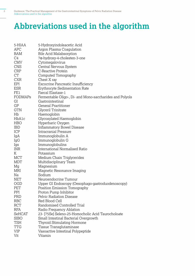
Abbreviations used in the algorithm
Abbreviations used in the algorithm
5-HIAA 5-Hydroxyindoleacetic Acid APC Argon Plasma CoagulationBAM Bile Acid Malabsorption C4 7α-hydroxy-4-cholesten-3-one CMV CytomegalovirusCNS Central Nervous System CRP C-Reactive ProteinCT Computed TomographyCXR Chest X ray EPI Excocrine Pancreatic InsufficiencyESR Erythrocyte Sedimentation RateFE1 Faecal Elastase-1 FODMAPs Fermentable Oligo-, Di- and Mono-saccharides and PolyolsGI GastrointestinalGP General Practitioner GTN Glyceril TrinitrateHb HaemoglobinHbA1c Glycosylated HaemoglobinHBO Hyperbaric Oxygen IBD Inflammatory Bowel Disease ICP Intracranial Pressure IgA Immunoglobulin A IgG Immunoglobulin GIgs Immunoglobulins INR International Normalised RatioK Potassium MCT Medium Chain TriglyceridesMDT Multidisciplinary Team Mg Magnesium MRI Magnetic Resonance ImagingNa Sodium NET Neuroendocrine TumourOGD Upper GI Endoscopy (Oesophago-gastroduodenoscopy)PET Position Emission TomographyPPI Proton Pump Inhibitor PRD Pelvic Radiation DiseaseRBC Red Blood Cell RCT Randomised Controlled Trial RFA Radio Frequency Ablation SeHCAT 23- [75Se] Seleno-25-Homocholic Acid Taurocholoate SIBO Small Intestinal Bacterial OvergrowthTSH Thyroid Stimulating HormoneTTG Tissue Transglutaminase VIP Vasoactive Intestinal Polypeptide Vit Vitamin
2 Guidance: The Practical Management of the Gastrointestinal Symptoms of Pelvic Radiation Disease
Guidance for blood tests used within the Guide
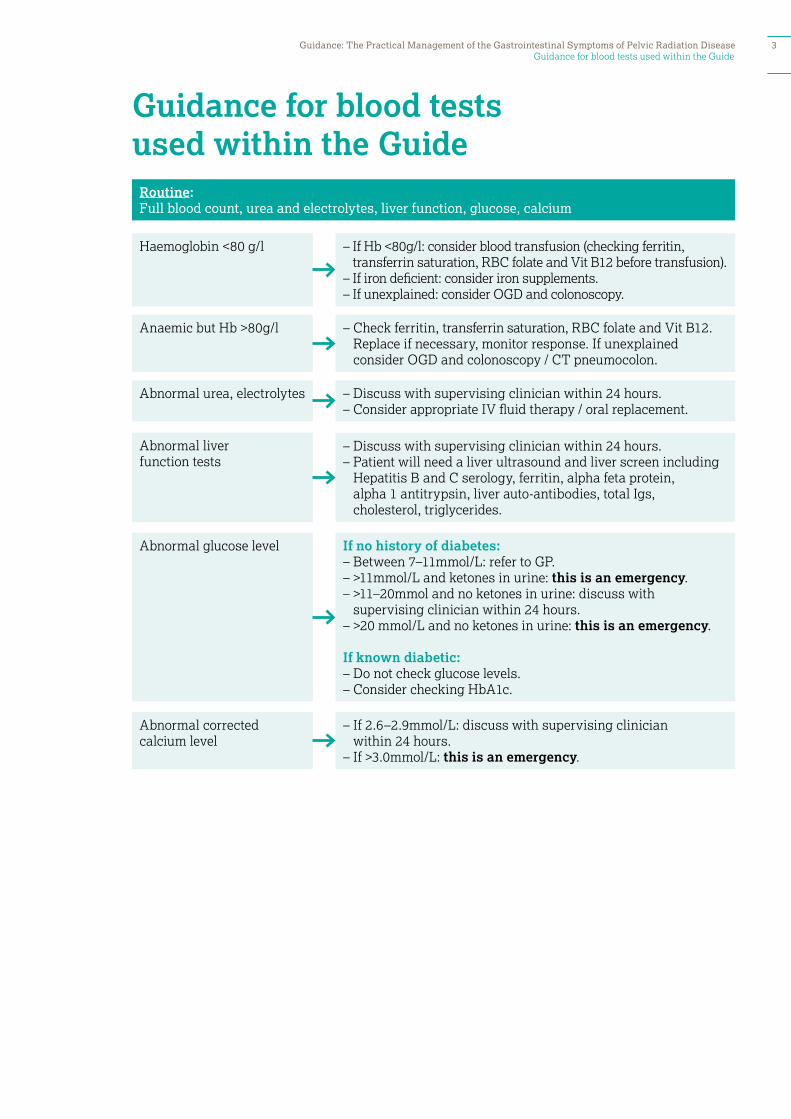
Guidance for blood tests used within the Guide
Haemoglobin <80 g/l
Anaemic but Hb >80g/l
Abnormal urea, electrolytes
Abnormal liver function tests
Abnormal glucose level
Abnormal corrected calcium level
Routine: Full blood count, urea and electrolytes, liver function, glucose, calcium
– If Hb <80g/l: consider blood transfusion (checking ferritin, transferrin saturation, RBC folate and Vit B12 before transfusion).
– If iron deficient: consider iron supplements.– If unexplained: consider OGD and colonoscopy.
– Check ferritin, transferrin saturation, RBC folate and Vit B12. Replace if necessary, monitor response. If unexplained consider OGD and colonoscopy / CT pneumocolon.
– Discuss with supervising clinician within 24 hours.– Consider appropriate IV fluid therapy / oral replacement.
– Discuss with supervising clinician within 24 hours.– Patient will need a liver ultrasound and liver screen including
Hepatitis B and C serology, ferritin, alpha feta protein, alpha 1 antitrypsin, liver auto-antibodies, total Igs, cholesterol, triglycerides.
If no history of diabetes:– Between 7–11mmol/L: refer to GP.– >11mmol/L and ketones in urine: this is an emergency.– >11–20mmol and no ketones in urine: discuss with
supervising clinician within 24 hours. – >20 mmol/L and no ketones in urine: this is an emergency.
If known diabetic:– Do not check glucose levels.– Consider checking HbA1c.
– If 2.6–2.9mmol/L: discuss with supervising clinician within 24 hours.
– If >3.0mmol/L: this is an emergency.
Guidance: The Practical Management of the Gastrointestinal Symptoms of Pelvic Radiation Disease 3
Guidance for blood tests used within the Guide
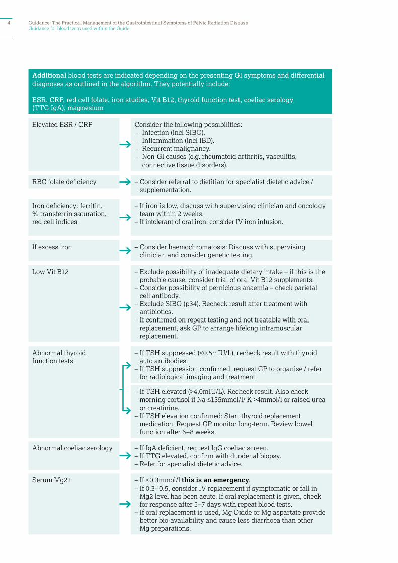
– If TSH elevated (>4.0mIU/L). Recheck result. Also check morning cortisol if Na ≤135mmol/l/ K >4mmol/l or raised urea or creatinine.
– If TSH elevation confirmed: Start thyroid replacement medication. Request GP monitor long-term. Review bowel function after 6–8 weeks.
Elevated ESR / CRP
Additional blood tests are indicated depending on the presenting GI symptoms and differential diagnoses as outlined in the algorithm. They potentially include:
ESR, CRP, red cell folate, iron studies, Vit B12, thyroid function test, coeliac serology (TTG IgA), magnesium
RBC folate deficiency
If excess iron
Low Vit B12
Abnormal thyroid function tests
Abnormal coeliac serology
Serum Mg2+
Iron deficiency: ferritin, % transferrin saturation, red cell indices
Consider the following possibilities:– Infection (incl SIBO).– Inflammation (incl IBD). – Recurrent malignancy.– Non-GI causes (e.g. rheumatoid arthritis, vasculitis,
connective tissue disorders).
– Consider referral to dietitian for specialist dietetic advice / supplementation.
– If iron is low, discuss with supervising clinician and oncology team within 2 weeks.
– If intolerant of oral iron: consider IV iron infusion.
– Consider haemochromatosis: Discuss with supervising clinician and consider genetic testing.
– Exclude possibility of inadequate dietary intake – if this is the probable cause, consider trial of oral Vit B12 supplements.
– Consider possibility of pernicious anaemia – check parietal cell antibody.
– Exclude SIBO (p34). Recheck result after treatment with antibiotics.
– If confirmed on repeat testing and not treatable with oral replacement, ask GP to arrange lifelong intramuscular replacement.
– If TSH suppressed (<0.5mIU/L), recheck result with thyroid auto antibodies.
– If TSH suppression confirmed, request GP to organise / refer for radiological imaging and treatment.
– If IgA deficient, request IgG coeliac screen. – If TTG elevated, confirm with duodenal biopsy.– Refer for specialist dietetic advice.
– If <0.3mmol/l this is an emergency.– If 0.3–0.5, consider IV replacement if symptomatic or fall in
Mg2 level has been acute. If oral replacement is given, check for response after 5–7 days with repeat blood tests.
– If oral replacement is used, Mg Oxide or Mg aspartate provide better bio-availability and cause less diarrhoea than other Mg preparations.
4 Guidance: The Practical Management of the Gastrointestinal Symptoms of Pelvic Radiation Disease
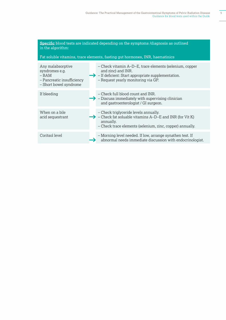
Guidance for blood tests used within the Guide
Any malabsorptive syndromes e.g. – BAM– Pancreatic insufficiency– Short bowel syndrome
If bleeding
When on a bile acid sequestrant
Coritsol level
Specific blood tests are indicated depending on the symptoms /diagnosis as outlined in the algorithm:
Fat soluble vitamins, trace elements, fasting gut hormones, INR, haematinics
– Check vitamin A–D–E, trace elements (selenium, copper and zinc) and INR.
– If deficient: Start appropriate supplementation.– Request yearly monitoring via GP.
– Check full blood count and INR.– Discuss immediately with supervising clinician
and gastroenterologist / GI surgeon.
– Check triglyceride levels annually.– Check fat soluable vitamins A–D–E and INR (for Vit K)
annually. – Check trace elements (selenium, zinc, copper) annually.
– Morning level needed. If low, arrange synathen test. If abnormal needs immediate discussion with endocrinologist.
Guidance: The Practical Management of the Gastrointestinal Symptoms of Pelvic Radiation Disease 5
How to use the algorithm
1. Identify the symptoms by systematic history taking.2. Examine the patient appropriately. 3. Use the algorithm to plan investigations for troublesome / severe
symptoms.4. Most patients have more than one symptom and so investigations
need to be requested for each symptom. 5. Usually all investigations are ordered at the same time and the
patient reviewed with all the results.6. When investigations should be ordered sequentially, the algorithm
indicates this by stating first line, second line etc.7. Treatment options are generally offered sequentially but clinical
judgement should be used.
How to use the algorithm
6 Guidance: The Practical Management of the Gastrointestinal Symptoms of Pelvic Radiation Disease
Taking an appropriate history
Patients cannot be helped without an accurate history being taken. • Taking a history of GI symptoms is a skill that must be learnt. • Tools such as a Bristol Stool Chart can often clarify exactly what
patients mean. • Specialist units find that symptom questionnaires completed by the
patient before the consultation often help clarify which issues are really troubling the patient.
Taking an appropriate history
Taking a history needs to elicit: • What was bowel function like before
the cancer emerged? • How have the symptoms changed
over time? • Are key features indicative of reversible
underlying pathology present, for example, • Steatorrhoea? • Nocturnal waking to defecate? • Rapid progressive worsening of
symptoms? • Rapid weight loss? • Has the patient noticed any masses?
• Patients and clinicians alike often miss the presence of intermittent steatorrhoea – ask:
• Is there an oily film in the lavatory water?
• Is the stool ever pale / putty-like / foul smelling / difficult to flush/ floating?
• A very clear definition of what a patient means when they use specific terms – for example, ‘diarrhoea’ / ‘loose stool’ – what type on the Bristol Stool Chart?; ‘frequency’ – true bowel opening or tenesmus and incomplete evacuation?
• Is there a consistent impact of a specific component of diet on their symptoms, especially:
• Fibre: how much are they eating – too much / too little?
• Fat: does this promote type 6–7 stool / steatorrhoea?
• Lactose-containing foods? • Gluten-containing foods? • Alcohol intake?
• Is there an association between the start of specific medication or increase in its dose and their symptoms – for example, metformin, lansoprazole, beta-blockers?
Guidance: The Practical Management of the Gastrointestinal Symptoms of Pelvic Radiation Disease 7

8 Guidance: The Practical Management of the Gastrointestinal Symptoms of Pelvic Radiation Disease

Bleeding (rectal): Dark bleeding
GI symptoms
Guide to managing Gastrointestinal Pelvic Radiation Disease 9
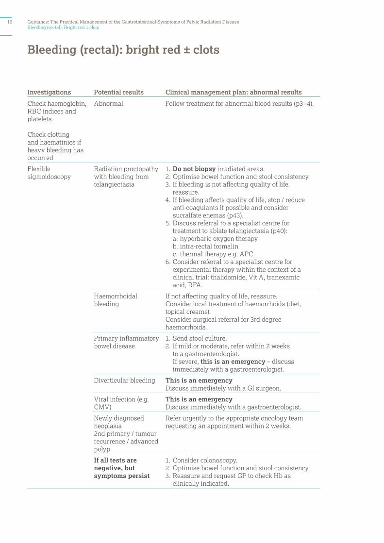
Investigations Potential results Clinical management plan: abnormal results
Check haemoglobin, RBC indices and platelets
Check clotting and haematinics if heavy bleeding has occurred
Abnormal Follow treatment for abnormal blood results (p3–4).
Flexible sigmoidoscopy
Radiation proctopathy with bleeding from telangiectasia
1. Do not biopsy irradiated areas.2. Optimise bowel function and stool consistency. 3. If bleeding is not affecting quality of life,
reassure.4. If bleeding affects quality of life, stop / reduce
anti-coagulants if possible and consider sucralfate enemas (p43).
5. Discuss referral to a specialist centre for treatment to ablate telangiectasia (p40):
a. hyperbaric oxygen therapy b. intra-rectal formalin c. thermal therapy e.g. APC.6. Consider referral to a specialist centre for
experimental therapy within the context of a clinical trial: thalidomide, Vit A, tranexamic acid, RFA.
Haemorrhoidal bleeding
If not affecting quality of life, reassure.Consider local treatment of haemorrhoids (diet, topical creams).Consider surgical referral for 3rd degree haemorrhoids.
Primary inflammatory bowel disease
1. Send stool culture.2. If mild or moderate, refer within 2 weeks
to a gastroenterologist. If severe, this is an emergency – discuss immediately with a gastroenterologist.
Diverticular bleeding This is an emergency Discuss immediately with a GI surgeon.
Viral infection (e.g. CMV)
This is an emergencyDiscuss immediately with a gastroenterologist.
Newly diagnosed neoplasia 2nd primary / tumour recurrence / advanced polyp
Refer urgently to the appropriate oncology team requesting an appointment within 2 weeks.
If all tests are negative, but symptoms persist
1. Consider colonoscopy.2. Optimise bowel function and stool consistency.3. Reassure and request GP to check Hb as
clinically indicated.
Bleeding (rectal): bright red ± clots
Bleeding (rectal): Bright red ± clots10 Guidance: The Practical Management of the Gastrointestinal Symptoms of Pelvic Radiation Disease
Bleeding (rectal): dark bleeding
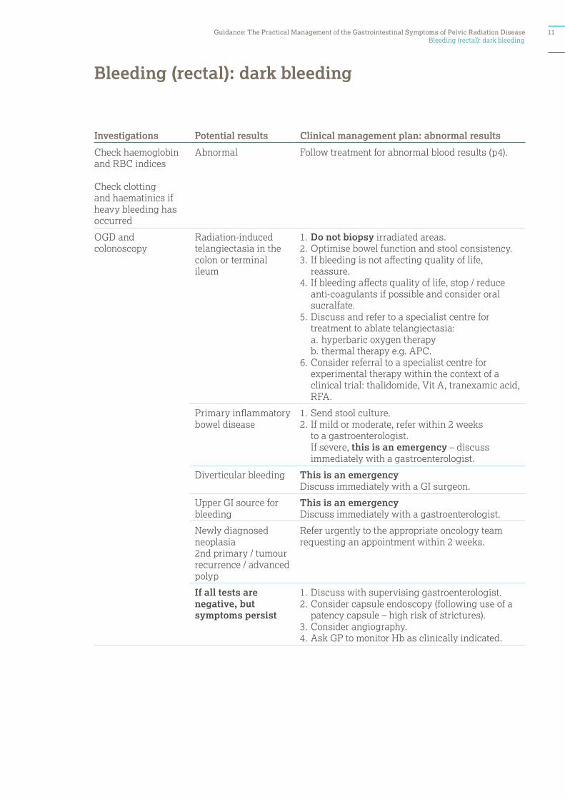
Investigations Potential results Clinical management plan: abnormal results
Check haemoglobin and RBC indices
Check clotting and haematinics if heavy bleeding has occurred
Abnormal Follow treatment for abnormal blood results (p4).
OGD and colonoscopy
Radiation-induced telangiectasia in the colon or terminal ileum
1. Do not biopsy irradiated areas.2. Optimise bowel function and stool consistency. 3. If bleeding is not affecting quality of life,
reassure.4. If bleeding affects quality of life, stop / reduce
anti-coagulants if possible and consider oral sucralfate.
5. Discuss and refer to a specialist centre for treatment to ablate telangiectasia:
a. hyperbaric oxygen therapy b. thermal therapy e.g. APC.6. Consider referral to a specialist centre for
experimental therapy within the context of a clinical trial: thalidomide, Vit A, tranexamic acid, RFA.
Primary inflammatory bowel disease
1. Send stool culture.2. If mild or moderate, refer within 2 weeks
to a gastroenterologist. If severe, this is an emergency – discuss immediately with a gastroenterologist.
Diverticular bleeding This is an emergency Discuss immediately with a GI surgeon.
Upper GI source for bleeding
This is an emergencyDiscuss immediately with a gastroenterologist.
Newly diagnosed neoplasia 2nd primary / tumour recurrence / advanced polyp
Refer urgently to the appropriate oncology team requesting an appointment within 2 weeks.
If all tests are negative, but symptoms persist
1. Discuss with supervising gastroenterologist.2. Consider capsule endoscopy (following use of a
patency capsule – high risk of strictures).3. Consider angiography. 4. Ask GP to monitor Hb as clinically indicated.
Bleeding (rectal): dark bleeding
Guidance: The Practical Management of the Gastrointestinal Symptoms of Pelvic Radiation Disease 11
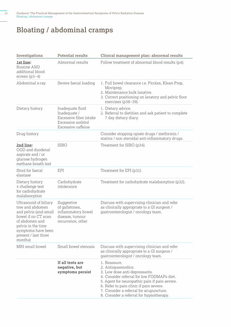
Bloating / abdominal cramps
Investigations Potential results Clinical management plan: abnormal results
1st line: Routine AND additional blood screen (p3–4)
Abnormal results Follow treatment of abnormal blood results (p4).
Abdominal x-ray Severe faecal loading 1. Full bowel clearance i.e. Picolax, Klean-Prep, Moviprep.
2. Maintenance bulk laxative.3. Correct positioning on lavatory and pelvic floor
exercises (p38–39).
Dietary history Inadequate fluid Inadequate / Excessive fibre intakeExcessive sorbitolExcessive caffeine
1. Dietary advice.2. Referral to dietitian and ask patient to complete
7 day dietary diary.
Drug history Consider stopping opiate drugs / metformin / statins / non steroidal anti-inflammatory drugs.
2nd line: OGD and duodenal aspirate and / or glucose hydrogen methane breath test
SIBO Treatment for SIBO (p34).
Stool for faecal elastase
EPI Treatment for EPI (p31).
Dietary history ± challenge test for carbohydrate malabsorption
Carbohydrate intolerance
Treatment for carbohydrate malabsorption (p32).
Ultrasound of biliary tree and abdomen and pelvis (and small bowel if no CT scan of abdomen and pelvis in the time symptoms have been present / last three months)
Suggestive of gallstones, inflammatory bowel disease, tumour recurrence, other
Discuss with supervising clinician and refer as clinically appropriate to a GI surgeon /gastroenterologist / oncology team.
MRI small bowel Small bowel stenosis Discuss with supervising clinician and refer as clinically appropriate to a GI surgeon / gastroenterologist / oncology team.
If all tests are negative, but symptoms persist
1. Reassure.2. Antispasmodics.3. Low dose anti-depressants.4. Consider referral for low FODMAPs diet.5. Agent for neuropathic pain if pain severe.6. Refer to pain clinic if pain severe.7. Consider a referral for acupuncture.8. Consider a referral for hypnotherapy.
Bloating / abdominal cramps
12 Guidance: The Practical Management of the Gastrointestinal Symptoms of Pelvic Radiation Disease
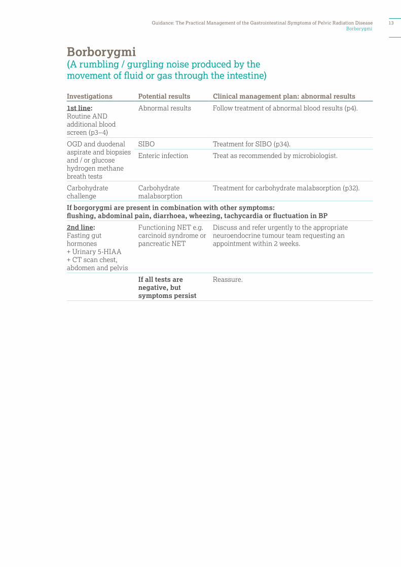
Borborygmi
Investigations Potential results Clinical management plan: abnormal results
1st line:Routine AND additional blood screen (p3–4)
Abnormal results Follow treatment of abnormal blood results (p4).
OGD and duodenal aspirate and biopsies and / or glucose hydrogen methane breath tests
SIBO Treatment for SIBO (p34).
Enteric infection Treat as recommended by microbiologist.
Carbohydrate challenge
Carbohydrate malabsorption
Treatment for carbohydrate malabsorption (p32).
If borgorygmi are present in combination with other symptoms: flushing, abdominal pain, diarrhoea, wheezing, tachycardia or fluctuation in BP
2nd line: Fasting gut hormones+ Urinary 5-HIAA + CT scan chest, abdomen and pelvis
Functioning NET e.g. carcinoid syndrome or pancreatic NET
Discuss and refer urgently to the appropriate neuroendocrine tumour team requesting an appointment within 2 weeks.
If all tests are negative, but symptoms persist
Reassure.
Borborygmi (A rumbling / gurgling noise produced by the movement of fluid or gas through the intestine)
Guidance: The Practical Management of the Gastrointestinal Symptoms of Pelvic Radiation Disease 13
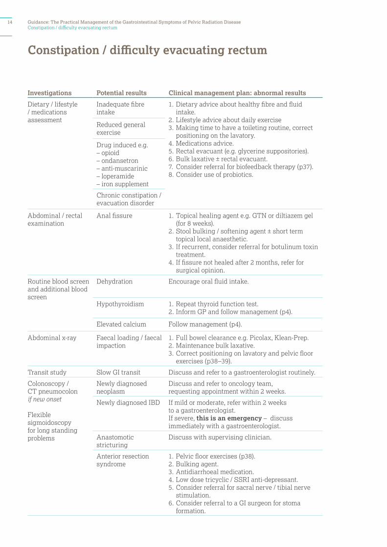
Constipation / difficulty evacuating rectum
Investigations Potential results Clinical management plan: abnormal results
Dietary / lifestyle / medications assessment
Inadequate fibre intake
1. Dietary advice about healthy fibre and fluid intake.
2. Lifestyle advice about daily exercise3. Making time to have a toileting routine, correct
positioning on the lavatory.4. Medications advice. 5. Rectal evacuant (e.g. glycerine suppositories).6. Bulk laxative ± rectal evacuant.7. Consider referral for biofeedback therapy (p37).8. Consider use of probiotics.
Reduced general exercise
Drug induced e.g.– opioid– ondansetron– anti-muscarinic– loperamide– iron supplement
Chronic constipation / evacuation disorder
Abdominal / rectal examination
Anal fissure 1. Topical healing agent e.g. GTN or diltiazem gel (for 8 weeks).
2. Stool bulking / softening agent ± short term topical local anaesthetic.
3. If recurrent, consider referral for botulinum toxin treatment.
4. If fissure not healed after 2 months, refer for surgical opinion.
Routine blood screen and additional blood screen
Dehydration Encourage oral fluid intake.
Hypothyroidism 1. Repeat thyroid function test.2. Inform GP and follow management (p4).
Elevated calcium Follow management (p4).
Abdominal x-ray Faecal loading / faecal impaction
1. Full bowel clearance e.g. Picolax, Klean-Prep.2. Maintenance bulk laxative.3. Correct positioning on lavatory and pelvic floor
exercises (p38–39).
Transit study Slow GI transit Discuss and refer to a gastroenterologist routinely.
Colonoscopy / CT pneumocolonif new onset
Flexible sigmoidoscopy for long standing problems
Newly diagnosed neoplasm
Discuss and refer to oncology team, requesting appointment within 2 weeks.
Newly diagnosed IBD If mild or moderate, refer within 2 weeks to a gastroenterologist. If severe, this is an emergency – discuss immediately with a gastroenterologist.
Anastomotic stricturing
Discuss with supervising clinician.
Anterior resection syndrome
1. Pelvic floor exercises (p38).2. Bulking agent.3. Antidiarrhoeal medication.4. Low dose tricyclic / SSRI anti-depressant.5. Consider referral for sacral nerve / tibial nerve
stimulation.6. Consider referral to a GI surgeon for stoma
formation.
Constipation / difficulty evacuating rectum
14 Guidance: The Practical Management of the Gastrointestinal Symptoms of Pelvic Radiation Disease

Diarrhoea
Investigations Potential results Clinical management plan: abnormal results
Dietary / lifestyle / medications assessment
High dietary fat intakeLow / high fibre intakeHigh fizzy drink intakeHigh use of sorbitol – containing sugar chewing gum or sweetsHigh caffeine intake High alcohol intake
1. Dietary advice about healthy fibre and dietary fat intake.
2. Referral to dietitian and ask patient to complete 7 day dietary diary beforehand.
3. Lifestyle advice about smoking cessation.4. Consider referral for psychological support.5. Medications advice.6. Anti-diarrhoeal ± bulk laxative.
Anxiety
Drug induced: e.g.– PPIs– Laxatives– Beta blockers– Metformin
Routine AND additional blood screen (p3–4)
Abnormal results Follow treatment of abnormal blood results (p4).
Mg2+ low Follow treatment of abnormal blood results (p4).
Coeliac disease 1. If IgA deficient, request IgG coeliac screen. 2. Confirm with duodenal biopsy.3. Refer to dietitian for gluten free diet. 4. Liaise with GP regarding long term monitoring of
bone densitometry and referral to a coeliac clinic.
Stool sample: for microscopy, culture and Clostridium Difficile toxin
Stool contains pathogen
Treat as recommended by the microbiologist and local protocols.
Stool sample: for faecal elastase
EPI See EPI (p31)
OGD with duodenal aspirate and biopsies and / or glucose hydrogen (methane) breath test
SIBO Treatment for SIBO (p34).
Carbohydrate challenge
Specific disacharride intolerance
Appropriate treatment (p32).
SeHCAT scan BAM Treatment for BAM (p30).
Diarrhoea (stool type 6–7 Bristol Stool Chart) Also use this section if patient has ‘frequency of defecation’, ‘nocturnal defecation’, or ‘urgency of defecation’
Guidance: The Practical Management of the Gastrointestinal Symptoms of Pelvic Radiation Disease 15
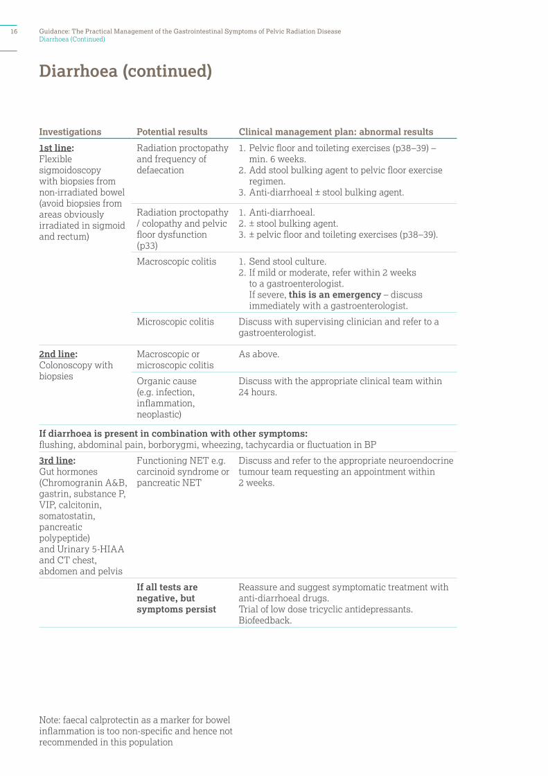
Diarrhoea (Continued)
Investigations Potential results Clinical management plan: abnormal results
1st line: Flexible sigmoidoscopy with biopsies from non-irradiated bowel (avoid biopsies from areas obviously irradiated in sigmoid and rectum)
Radiation proctopathy and frequency of defaecation
1. Pelvic floor and toileting exercises (p38–39) – min. 6 weeks.
2. Add stool bulking agent to pelvic floor exercise regimen.
3. Anti-diarrhoeal ± stool bulking agent.
Radiation proctopathy / colopathy and pelvic floor dysfunction (p33)
1. Anti-diarrhoeal. 2. ± stool bulking agent.3. ± pelvic floor and toileting exercises (p38–39).
Macroscopic colitis 1. Send stool culture.2. If mild or moderate, refer within 2 weeks
to a gastroenterologist. If severe, this is an emergency – discuss immediately with a gastroenterologist.
Microscopic colitis Discuss with supervising clinician and refer to a gastroenterologist.
2nd line: Colonoscopy with biopsies
Macroscopic or microscopic colitis
As above.
Organic cause (e.g. infection, inflammation, neoplastic)
Discuss with the appropriate clinical team within 24 hours.
If diarrhoea is present in combination with other symptoms: flushing, abdominal pain, borborygmi, wheezing, tachycardia or fluctuation in BP
3rd line: Gut hormones (Chromogranin A&B, gastrin, substance P, VIP, calcitonin, somatostatin, pancreatic polypeptide)and Urinary 5-HIAA and CT chest, abdomen and pelvis
Functioning NET e.g. carcinoid syndrome or pancreatic NET
Discuss and refer to the appropriate neuroendocrine tumour team requesting an appointment within 2 weeks.
If all tests are negative, but symptoms persist
Reassure and suggest symptomatic treatment with anti-diarrhoeal drugs. Trial of low dose tricyclic antidepressants. Biofeedback.
Diarrhoea (continued)
Note: faecal calprotectin as a marker for bowel inflammation is too non-specific and hence not recommended in this population
16 Guidance: The Practical Management of the Gastrointestinal Symptoms of Pelvic Radiation Disease
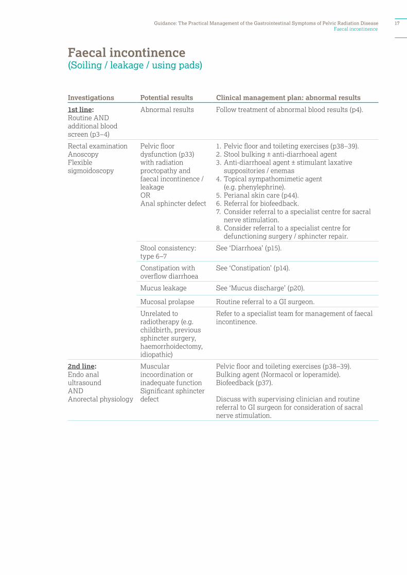
Faecal incontinence
Investigations Potential results Clinical management plan: abnormal results
1st line: Routine AND additional blood screen (p3–4)
Abnormal results Follow treatment of abnormal blood results (p4).
Rectal examinationAnoscopy Flexible sigmoidoscopy
Pelvic floor dysfunction (p33) with radiation proctopathy and faecal incontinence / leakage OR Anal sphincter defect
1. Pelvic floor and toileting exercises (p38–39).2. Stool bulking ± anti-diarrhoeal agent3. Anti-diarrhoeal agent ± stimulant laxative
suppositories / enemas4. Topical sympathomimetic agent
(e.g. phenylephrine). 5. Perianal skin care (p44).6. Referral for biofeedback.7. Consider referral to a specialist centre for sacral
nerve stimulation.8. Consider referral to a specialist centre for
defunctioning surgery / sphincter repair.
Stool consistency: type 6–7
See ‘Diarrhoea’ (p15).
Constipation with overflow diarrhoea
See ‘Constipation’ (p14).
Mucus leakage See ‘Mucus discharge’ (p20).
Mucosal prolapse Routine referral to a GI surgeon.
Unrelated to radiotherapy (e.g. childbirth, previous sphincter surgery, haemorrhoidectomy, idiopathic)
Refer to a specialist team for management of faecal incontinence.
2nd line:Endo anal ultrasoundAND Anorectal physiology
Muscular incoordination or inadequate functionSignificant sphincter defect
Pelvic floor and toileting exercises (p38–39).Bulking agent (Normacol or loperamide).Biofeedback (p37).
Discuss with supervising clinician and routine referral to GI surgeon for consideration of sacral nerve stimulation.
Faecal incontinence (Soiling / leakage / using pads)
Guidance: The Practical Management of the Gastrointestinal Symptoms of Pelvic Radiation Disease 17
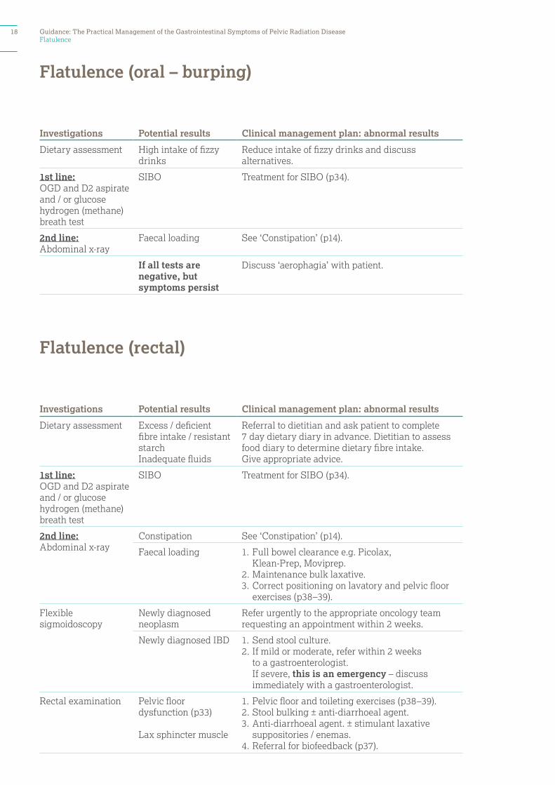
Flatulence
Investigations Potential results Clinical management plan: abnormal results
Dietary assessment High intake of fizzy drinks
Reduce intake of fizzy drinks and discuss alternatives.
1st line:OGD and D2 aspirate and / or glucose hydrogen (methane) breath test
SIBO Treatment for SIBO (p34).
2nd line:Abdominal x-ray
Faecal loading See ‘Constipation’ (p14).
If all tests are negative, but symptoms persist
Discuss ‘aerophagia’ with patient.
Investigations Potential results Clinical management plan: abnormal results
Dietary assessment Excess / deficient fibre intake / resistant starchInadequate fluids
Referral to dietitian and ask patient to complete 7 day dietary diary in advance. Dietitian to assess food diary to determine dietary fibre intake. Give appropriate advice.
1st line:OGD and D2 aspirate and / or glucose hydrogen (methane) breath test
SIBO Treatment for SIBO (p34).
2nd line:Abdominal x-ray
Constipation See ‘Constipation’ (p14).
Faecal loading 1. Full bowel clearance e.g. Picolax, Klean-Prep, Moviprep.
2. Maintenance bulk laxative.3. Correct positioning on lavatory and pelvic floor
exercises (p38–39).
Flexible sigmoidoscopy
Newly diagnosed neoplasm
Refer urgently to the appropriate oncology team requesting an appointment within 2 weeks.
Newly diagnosed IBD 1. Send stool culture.2. If mild or moderate, refer within 2 weeks
to a gastroenterologist. If severe, this is an emergency – discuss immediately with a gastroenterologist.
Rectal examination Pelvic floor dysfunction (p33)
Lax sphincter muscle
1. Pelvic floor and toileting exercises (p38–39). 2. Stool bulking ± anti-diarrhoeal agent.3. Anti-diarrhoeal agent. ± stimulant laxative
suppositories / enemas.4. Referral for biofeedback (p37).
Flatulence (oral – burping)
Flatulence (rectal)
18 Guidance: The Practical Management of the Gastrointestinal Symptoms of Pelvic Radiation Disease

Loss of sensation
Investigations Potential results Clinical management plan: abnormal results
Neurological examination (including perianal sensation)
Abnormal examination (e.g. suspected spinal cord compression,cauda equina syndrome, neurogenic bladder)
This is an emergency Discuss immediately with an oncology or neurology team.
Routine blood screen and ESR, Vit B12, red cell folate
Abnormal results Follow treatment of abnormal blood results (p4).
Consider MRI pelvis Tumour recurrence or other cause for neurological dysfunction
Discuss immediately with supervising clinician.
Related to radiotherapy or surgery
1. Pelvic floor and toileting exercises (p38–39).2. Bulking agent ± anti diarrhoeal.3. Consider referral for biofeedback (p37).
Loss of sensation (Unable to discriminate between need to defecate and pass urine)
Guidance: The Practical Management of the Gastrointestinal Symptoms of Pelvic Radiation Disease 19
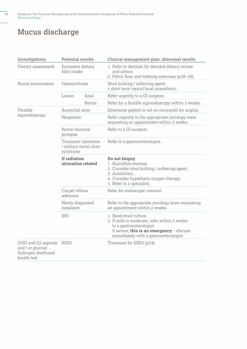
Mucus discharge
Investigations Potential results Clinical management plan: abnormal results
Dietary assessment Excessive dietary fibre intake
1. Refer to dietitian for detailed dietary review and advice.
2. Pelvic floor and toileting exercises (p38–39).
Rectal examination Haemorrhoids Stool bulking / softening agent. ± short term topical local anaesthetic.
Lesion Anal Refer urgently to a GI surgeon.
Rectal Refer for a flexible sigmoidoscopy within 2 weeks.
Flexible sigmoidoscopy
Anorectal ulcer Determine patient is not on nicorandil for angina.
Neoplastic Refer urgently to the appropriate oncology team requesting an appointment within 2 weeks.
Rectal mucosal prolapse
Refer to a GI surgeon.
Traumatic ulceration / solitary rectal ulcer syndrome
Refer to a gastroenterologist.
If radiation ulceration related
Do not biopsy1. Sucralfate enemas.2. Consider stool bulking / softening agent.3. Antibiotics.4. Consider hyperbaric oxygen therapy.5. Refer to a specialist.
Carpet villous adenoma
Refer for endoscopic removal.
Newly diagnosed neoplasm
Refer to the appropriate oncology team requesting an appointment within 2 weeks.
IBD 1. Send stool culture.2. If mild or moderate, refer within 2 weeks
to a gastroenterologist. If severe, this is an emergency – discuss immediately with a gastroenterologist.
OGD and D2 aspirate and / or glucose hydrogen (methane) breath test
SIBO Treatment for SIBO (p34).
Mucus discharge
20 Guidance: The Practical Management of the Gastrointestinal Symptoms of Pelvic Radiation Disease
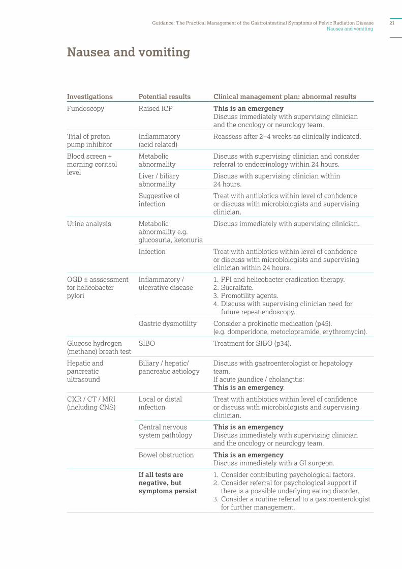
Nausea and vomiting
Investigations Potential results Clinical management plan: abnormal results
Fundoscopy Raised ICP This is an emergencyDiscuss immediately with supervising clinician and the oncology or neurology team.
Trial of proton pump inhibitor
Inflammatory (acid related)
Reassess after 2–4 weeks as clinically indicated.
Blood screen + morning coritsol level
Metabolic abnormality
Discuss with supervising clinician and consider referral to endocrinology within 24 hours.
Liver / biliary abnormality
Discuss with supervising clinician within 24 hours.
Suggestive of infection
Treat with antibiotics within level of confidence or discuss with microbiologists and supervising clinician.
Urine analysis Metabolic abnormality e.g. glucosuria, ketonuria
Discuss immediately with supervising clinician.
Infection Treat with antibiotics within level of confidence or discuss with microbiologists and supervising clinician within 24 hours.
OGD ± asssessment for helicobacter pylori
Inflammatory / ulcerative disease
1. PPI and helicobacter eradication therapy.2. Sucralfate. 3. Promotility agents.4. Discuss with supervising clinician need for
future repeat endoscopy.
Gastric dysmotility Consider a prokinetic medication (p45).(e.g. domperidone, metoclopramide, erythromycin).
Glucose hydrogen (methane) breath test
SIBO Treatment for SIBO (p34).
Hepatic and pancreatic ultrasound
Biliary / hepatic/ pancreatic aetiology
Discuss with gastroenterologist or hepatology team.If acute jaundice / cholangitis: This is an emergency.
CXR / CT / MRI (including CNS)
Local or distal infection
Treat with antibiotics within level of confidence or discuss with microbiologists and supervising clinician.
Central nervous system pathology
This is an emergencyDiscuss immediately with supervising clinician and the oncology or neurology team.
Bowel obstruction This is an emergency Discuss immediately with a GI surgeon.
If all tests are negative, but symptoms persist
1. Consider contributing psychological factors.2. Consider referral for psychological support if
there is a possible underlying eating disorder.3. Consider a routine referral to a gastroenterologist
for further management.
Nausea and vomiting
Guidance: The Practical Management of the Gastrointestinal Symptoms of Pelvic Radiation Disease 21
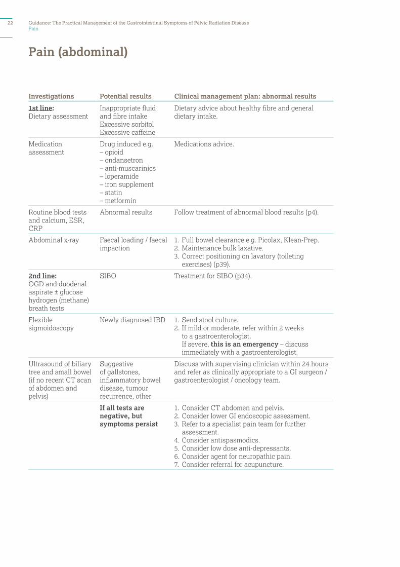
Pain
Investigations Potential results Clinical management plan: abnormal results
1st line:Dietary assessment
Inappropriate fluid and fibre intakeExcessive sorbitolExcessive caffeine
Dietary advice about healthy fibre and general dietary intake.
Medication assessment
Drug induced e.g.– opioid– ondansetron– anti-muscarinics– loperamide– iron supplement– statin – metformin
Medications advice.
Routine blood tests and calcium, ESR, CRP
Abnormal results Follow treatment of abnormal blood results (p4).
Abdominal x-ray Faecal loading / faecal impaction
1. Full bowel clearance e.g. Picolax, Klean-Prep.2. Maintenance bulk laxative.3. Correct positioning on lavatory (toileting
exercises) (p39).
2nd line: OGD and duodenal aspirate ± glucose hydrogen (methane) breath tests
SIBO Treatment for SIBO (p34).
Flexible sigmoidoscopy
Newly diagnosed IBD 1. Send stool culture.2. If mild or moderate, refer within 2 weeks
to a gastroenterologist. If severe, this is an emergency – discuss immediately with a gastroenterologist.
Ultrasound of biliary tree and small bowel (if no recent CT scan of abdomen and pelvis)
Suggestive of gallstones, inflammatory bowel disease, tumour recurrence, other
Discuss with supervising clinician within 24 hours and refer as clinically appropriate to a GI surgeon / gastroenterologist / oncology team.
If all tests are negative, but symptoms persist
1. Consider CT abdomen and pelvis.2. Consider lower GI endoscopic assessment.3. Refer to a specialist pain team for further
assessment.4. Consider antispasmodics.5. Consider low dose anti-depressants.6. Consider agent for neuropathic pain.7. Consider referral for acupuncture.
Pain (abdominal)
22 Guidance: The Practical Management of the Gastrointestinal Symptoms of Pelvic Radiation Disease

Pain
Investigations Potential results Clinical management plan: abnormal results
Neurological examination (including perianal sensation)
Abnormal examination (e.g. suspected spinal cord compression)
This is an emergency Discuss immediately with an oncology or neurology team.
Symptom assessment
Pain over the renal angle
1. Consider pyelonephritis kidney infection / stone / urinary tract infection.
2. Urine dip stick and urine sample for culture and sensitivity.
3. Consider renal ultrasound.
Pain in the lower flank
Consider constipation, faecal loading and faecal impaction (p14).
Pain in the lower back 1. Consider lower back fracture.2. Request a spinal (thoracic / lumbar) x-ray.3. Consider MRI.
Bone pain Consider a bone scan and myeloma screen.
Routine blood screen and additional blood screen
Abnormal results suggesting cancer relapse
Discuss immediately with supervising clinician.
CT / MRI / PET scan abdomen and pelvis
Colonic faecal loading See ‘Constipation’ (p14).
Acute bowel obstruction
This is an emergencyDiscuss immediately with a GI surgeon.
Spinal fracture Discuss immediately with supervising clinician.
Pain (back – new onset)
Investigations Potential results Clinical management plan: abnormal results
Symptom assessment
Spasm of the levator ani muscles
Treatment for rectal spasm 1. Pelvic floor and toileting exercises (p38–39).2. Consider a low dose anti-depressant.3. Consider a trial of an inhaled beta 2 agonist.4. Consider referral to a specialist centre for
biofeedback (p37).5. Consider referral for acupuncture.
Pain (anal / perianal / rectal): typical proctalgia fugax (A sudden, severe pain in the anorectal region lasting less than 20 minutes, resolving spontaneously)
Guidance: The Practical Management of the Gastrointestinal Symptoms of Pelvic Radiation Disease 23
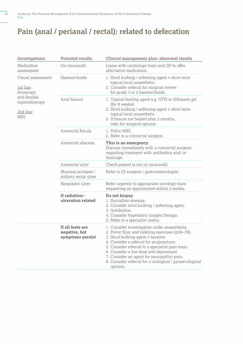
Pain
Investigations Potential results Clinical management plan: abnormal results
Medication assessment
On nicorandil Liaise with cardiology team and GP to offer alternative medication.
Visual assessment
1st line: Anoscopy and flexible sigmoidoscopy
2nd line: MRI
Haemorrhoids 1. Stool bulking / softening agent ± short term topical local anaesthetic.
2. Consider referral for surgical review for grade 3 or 4 haemorrhoids.
Anal fissure 1. Topical healing agent e.g. GTN or diltiazem gel (for 8 weeks).
2. Stool bulking / softening agent ± short term topical local anaesthetic.
3. If fissure not healed after 2 months, refer for surgical opinion.
Anorectal fistula 1. Pelvic MRI.2. Refer to a colorectal surgeon.
Anorectal abscess This is an emergencyDiscuss immediately with a colorectal surgeon regarding treatment with antibiotics and/ or drainage.
Anorectal ulcer Check patient is not on nicorandil.
Mucosal prolapse / solitary rectal ulcer
Refer to GI surgeon / gastroenterologist.
Neoplastic ulcer Refer urgently to appropriate oncology team requesting an appointment within 2 weeks.
If radiation–ulceration related
Do not biopsy1. Sucralfate enemas.2. Consider stool bulking / softening agent.3. Antibiotics.4. Consider hyperbaric oxygen therapy.5. Refer to a specialist centre.
If all tests are negative, but symptoms persist
1. Consider investigation under anaesthesia. 2. Pelvic floor and toileting exercises (p38–39). 3. Stool bulking agent ± laxative.4. Consider a referral for acupuncture.5. Consider referral to a specialist pain team.6. Consider a low dose anti-depressant.7. Consider an agent for neuropathic pain.8. Consider referral for a urological / gynaecological
opinion.
Pain (anal / perianal / rectal): related to defecation
24 Guidance: The Practical Management of the Gastrointestinal Symptoms of Pelvic Radiation Disease
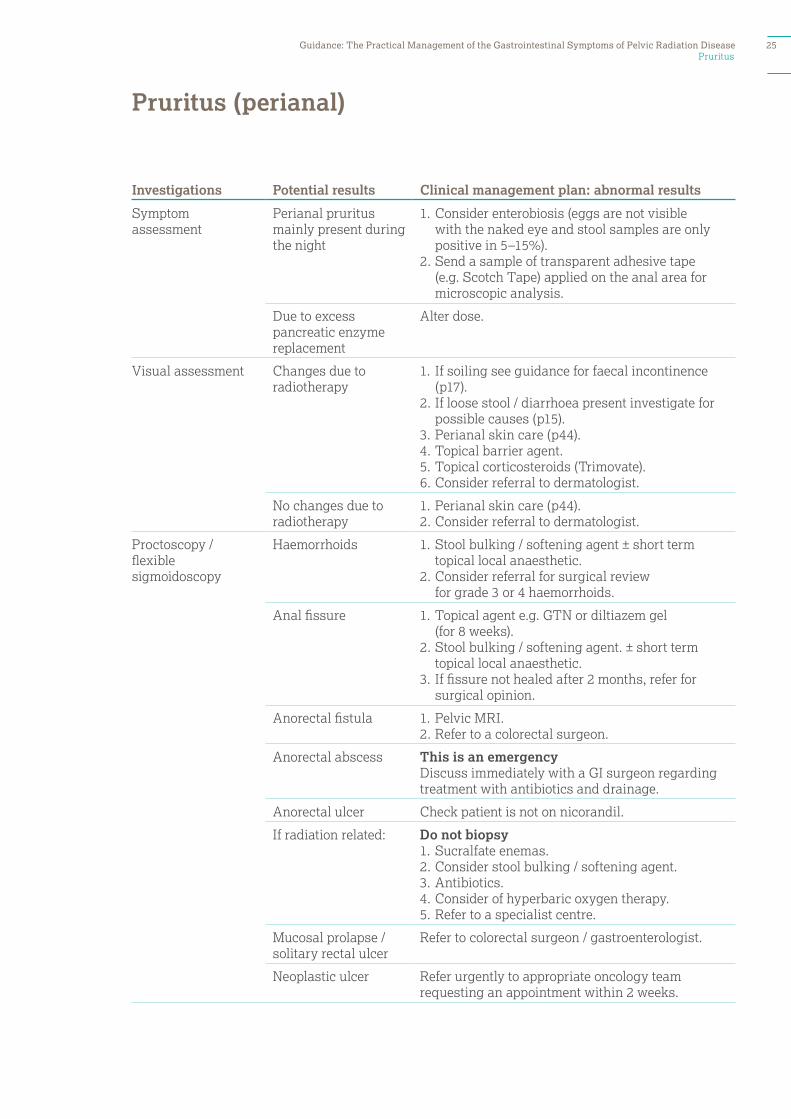
Pruritus
Investigations Potential results Clinical management plan: abnormal results
Symptom assessment
Perianal pruritus mainly present during the night
1. Consider enterobiosis (eggs are not visible with the naked eye and stool samples are only positive in 5–15%).
2. Send a sample of transparent adhesive tape (e.g. Scotch Tape) applied on the anal area for microscopic analysis.
Due to excess pancreatic enzyme replacement
Alter dose.
Visual assessment Changes due to radiotherapy
1. If soiling see guidance for faecal incontinence (p17).
2. If loose stool / diarrhoea present investigate for possible causes (p15).
3. Perianal skin care (p44).4. Topical barrier agent.5. Topical corticosteroids (Trimovate).6. Consider referral to dermatologist.
No changes due to radiotherapy
1. Perianal skin care (p44).2. Consider referral to dermatologist.
Proctoscopy / flexible sigmoidoscopy
Haemorrhoids 1. Stool bulking / softening agent ± short term topical local anaesthetic.
2. Consider referral for surgical review for grade 3 or 4 haemorrhoids.
Anal fissure 1. Topical agent e.g. GTN or diltiazem gel (for 8 weeks).
2. Stool bulking / softening agent. ± short term topical local anaesthetic.
3. If fissure not healed after 2 months, refer for surgical opinion.
Anorectal fistula 1. Pelvic MRI. 2. Refer to a colorectal surgeon.
Anorectal abscess This is an emergencyDiscuss immediately with a GI surgeon regarding treatment with antibiotics and drainage.
Anorectal ulcer Check patient is not on nicorandil.
If radiation related: Do not biopsy1. Sucralfate enemas.2. Consider stool bulking / softening agent.3. Antibiotics.4. Consider of hyperbaric oxygen therapy.5. Refer to a specialist centre.
Mucosal prolapse / solitary rectal ulcer
Refer to colorectal surgeon / gastroenterologist.
Neoplastic ulcer Refer urgently to appropriate oncology team requesting an appointment within 2 weeks.
Pruritus (perianal)
Guidance: The Practical Management of the Gastrointestinal Symptoms of Pelvic Radiation Disease 25
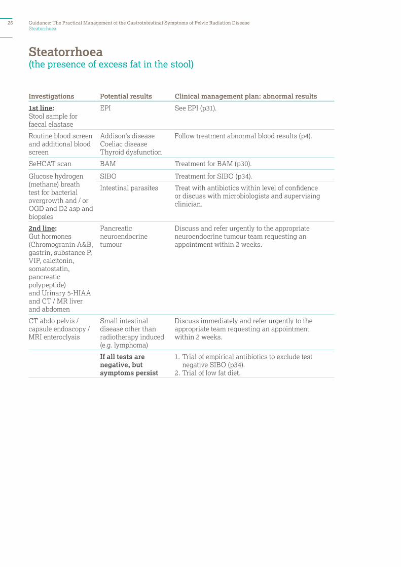
Steatorrhoea
Investigations Potential results Clinical management plan: abnormal results
1st line: Stool sample for faecal elastase
EPI See EPI (p31).
Routine blood screen and additional blood screen
Addison’s disease Coeliac disease Thyroid dysfunction
Follow treatment abnormal blood results (p4).
SeHCAT scan BAM Treatment for BAM (p30).
Glucose hydrogen (methane) breath test for bacterial overgrowth and / or OGD and D2 asp and biopsies
SIBO Treatment for SIBO (p34).
Intestinal parasites Treat with antibiotics within level of confidence or discuss with microbiologists and supervising clinician.
2nd line: Gut hormones (Chromogranin A&B, gastrin, substance P, VIP, calcitonin, somatostatin, pancreatic polypeptide)and Urinary 5-HIAAand CT / MR liver and abdomen
Pancreatic neuroendocrine tumour
Discuss and refer urgently to the appropriate neuroendocrine tumour team requesting an appointment within 2 weeks.
CT abdo pelvis / capsule endoscopy / MRI enteroclysis
Small intestinal disease other than radiotherapy induced (e.g. lymphoma)
Discuss immediately and refer urgently to the appropriate team requesting an appointment within 2 weeks.
If all tests are negative, but symptoms persist
1. Trial of empirical antibiotics to exclude test negative SIBO (p34).
2. Trial of low fat diet.
Steatorrhoea (the presence of excess fat in the stool)
26 Guidance: The Practical Management of the Gastrointestinal Symptoms of Pelvic Radiation Disease

Tenesmus
Investigations Potential results Clinical management plan: abnormal results
Flexible sigmoidoscopy
Radiation proctopathy
Anterior resection syndrome
1. Pelvic floor and toileting exercises (p38–39).2. Stool bulking agent.3. Low dose anti-depressants.4. Consider referral to a specialist centre for
biofeedback (p37).5. Consider referral for acupuncture.
Polyp Arrange endoscopic / surgical removal.
Newly diagnosed neoplasm
Refer urgently to the appropriate oncology team requesting an appointment within 2 weeks.
Newly diagnosed IBD / infection
1. Send stool culture.2. If mild or moderate, refer within 2 weeks
to a gastroenterologist. If severe, this is an emergency – discuss immediately with a gastroenterologist.
Tenesmus (A feeling of constantly needing to pass stools, despite an empty rectum)
Guidance: The Practical Management of the Gastrointestinal Symptoms of Pelvic Radiation Disease 27
Weight loss
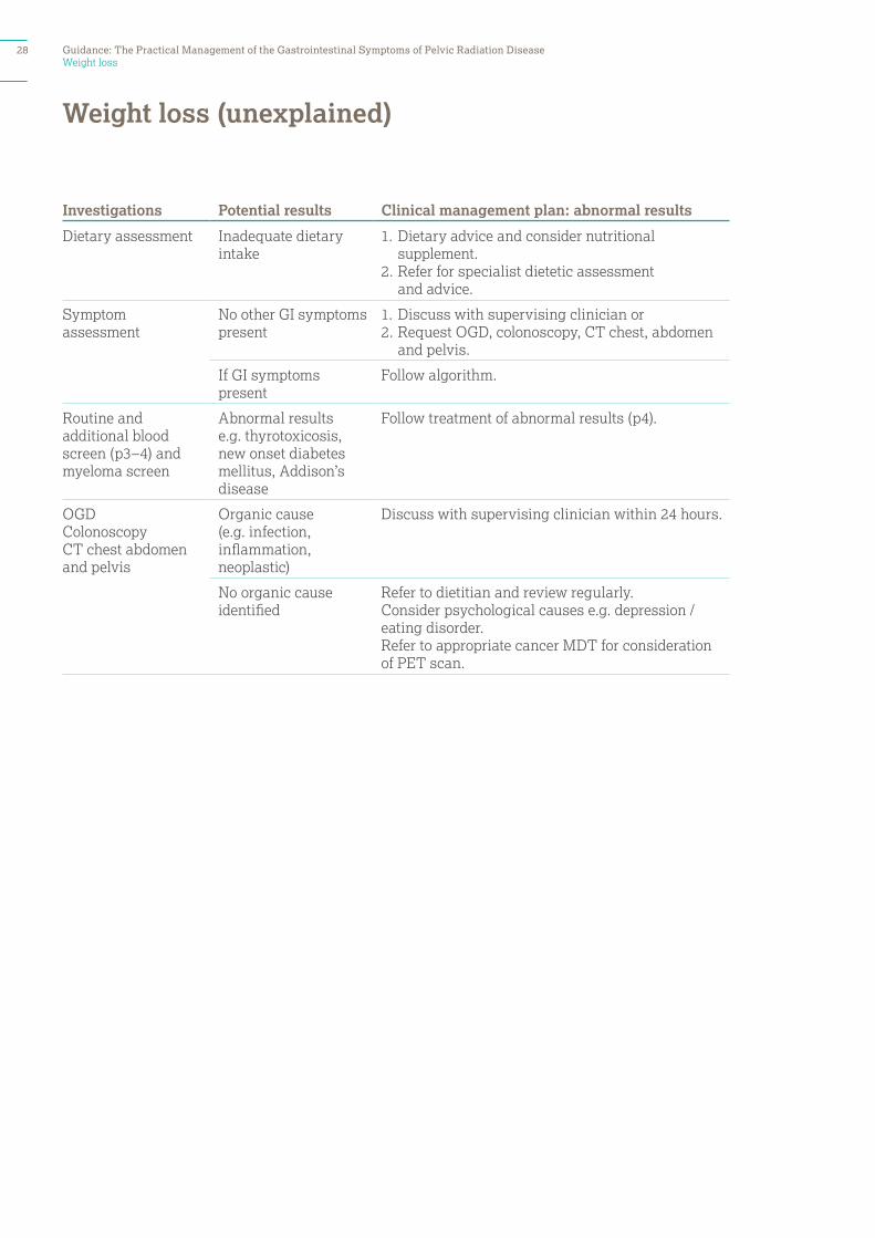
Investigations Potential results Clinical management plan: abnormal results
Dietary assessment Inadequate dietary intake
1. Dietary advice and consider nutritional supplement.
2. Refer for specialist dietetic assessment and advice.
Symptom assessment
No other GI symptoms present
1. Discuss with supervising clinician or 2. Request OGD, colonoscopy, CT chest, abdomen
and pelvis.
If GI symptoms present
Follow algorithm.
Routine and additional blood screen (p3–4) and myeloma screen
Abnormal results e.g. thyrotoxicosis, new onset diabetes mellitus, Addison’s disease
Follow treatment of abnormal results (p4).
OGD ColonoscopyCT chest abdomen and pelvis
Organic cause (e.g. infection, inflammation, neoplastic)
Discuss with supervising clinician within 24 hours.
No organic cause identified
Refer to dietitian and review regularly. Consider psychological causes e.g. depression / eating disorder.Refer to appropriate cancer MDT for consideration of PET scan.
Weight loss (unexplained)
28 Guidance: The Practical Management of the Gastrointestinal Symptoms of Pelvic Radiation Disease

Appendix: Common conditions in this group
Bile acid malabsorption (BAM) Exocrine pancreatic insufficiency (EPI) Carbohydrate malabsorption – for example, lactoseor other disaccharide intolerance Pelvic floor dysfunction Small intestinal bacterial overgrowth (SIBO)
Guidance: The Practical Management of the Gastrointestinal Symptoms of Pelvic Radiation Disease 29
Bile acid malabsorption (BAM)
Bile acid malabsorption (BAM)
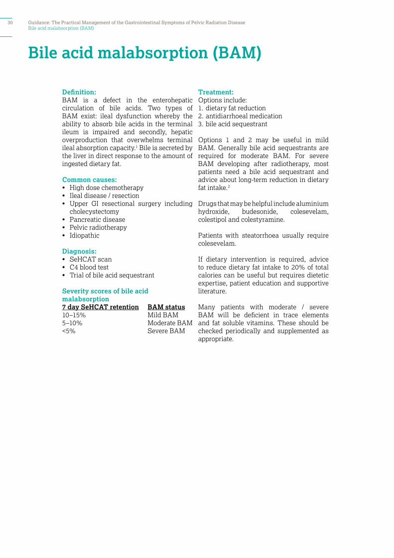
Definition: BAM is a defect in the enterohepatic circulation of bile acids. Two types of BAM exist: ileal dysfunction whereby the ability to absorb bile acids in the terminal ileum is impaired and secondly, hepatic overproduction that overwhelms terminal ileal absorption capacity.1 Bile is secreted by the liver in direct response to the amount of ingested dietary fat.
Common causes: • High dose chemotherapy• Ileal disease / resection• Upper GI resectional surgery including
cholecystectomy • Pancreatic disease• Pelvic radiotherapy• Idiopathic
Diagnosis:• SeHCAT scan• C4 blood test • Trial of bile acid sequestrant
Severity scores of bile acid malabsorption7 day SeHCAT retention BAM status10–15% Mild BAM5–10% Moderate BAM<5% Severe BAM
Treatment:Options include: 1. dietary fat reduction 2. antidiarrhoeal medication 3. bile acid sequestrant
Options 1 and 2 may be useful in mild BAM. Generally bile acid sequestrants are required for moderate BAM. For severe BAM developing after radiotherapy, most patients need a bile acid sequestrant and advice about long-term reduction in dietary fat intake.2
Drugs that may be helpful include aluminium hydroxide, budesonide, colesevelam, colestipol and colestyramine.
Patients with steatorrhoea usually require colesevelam.
If dietary intervention is required, advice to reduce dietary fat intake to 20% of total calories can be useful but requires dietetic expertise, patient education and supportive literature.
Many patients with moderate / severe BAM will be deficient in trace elements and fat soluble vitamins. These should be checked periodically and supplemented as appropriate.
30 Guidance: The Practical Management of the Gastrointestinal Symptoms of Pelvic Radiation Disease
Exocrine pancreatic insufficiency (EPI)
Exocrine pancreatic insufficiency (EPI)
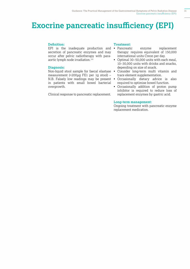
Definition: EPI is the inadequate production and secretion of pancreatic enzymes and may occur after pelvic radiotherapy with para-aortic lymph node irradiation.3,4
Diagnosis:Non-liquid stool sample for faecal elastase measurement (<200µg FE1 per 1g stool) – N.B. Falsely low readings may be present in patients with small bowel bacterial overgrowth.
Clinical response to pancreatic replacement.
Treatment:• Pancreatic enzyme replacement
therapy: requires equivalent of 150,000 international units Creon per day.
• Optimal 30–50,000 units with each meal, 10–30,000 units with drinks and snacks, depending on size of snack.
• Consider long-term multi vitamin and trace element supplementation.
• Occasionally dietary advice is also required to optimise bowel function.
• Occasionally addition of proton pump inhibitor is required to reduce loss of replacement enzymes by gastric acid.
Long-term management:Ongoing treatment with pancreatic enzyme replacement medication.
Guidance: The Practical Management of the Gastrointestinal Symptoms of Pelvic Radiation Disease 31
Carbohydrate malabsorption – e.g. lactose or other disaccharide intolerance
Carbohydrate malabsorption – for example lactose or other disaccharide intolerance
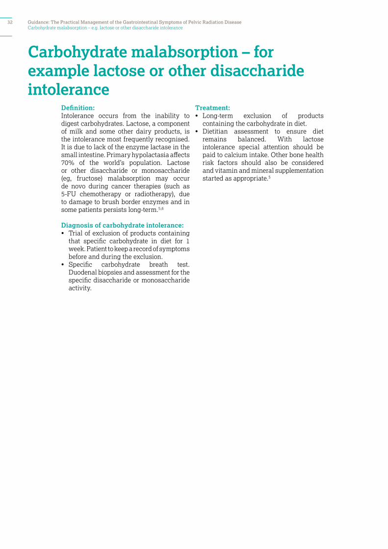
Definition: Intolerance occurs from the inability to digest carbohydrates. Lactose, a component of milk and some other dairy products, is the intolerance most frequently recognised. It is due to lack of the enzyme lactase in the small intestine. Primary hypolactasia affects 70% of the world’s population. Lactose or other disaccharide or monosaccharide (eg, fructose) malabsorption may occur de novo during cancer therapies (such as 5-FU chemotherapy or radiotherapy), due to damage to brush border enzymes and in some patients persists long-term.5,6
Diagnosis of carbohydrate intolerance:• Trial of exclusion of products containing
that specific carbohydrate in diet for 1 week. Patient to keep a record of symptoms before and during the exclusion.
• Specific carbohydrate breath test. Duodenal biopsies and assessment for the specific disaccharide or monosaccharide activity.
Treatment:• Long-term exclusion of products
containing the carbohydrate in diet.• Dietitian assessment to ensure diet
remains balanced. With lactose intolerance special attention should be paid to calcium intake. Other bone health risk factors should also be considered and vitamin and mineral supplementation started as appropriate.5
32 Guidance: The Practical Management of the Gastrointestinal Symptoms of Pelvic Radiation Disease
Pelvic floor dysfunction / Biofeedback
Pelvic floor dysfunction
Definition: Symptoms of pelvic floor dysfunction include urinary incontinence, bladder storage problems, altered bladder sensation, voiding and post micturition problems, anorectal symptoms, pelvic pain, sexual difficulties and pelvic organ prolapse (in females). Anorectal symptoms can include faecal incontinence, flatal incontinence, faecal urgency, straining to defecate, tenesmus, diminished rectal sensation, constipation, rectal prolapse, rectal bleeding and mucus discharge.7
Diagnosis:• rectal examination (sphincter tone and squeeze).• endo-anal ultrasound.• anorectal physiology investigations: • anal resting pressure. • sphincter muscle squeeze. • 15 seconds squeeze. • rectal sensitivity to rectal distension.
Treatment:• pelvic floor exercises (p38).• toileting posture exercises (p39).• biofeedback (p37).
A contributing factor is often constipation and a non-fermentable stool bulking agent such as Normacol can be helpful to restore rectal volume and is less likely to cause flatulence than other fibre supplements.
Guidance: The Practical Management of the Gastrointestinal Symptoms of Pelvic Radiation Disease 33
Small intestinal bacterial overgrowth (SIBO)
Small intestinal bacterial overgrowth (SIBO)
Definition: SIBO is the presence of excessive bacteria in the small intestine. Small bowel bacterial overgrowth occurs in 25% of patients during the acute phase of radiotherapy and is a cause of diarrhoea in up to 15% of patients after radiotherapy.8,9,10
Diagnosis:11
• There is no gold standard for diagnosing SIBO.12
• Glucose hydrogen / methane breath testing ± duodenal (D2) aspirate via upper GI endoscopy.
• RBC folate and total serum bile acid levels may be elevated and vitamin B12 levels and faecal elastase may be low.
• 10–15% patients with negative tests still have SIBO.
Suggested antibiotic treatment options if no growth on culture to direct treatment 7–10 days treatment with:• Ciprofloxacin 500mg bd• Doxycycline 200mg day 1,
100mg days 2–7 / 10• Clarithromycin 500mg bd• Metronidazole 400mg tds• Rifaximin 550mg bd.
Symptoms can recur any time after antibiotics are stopped because the underlying cause of bacterial overgrowth can not always be addressed. If symptoms return, repeat treatment with antibiotics for a few days every month or continually at the lowest effective dose may be helpful in managing symptoms long-term. Some clinicians recommend rotating antibiotics but this may not be effective if the organisms involved are not sensitive to the antibiotics used.
Treatment decisions should be individualised and consider the risks of long term antibiotic therapy such as Clostridium difficile infection, cumulative irreversible neuropathy with metronidazole, Achilles tendon rupture with ciprofloxacin, intolerance, side-effects, bacterial resistance and costs.8,9,10,11,12
34 Guidance: The Practical Management of the Gastrointestinal Symptoms of Pelvic Radiation Disease
Appendix: Management techniques
Written information is often helpful to supplement the management of specific diagnoses. If information sheets are not available locally, information sheets on the following can be obtained from Dr. Andreyev’s office at The Royal Marsden (020 7811 8216):
1. advice for those with constipation or who often need to strain2. having a SeHCAT scan3. having a glucose hydrogen / methane breath test4. pancreatic insufficiency.
Specific leaflets are also available on the following treatments:
1. lactose free diet 2. managing fibre in your diet3. taking anti-diarrhoeal medication4. taking colesevelam5. taking loperamide5. taking Normacol6. treatment of radiation-induced gastrointestinal bleeding.
Guidance: The Practical Management of the Gastrointestinal Symptoms of Pelvic Radiation Disease 35

Dietary fibre manipulation
The mean UK average consumption of non-starch polysaccharides for healthy adults (aged 19–64 years) is 14.9g / day for men and 12.8g /day for women.13 The current dietary recommended daily intake for fibre – 18g non-starch polysaccharides per day – is based on the effect that total dietary fibre has on stool weight. The rationale for this is that daily stool output of <100g / day is associated with a non-starch polysaccharides intake of below 12g /day and with an increased risk of bowel disease. In healthy populations, increasing non-starch polysaccharides
intake from 13 to 18g /day is associated with a 25% increase in stool weight. Some patients after pelvic radiotherapy cannot tolerate as much fibre as this.
Reduction in dietary fibre intake can be helpful when patients complain of any of the following symptoms: bloating, constipation, bowel obstruction, diarrhoea, rectal flatulence, mucus discharge and abdominal pain and may require help from a dietitian.13,14,15
Dietary fibre manipulation 36 Guidance: The Practical Management of the Gastrointestinal Symptoms of Pelvic Radiation Disease

Biofeedback
Biofeedback is widely regarded as a useful, non-invasive treatment in constipation, evacuatory disorders and faecal incontinence. Biofeedback is a behavioural approach to which there are no side effects and offers a non-surgical approach
for patients with bowel dysfunction. It includes toileting exercises and pelvic floor exercises.16,17
Biofeedback services are available locally around the UK.
Biofeedback Guidance: The Practical Management of the Gastrointestinal Symptoms of Pelvic Radiation Disease 37
Pelvic floor exercises
The pelvic floor muscles include the levator ani, the coccygeus and associated connective tissue and, if weakened, can cause several symptoms associated with pelvic floor dysfunction (p33). Exercises can strengthen the pelvic muscles so that they give support and are better coordinated. Pelvic floor exercises can improve problems with urinary incontinence, faecal incontinence or leakage and sexual function.18,19
Technique:1. Sit, stand or lie with your knees slightly
apart. Tighten and pull up your bottom muscles as tightly as you can. Hold for at least 5 seconds and then relax for at least 10 seconds. Repeat at least five times. This will work on the strength of your muscles.
2. Next, pull the muscles up to about half of their maximum squeeze. See how long you can hold this for. Then relax for at least 10 seconds. Repeat at least five times. This will work on the endurance or staying power of your muscles and will improve their coordination.
3. Pull up the muscles as quickly and tightly as you can and then relax and then pull up again, and see how many times you can do this before you get tired. Try for at least five quick pull-ups.
4. Repeat exercises 1, 2 and 3 at least 10 times every day.
5. As the muscles get stronger, you will find that you can hold for longer than 5 seconds, and that you can do more pull-ups each time without the muscle getting tired.
Patient information:www.yourpelvicfloor.co.uk
Pelvic floor exercises38 Guidance: The Practical Management of the Gastrointestinal Symptoms of Pelvic Radiation Disease
Toileting exercises
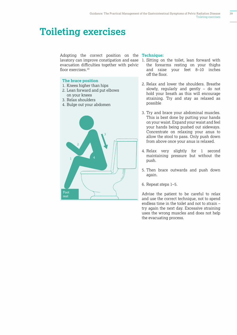
Adopting the correct position on the lavatory can improve constipation and ease evacuation difficulties together with pelvic floor exercises.20
Technique:1. Sitting on the toilet, lean forward with
the forearms resting on your thighs and raise your feet 8–10 inches off the floor.
2. Relax and lower the shoulders. Breathe slowly, regularly and gently – do not hold your breath as this will encourage straining. Try and stay as relaxed as possible
3. Try and brace your abdominal muscles. This is best done by putting your hands on your waist. Expand your waist and feel your hands being pushed out sideways. Concentrate on relaxing your anus to allow the stool to pass. Only push down from above once your anus is relaxed.
4. Relax very slightly for 1 second maintaining pressure but without the push.
5. Then brace outwards and push down again.
6. Repeat steps 1–5.
Advise the patient to be careful to relax and use the correct technique, not to spend endless time in the toilet and not to strain – try again the next day. Excessive straining uses the wrong muscles and does not help the evacuating process.
The brace position1. Knees higher than hips2. Lean forward and put elbows
on your knees3. Relax shoulders4. Bulge out your abdomen
Toileting exercises Guidance: The Practical Management of the Gastrointestinal Symptoms of Pelvic Radiation Disease 39
Treating bleeding telangiectasia
Radiation-induced bleeding typically starts 6–12 months after radiotherapy, is at its worse 4 years after the end of radiotherapy and has disappeared by 8–10 years.
1. Investigate with flexible endoscopy to determine the cause of the bleeding.
2. Optimise bowel function and stool consistency.
3. If bleeding is not staining clothes, causing anaemia or interfering with daily activities, reassure and do nothing.
4. If bleeding affects quality of life, stop anticoagulants if possible and consider sucralfate enemas ± metronidazole 400mg tds for 4 weeks.
5. Discuss definitive treatment to ablate the telangiectasia:
A. Hyperbaric oxygen therapy: Advantages: supported by RCT evidence, may improve other symptoms, for example, urinary; disadvantages: time consuming (8 weeks of daily treatment), expensive and patients may need to travel long distances to their nearest unit.
B. Any thermal therapy (eg APC): Advantages: easily available and simple; disadvantages: significant risk of non-healing ulceration / perforation as the tissue is ischaemic and unproven efficacy in heavy bleeding. Absolutely contraindicated in patients treated with brachytherapy. If used, the bowel must be fully prepared (as for colonoscopy).
C. Formalin therapy: Advantages: simple to perform; disadvantages: long-term outcomes poorly known, small risks of serum sickness, severe proctitis or chemical burn to the skin if there is spillage.
Treating bleeding telangiectasia40 Guidance: The Practical Management of the Gastrointestinal Symptoms of Pelvic Radiation Disease

Using intra-rectal formalin for radiation induced telangiectasia
Using intra-rectal formalin for radiation induced telangiectasia
Formalin chemically cauterises by hydrolysing protein and superficially coagulating the tissue. In general, the procedure seems to be effective, safe, well tolerated by the patient, inexpensive and technically simple.21,22,23,24,25
The use of intrarectal formalin for radiation-induced bleeding is contraindicated if the rectal mucosa is ulcerated.
Preparation:• Prepare the bowel as for colonoscopy
with full bowel preparation. • Use a gastroscope not a colonoscope.• 30–35mL of 5% formalin is usually
sufficient to cover the telangiectasia.• Initial instillation of saline can help
assess how much formalin will be required.
Technique:• Patient position: prone.• Instil the formalin through a catheter
passed through the gastroscope channel into the rectum.
• Apply with wet cloths and continued pressure to the perianal area to prevent leakage of formalin during the procedure (by endoscopy nurse).
• Keep the gastroscope in place during the procedure.
• Leave the formalin in the rectum for 3 minutes exactly.
• Then remove the formalin with copious washes of water.
• Continue the perianal pressure / pads until all of the rectal formalin removed.
Considerations after treatment:• Advise patients they may not notice any
improvement in bleeding for 1–2 weeks.• Consider the use sucralfate enemas for
2–3 weeks twice daily to help healing.• Consider retreating if necessary
6–8 weeks later and repeat if necessary a third time after a further 6–8 weeks.
Guidance: The Practical Management of the Gastrointestinal Symptoms of Pelvic Radiation Disease 41

How to refer for hyperbaric oxygen therapy
How to refer for hyperbaric oxygen therapy
Hyperbaric oxygen for radiation-induced damage requires a funding application to the Specialised Commissioning Group. The local hyperbaric oxygen unit and the referring clinician need to fill in the application jointly.26
Hyperbaric units that provide medically supervised hyperbaric oxygen therapy with the correct pressures believed to be useful for treating radiation injury are only found at the following centres:
• DDRC Hyperbaric Medical Centre, Tamar Science Park, Plymouth
• James Paget Hospital, Great Yarmouth• Midlands Diving Chamber,
Hospital of St Cross, Rugby• Spire Hospital, Cardiff• Spire Hull and East Riding Hospital, Hull• Spire Murrayfield Hospital, Wirral• St John and Elizabeth Hospital,
North London• St Richards Hospital, Chichester• Whipps Cross Hospital, East London
There is no evidence at all that non-medically supervised hyperbaric oxygen therapy as provided by MS treatment centres is of any benefit for treating radiation-induced toxicity.
For further information on managing GI bleeding, please see the BSG / ASCPGBI / AUGIS / Royal College of Radiology Guidance published: Gut 2012;61:179–192: Practice guidance on the management of acute and chronic gastrointestinal problems arising as a result of treatment for cancer (open access publication).
42 Guidance: The Practical Management of the Gastrointestinal Symptoms of Pelvic Radiation Disease

Sucralfate enemas
Sucralfate forms a mechanical protective layer over radiation-induced telangiectasia and improves healing.
1. Use 2g (10mL) sucralfate suspension (1g in 5mL) made up to 50mL using warm tap water in a bladder syringe.
2. Attach a soft, lubricated Foley catheter to the syringe.
3. The patient should insert the catheter gently into their rectum and instil the enema twice a day until the bleeding has stopped.
4. The enema should be held in the rectum as long as possible.
5. The patient should roll over at least once to coat the entire rectum but spend the majority of the time lying prone so the solution treats the anterior rectal wall.
6. Long-term once-daily enemas may help prevent the bleeding recurring.
7. If bleeding starts again go back to using sucralfate enemas twice a day.
Sucralfate enemas Guidance: The Practical Management of the Gastrointestinal Symptoms of Pelvic Radiation Disease 43

Perianal skin care
Perianal skin care
Radiotherapy skin reactions may present as skin irritation, erythema and ulceration and atrophy of the skin within the radiotherapy field that may be worsened by enzymes present in faecal fluid when incontinent or leaking (p17).
Key principles:27,28
• Keep skin dry• Keep skin free of faeces• Prevent the development of perianal
dermatitis by:
1. Use ‘simple’ soap or ‘Dove Sensitive’ soap that will not affect the pH of the skin (normally 5.5). Regular soap has a pH of nine and can disrupt the skin pH which inhibits the growth of bacteria and thus increases dermatitis.
2. Treating the underlying cause:• Use a skin barrier: cream or film. • Please note whether the patient has
any allergies to any of the constituents. • If the skin is damaged, Cavilon No
Sting Barrier Film or Epaderm can be very useful.
• Consider the use of antifungal creams or corticosteroids (for a short period only).
• Consider referral for specialist dermatology assessment and advice.
Patient information:www.stmarkshospital.org.uk/patient-information-leaflets
44 Guidance: The Practical Management of the Gastrointestinal Symptoms of Pelvic Radiation Disease
Using prokinetics
Using prokinetics
Effects on stomach:29,30
• Erythromycin: largely ineffective after 4–8 weeks through tachyphylaxis. Recommended dose 250mg bd as a syrup 30 min before food.
• Domperidone: no tachyphylaxis for 8 weeks, may occur after longer use. Recommended dose 10mg qds 30 min before food as a syrup orally or 30mg qds as a rectal suppository. Increased risk of cardiac arrhythmia (NB MHRA advice issued 2014).
• Metoclopramide: risk of tardive dyskinesia with use >3 months.
• Naloxone by subcutaneous infusion• Paroxetine that stimulates small intestinal
motility only.
Guidance: The Practical Management of the Gastrointestinal Symptoms of Pelvic Radiation Disease 45

How to perform a duodenal aspirate
How to perform a duodenal aspirate
Treatment: 1. Flush 100mL of sterile saline into the
duodenum via the endoscope channel
2. Follow this by 20mL of air to ensure no saline remains in the endoscope channel
3. Turn down the suction
4. Leave the fluid to equilibrate with the duodenal contents for 10–20 s
5. Aspirate 20mL of fluid into a sterile trap
6. Send the duodenal aspirate sample directly to microbiology.
46 Guidance: The Practical Management of the Gastrointestinal Symptoms of Pelvic Radiation Disease
References
1. Walters J & Pattni S. Managing bile acid malabsorption. Therap Adv Gastroenterol 2010; 3: 349–357.
2. Wedlake L, Thomas K, Lalji A, Anagnostopoulos C. Effectiveness and tolerability of colesevelam hydrochloride for bile acid malabsorption in patients with cancer: a retrospective chart review and patient questionnaire. Clinical Therapeutics. 2009; 31: 2549–2558.
3. Dookeran KA, Thompson MM & Allum WH. Pancreatic insufficiency secondary to abdominal radiotherapy. Eur J Surg Oncol. 1993; 19: 95–6.
4. Kingham JG, Barrett A. Pancreatic insufficiency following abdominal irradiation. Postgrad Med J. 1980; 56: 804–5.
5. Andreyev J. Gastrointestinal symptoms after pelvic radiotherapy: a new understanding to improve management of symptomatic patients. Lancet Oncol 2007; 8: 1007–1017.
6. Fabrizis L. Suarez, Dennis A. Savaiano, and Michael D. Levitt. A Comparison of Symptoms after the Consumption of Milk or Lactose-Hydrolyzed Milk by People with Self-Reported Severe Lactose Intolerance. N Engl J Med 1995; 333:1–4.
7. Haylen B, de Ridder D, Freeman R, Swift S et al. An International Urogynaecological Association (IUGA)/ International Continence Society (ICS) joint report on the terminology for female pelvic floor dysfunction. Neurology and Urodynamics 2010; 29: 24–29
8. Andreyev HJ, Davidson S, Gillespie C, et al. Practice guidance on the management of acute and chronic gastrointestinal problems arising as a result of treatment for cancer. Gut 2012; 61: 179–192.
9. Andreyev J. Gastrointestinal symptoms after pelvic radiotherapy: a new understanding to improve management of symptomatic patients. Lancet Oncol 2007; 8: 1007–1017.
10. Dukowicz A, Lacy B & Levine G. Small bowel bacterial overgrowth: a comprehensive review. Gastroenterol Hepatol 2007; 3: 112–122.
11. Gasbarrini A, Lauritano EC, Gabrielli M, Scarpellini E, Lupascu A, Ojetti V et al. Small intestinal bacterial overgrowth: diagnosis and treatment. Dig Dis. 2007; 25: 237–40.
12. Grace E, Shaw C, Whelan K, Andreyev HJ. Small intestinal bacterial overgrowth prevalence, clinical features, current and developing diagnostic tests, and treatment. Aliment Pharmacol Ther. 2013; 38(7): 674–88
13. Abayomi J, Kirwan J, Hackett A. Coping mechanisms used by women in an attempt to avoid symptoms of chronic radiation enteritis. J Hum Nutr Diet 2009; 22: 310–16.
14. Department of Health. (2011). National Diet and Nutrition Survey: Headline results from Years 1 and 2 (combined) of the rolling programme 2008/9–2009/10. www.dh.gov.uk/en/Publicationsandstatistics/Publications/PublicationsStatistics/DH_128166
15. Gami B, Harrington K, Blake P, et al. How patients manage gastrointestinal symptoms after pelvic radiotherapy. Aliment Pharmacol Ther 2003; 18: 987–94.
References Guidance: The Practical Management of the Gastrointestinal Symptoms of Pelvic Radiation Disease 47
16. Wald A (2007) Chronic constipation: advances in management. Neurogastroenterology and Motility; 19: 1, 4–10.
17. National Institute of Clinical Excellence. Management of faecal incontinence in adults: CG 49. London: NICE; 2007.
18. Law Y & Fielding J. MRI of pelvic floor dysfunction: review. AJR Online 2008; 191: S45–S53.
19. Rao, S.S.C. (2008) Disorders of the Pelvic Floor and Anorectum. Gastorenterology Clinic of North America. 37(3): 493–506.
20. Sikirov D (2003) Comparison of straining during defecation in three positions: results and implications for human health. Digestive Diseases and Sciences; 48: 1201–1205
21. For further information on managing GI bleeding, please see the BSG / ASCPGBI / AUGIS / Royal College of Radiology Guidance published: Gut 2012; 61: 179–192: Practice guidance on the management of acute and chronic gastrointestinal problems arising as a result of treatment for cancer (open access publication).
22. Cullen SN, Frenz M, Mee A. Treatment of haemorrhagic radiation-induced proctopathy using small volume topical formalin instillation. Aliment Pharmacol Ther. 2006 1; 23(11): 1575–9.
23. Raman R. Two percent formalin retention enemas for haemorrhagic radiation proctitis: A preliminary report. Dis Col Rectum 2007; 50: 1–8.
24. Rubinstein E, Ibsen T, Rasmussen R, Reimer E & Sorensen B. Formalin treatment of radiation-induced hemorrhagic proctitis. Am J Gastroenterol. 1986; 81: 44–5.
25. Botten RJ, Di Matteo AC, Butters J et al. Randomised trial of argo plasma coagulation therapy versus topical formalin for persistent rectal bleeding. Int J Radiol Biol Phys 2013 87 (5) 954–9.
26. For further information on managing GI bleeding, please see the BSG / ASCPGBI / AUGIS / Royal College of Radiology Guidance published: Gut 2012; 61: 179–192: Practice guidance on the management of acute and chronic gastrointestinal problems arising as a result of treatment for cancer (open access publication).
27. Nazarko L. Managing a common dermatological problem: incontinence dermatitis. British Journal of Community Nursing 2007; 12: 358–363.
28. The Royal Marsden NHS Foundation Trust. The Royal Marsden Hospital Manual of Clinical Procedures. Eighth Edition. Edited by Dougherty L. & Lister S. Chapter 19: Wound Management: Radiotherapy skin reactions: 1180–1183.
29. Park MI, Camilleri M. Gastroparesis: clinical update. Am J Gastroenterol. 2006; 101(5): 1129–39.
30. Khoo J, Rayner CK, Jones KL, Horowitz M. Pathophysiology and management of gastroparesis. Expert Rev Gastroenterol Hepatol. 2009; 3(2): 167–81. doi: 10.1586/egh.09.10.
References48 Guidance: The Practical Management of the Gastrointestinal Symptoms of Pelvic Radiation Disease

Guidance: The Practical Management of the Gastrointestinal Symptoms of Pelvic Radiation Disease 49

50 Guidance: The Practical Management of the Gastrointestinal Symptoms of Pelvic Radiation Disease

Guidance: The Practical Management of the Gastrointestinal Symptoms of Pelvic Radiation Disease 51