Embed Size (px)
Citation preview
1
The Harvard Orthopaedic Journal
Volume 15 · December 2013
http://www.orthojournalhms.org
Growth Disturbance Following Intraarticular Distal Radius Fractures in
the Skeletally-Immature PatientEric C. Fu M.D.1, Apurva S. Shah M.D., M.B.A.2, Peter M. Waters M.D.1, Donald S. Bae M.D.1
1Boston Children’s Hospital, Boston, MA, 2University of Iowa Hospitals and Clinics, Iowa City, IA
Introduction: The purpose of this study was to defi ne the incidence of growth disturbance following intraarticular distal radius fractures in the skeletally immature and assess radiographic and functional outcomes.
Methods: A retrospective investigation of all pediatric patients with intraarticular distal radius fractures over a fi ve year period at a single institution was performed. Fractures in skeletally-mature patients and pathologic fractures were excluded. Twenty-nine patients with a mean age of 13.9 years and mean follow-up of 17.7 months met the inclusion criteria. Fractures were categorized according to the Salter-Harris classifi cation and all radiographs were assessed for evidence of physeal disturbance. Information regarding treatment and early clinical results were obtained from medical record review. Functional outcomes using the Disabilities of the Arm, Shoulder, and Hand (DASH) and Modifi ed Mayo Wrist Score (MMWS) were collected. Fisher’s exact test was used to compare incidence of physeal arrest in the study population to previously published rates of physeal arrest in fractures involving the distal radius.1,2 The Mann-Whitney-Wilcoxon test was to compare those who did and did not develop physeal arrest.
Results: Of the 29 patients, 9 (31%) sustained Salter-Harris III fractures and 20 (69%) sustained Salter-Harris IV fractures. Growth disturbance occurred in 12 (41%) patients. All children age 10 years or younger had growth arrests that underwent subsequent procedures to address deformity. By DASH and MMWS, all patients had minimal disability and excellent functional outcomes after fi nal treatments.
Conclusion: Intraarticular distal radius fractures in skeletally-immature patients have a considerably higher rate of physeal growth arrest than extraarticular physeal fractures.1,2 The treating surgeon should make an aggressive effort to restore/preserve anatomic physeal alignment. Follow-up radiographs should be obtained to evaluate for physeal arrest in this cohort. Patients and families should be counseled regarding the high rate of growth disturbance and the potential need for deformity correction in the future, particularly in younger children.
Level of Evidence: IV – Case Series
1. Mizuta T, Benson WM, Foster BK, et al. Statistical analysis of the incidence of physeal injuries. J Pediatr Orthop. 1987; 7:518–523.
2. Cannata G, De Maio F, Mancini F, Ippolito E. Physeal Fractures of the Distal Radius and Ulna: Long-Term Prognosis. J Orthop Trauma. 2003; 17(3): 172-179.
Citations
2
The Harvard Orthopaedic Journal
Volume 15 · December 2013
http://www.orthojournalhms.org
Simpli ied and Strong: Abductor Pollicus Longus Suspension Arthroplasty with
Biotenodesis Screw Fixation in the Base of the Index Metacarpal
Terrill P. Julien, M.D., Brandon E. Earp, M.D., Philip E. Blazar, M.D. Orthopaedic Hand Service, Brigham and Women’s Hospital, Harvard Combined Orthopaedic Residency Program,
Boston, MA 02114
Abstract: Surgical treatment of thumb basal joint arthritis is aimed at pain relief, restoration of pinch strength and stability. We describe a variation of the abductor pollicus longus (APL) suspension arthroplasty originally described by Thompson that maximizes strength of repair and is technically simple to perform. The technique consists of a trapeziectomy followed by resection of a slip of the APL and secure docking of the tendon into the base of the index metacarpal base. Stability is enhanced with a 3.0mm biotenodesis screw (Arthrex Inc, Naples Florida). From March 2009 to September 2011, 51 patients treated at our institution. Early results showed consistent pain relief, functional improvement and minimal complications. The data support APL suspension arthroplasty with tenodesis screw fi xation as a safe and effective treatment for CMC arthritis.
Corresponding AuthorTerrill P. Julien MDResident Harvard Combined Orthopedic ProgramBrigham and Women’s Hospital75 Francis Street, Boston, MA 02115Telephone: 617-732-5384Facsimile: 617-730-2818Email: [email protected]
Brandon E. Earp, MDInstructor, Orthopaedic Surgery, Harvard Medical SchoolOrthopaedic Hand Service, Brigham and Women’s Hospital,Boston, MA.
Philip E. Blazar, MDAssistant Professor, Orthopaedic Surgery, Harvard Medical SchoolDirector, Hand Fellowship ProgramOrthopaedic Hand Service, Brigham and Women’s Hospital,Boston, MA.
3
The Harvard Orthopaedic Journal
Volume 15 · December 2013
http://www.orthojournalhms.org
Durability of Partial Humeral Head Resurfacing
Ruth A. Delaney M.D.1, Michael T. Freehill M.D.1,2, Laurence D. Higgins M.D.2, Jon J.P. Warner M.D.1
Harvard Shoulder Service, 1Massachusetts General Hospital, 2Brigham & Women’s Hospital, Boston, MA, 02114
Background: Partial humeral head resurfacing arthroplasty utilizes a stemless device, which conserves bone and restores normal anatomy. We hypothesized that this does not offer a reasonable alternative to full resurfacing or total shoulder arthroplasty.
Methods: This is a retrospective study of 39 shoulders with focal chondral defects of the humeral head treated with partial resurfacing arthroplasty. Minimum 2-year follow-up is reported, unless failure and operative intervention superceded this duration. Mean follow-up was 51.3 months. Mean age was 45.6 years (range 27–76). Pre and post-operative evaluation included history,physical examination,radiographs, and clinical scoring using ASES Shoulder Score Index(SSI) and Subjective Shoulder Value(SSV). Results: Twenty-fi ve of 39 shoulders (64.1%) demonstrated functional improvement and decreased pain. Signifi cant mean improvements were observed in forward fl exion (121.3o to 151.6o;p = 0.002), external rotation(37.1o to 58.3o;p=0.0003), mean SSV(30.75% to 73.6%;p < 0.0001),and ASES score (29.35 to 70;p <0.0001). However, at an average of 26.6 months follow up, the failure group included 6 patients (15.3%) who underwent revision and another four (10.2%) who were recommended revision. Patients with no prior or concomitant procedure were rare (n=5) but had the most reliable outcomes with partial resurfacing, with no failures in that group. Of the 24 patients with prior procedures, 5 had been revised, and the clinical outcome scores for the remaining patients were consistently lower than those seen in patients without prior procedures.
Conclusion: Concomitant pathology and prior or concomitant surgical procedures potentially impair the outcome of the resurfacing procedure and could be a contraindication. Long-term success remains guarded with this treatment modality, especially in patients whose chondral injury is not an isolated fi nding.
Partners Healthcare Institutional Review Board approval: Protocol # 2009P000960
Financial Disclaimer: None
Corresponding AuthorJon J.P. Warner, MDChief, The Harvard Shoulder Service 55 Fruit Street Yawkey Center for Outpatient Care Suite 3200, 3G, Room 3-046Boston, MA 02114Telephone: (617) 724-7300 (of ice) Facsimile: (617) 724-3846Email: [email protected]

4
The Harvard Orthopaedic Journal
Volume 15 · December 2013
http://www.orthojournalhms.org
Intra-operative Neuromonitoring During the Latarjet Procedure
Ruth A. Delaney, M.D.1, Michael T. Freehill, M.D.1,2, David R. Janfaza, M.D.2, Mirela V. Simon, M.D.2, Kamen V. Vlassakov, M.D.3, Laurence D. , Higgins, M.D.4, Jon J.P. Warner, M.D.1
1Harvard Shoulder Service, Massachusetts General Hospital, 2Department of Neurology, Massachusetts General Hospital, 3Department of Anesthesiology, Brigham & Women’s Hospital, 4Harvard Shoulder Service, Brigham &
Women’s Hospital, Boston, MA
Background: The purpose of this study was to use intra-operative neuromonitoring to defi ne the stages of the Latarjet procedure during which the nerves are at greatest risk, allowing the surgeon to take intra-operative measures to reduce this risk.
Methods: 34 patients were included in this prospective study. Mean patient age was 28.4 years. Upper extremity neurologic function was assessed pre-operatively, immediately post-operatively in the recovery room before any neurologic block was performed, and at routine follow up visits. The Latarjet procedure was divided into 9 defi ned stages. All surgeries were performed under total intravenous anesthesia. Bilateral median and ulnar somatosensory evoked responses (SSEPs) and transcranial motor evoked potentials (tcMEPs) from all arm myotomes were continuously monitored. A ‘nerve alert’ was defi ned as averaged 50% amplitude attenuation, or 10% latency prolongation of ipsilateral SSEPs and tcMEPs. For each nerve alert, the surgeon altered retractor placement, and if no response to this, then changed the position of the operative extremity.
Results: 26 of 34 patients (76.5%) had 45 separate nerve alert episodes. Forty-one of these alerts were based on attenuation of tcMEPs. Thirteen patients (38.2%) had 2 or more nerve alerts, with 2 patients having 4 nerve alert episodes. The most common stages of the procedure for a nerve alert to occur were glenoid exposure (12 alerts) and graft insertion (17 alerts). The axillary nerve was involved in 35 alerts; the musculocutaneous nerve in 22. Fourteen alerts involved both nerves.Seven of the 34 patients (20.6%) had a clinically detectable nerve defi cit post-operatively. In all 7 cases, the neurapraxia correlated with an intra-operative nerve alert. All cases involved the axillary nerve. Four nerve palsies resolved completely, 3 were improving at latest follow up but had not completely resolved.Prior surgery, BMI and number of nerve alerts during surgery were not predictive of a clinically detectable neurologic defi cit post-operatively, however total operative time (p = 0.042) and duration of the stage of procedure in which the concordant nerve alert occurred (p = 0.010) were statistically signifi cant predictors of a post-op nerve defi cit.Before we implemented neuromonitoring, 5 in a series of 48 shoulders (10.4%) had nerve palsies after the Latarjet procedure, 2 of which did not improve and required return to the operating room for neurolysis. In this series, no post-operative nerve palsy was severe enough to require intervention, however there was a 20.6% rate of clinically detectable nerve palsy, despite neuromonitoring.Conclusion: The nerves are at risk during the Latarjet procedure, in particular the axillary nerve and the musculocutaneous nerve. The most common stages of the Latarjet procedure during which the nerves are under excessive tension are glenoid exposure and graft insertion. The surgeon should be especially meticulous and consider duration of retraction during these stages. Attention to graft placement is important. A more superior position of the graft may be protective with regard to axillary nerve palsy. It is possible that neuromonitoring leads to decreased severity of post-operative nerve defi cits, but in this series it did not reduce the actual rate of clinically detectable defi cits.
Partners Healthcare Institutional Review Board approval: Protocol # 2011P000188Financial Disclaimer: None
Corresponding AuthorJon J.P. Warner, MDChief, The Harvard Shoulder Service 55 Fruit Street Yawkey Center for Outpatient Care Suite 3200, 3G, Room 3-046Boston, MA 02114
Telephone: (617) 724-7300 (of ice) Facsimile: (617) 724-3846Email: [email protected]
5
The Harvard Orthopaedic Journal
Volume 15 · December 2013
http://www.orthojournalhms.org
Safety and Ef icacy of Derotational Osteotomy for Congenital
Radioulnar SynostosisXavier Simcock, M.D.1, Apurva S. Shah M.D., M.B.A.2, Donald S. Bae M.D.1, Peter M. Waters M.D.1
1Boston Children’s Hospital, Boston, MA, 2University of Iowa Hospitals and Clinics, Iowa City, IA
The historical complication rate following derotational osteotomy for congenital radioulnar synostosis exceeds 35% and includes a high risk of compartment syndrome and loss of fi xation. Standardization of surgical technique can improve the safety and effi cacy of this procedure by signifi cantly diminishing the risk of neurovascular compromise and loss of fi xation.
Methods:A retrospective evaluation of consecutive patients who underwent derotational osteotomy for congenital radioulnar synostosis at a single institution between 1995 and 2011 was performed. Patients with substantial functional limitations secondary to excessive pronation were indicated for surgery with a goal correction to 0 to 20 degrees of pronation. All patients were treated with a standardized surgical technique including careful subperiosteal elevation, rotational osteotomy at the level of the radioulnar synostosis, control of the osteotomy fragments, appropriate pinning techniques (with 2 to 4 Kirschner-wires), and prophylactic forearm fasciotomies. Pre-operative radiographs were reviewed to confi rm the diagnosis. Electronic medical records were evaluated for pre-operative and post-operative forearm position, elbow motion, post-operative complications, and need for additional surgery. Post-operative radiographs were assessed for union.
Results: During the collection period, a derotational osteotomy utilizing the standardized technique was performed in 31 forearms in 26 children (13 bilateral, 13 unilateral) with a mean age of 6.8 years (range 3.0-18.8 years). The mean clinical follow-up was 30 months. The mean initial pronation deformity was 85 degrees (range 60-100 degrees). The mean correction achieved was 77 degrees (range 40-95 degrees), resulting in a mean fi nal position of 8 degrees of pronation (range 0-30 degrees). All patients successfully achieved union by 8 weeks. There were no cases of compartment syndrome, vascular compromise, or loss of fi xation. The overall complication rate was 12% (3 transient nerve palsies, 1 symptomatic muscle herniation). Two transient anterior interosseous nerve palsies were identifi ed, with both occurring in patients with rotational corrections exceeding 80 degrees. One transient radial nerve palsy was observed, and was attributed to retractor placement.
67th Annual Meeting of The American Society for Surgery of the HandSeptember 6-8, 2012, Chicago, IL

6
The Harvard Orthopaedic Journal
Volume 15 · December 2013
http://www.orthojournalhms.org
Displaced Femoral Neck Fractures in Patients <60 Years of Age
Stephen T. Gardner, M.D., Michael J. Weaver, M.D., Seth A. Jerabek, M.D., Mark S. Vrahas, M.D., Paul T. Appleton, M.D., Mitchel B. Harris, M.D.
Brigham and Women’s Hospital, Massachusetts General Hospital, Beth Isreal Deaconess Hospital,
Boston, MA, USA
Purpose: Displaced femoral neck fractures in young patients are relatively rare and potentially devastating injuries. Primary arthroplasty is increasingly advocated for the older patient but is rarely indicated for patients <60 years of age. The two most common methods of fi xation of these fractures are percutaneous cannulated lag screw (PCS) fi xation and a sliding hip screw (SHS). This paper reports the outcomes of these two different fi xation techniques in displaced femoral neck fractures in patients younger than 60 years of age.
Methods: A retrospective review of a prospectively enrolled trauma database was performed at three Level I trauma centers spanning the years 2000–2010. The electronic medical records and radiographs of all patients <60 years of age with displaced femoral neck fractures (OTA 31.B) treated by open or closed reduction and PCS fi xation or SHS were individually reviewed. Quality of reduction was recorded for each fracture fi xation construct and tip-apex distance (TAD) was recorded for each SHS construct. Exclusion criteria included all patients treated primarily with hip arthroplasty, follow-up <6 months, pathologic fracture through a bone lesion, or femoral neck fractures in association with acetabular or femoral head fractures. The primary outcome measurement was a return to the operating room within 6 months of the index procedure due to implant failure, or loss of reduction. Secondary outcomes were defi ned as loss of fi xation after 6 months, symptomatic osteonecrosis requiring surgery, or nonunion requiring repair or conversion to total hip arthroplasty. A two-tailed Fisher exact test was used to compare independent outcome variables. P value was set at 0.05 to indicate statistical signifi cance.
Results: 133 displaced femoral neck fractures were identifi ed in 132 patients <60 years of age. 64 patients were excluded: primary arthroplasty (n = 41), follow-up <6 months (n = 17), pathologic fracture through a bone lesion (n = 3), and complex combined injury (n = 3). Our fi nal study cohort was 69 femoral neck fractures in 68 patients. 40 patients were treated with SHS and 29 were treated by PCS fi xation. Mean age in the groups was similar (SHS: 42.4 years, PCS: 43.7 years). Excluding patients with early failure, follow-up ranged 6 to 84 months (median, 18 months). TAD was <25 mm in 34 of 40 SHS patients. Reduction quality was graded as excellent (n = 19), good (n = 31), or fair (n = 8). 11 patients did not have immediate postoperative radiographs and quality of reduction could not be determined. At 6 months, only 1 (3%) patient in the SHS group lost fi xation compared to 6 (21%) patients in the PCS group (P = 0.02). However, overall complication rates at most recent follow-up were similar between patients treated with SHS (25%) or PCS (31%) (P = 0.60).
Conclusion: There remains controversy regarding the optimal fi xation method for displaced femoral neck fractures in younger patients. Biomechanical data suggest that SHSs are stronger than PCS constructs. It has been our clinical experience that fi xation with SHS leads to a signifi cantly lower short-term mechanical failure rate. The longer-term failure rate in our series is similar to other published reports and appears to be independent of fi xation method. This suggests that biologic, and not mechanical, factors are most important in determining long-term outcome in these injuries.
Current Faculty and Residents28th Annual Meeting of The Orthopaedic Trauma AssociationOctober 3-6, 2012, Minneapolis, MN
7
The Harvard Orthopaedic Journal
Volume 15 · December 2013
http://www.orthojournalhms.org
A Prospective, Randomized Controlled Trial of a Fibular Nail Versus Standard Open Reduction and Internal Fixation
for Fixation of Ankle Fractures Timothy O. White, M.D., F.R.C.S., Kate E. Bugler, Paul T. Appleton, M.D.,
Margaret M. McQueen, M.D., Charles M. Court-Brown, M.D
Edinburgh, Scotland, United Kingdom
Purpose: The technique of open reduction and internal fi xation (ORIF) of ankle fractures with plates and screws has not changed substantially since the 1960s. Three principal complications are associated with this type of surgery. Firstly, wound dehiscence and infection, with published rates of up to 30%, and higher rates in patients with diabetes and neuropathy. Secondly, there is a risk of construct failure, particularly in osteoporotic bone. Thirdly, the scar or prominent hardware may cause later irritation and require further surgery. We have developed a technique of intramedullary fi bular nailing that is biomechanically stronger than ORIF, requires only minimal incisions, and has low-profi le hardware. We hypothesized that fi bular nailing would result in a rate of reduction and union comparable to fi xation, with a reduced rate of wound and hardware problems.
Methods: 100 patients over the age of 65 years with unstable ankle fractures requiring fi xation were recruited and randomized to undergo fi bular nailing or standard stabilization using AO techniques. Immediate weight bearing in cast was permitted. Outcome measures assessed over the 12 postoperative months were: the accuracy of reduction, development of wound complications or radiographic arthritis, range of movement, Olerud and Molander score (OMS), and the total cost of treatment. The mean age was 74 years (range, 65-93) and 75 patients were women. Twelve patients were smokers, two were diabetic, and all had some form of comorbidity, most commonly hypertension or ischaemic heart disease. Three injuries occurred during sport and one after a fall from a height; the remainder occurred after a simple fall from a standing height. 72 patients underwent additional medial fi xation.
Results: Signifi cantly fewer wound infections occurred in the fi bular nail group (P = 0.002). Eight patients (16%) in the ORIF group developed lateral-sided wound infections and required antibiotics. Two of these developed a wound dehiscence and required readmission for surgical débridement and removal of metalwork. In addition, six further patients complained of discomfort related to their wounds or hardware. One patient suffered surgical division of the superfi cial peroneal nerve and one patient went on to a malunion. No infections or wound problems occurred in the fi bular nail group. One patient underwent reoperation during the index admission for loss of reduction, one patient complained of a prominent locking screw, and one patient developed a malunion. The overall cost of treatment in the fi bular nail group was less despite the higher initial cost of the implant. At 1 year, fi bular nail patients were signifi cantly more happy with the condition of their scar (P = 0.02), and had slightly better OMS scores (63 vs 61, not signifi cant [P = 0.61]).
Conclusion: The fi bular nail allows accurate reduction and secure fi xation of ankle fractures with a signifi cantly reduced rate of soft-tissue complications when compared with standard ORIF.
Current Faculty and Residents28th Annual Meeting of The Orthopaedic Trauma AssociationOctober 3-6, 2012, Minneapolis, MN

8
The Harvard Orthopaedic Journal
Volume 15 · December 2013
http://www.orthojournalhms.org
Why are Reported Nonunion Rates After Locked Plate Fixation of Distal Femur Fractures so Variable? A Multicenter Retrospective Study of 284 Fractures
Edward K. Rodriguez, M.D., Ph.D.1, Michael J. Weaver, M.D.2, Lindsay M. Herder, B.A.1,Jordan H. Morgan, B.S.2,3, David Zurakowski, M.D.4, Paul T. Appleton, M.D.1,
Mark S. Vrahas, M.D.2,3
1Beth Israel Deaconess Medical Center, 2Brigham and Women’s Hospital, 3Massachusetts General Hospital, 4Children’s Hospital, Boston, MA, USA
Background/Purpose: Reported initial success rates after lateral locked plating (LLP) of distal femur fractures have given way to more concerning outcomes with reported nonunion rates now ranging from 0% to 21%. Reported factors associated with nonunion include comorbidities such as obesity, age, and diabetes, as well as technical factors such as plate length and screw density of constructs. Our goal was to examine variation in institutional nonunion rates at three Level I trauma centers treating a similar patient population in order to defi ne a set of patient characteristics that identify nonunion risk and to determine if nonunion rates are related to the management approach. We hypothesized that institutions with a more aggressive approach to nonunion management based on radiographic fi ndings and patient symptoms would have higher nonunion rates and shorter times to intervention than those where nonunion is primarily managed only after hardware failure.
Methods: A retrospective review was conducted of all distal femoral fractures treated with LLP at the three institutions (A, B, C) comprising our Combined Trauma Service (August 2004–December 2010). Nonunion was defi ned as the need for a secondary procedure to manage poor healing based on individual surgeon criteria (hardware failure, radiographic fi ndings, and/or patient symptoms). 284 fractures met inclusion criteria and each patient’s chart and radiographs was reviewed to extract age, gender, medical comorbidities (obesity, diabetes, tobacco use, steroid usage, dialysis), and injury characteristics (AO fracture type, open vs closed, mechanism of injury, periprosthetic). Multivariate analysis was performed using the Cox regression model.
Results: 29 of the 284 fractures analyzed went on to nonunion (10.2%). Only obesity, diabetes, and an open fracture were signifi cant independent risk factors. 38% of patients with nonunion had diabetes compared with 23% of patients in the healed group. 57% of patients with nonunion were obese compared with 37% of patients in the healed group. 23% of patients with open fractures went on to nonunion. Institution A had 14.1% of all nonunions, B had 8.8%, and C had 8.1%. While χ2 testing suggested no differences in nonunion rates between the institutions, the time to intervention for nonunions varied inversely with nonunion rates. Institution A intervened on average at 9 ± 4 months, institution B at 11 ± 7 months, and C at 20 ± 10 months. Institution C had a signifi cantly longer time to intervention for nonunion than A (P = 0.02) and B (P = 0.04).
Conclusion: Obesity, diabetes, and an open fracture are all predictors of nonunion in distal femoral fractures treated with LLP despite differences in how surgeons defi ne and manage nonunion. The institutional difference in nonunion rates, and perhaps in the literature, may be explained in part by individual surgeon approaches to the management of the nonunion patient. Without a consistent defi nition of nonunion, comparisons between institutions and surgeons are diffi cult.
Current Faculty and Residents28th Annual Meeting of The Orthopaedic Trauma AssociationOctober 3-6, 2012, Minneapolis, MN
9
The Harvard Orthopaedic Journal
Volume 15 · December 2013
http://www.orthojournalhms.org
Pathoanatomical Considerations and Implications of Heterotopic Ossi ication
Following Surgical Treatmentof Elbow Trauma
Bryce T. Gillespie, M.D., George S.M. Dyer, M.D.
Division of Hand and Upper Extremity Surgery, Department of Orthopaedic Surgery, Brigham and Women’s Hospital, Boston, MA, USA
Purpose: This work was undertaken to develop a pathoanatomical classifi cation system of heterotopic ossifi cation (HO) following surgical treatment of elbow trauma based upon pre-excision imaging.
Methods: 36 patients who had undergone excision of HO following initial surgical treatment of periarticular skeletal elbow trauma were identifi ed. Pre-excision imaging studies (including elbow radiographs alone or combined with CT scans) were reviewed independently to identify common patterns of HO. Injury pattern, elbow range of motion (ROM) data, and surgical characteristics were analyzed. One-way analysis of variance with Tukey’s post-hoc type 1 error adjustment was used to determine pairwise differences for the ROM data. Fisher’s exact test was used to compare the relationship between surgical characteristics and AO fracture classifi cation with the fi ve HO patterns.
Results: Five patterns of HO were identifi ed, including anterolateral elbow, anterior distal humerus, coronoid and olecranon fossae, proximal radioulnar joint (PRUJ), and posteromedial elbow/other. Signifi cant differences were found between the fi ve patterns when comparing pre-excision fl exion arc (P = 0.0355), fl exion arc gain (P = 0.0386), pre-excision rotation arc (P = 0.0014), and rotation arc gain (P = 0.0004). The PRUJ pattern had a signifi cantly greater pre-excision fl exion arc than the anterolateral pattern (95% confi dence interval [CI]: 2.5-96.1). Comparing pre-excision rotation arc, the anterior pattern was signifi cantly greater than the PRUJ pattern (95% CI: 25.6-228.4) and the fossae pattern was signifi cantly greater than the anterolateral and PRUJ patterns (95% CI: 1.7-143.8 and 35.2-218.0, respectively). For rotation arc gained, the PRUJ pattern gained signifi cantly more than the anterior and fossae patterns (95% CI: 19.8-192.2 and 42.8-198.2, respectively). Overall, the mean pre-excision fl exion arc was 58° and improved to 100° at fi nal follow-up (mean of 41 weeks) after excision of HO. The mean forearm rotation arc improved from 97° to 146°. The postexcision fl exion and rotation arcs were not signifi cant differently between the fi ve patterns. There is a signifi cant association between the fi ve patterns and AO fracture classifi cation. The anterior and fossae patterns were more often AO 13 than AO 21. Subjects with PRUJ and posteromedial/other patterns were exclusively AO 21, while subjects with the anterolateral pattern were divided between AO 13 (5 subjects) and AO 21 (9 subjects).
Conclusion: Several distinct patterns of HO about the elbow are identifi able and may have implications on elbow ROM and expected outcomes. Anterolateral HO appears to have restricted ulnohumeral and forearm motion. Anterior and fossae patterns were related to restricted ulnohumeral motion, while PRUJ HO was related to restricted forearm rotation. The postexcision fl exion and rotation arcs are similar for all fi ve patterns and comparable to previously published data regarding surgical treatment of elbow HO. Injury pattern may also be related to the subsequent morphology of HO. Anterior and fossae patterns develop more frequently following distal humerus fractures (AO 13) than proximal forearm injuries (AO 21). PRUJ and posteromedial/other patterns develop exclusively following proximal forearm injuries. The anterolateral pattern developed following either an AO 13 or AO 21 injury.
Current Faculty and Residents28th Annual Meeting of The Orthopaedic Trauma AssociationOctober 3-6, 2012, Minneapolis, MN
10
The Harvard Orthopaedic Journal
Volume 15 · December 2013
http://www.orthojournalhms.org
Cast Immobilization With and Without Immobilization of the Thumb for
Nondisplaced Scaphoid Waist Fractures: A Multicenter Randomized Controlled Trial
Geert A. Buijze, M.D., J. Carel Goslings, M.D., Steven J. Rhemrev, M.D.,Alexander A. Weening, M.D., Bart Van Dijkman, M.D., Job N. Doornberg, M.D.,
David C. Ring, M.D., Ph.D.
CAST (Collaborative Ankle Support Trial) Collaboration,Massachusetts General Hospital, Boston, MA, USA
Purpose: This trial was conducted to test the null hypothesis that there is no difference in union or arm-specifi c disability between nondisplaced scaphoid waist fractures treated in a below-elbow cast including or excluding the thumb.
Methods: 55 patients with a nondisplaced fracture of the scaphoid waist were enrolled in a prospective multicenter randomized controlled trial comparing treatment in a below-elbow cast including the thumb with a below-elbow cast excluding the thumb. Due to a misunderstanding at some centers, 7 distal scaphoid fractures were enrolled during the early part of the trial for a total of 7 distal and 55 waist fractures. We adhered to strict intention-to-treat principles. The primary study question addressed the extent of union on CT performed after 10 weeks of cast treatment and expressed as a percentage of the fracture line that had bridging bone by musculoskeletal radiologists blinded to treatment. Secondary study questions addressed wrist motion, grip strength, Mayo wrist scores, DASH (Disabilities of the Arm, Shoulder and Hand) scores, pain, and union at 6 months after treatment.
Results: There was a signifi cant, but clinically irrelevant, difference in the extent of union on CT at 10 weeks (85% vs 70%), favoring treatment with a cast excluding the thumb. One waist fracture treated with the thumb immobilized elected operative treatment 1 week after enrollment used crutches and developed nonunion (98% union overall; 100% union with nonoperative treatment). There were no signifi cant differences between the groups for wrist motion, grip strength, Mayo wrist scores, DASH scores, pain scores, or union.
Conclusion: Fractures of the scaphoid waist can be adequately treated in a below-elbow cast excluding the thumb.
Current Faculty and Residents28th Annual Meeting of The Orthopaedic Trauma AssociationOctober 3-6, 2012, Minneapolis, MN
11
The Harvard Orthopaedic Journal
Volume 15 · December 2013
http://www.orthojournalhms.org
Sustentaculum Screw Placement During Calcaneal Open Reduction and Internal
Fixation: When Is the Screw Out?Ida L. Gitajn, M.D., R. James Toussaint, M.D., John Y. Kwon, M.D.
Massachusetts General Hospital, Boston, MA, USA
Background/Purpose: During fi xation of calcaneal fractures a screw is often placed from lateral to medial into the sustentaculum as the constant fragment. This can be technically diffi cult as the sustentaculum is a small anatomic structure and this screw is generally placed from lateral to medial under fl uoroscopic guidance using the axial Harris calcaneal heel view. Misplacement of this screw can result in signifi cant complications given the high density of functionally important structures in this anatomic area, including the fl exor digitorum longus, fl exor hallucis longus, subtalar joint, tarsal canal, and neurovascular structures. Therefore the aims of this study were to determine whether there are certain fl uoroscopic axial heel views taken at specifi c angles that can accurately confi rm correct placement or misplacement of the sustentacular screw.
Methods: Lateral and medial dissection was performed on one cadaver foot specimen to remove skin and subcutaneous tissues. A 4.0-mm cancellous screw was placed from lateral to medial in fi ve different confi gurations: (1) screw placed anatomically within the sustentaculum, (2) screw misdirected inferior to the sustentaculum, (3) screw misdirected superior to the sustentaculum, (4) screw misdirected anterior to the sustentaculum, and (5) screw misdirected posterior to the sustentaculum. A large C-arm was used to obtain Harris heel views at fi ve different angulations (10°-50°). Two orthopaedic residents and one orthopaedic attending analyzed the C-arm images to determine at which angulation the screw placement could be confi rmed in each cadaver.
Results: A screw placed anatomically was noted to be radiographically within the sustentaculum in all fi ve views (Harris heel view at 10°, 20°, 30°, 40°, and 50°). An inferiorly misdirected screw appeared to be radiographically within the sustentaculum at 30°, 40°, and 50° but was confi rmed to be misplaced inferiorly on the 10° and 20° views. A posteriorly misdirected screw was confi rmed to be misplaced posteriorly on all fi ve views (10°, 20°, 30°, 40°, and 50° Harris heel views). An anteriorly misdirected screw appeared to be radiographically within the sustentaculum on the 10° view but was confi rmed to be misplaced anteriorly on all other views (20°, 30°, 40°, and 50° views). A superiorly misdirected screw was confi rmed to be misplaced on all views (10°, 20°, 30°, 40°, and 50° Harris heel views). Interobserver agreement on placement of the sustentacular screw was 100% among all three authors.
Conclusion: Clinicians should be aware that in order to verify correct placement of the sustentacular screw several axial Harris heel views are required. Axial heel views must be obtained at 10° to 20° to assess for inferior misplacement of the screw and views must be obtained at 20° to 50° to evaluate for anterior misplacement of the screw.
Current Faculty and Residents28th Annual Meeting of The Orthopaedic Trauma AssociationOctober 3-6, 2012, Minneapolis, MN
12
The Harvard Orthopaedic Journal
Volume 15 · December 2013
http://www.orthojournalhms.org
Peroneal Tendon Dislocation Associated With Intra-Articular Calcaneus Fractures:
An Underappreciated ProblemR. James Toussaint, M.D.1, Darius Lin, M.D.1, Lauren K. Ehrlichman, M.D.1,
Seenu Susarla, M.D., D.M.D.1, J. Kent Ellington, M.D., M.S.2, Nicholas Strasser, M.D.2, John Y. Kwon, M.D.1
1Massachusetts General Hospital, Boston, MA, USA, 2Carolinas Medical Center, Charlotte, NC, USA
Background/Purpose: Peroneal tendon dislocations (PTDs) are often undetected and undertreated complications of intra-articular calcaneus fractures. Existing studies demonstrate an association of PTD with intra-articular calcaneus fractures using CT. However, these studies are limited by small sample sizes, making the determination of true incidence unreliable. Further, existing research does not correlate fracture classifi cation with PTD and therefore offers little prognostic value. The goals of this multicenter, retrospective study are to determine: (1) incidence of PTD associated with intra-articular calcaneus fractures, (2) correlation of PTD with fracture classifi cation, (3) association of PTD and heel width, and (4) the rate of missed radiographic diagnosis and subsequent lack of treatment of PTD.
Methods: An IRB-approved review of calcaneus fractures from June 30, 2006 to June 30, 2011 was performed. Cases of intra-articular calcaneus fractures on plain fi lm and CT imaging were included. Fractures were classifi ed by the Essex-Lopresti and Sanders classifi cations. CT imaging was used to measure heel width and to identify PTD using available techniques. Plain radiographs were examined for signs of PTD (ie, “fl eck” sign, distal fi bular avulsion fracture). Radiology reports were reviewed for identifi cation of PTD. Medical records of PTD cases were reviewed for operative treatment of PTD at initial fracture fi xation or at a later date.
Results: Of 354 calcaneus fractures, 269 (76%) were intra-articular. 63.2% were classifi ed as joint depression, the remainder were tongue type. 13.8% were Sanders I, 37.1% Sanders II, 27.5% Sanders III, and 17.1% were Sanders IV. 31.7% of intra-articular fractures had peroneal tendon dislocations. There was a statistically signifi cant correlation between heel width (P <0.001) and joint depression fractures (P = 0.003) with PTD. Sanders IV fractures were statistically signifi cant and more likely to have PTD than Sanders Class I to III fractures (P = 0.003). Radiologists identifi ed 9.2% of PTDs on CT scans. None (0%) of the fractures with PTD taken for surgical fi xation had the peroneal tendons surgically addressed. The “fl eck” sign was seen in 5.6% of patients with PTD. This diagnostic test for PTD had a sensitivity of 21%, a specifi city of 100%, a positive predictive value of 100%, and a negative predictive value of 73%.
Conclusions: The results of our study demonstrate a statistically signifi cant and high incidence of PTD with intra-articular calcaneus fractures. This injury is often overlooked by radiologists and undertreated by orthopedists. Further research is required to determine if this fi nding is linked with increased patient morbidity.
Current Faculty and Residents28th Annual Meeting of The Orthopaedic Trauma AssociationOctober 3-6, 2012, Minneapolis, MN
13
The Harvard Orthopaedic Journal
Volume 15 · December 2013
http://www.orthojournalhms.org
Accuracy and Reliability of Bohler’s Angle Measurements With Oblique Lateral Radiographs Taken in the
Trauma SettingR. James Toussaint, M.D., Ida L. Gitajn, M.D., John Y. Kwon, M.D.
Massachusetts General Hospital, Boston, MA, USA
Background/Purpose: First described in 1931, Bohler’s angle is used to determine the amount of posterior facet displacement and severity of injury in calcaneus fractures. It is used to guide management or the need for additional imaging. Lateral images used to obtain Bohler’s angle in the trauma setting are often oblique due to diffi culties in positioning of the traumatized extremity or limitations from splint materials. Inaccurate Bohler’s angles in this setting can lead to under/overtreatment of patients. The purpose of this study is to assess the accuracy and reliability of measuring Bohler’s angle on oblique lateral imaging and to determine how improper imaging infl uences measurement.
Methods: A cadaver specimen was imaged using a large C-arm to obtain multiple fl uoroscopic images. First, a perfect lateral was obtained. Next, a series of oblique images was taken with the beam directed anteriorly, posteriorly, cephalad, and caudad. The images were taken in 5° increments from 5° to 25° in each direction. Orthopaedic staff and residents were asked to measure the observed Bohler’s angles. To defi ne the true Bohler’s angles, metallic markers were then placed on the anterior calcaneal process, the superior most portion of the posterior facet, and the superior posterior tuberosity of the same cadaver calcaneus. The same series of images were repeated to measure true Bohler’s angles using the marked specimen. The senior author then measured the true Bohler’s angles from the marked specimen.
Results: 41 orthopaedic staff and residents participated in the study. The mean values for the observed Bohler’s angles were signifi cantly different (P <0.05) from the true Bohler’s angles for all series of images except a posteriorly directed x-ray beam at 20° from the horizontal (P = 0.43). The mean value for observed Bohler’s angles deviated further from the true Bohler’s angles with increasing image obliquity for all series except the posteriorly directed x-ray beam. The true Bohler’s angle on a perfect lateral image was 35°. The true Bohler’s angle was found to vary based on the obliquity of the fl uoroscopic image (see table below).
Conclusion: The study fi ndings reveal that orthopaedic staff and resident physicians’ ability to accurately measure Bohler’s angle signifi cantly decreases with increasing obliquity of lateral radiographs. The true Bohler’s angle also varies with image obliquity. Understanding these changes with oblique lateral radiographs taken in the trauma setting should decrease reliance on only Bohler’s angle to determine management and need for additional imaging.
Current Faculty and Residents28th Annual Meeting of The Orthopaedic Trauma AssociationOctober 3-6, 2012, Minneapolis, MN
Changes in true Bohler’s angles with oblique luoroscopic images
Fluoro Direction (5o-25o)
True Bohler’s Angle Range, degrees
Fluoro Direction (5o-25o)
True Bohler’s Angle Range, degrees
Anteriorly 36-40 Cephalad 24-34Posteriorly 34-37 Caudad 37-39

14
The Harvard Orthopaedic Journal
Volume 15 · December 2013
http://www.orthojournalhms.org
The Role of Preoperative CT Scans in Operative Planning and Fixation of
Malleolar Ankle FracturesE. M. Black, V. Antoci, J. T. Lee, M. J. Weaver, A. H. Johnson, S. M. Susarla, John Y. Kwon, M.D.
Massachusetts General Hospital and Brigham and Women’s Hospital, Boston, MA , USA
Purpose: The purpose of this study was to determine the role of preoperative CT scans on surgical planning in malleolar ankle fractures. We hypothesized that CT would play an increasing role in surgical planning with fractures of higher energy and with lesser-quality preoperative radiographs.
Methods: A retrospective analysis was performed on the records of 100 consecutive patients treated at our institution between 2006 and 2010 for malleolar ankle fractures (AO Type 44) who had both preoperative radiographs as well as CT scans. Three fellowship-trained orthopaedic attending surgeons and three orthopaedic residents reviewed available preoperative radiographs and formulated an operative (or nonoperative) plan including patient positioning, surgical approach, and fi xation methods for all applicable components of the fracture. The six reviewers then analyzed CT scans of the same fractures and decided whether or not and how they would alter their operative strategy based on the CT scan. Fracture characteristics including number of involved malleoli, AO classifi cation, quality and nature of preoperative radiographs, and presence of dislocation on hospital presentation were noted and correlated with changes in operative strategy.
Results: Operative strategy was signifi cantly changed in 24% of cases after review of the CT scan. There was strong intraclass correlation between all reviewers (0.733), and no signifi cant difference based on level of training (P = 0.57). The most common changes in operative strategy involved fi xation of the medial malleolus (21%), posterior malleolus (15%), and fi xation of an occult anterolateral plafond fracture (9%). Predictors of changes in operative strategy included trimalleolar over unimalleolar fractures (29% vs 10% rate of change), preoperative dislocation over no dislocation (31% vs 20%), the presence of only radiographs with overlying plaster versus fractures with at least one set of radiographs without plaster (25% vs 14%), and suprasyndesmotic fractures vs. trans- and infrasyndesmotic fractures (40% vs 20% and 4%, respectively).
Conclusions: CT scans may be useful adjuncts in preoperative planning for malleolar ankle fractures, most notably in fracture dislocations, poor-quality preoperative radiographs, trimalleolar fracture patterns, and suprasyndesmotic (AO 44-C) ankle fractures.
Current Faculty and Residents28th Annual Meeting of The Orthopaedic Trauma AssociationOctober 3-6, 2012, Minneapolis, MN

15
The Harvard Orthopaedic Journal
Volume 15 · December 2013
http://www.orthojournalhms.org
Massachusetts Health Care Reform: Its Effect on the Percentage of Uninsured Patients and the Reimbursement for Provider Services at Academic Urban
Level I Trauma CentersR. James Toussaint, M.D.1, Michael J. Weaver, M.D.1, Paul Tornetta, III, M.D.2,
Mark S. Vrahas, M.D.1, Mitchel B. Harris, M.D.1
1Brigham and Women’s Hospital, 2Boston Medical Center, Boston, MA, USA
Background/Purpose: On April 12, 2006, the Massachusetts legislature approved its landmark health care reform law (also known as Chapter 58 of the Acts of 2006 or “Mass Health Reform” [MHR]) to provide health insurance coverage for all of its residents. MHR sought not only to provide uninsured residents with insurance coverage, but also to appropriately compensate hospitals and providers for their services. The purpose of this study is to evaluate the demographic and economic effects of Chapter 58 on the orthopaedic trauma population of three metropolitan Level I trauma centers.
Methods: We performed an IRB-approved retrospective review of all non–spine fracture and dislocation cases for patients less than 65 years old treated by the orthopaedic trauma services of three of the four Level I trauma centers in a major metropolitan area from 2003 to 2010. All out-of-state patients were excluded since they are not under the jurisdiction of MHR. The years 2006 and 2007 were excluded in order to remove the effects of volatility in the insurance market during the enrollment period. The population was divided into two groups: Group 1 included cases from January 2003 to December 2005 and consisted of patients treated for fractures and dislocations prior to the enactment of MHR; Group 2 consisted of patients from January 2008 to December 2010 who were treated subsequent to MHR. Relative Value Unit (RVU) data, CPT codes, fi nancial data, and patient insurance classifi cation were compared between the two groups.
Results: The fi nal study cohort included 12,955 patients (5318 in Group 1 and 7637 in Group 2, a 43.6% increase). There were 18,210 procedures (,462 in Group 1 and 10,748 in Group 2, a 44.0% increase). The percentage of patients who were uninsured in Group 1 was 27.4%; the percentage of uninsured patients in Group 2 decreased to 17.3%. There was a 44.0% increase in RVUs from Group 1 to Group 2. After analyzing two-thirds of the available data, the annual growth rate in revenue collections per RVU was 1.0% from Group 1 to Group 2 compared to 2.2% for the Medicare Economic Index during the same period.
Conclusion: Massachusetts health care reform has resulted in a 36.8% decrease in the percentage of uninsured patients treated for fractures and dislocations at three of four Level I trauma centers in a major metropolitan area. Despite this improvement, nearly one out of fi ve trauma patients at the urban trauma centers did not have health insurance. During this same time period, the Medicare Economic Index grew at a pace over twice that of the centers’ collections per RVU.
Current Faculty and Residents28th Annual Meeting of The Orthopaedic Trauma AssociationOctober 3-6, 2012, Minneapolis, MN
16
The Harvard Orthopaedic Journal
Volume 15 · December 2013
http://www.orthojournalhms.org
Corrective Osteotomy for Combined Intra- and Extra-Articular Distal
Radius MalunionGeert A. Buijze, M.D.1, Karl-Josef Prommersberger, M.D.2, Juan González del Pino, M.D., Ph.D.3,
Diego L. Fernandez, M.D.4, Jesse B. Jupiter, M.D.1
1Massachusetts General Hospital, Boston, MA, USA, 2Rhön-Klinikum, Bad Neustadt an der Saale, Germany,
3Santa Cristina University Hospital, Madrid, Spain, 4Lindenhof Hospital, Bern, Switzerland
Purpose: This study evaluated the functional outcome of corrective osteotomy for combined intra- and extra-articular malunions of the distal radius using multiple outcome scores.
Methods: 18 skeletally mature patients were evaluated at an average of 78 months after corrective osteotomy for a combined intra- and extra-articular malunion of the distal part of the radius. The indication for osteotomy in all patients was the combination of an extra-articular deformity (≥15° volar or ≥10° dorsal angulation or ≥3 mm radial shortening) and intra-articular incongruity of ≥2 mm (maximum step-off or gap) as measured on lateral and posteroanterior radiographs.The average interval from the injury to the osteotomy was 9 months. The average maximum step-off or gap of the articular surface prior to surgery was 4 mm.
Results: All 18 patients healed uneventfully and the fi nal articular incongruity was reduced to 2 mm or less. Final range of motion and grip strength signifi cantly improved (P <0.05), averaging 89% and 84% of the uninjured side, and 185% and 241% of the preoperative measures, respectively. The rate of excellent or good results was 72% according to the validated rating system Mayo Modifi ed Wrist Score, and 89% according to the unvalidated system of Gartland and Werley. The mean Disabilities of the Arm, Shoulder and Hand (DASH) score was 11, which corresponds to mild perceived disability. 11 of the 18 cases normalized their upper limb function. Four patients had complications that were successfully treated. According to the rating system of Knirk and Jupiter, four had a Grade 1 and one had a Grade 2 osteoarthritis of the radiocarpal joint on radiographs. Only two of these patients reported occasional mild pain. Radiographic osteoarthritis did not correlate with strength, motion and wrist scores.
Conclusion: Outcomes of corrective osteotomy for combined intra- and extra-articular malunions are comparable to those of osteotomy for isolated intra- and extra-articular malunions. If carefully planned, a corrective osteotomy for the treatment of complex intra- and extra-articular distal radius malunions can improve wrist function.
Current Faculty and Residents28th Annual Meeting of The Orthopaedic Trauma AssociationOctober 3-6, 2012, Minneapolis, MN
17
The Harvard Orthopaedic Journal
Volume 15 · December 2013
http://www.orthojournalhms.org
Optimizing the Biomechanics of Iliosacral Screw Fixation: The
Importance of Washers and Avoiding Lateral Cortex Perforation
Julius A. Bishop, M.D., Anthony W. Behn, M.S., Tiffany N. Castillo, M.D.
Stanford University Medical Center, Palo Alto, CA, USA
Purpose: Percutaneous iliosacral screws are frequently used to stabilize posterior pelvic ring injuries and can be placed with or without washers. Because the cortical bone of the outer table of the posterior ilium is thin, it is possible for the surgeon to unintentionally perforate this cortex during screw insertion, theoretically compromising fi xation. The purpose of this study was to detail the biomechanical consequences of washer use and iliosacral screw intrusion.
Methods: Partially threaded 7.0-mm cannulated screws with and without washers were placed through a synthetic bone test block fabricated to approximate the cortical and cancellous bone of the posterior ilium. A load cell was used to measure the compression generated before and after perforation of the outer cortex. 24 screws were tested under three different conditions: with a washer, without a washer, and with a washer after intrusion.
Results: Screws inserted with washers generated signifi cantly more compressive force than screws inserted without washers before screw intrusion. After intrusion, compressive force decreased signifi cantly under all conditions but screws inserted with washers maintained greater compressive force than screws inserted without washers. After intrusion of screws without washers, screws with washers reinserted through the same holes produced almost as much compressive force as screws inserted with washers primarily.
Conclusion: Screw intrusion during iliosacral screw insertion can compromise fi xation quality. Washers are advantageous in that they allow for more compression to be generated before intrusion occurs and can be used to salvage intruded screws initially placed without them. Washers can also be monitored fl uoroscopically as they seat against the ilium, providing an additional safeguard against intrusion.
Alumni28th Annual Meeting of The Orthopaedic Trauma AssociationOctober 3-6, 2012, Minneapolis, MN
18
The Harvard Orthopaedic Journal
Volume 15 · December 2013
http://www.orthojournalhms.org
Role of HtrA1 in the Transition From Cartilage to Bone in Fracture HealingMarie E. Walcott, M.D., John J. Wixted, M.D., Monica Thim, B.A., David C. Ayers, M.D.,
Paul J. Fanning, Ph.D.
University of Massachusetts Medical School, Worcester, MA, USA
Background/Purpose: Endochondral bone formation is fundamental to the process of normal fracture healing. Central to the process of endochondral ossifi cation is the production and destruction of chondrocytes along with their associated extracellular matrices (ECMs). The effects of mechanical stability have been studied extensively in bone remodeling but are lacking in the study of cartilage tissue turnover in fracture healing. Interestingly, a serine protease HtrA1 (high-temperature requirement protein A1) with cartilage ECM-degrading activity and mechanoresponsiveness has recently been identifi ed. HtrA1 expression is restricted to the pericellular matrix (PCM) that immediately surrounds the chondrocyte. Taken together, these data strongly suggest that an understanding of the role of HtrA1 and the chondrocyte PCM in fracture healing is needed. We hypothesized that HtrA1 expression levels would vary temporally and spatially during the progression of fracture healing in mice. Additionally, we have asked whether these levels differ between rigid versus fl exible internal femur fracture fi xation situations. Such differences could implicate relationships between chondrocytes, HtrA1 expression, PCM turnover, and the inception of delayed fracture healing.
Methods: Fracture technique: Reproducible transverse femur fractures were generated via a traumatic three-point bending method with reproducible energy of injury in 8-week-old C57B/6 male mice. Quantitative real-time polymerase chain reaction (PCR): On sacrifi ce days 6, 8, and 10 the fractured limb callus was dissected free and callus tissue alone was placed in TRIzol reagent. The tissue was ground using a Polytron homogenizer and total RNA isolated using a commercially available kit. Potential DNA contamination was removed by RNase-free DNase treatment. Reverse transcription reaction was performed using 1ug of total RNA and random hexamer primers. Relative transcript levels were measured by quantitative PCR (qPCR) in an ABI PRISM 7000 FAST sequence detection system and were normalized to GAPDH (glyceraldehyde-3-phosphate dehydrogenase) mRNA levels using commercially available primers and SYBR-Green master mix.
Results: HtrA1 mRNA levels were found to increase over the fi rst 2 phases of fracture healing (infl ammatory and cartilage formation) and peaked at day14 during the transition period from peak cartilage formation to primary bone formation/coupled resorption. qPCR revealed differences in gene expression events involved in chondrocyte pericellular matrix degradation between rigid versus fl exible fractures. Flexible fi xed fractures showed higher levels of mRNA for HtrA1 and discoid domain receptor 2 (DDR2) versus rigid fractures at day 7 of fracture healing. In addition, markers for early endochondral bone formation (vascular endothelial growth factor, collagen type II and collagen type X) were reduced in fl exible femur fractures compared to rigid fractures.
Conclusion: In this study, we examined the effect of mechanical stability on chondrocyte behavior during the early stages of endochondral ossifi cation in fractures. Our data validate a new methodology for studying the molecular mechanisms of mechanotransduction during fracture repair using relatively rigid or fl exible fi xation. In doing so, we are able to demonstrate an important role for the chondrocyte PCM in regulating the behavior of chondrocytes. These data provide insight into the potential role of both HtrA1 and DDR in regulating these early processes and suggest a potential role for pericellular chondrocyte signaling along the continuum of fracture healing from union to nonunion.
Alumni28th Annual Meeting of The Orthopaedic Trauma AssociationOctober 3-6, 2012, Minneapolis, MN
19
The Harvard Orthopaedic Journal
Volume 15 · December 2013
http://www.orthojournalhms.org
Can Glucose Levels Diagnose Compartment Syndrome?
Christopher J. Doro, M.D.1, Thomas J. Sitzman, M.D.2, Robert V. O’Toole, M.D.3
1Department of Orthopaedics, University of Wisconsin School of Medicine and Public Health, Madison, WI, USA, 2Division of Plastic Surgery, University of Wisconsin School of Medicine and Public Health, Madison, WI, USA,
3R Adams Cowley Shock Trauma Center, Department of Orthopaedics, University of Maryland School of Medicine, Baltimore, MD, USA
Purpose: Compartment syndrome is a diffi cult condition to diagnose, particularly in patients without a reliable clinical examination. Current objective methods rely on intracompartmental pressure measurements. We hypothesize that intracompartmental glucose levels can be used to diagnose compartment syndrome.
Methods: A compartment syndrome was created in 12 adult mixed-gender beagles using a previously described and validated model. Compartment syndrome was created in the anterior compartment of a lower leg in anesthetized dogs by infusion of lactated Ringer’s solution with normal serum concentration of glucose until intracompartment pressure exceeded 20 mm Hg above diastolic blood pressure. The contralateral leg was used as a control. Intracompartmental pressure, oxygen tension, and glucose concentration were recorded within each compartment using commercially available probes (Medtronic Diabetes; Oxford Optronix). Compartment syndrome was confi rmed in all cases by histologic analysis at 2 weeks after the infusion.
Results: Within 15 minutes of creating the compartment syndrome, glucose concentration in the experimental limb measured signifi cantly lower than the control limb (glucose P = 0.02; 2-tailed t test). Intramuscular glucose concentration less than 97 mg/dL was 100% (95% confi dence interval [CI]: 73%-100%) sensitive and 75% (95% CI: 40%-94%) specifi c for the presence of compartment syndrome.
Conclusion: Our results show that the intracompartmental glucose concentration appears to rapidly identify muscle ischemia after an experimentally created compartment syndrome. Real-time glucose sensors could provide a signifi cant advancement to the diagnosis of compartment syndrome by providing objective data that accurately and quickly indicate the presence of muscle ischemia, as opposed to relying on pressure measurements that are a much less direct indicator of ischemia.
Alumni28th Annual Meeting of The Orthopaedic Trauma AssociationOctober 3-6, 2012, Minneapolis, MN
20
The Harvard Orthopaedic Journal
Volume 15 · December 2013
http://www.orthojournalhms.org
Functional Outcomes After Nonoperative Treatment of Lateral Compression
Type 1 (LC-1) Pelvic Ring Injuries With Complete Sacral Fractures
Greg Gaski, M.D., Robert V. O’Toole, M.D., Renan Castillo, M.S., Gerard P. Slobogean, M.D., Theodore T. Manson, M.D.
R Adams Cowley Shock Trauma Center, Baltimore, MD, USA
Purpose: There is controversy regarding the optimum management of LC-1 fractures (OTA 61-B2), particularly for those with complete sacral fractures. Our hypothesis was that nonoperative treatment of these injuries would result in acceptable functional outcomes.
Methods: We conducted a review of a prospectively maintained database at a Level I trauma center over 3 years (2008-2011) to identify all LC-1 fractures (n = 315). We identifi ed a subset of “more severe” LC-1 injuries characterized by complete fracture through Denis zones 2 or 3 of the sacrum (n = 76). Of these, 12 patients were managed operatively at the discretion of the treating surgeon due to fracture displacement greater than 1 cm or severe comminution, and are not included in this analysis. The 64 remaining patients with complete sacral fracture and displacement less than 1 cm were treated nonsurgically and form the population of interest. Two patients were excluded due to spinal cord injury and 6 patients were deceased, leaving 56 potential patients. 30 patients were successfully contacted for functional outcome assessment at an average follow-up of 24.4 months (range, 15-37). The mean age at time of injury was 38.8 years (range, 17-80). Primary outcome measures were the Majeed Pelvic Score and the Physical and Mental Component Summary scores (PCS and MCS) of the Short Form-12 v.2. Bivariate analyses were performed with respect to age, ISS, anterior pelvic ring injury (none/unilateral rami fractures vs bilateral), associated lower extremity (LE) injuries, and initial weight-bearing status (non vs weight bearing as tolerated).
Results: The average Majeed Pelvic Score was 81.7 (95% confi dence interval [CI]: 75.1 to 88.4) yielding 19 excellent, 4 good, 3 fair, and 4 poor graded outcomes. Mean PCS and MCS scores were 46.9 (95% CI: 42.4, 51.3) and 48.2 (95% CI: 44.5, 51.8), respectively. Both intervals included 50, the mean score for a healthy, normative population. Patients with LE injuries had lower PCS scores than patients without LE injuries (38.1 vs 50.0, P = 0.04), and were less likely to have an “excellent” Majeed score (2 of 8 vs 17 of 22 in the non-LE injury group, P = 0.009). Additionally, all Majeed “poor” outcomes were in the subgroup of patients with concomitant LE injuries. There were no statistically signifi cant differences in regard to weight-bearing status, anterior ring injury, or ISS.
Conclusion: Recent studies have shown that “more severe” LC-1 fractures are susceptible to future displacement, but the clinical consequence of nonoperative treatment is unknown. This study suggests that good outcomes can be expected with nonsurgical management of “more severe” LC-1 fractures with less than 1 cm of initial displacement. Improvement in long-term functional outcome does not seem to be a valid rationale to treat these injuries operatively.
Alumni28th Annual Meeting of The Orthopaedic Trauma AssociationOctober 3-6, 2012, Minneapolis, MN
21
The Harvard Orthopaedic Journal
Volume 15 · December 2013
http://www.orthojournalhms.org
Is Closed Reduction and Percutaneous Fixation of Type 3 Posterior Ring Injuries
as Accurate as Open Reduction and Internal Fixation?
Adam Lindsay, M.D.1, Paul Tornetta, III, M.D.1, Amna Diwan, M.D.2, David C. Templeman, M.D.2
1Boston University Medical Center, Boston, MA, USA, 2Hennepin County Medical Center, Minneapolis, MN, USA
Background/Purpose: Type 3 posterior ring injuries are complete injuries and typically treated with reduction and internal fixation. While open treatment through a posterior approach allows for direct fracture reduction and is the gold standard, many surgeons are employing more percutaneous approaches. We compared closed reduction and percutaneous fixation (CRPP) versus open reduction and internal fixation (ORIF) of type 3 posterior ring injuries with the hypothesis that CRPP would be equivalent to ORIF in quality of reduction.
Methods: We reviewed 113 consecutive cases of unilateral unstable posterior ring injuries treated by two physicians in two different centers with iliosacral (IS) screws. Only true type 3 injuries were included. One surgeon routinely performed ORIF (n = 60); and the other, CRPP (n = 53). These two groups were compared. For all cases, demographic information, time to surgery, type of injury, and type of fixation was documented. Displacements were measured on the initial presentation and postoperative AP, inlet, and outlet views. These included the differences in the affected side from the normal side for: iliac wing height (AP, outlet), sacral height (AP), posterior superior iliac spine (PSIS) displacement (inlet), ischial height (AP, outlet), ring width (AP, inlet), and sacral width (inlet). These were compared with two-tailed t test assuming 0.05 for significance. CRPP Technique: A special traction setup was used that has not been previously described. The patient is positioned supine with a bump under the affected thorax and all the way to the edge of the table to allow easy placement of IS screws. The normal side foot is placed in a boot and the heel elevated to keep the knee in extension and locked there. Skeletal traction with ~20° to 30° of flexion is used on the affected side. A specially adapted hip positioning pad is locked to the table and abuts the affected thorax to prevent lateral displacement of the thorax when traction is applied. Under fluoroscopy, the posterior reduction can be titrated using traction as the unaffected leg acts as a post and the thorax pad prevents angulation of the body as the traction is applied to the affected side. Once the reduction is obtained, IS screws are placed. ORIF Technique: The patient is positioned prone on pads to allow free manipulation of the pelvis. A gluteus maximus–sparing approach was used in all cases. Clamps are used to directly reduce the posterior ring, augmented by fluoroscopy, and IS screws are placed.
Results: There were 53 patients treated with CRPP and 60 with ORIF during the study period. The ages (average, 36.9 years; range, 13-71), gender (62 female, 51 male), and ISS scores (average, 22.4; range, 4-59) did not differ between the groups. Initial displacements were also not different for the two groups (see table). The time to surgery was 4 days (range, 0-16) for the CRPP group and 5 (range, 0-36) for the ORIF group. Overall reduction quality was slightly better for the CRPP method for most parameters tested.
(cont.)
22
The Harvard Orthopaedic Journal
Volume 15 · December 2013
http://www.orthojournalhms.org
Alumni28th Annual Meeting of The Orthopaedic Trauma AssociationOctober 3-6, 2012, Minneapolis, MN
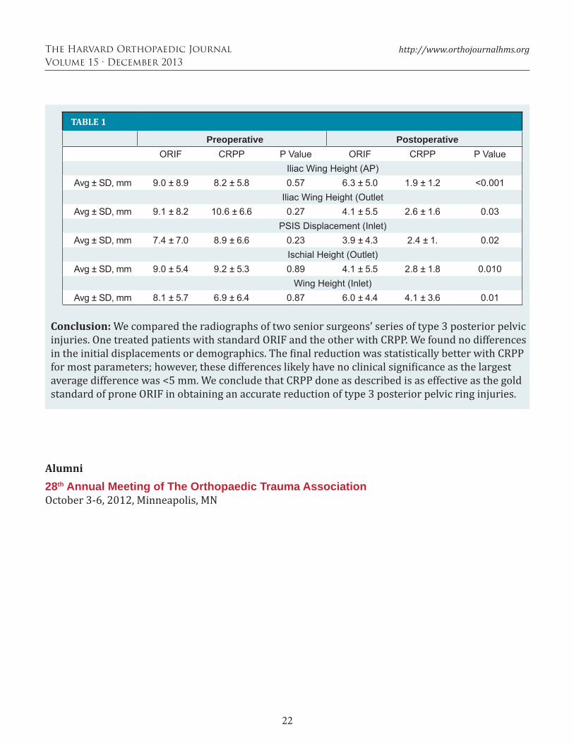
Conclusion: We compared the radiographs of two senior surgeons’ series of type 3 posterior pelvic injuries. One treated patients with standard ORIF and the other with CRPP. We found no differences in the initial displacements or demographics. The inal reduction was statistically better with CRPP for most parameters; however, these differences likely have no clinical signi icance as the largest average difference was <5 mm. We conclude that CRPP done as described is as effective as the gold standard of prone ORIF in obtaining an accurate reduction of type 3 posterior pelvic ring injuries.
TABLE 1
Preoperative PostoperativeORIF CRPP P Value ORIF CRPP P Value
Iliac Wing Height (AP)Avg ± SD, mm 9.0 ± 8.9 8.2 ± 5.8 0.57 6.3 ± 5.0 1.9 ± 1.2 <0.001
Iliac Wing Height (OutletAvg ± SD, mm 9.1 ± 8.2 10.6 ± 6.6 0.27 4.1 ± 5.5 2.6 ± 1.6 0.03
PSIS Displacement (Inlet)Avg ± SD, mm 7.4 ± 7.0 8.9 ± 6.6 0.23 3.9 ± 4.3 2.4 ± 1. 0.02
Ischial Height (Outlet)Avg ± SD, mm 9.0 ± 5.4 9.2 ± 5.3 0.89 4.1 ± 5.5 2.8 ± 1.8 0.010
Wing Height (Inlet)Avg ± SD, mm 8.1 ± 5.7 6.9 ± 6.4 0.87 6.0 ± 4.4 4.1 ± 3.6 0.01
23
The Harvard Orthopaedic Journal
Volume 15 · December 2013
http://www.orthojournalhms.org
Radiographic Predictors of Compartment Syndrome after Tibial Fracture
Chris Allmon, M.D., Ebrahim Paryavi, M.D., M.P.H., Andrew Dubina, Robert V. O’Toole, M.D.
R Adams Cowley Shock Trauma Center, Department of Orthopaedics, University of Maryland Medical School, Baltimore, MD, USA
Purpose: Compartment syndrome (CS) is a potentially devastating injury that has been associated with tibial fractures. Little data exist regarding the radiographic predictors of CS. Our hypothesis was that radiographic measures of the fracture would be associated with the development of CS.
Methods: Our study group was a consecutive series of patients with tibial fractures with CS (n = 40) and without CS (n = 341) at a single Level I trauma center. Radiographs were reviewed and the following parameters were recorded: fracture classifi cation according to the AO/OTA system and the Schatzker system for plateaus, proximal extent of fracture, distal extent of fracture, location of center of fracture, length of fracture, and location of fracture. Bivariate logistic regression was used to determine the relationship between the radiographic parameters and likelihood of compartment syndrome. Medical records were then reviewed for evidence of CS diagnosed by an attending orthopaedic surgeon and treated by emergent fasciotomy.
Results: Consistent with existing dogma, CS was most likely with more proximal fractures such as those in the second decile of the tibia, occurring at a rate of 38%. What has not been previously reported is that the rate of CS rose monotonically according to length of the fracture line, peaking at 38% when the fracture comprised between 40% and 60% of the total tibial length. Schatzker VI fractures developed CS at a rate of 27%, whereas only 4% of 25 Schatzker IV medial plateau fracture dislocations developed CS. Further analysis demonstrated that odds of CS increased by a factor of 1.91 (95% confi dence interval [CI] 1.46 to 2.49) for every 10% of the total tibial length the fracture occupied. The odds of CS decreased by 27% (95% CI 16%, 37%), 23% (95% CI 32%, 13%), and 18% (95% CI 27%, 9%) for every 10% of the total tibial length the proximal fracture extent, fracture middle, and distal fracture extent were away from the proximal end of the tibia, respectively. In comparison to all plateau fractures, Schatzker VI plateaus have an odds ratio of CS of 3.98 (95% CI 1.68, 9.45), whereas in contrast to previous case series we did not observe Shatzker IV to have a statistically signifi cant association with CS: odds ratio is 0.17 (95% CI 0.02, 1.29).
Conclusion: To our knowledge this is the largest series to rigorously examine radiographic predictors of CS. In keeping with expectations, we observed that Schatzker VI plateau fractures and more proximal fractures are more likely to develop CS. However, to our knowledge this is the fi rst study to propose a powerful new predictor of CS, the total length of the fracture. This parameter may be of use to clinicians as they evaluate patients for their risk of CS and in helping to diagnose patients with CS.
Alumni28th Annual Meeting of The Orthopaedic Trauma AssociationOctober 3-6, 2012, Minneapolis, MN

24
The Harvard Orthopaedic Journal
Volume 15 · December 2013
http://www.orthojournalhms.org
Alignment in Nonperatively Treated Distal Radius Fractures: Are Our Current
Predictors Predictive?Joey Lamartina, M.D., Charlton Stucken, M.D., Andrew Jawa, M.D., Paul Tornetta III, M.D.
Boston University Medical Center, Boston, MA, USA
Background/Purpose: Multiple methods have been described to predict loss of reduction in distal radius fractures treated nonoperatively. We sought to independently validate the McQueen equation and LaFontaine’s criteria in a large series of distal radius fractures treated nonoperatively. Additionally, we wished to evaluate postreduction volar cortical alignment (volar hook) and a specifi c defi nition for dorsal comminution on the fi nal reduction of these patients. We hypothesized that restoring the volar cortical integrity would aid in maintenance of the volar tilt by a standard three-point molded cast.
Methods: We prospectively screened 546 consecutive distal radius fractures using the McQueen equation and LaFontaine’s criteria for instability. We excluded patients with <10° of dorsal tilt upon presentation, leaving 275 fractures of which 168 were treated nonoperatively and form the basis of this study. Patients were managed with short arm casts and seen every other week in the clinic by an attending orthopaedic trauma surgeon. Patients were recasted if there was thought to be a shift in the fracture position or if the cast became loose. We measured the following parameters on the initial reduction and fi nal radiographs: dorsal tilt, radial height, radial inclination, ulnar variance, and the presence of carpal malalignment. We defi ned dorsal comminution as having a loss of the dorsal cortex of ≥5mm on the postreduction lateral radiograph. We defi ned “volar hook” as having collinear alignment of the cortical edges of the fracture at the volar surface. We performed univariate analysis to determine how predictive the McQueen percentage and the number of Lafontaine’s criteria present were on each radiographic parameter. Additional univariate analyses were done on the radiographic components of each score, volar hook, sex, and age. Based on the univariate analysis of the various predictors, a multivariate analysis was done including age, dorsal comminution (DC), volar hook (VH), and intra-articular fracture (IAF) against all radiographic outcome parameters and any change in those parameters during healing.
Results: In the univariate analysis, the McQueen percentage and the total number of LaFontaine’s criteria present predicted the change in radial height and inclination. The change in dorsal tilt was predicted by VH and DC. The change in ulnar variance was predicted by DC and IAF. The change in radial height was predicted by IAF and age and the change in radial inclination was predicted only by age. Final dorsal tilt was predicted by VH, DC, and sex. Carpal malalignment at healing was predicted by VH and age. The table details the results of the multivariate analysis.
Factors that were statistically signi icant in the miltivariate analysis for each radiographic outcome
Dorsal Tilt Ulnar Variance Radial Height Radial Inclination
Final Position VH, DC DC Age Age
Change During Treatment VH, DC Age Age, IAF Age, DC
25
The Harvard Orthopaedic Journal
Volume 15 · December 2013
http://www.orthojournalhms.org
Alumni28th Annual Meeting of The Orthopaedic Trauma AssociationOctober 3-6, 2012, Minneapolis, MN
Discussion: We attempted to validate the McQueen percentage and Lafontaine’s criteria on the fi nal radiographic position and the change in position over time in 168 consecutive patients with displaced distal radius fractures treated nonoperatively. Additionally, using a multivariate tech-nique, we found that VH and DC were strong predictors of fi nal dorsal tilt and the change in angu-lation during healing. Age was the most important factor in predicting ulnar variance, radial height, and inclination as well as the change in these parameters during treatment. Most important, VH (P = 0.001) and age (P = 0.03) were both predictive of carpal malalignment.
Conclusion: We were able to validate the McQueen equation and LaFontaine’s criteria on ra-dial height and inclination. However, neither method was predictive of fi nal dorsal tilt or carpal malalignment. Hooking the volar cortex (restoring the volar cortical integrity) was the strongest predictor of fi nal volar tilt, the change in volar tilt, and carpal malalignment at union. These data suggest that restoration of volar cortical alignment is an important predictor of success in nonop-erative treatment of distal radius fractures.