Embed Size (px)
Citation preview
Gross Anatomy andGross Anatomy andEmbryology of the JointsEmbryology of the Joints
Lawrence M. Witmer, PhDLawrence M. Witmer, PhDDepartment of Biomedical SciencesCollege of Osteopathic MedicineOhio UniversityAthens, Ohio [email protected]
Handout download:http://www.oucom.ohiou.edu/dbms-witmer/peds-joints.pdf
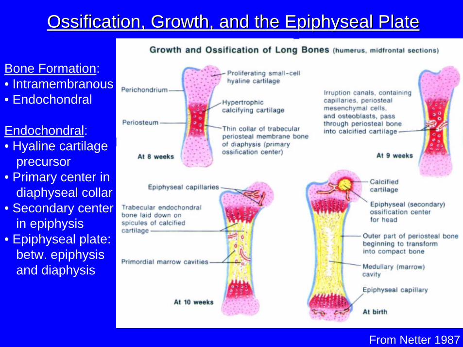
Ossification, Growth, and the Epiphyseal PlateOssification, Growth, and the Epiphyseal Plate
Bone Formation:• Intramembranous• Endochondral
Endochondral: • Hyaline cartilage
precursor• Primary center in
diaphyseal collar• Secondary center
in epiphysis• Epiphyseal plate:
betw. epiphysisand diaphysis
From Netter 1987
From Netter 1987
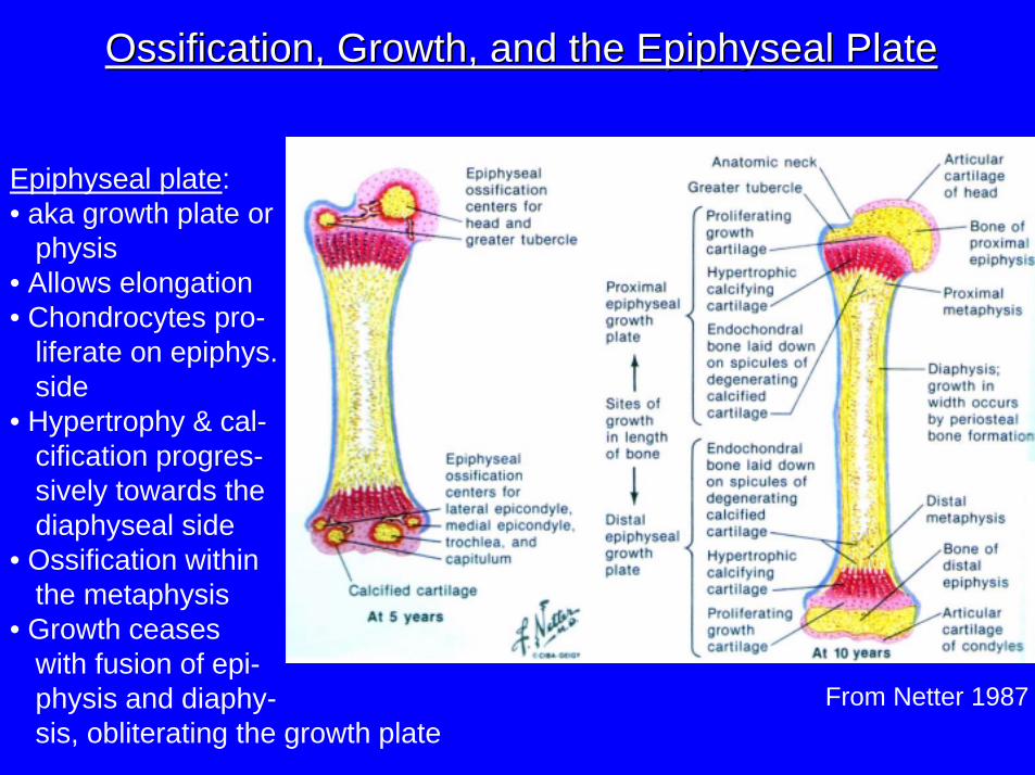
Ossification, Growth, and the Epiphyseal PlateOssification, Growth, and the Epiphyseal Plate
Epiphyseal plate:• aka growth plate or
physis• Allows elongation• Chondrocytes pro-
liferate on epiphys.side
• Hypertrophy & cal-cification progres-sively towards thediaphyseal side
• Ossification withinthe metaphysis
• Growth ceases with fusion of epi-physis and diaphy-sis, obliterating the growth plate
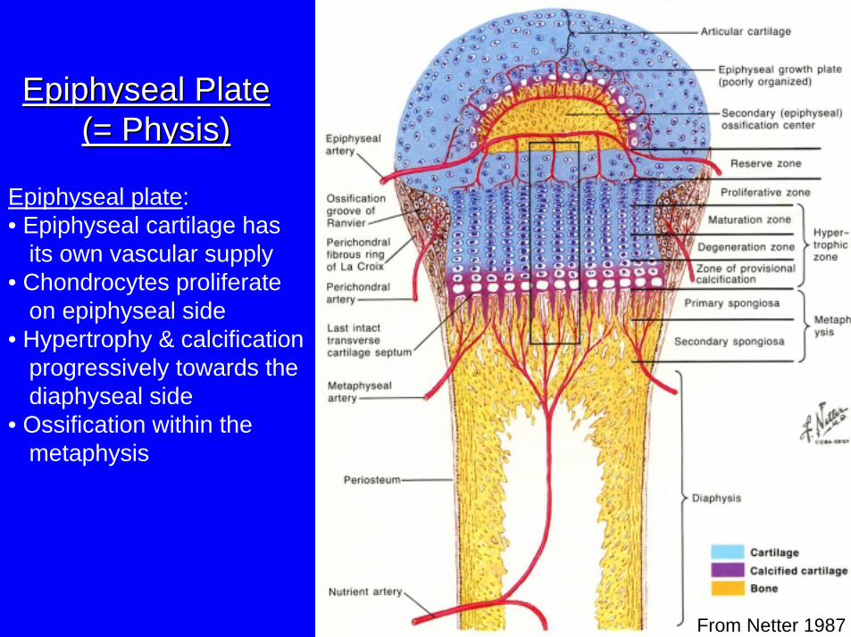
Epiphyseal PlateEpiphyseal Plate(= Physis)(= Physis)
Epiphyseal plate:• Epiphyseal cartilage has
its own vascular supply• Chondrocytes proliferate
on epiphyseal side• Hypertrophy & calcification
progressively towards thediaphyseal side
• Ossification within the metaphysis
From Netter 1987
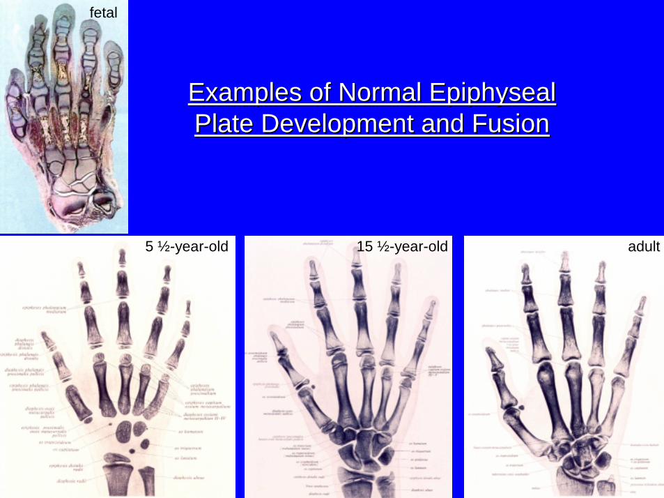
5 ½-year-old 15 ½-year-old adult
fetal
Examples of Normal Epiphyseal Examples of Normal Epiphyseal Plate Development and FusionPlate Development and Fusion
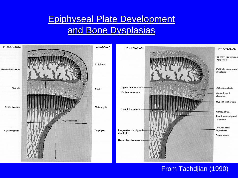
Epiphyseal Plate Development Epiphyseal Plate Development and Bone Dysplasiasand Bone Dysplasias
From Tachdjian (1990)
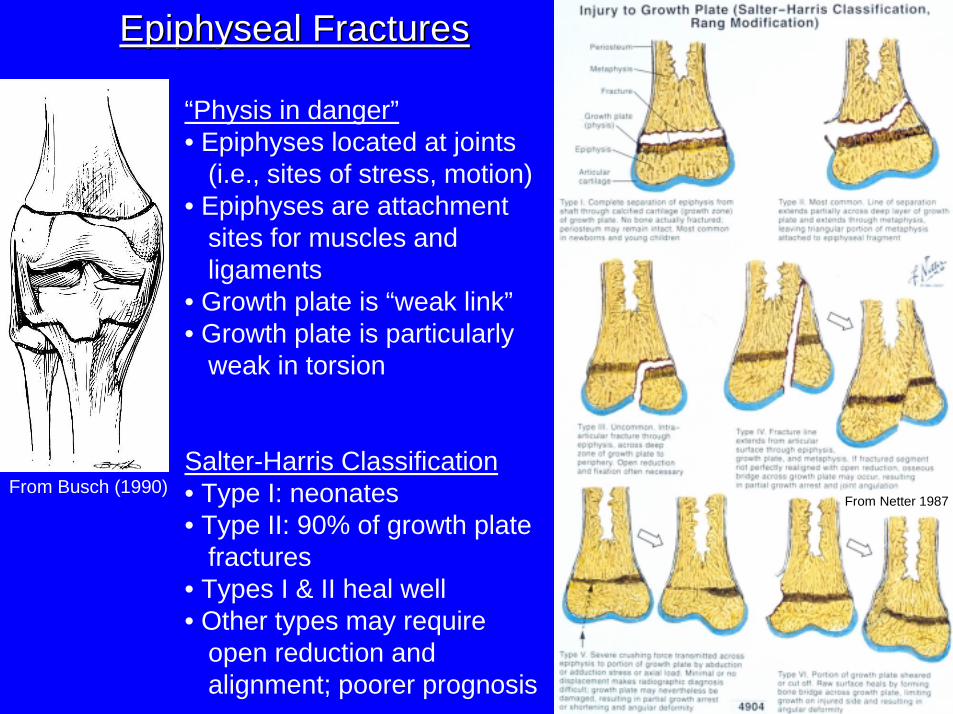
Epiphyseal FracturesEpiphyseal Fractures
From Netter 1987
“Physis in danger”• Epiphyses located at joints
(i.e., sites of stress, motion)• Epiphyses are attachment
sites for muscles and ligaments
• Growth plate is “weak link”• Growth plate is particularly
weak in torsion
Salter-Harris Classification• Type I: neonates• Type II: 90% of growth plate
fractures• Types I & II heal well• Other types may require
open reduction and alignment; poorer prognosis
From Busch (1990)
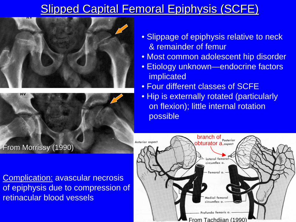
Slipped Capital Femoral Epiphysis (SCFE)Slipped Capital Femoral Epiphysis (SCFE)
From Morrissy (1990)From Morrissy (1990)
branch ofobturator a.
From Tachdjian (1990)
• Slippage of epiphysis relative to neck & remainder of femur
• Most common adolescent hip disorder• Etiology unknown—endocrine factors
implicated• Four different classes of SCFE• Hip is externally rotated (particularly
on flexion); little internal rotation possible
Complication: avascular necrosis of epiphysis due to compression of retinacular blood vessels
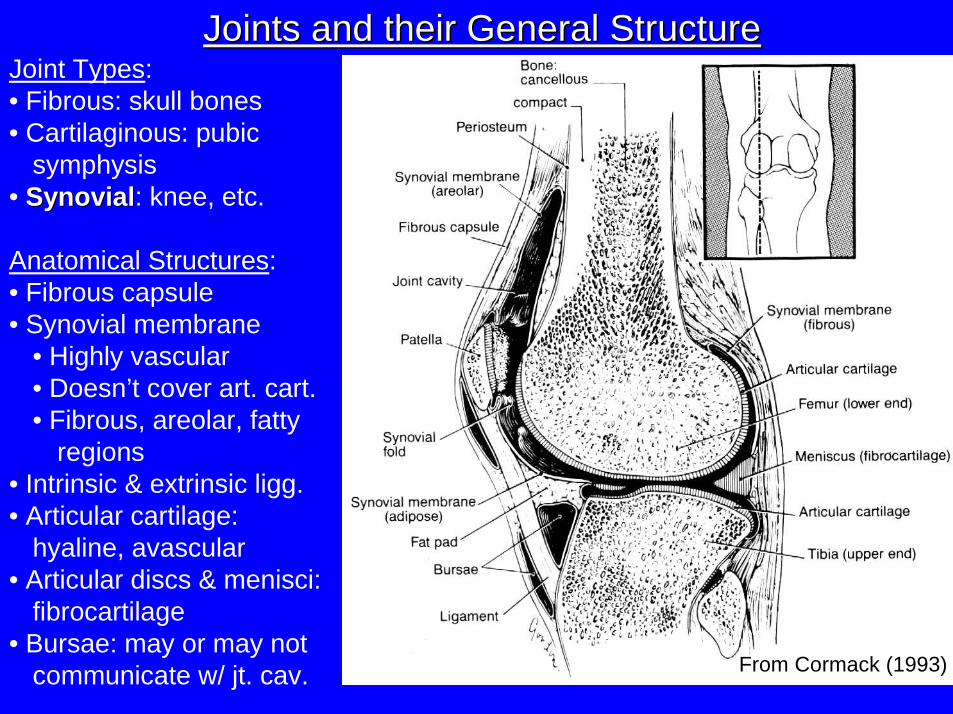
Joints and their General StructureJoints and their General StructureJoint Types:• Fibrous: skull bones• Cartilaginous: pubic
symphysis• SynovialSynovial: knee, etc.
Anatomical Structures:• Fibrous capsule• Synovial membrane
• Highly vascular• Doesn’t cover art. cart.• Fibrous, areolar, fatty
regions• Intrinsic & extrinsic ligg.• Articular cartilage:
hyaline, avascular• Articular discs & menisci:
fibrocartilage• Bursae: may or may not
communicate w/ jt. cav. From Cormack (1993)
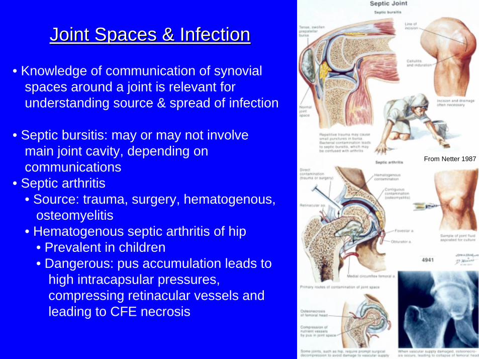
Joint Spaces & InfectionJoint Spaces & Infection
• Knowledge of communication of synovial spaces around a joint is relevant for understanding source & spread of infection
• Septic bursitis: may or may not involve main joint cavity, depending on communications
• Septic arthritis• Source: trauma, surgery, hematogenous,
osteomyelitis• Hematogenous septic arthritis of hip
• Prevalent in children• Dangerous: pus accumulation leads to
high intracapsular pressures, compressing retinacular vessels and leading to CFE necrosis
From Netter 1987
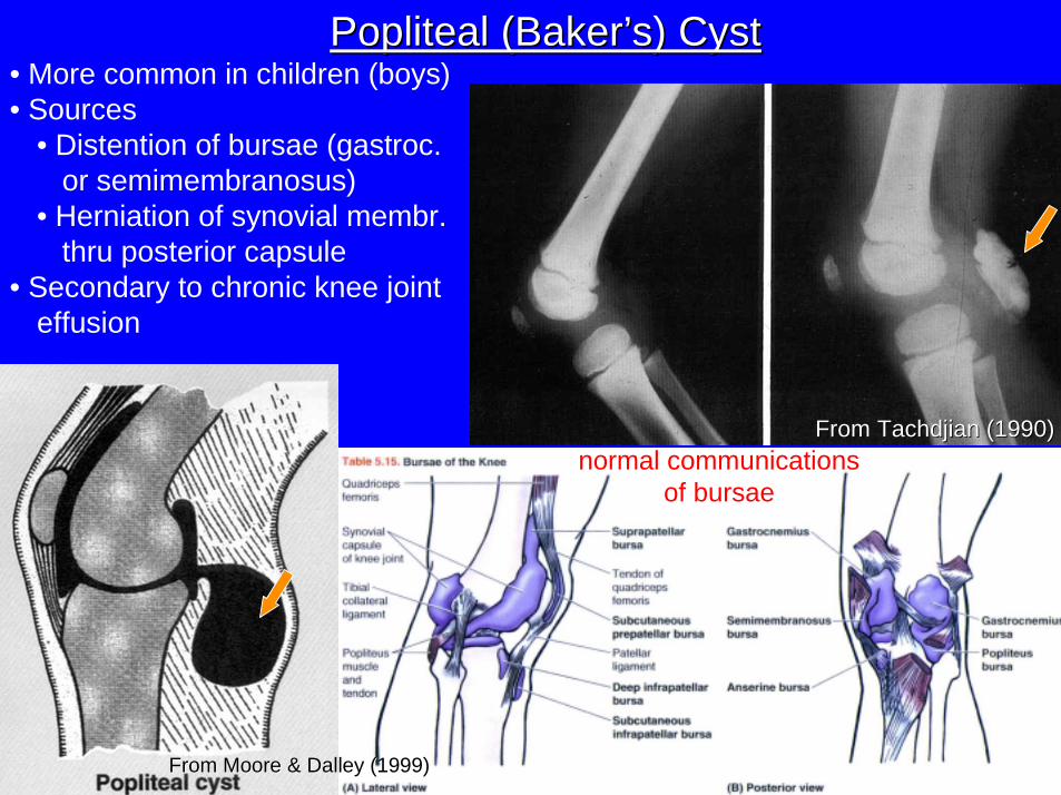
Popliteal (Baker’s) CystPopliteal (Baker’s) Cyst
From Moore & Dalley (1999)From Moore & Dalley (1999)
From Tachdjian (1990)From Tachdjian (1990)
• More common in children (boys)• Sources
• Distention of bursae (gastroc. or semimembranosus)
• Herniation of synovial membr. thru posterior capsule
• Secondary to chronic knee jointeffusion
normal communicationsof bursae
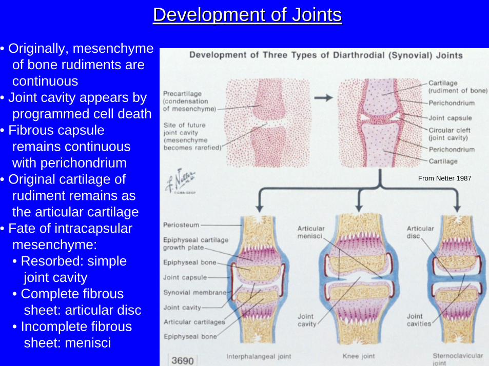
Development of JointsDevelopment of Joints
• Originally, mesenchyme of bone rudiments arecontinuous
• Joint cavity appears by programmed cell death
• Fibrous capsule remains continuous with perichondrium
• Original cartilage of rudiment remains as the articular cartilage
• Fate of intracapsular mesenchyme:• Resorbed: simple
joint cavity• Complete fibrous
sheet: articular disc• Incomplete fibrous
sheet: menisci
From Netter 1987
From Netter 1987
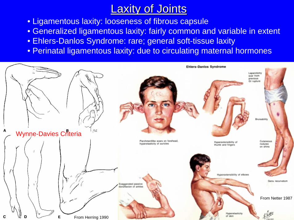
From Herring 1990
Wynne-Davies Criteria
Laxity of JointsLaxity of Joints• Ligamentous laxity: looseness of fibrous capsule• Generalized ligamentous laxity: fairly common and variable in extent• Ehlers-Danlos Syndrome: rare; general soft-tissue laxity• Perinatal ligamentous laxity: due to circulating maternal hormones
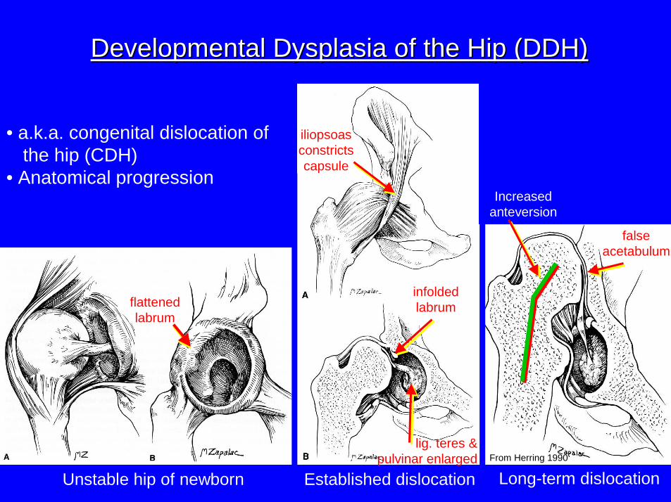
Unstable hip of newborn
flattenedlabrum
Established dislocation Long-term dislocation
iliopsoasconstrictscapsule
infoldedlabrum
falseacetabulum
Developmental Dysplasia of the Hip (DDH)Developmental Dysplasia of the Hip (DDH)
• a.k.a. congenital dislocation of the hip (CDH)
• Anatomical progression
lig. teres &pulvinar enlarged
Increasedanteversion
From Herring 1990
ReferencesReferencesBannister, L. H., et al. 1995. Gray’s Anatomy, 38th Edition. Churchill Livingstone,
New York.Busch, M. T. 1990. Sports medicine in children and adolescents, pp. 1091–1128
in R. T. Morrissy (ed.), Lovell and Winter’s Pediatric Orthopaedics, Volume 2, 3rd Edition. Lippincott, Philadelphia.
Cormack, D. H. 1993. Essential Histology. Lippincott, Philadelphia.Herring, J. A. 1990. Congenital dislocation of the hip, pp. 815-850 in R. T.
Morrissy (ed.), Lovell and Winter’s Pediatric Orthopaedics, Volume 2, 3rd
Edition. Lippincott, Philadelphia.Moore, K. L. and A. F. Dalley. 1999. Clinically Oriented Anatomy. Lippincott,
Williams, & Wilkins, Baltimore.Morrissy, R. T. 1990. Slipped capital femoral epiphysis, pp. 885–904 in R. T.
Morrissy (ed.), Lovell and Winter’s Pediatric Orthopaedics, Volume 2, 3rd
Edition. Lippincott, Philadelphia.Netter, F. H. 1987. The CIBA Collection of Medical Illustrations, Volume 8:
Musculoskeletal System. CIBA-Geigy, Summit.Staubesand, J. 1990. Sobotta Atlas of Human Anatomy. Urban &
Schwarzenberg, Baltimore.Tachdjian, M. O. 1990. Pediatric Orthopedics, Volumes 1–4, 2nd Edition. W. B.
Saunders, Philadelphia.Thompson, G. H. & P. V. Scoles. 1999. Bone and joint disorders, pp. 2055-2098
in Nelson’s Textbook of Pediatrics. Saunders, Philadelphia.