Embed Size (px)
Citation preview
Int. J. Adv. Res. Biol. Sci. (2018). 5(2): 44-65
44
International Journal of Advanced Research in Biological SciencesISSN: 2348-8069
www.ijarbs.comDOI: 10.22192/ijarbs Coden: IJARQG(USA) Volume 5, Issue 2 - 2018
Research Article
Gross Anatomical, Radiographic and Ultra-structuralIdentification of Splenic Vasculature in some Ruminants
(Camel, Buffalo Calf, Sheep and Goat)
Nawal, A. Noor and Maher, M.A.Anatomy & Embryology Department, Faculty of Veterinary Medicine, Cairo University, Egypt
*Corresponding author: [email protected]
Abstract
The current investigation was carried out on the spleens of camel, buffalo calf, sheep and goat. The number of spleens used foreach species was sixteen spleens divided as the following: five spleens for latex technique, five spleens for corrosion cast, fivespleens for radiographic technique and one spleen for histological and scanning electron microscopy. The spleen of camel hastwo hilus, four segments, two splenic arteries and two splenic veins while the other species having only one small hilus. Therewas no arterial anastomosis in the spleen of camel only resulting in the presence of arterial segmentation but there wasa venousconnection between the dorsal and intermediate segments in camel resulting in the absence of venous segmentation between thesesegments while the other species studied having an obvious arterial anastomosis and absence of the venous one. The aim of thisstudy is to illustrate the vascular segmentation of spleen through the vascular anastomosis which is of great importance invariable surgical applications as sample biopsy, partial or complete removal of spleen and localization of vascular emboli and toidentify by using the aid of scanning electron microscope, the sinusal or non-sinusal types of spleens.
Keywords: Spleen, Ruminants, Radiography, Scanning, Vascular segmentation, Sinusal type.
Introduction
The role of the spleen as a haemopiotic andimmunologic organ, especially for meat producinganimals as ruminants, gives it a great interest of manyinvestigators in anatomy and surgery. However, fordefense, spleen is the major secondary lymphaticorgan structurally specialized to filter, retain and dealwith the blood borne pathogens. In addition, it has theability to add to the erythrocyte and granulocytepopulation and can be acting as a reservoir of redblood cells during periods of unusual demand (Pabst,1993and Onkar and Govardhan, 2013) in camel.Histologically, the spleen classified by many authorsinto defensive and storage types (Banks,1993)according to trabeculae and smooth muscle
fibers, others into sinusal and non-sinusal types(Dellman and Brown, 1981) according to the type ofpost capillary vessels. There are sinusal type spleens,e.g. in horses, dogs and pigs (Brown and Dellmann,1976), man, rats and rabbits (Blue and Weiss, 1981b)and non-sinusal types, e.g. in cats, ruminants (Brownand Dellmann, 1976) and mice (Blue and Weiss,1981a) while (Bareedy et al., 1982 and Zidan et al.,2000) confirmed that the camel spleen classified as ofa sinusal type.
In this our investigation, we try to understand andavoiding the generally mis-talking a word of "sinusalor non-sinusal" spleens on all ruminants, so, this study
DOI: http://dx.doi.org/10.22192/ijarbs.2018.05.02.006
Int. J. Adv. Res. Biol. Sci. (2018). 5(2): 44-65
45
purposed to strictly knowing whose of studiedruminants of sinusal and whose of non-sinusal typespleens, in addition to anatomical segmentation andparenchymal distribution of splenic blood vesselswhich is of great importance in surgical localization ofvascular emboli, sample biopsy or for partial andcomplete removal of spleen.
Materials and Methods
The present investigation was conducted on sixty fourspleens of two large ruminants; the camel and buffalocalf and two small ruminants; the sheep and goat,obtaining sixteen spleens for each individual animal.The specimens were obtained fresh after slaughteringfrom the abattoir near Giza governorate.
Three different techniques were used in ourinvestigation; colored latex injection, corrosion castand radiography of the splenic vasculature throughusing five spleens from each species for eachtechnique and one spleen for microscopicexamination.
Each spleen was perfused with normal saline throughsplenic artery to remove blood from blood vessels.Later the spleen was infused with 10% bufferedformalin through splenic artery and splenic vein wasthen ligated. The splenic artery and vein were exposedand cannulated using a suitablecannula or catheteraccording to the size of vessels.
For latex technique, injecting suitable amount of 60%gum milk latex (diluted with ammonia) which coloredred for the artery and blue for the vein with Rotring®ink.
For corrosion cast technique, using (KEMAPOXY150 2A/1B); a transparent chemical mixture of thin(B) and thick (A) reagents obtained from chemicalmodern building (CMB) and mixed together in ratio of2A:1B then injected immediately before solidificationafter adding the color.
For radiography, we used two radio-opaque material;few milliliters of urograffin (75%) diluted 3:1 withwater (El-Zomor, 1991) then radiographedimmediately using 52 KVP 48 MA, 0.5 second andFFD70 cm. others were injected with red lead oxideand turpentine oil (Hagras, 1982) then radiographedimmediately using 55 KVP 30-70 MA, 0.5 second andFFD70 cm.
For scanning electron microscopy (SEM), small piecesof 1cm³ from each spleen at different regions weretaken fresh after slaughter and put in a fixativecontaining 2.5% glutheraldehyde in 0.1 M phosphatebuffer and adjusted to pH 7.4 then dehydrated inascending concentrations of ethanol using automatictissue processor (Leica EM TP).The dried specimenswere fixed on a metal stub with conductive pasteholding the fractured surface upward. In order toobtain sufficient electrical conductivity and yield ofsecondary electrons, the fractured surface of thespecimen was doubly coated by vacuum evaporatedcarbon and ion-sputtered gold (SPI-module-sputtercoater). The specimens were rotated and tilted duringthe evaporation. All the specimens were examined byscanning electron microscopy (JEOLJSM-5500 LV)JEOL Ltd, Japan, in 18 micrographs by using lowvaccum mode at the Regional Center of Mycologyand Biotechnology (RCMB), Al-Azhar University,Cairo, Egypt.
The obtained results were photographed using a digitalphoto camera Nikon COOLPIX L310 14.1Megapixels in 10photos. The nomenclature used wasthat recommended by the Nomina AnatomicaVeterinaria, 5th edition by (Frewein and Habel, 2012)as well as the previous literatures.
Results and Discussion
Anatomical Findings:
The current investigation revealed that the spleen ofcamel was C-shaped with a thin serrated cranialborder(fig.1a/h) and a thick wide fatty caudalborder(fig.1a/i) and blunt dorsal and ventralextremities, the thickness was not uniform, appearedthick at midpoint and hilus but thin at edges. Thesurface of the camel spleen was rough. It extendingfrom the caudodorsal part of rumen to the caudal endof left kidney and maintained on a ligamentoussystem; the reno-splenic ligament (fig.1a/e) and thesplenopancreatic ligament(fig.1a/f), the latter beingattached to the caudal thick margin of the spleen. Thespleen is also strongly related to rumen by means of arather short ligament, the gastrosplenic ligament. Aresult which was similar to that recorded by(Bennoune and Al-Samarrae, 2012 and Maina et al.,2014) in camel.
Int. J. Adv. Res. Biol. Sci. (2018). 5(2): 44-65
46
Our results were in agreement with that found by(Maina et al., 2014)and (Usende et al.,2014)that thecamel spleen was dark brown in color contrasting thebright red color in Nigeria indigenous pig and lightbrown colour in West African dwarf goats. They alsoadded that the C-shaped and blunt edge spleen ofcamel also contrast the slipper-shaped of that pig,quadrangular-shaped of that goat, tongue-shaped ofdog by (Onkar and Govardhan, 2013), a falciform-shape of horse by (FozFilho et al., 2013)and triangularcomma-shaped of the Bangladesh horse by (Alam etal., 2005).
The spleen of camel in this study has a length of 22.5to 31.5 cm, a width in the middle of 8.5 to 14cm and amaximum thickness from 1.25 to 2.5 cm. a resultwhich not simulates that recorded by (Bennoune andAl-Samarrae, 2012) that the camel spleen has alength of 17.5 to 28 cm, a width in the middle of 6.25to 12 cm and a maximum thickness from 1.84 to 4.96cm. and also differed than that reported by (Maina etal., 2014) that the camel spleen weighing of0.425±0.04kg.,while in our results weighing about0.837±0.03kg.
The splenic arterial vasculature of the camelsinvestigated was ensured by two branches of thesplenic artery; the body and the ventral extremity werevascularized by the first branch of the splenic artery(fig.1a/1). The second branch (fig.1a/7) ensured theirrigation of the dorsal end of the spleen. The hilum ofthe spleen was of broad type, the arteries and theirbranches penetrated at different points of the dorsalend to the middle of the caudal margin with thepresence of an enough fatty tissue that completelysurrounds the blood vessels.
Our observations revealed that the arterial vascularpattern of the camel spleen throughout its ventral twothirds was perpendicular to its long axis while beingparallel to the same axis in the dorsal third of thespleen. In addition, Gupta et al. (1978a through 1979)in the buffalo, dog, goat and sheep respectively andFozFilho et al. (2013) in horse reported that thevascular pattern was parallel to long axis of the spleenthroughout the entire length of the organ. Moreover,Gupta et al. also described two to three arterialsegments in the spleen of buffalo and goat and onlytwo in that of sheep and dog. Finally, the camel spleenrevealed cranial, middle and caudal arterial segmentsby Abu-Zaid et al. (1985) in camel while Osman etal. (1981)in ox, sheep, camel, pig and dog; simulateour results by describing them as dorsal(fig.1b/a),
intermediate(fig.1a/b) and ventral (fig.1a/d)arterialsegments.
All the previous literatures agreed that the arterialsupply of the camel spleen divided according to itssize and area of distribution into major or "first" andminor or "second" splenic arteries. The major splenicartery originating from the celiac trunk, it was largerthan the minor one and early gives rise to main twobranches(fig.1a/3,4) which running in thesplenopancreatic fold (fig.1a/f) and penetrate into thespleen at the level of the caudal thick margin, thesplenic hilus.
The major splenic artery (fig.1a/1) was responsible forblood irrigation of more than two third of the spleen,Then proceeds caudally and at the middle of pancreas(fig.1a/g), divided into two primary branches, thecranial (fig.1a/3) of which proceeds toward the middlethird of the spleen to which it gives 3-4smallerbranches (fig.1a/9). The caudalone (fig.1a/4) precedescaudally toward the posterior third of the spleen towhich it detaches 5-6 rami before it gains the posteriorsplenic hilus. The primary branches of this branchirrigate a well-defined segment without anastomosiswith the other branches; the segments were separatedby relatively avascular planes. A result which was inagreement with that found by (Abu-Zaid et al.,1985,Bennouneand Al-Samarrae, 2012, and Mainaet al., 2014) in camel and (FozFilho et al., 2013) inhorse.
Our results were in accordance with that found byAbu-Zaid et al. (1985) in camel that the entrance siteof the cranial branch of the major splenic artery wasvariable; it might be in front of the dorsal end orbetween the dorsal end and middle of the caudalmargin and supporting the intermediate segment ofspleen. The caudal branch was located just after thepenetration of cranial one, where it gives twosecondary dorsal and ventral arteries, supporting –from ventral to dorsal- two splenic segments; theventral segment and the additional segment (fig.1a and4b/9`).On the other hand, our results were disagreedwith Bennoune and Al-Samarrae (2012) whodescribed that the additional segment, which locatedbetween the ventral and the intermediate segments,irrigated by an artery from the cranial branch of themajor splenic artery and not from the caudal one.
Int. J. Adv. Res. Biol. Sci. (2018). 5(2): 44-65
47
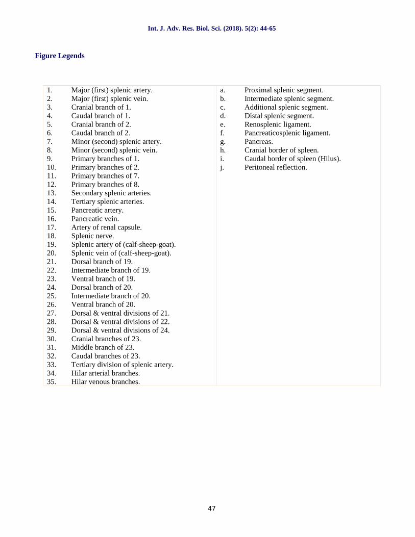
Figure Legends
1. Major (first) splenic artery.2. Major (first) splenic vein.3. Cranial branch of 1.4. Caudal branch of 1.5. Cranial branch of 2.6. Caudal branch of 2.7. Minor (second) splenic artery.8. Minor (second) splenic vein.9. Primary branches of 1.10. Primary branches of 2.11. Primary branches of 7.12. Primary branches of 8.13. Secondary splenic arteries.14. Tertiary splenic arteries.15. Pancreatic artery.16. Pancreatic vein.17. Artery of renal capsule.18. Splenic nerve.19. Splenic artery of (calf-sheep-goat).20. Splenic vein of (calf-sheep-goat).21. Dorsal branch of 19.22. Intermediate branch of 19.23. Ventral branch of 19.24. Dorsal branch of 20.25. Intermediate branch of 20.26. Ventral branch of 20.27. Dorsal & ventral divisions of 21.28. Dorsal & ventral divisions of 22.29. Dorsal & ventral divisions of 24.30. Cranial branches of 23.31. Middle branch of 23.32. Caudal branches of 23.33. Tertiary division of splenic artery.34. Hilar arterial branches.35. Hilar venous branches.
a. Proximal splenic segment.b. Intermediate splenic segment.c. Additional splenic segment.d. Distal splenic segment.e. Renosplenic ligament.f. Pancreaticosplenic ligament.g. Pancreas.h. Cranial border of spleen.i. Caudal border of spleen (Hilus).j. Peritoneal reflection.
Int. J. Adv. Res. Biol. Sci. (2018). 5(2): 44-65
48
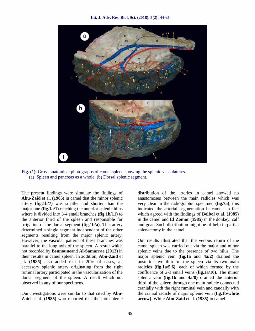
Fig. (1). Gross anatomical photographs of camel spleen showing the splenic vasculatures.(a) Spleen and pancreas as a whole. (b) Dorsal splenic segment.
The present findings were simulate the findings ofAbu-Zaid et al. (1985) in camel that the minor splenicartery (fig.1b/7) was smaller and shorter than themajor one (fig.1a/1) reaching the anterior splenic hiluswhere it divided into 3-4 small branches (fig.1b/11) tothe anterior third of the spleen and responsible forirrigation of the dorsal segment (fig.1b/a). This arterydetermined a single segment independent of the othersegments resulting from the major splenic artery.However, the vascular pattern of these branches wasparallel to the long axis of the spleen. A result whichnot recorded by Bennouneand Al-Samarrae (2012) intheir results in camel spleen. In addition, Abu-Zaid etal. (1985) also added that in 20% of cases, anaccessory splenic artery originating from the rightruminal artery participated in the vascularization of thedorsal segment of the spleen. A result which notobserved in any of our specimens.
Our investigations were similar to that cited by Abu-Zaid et al. (1985) who reported that the intrasplenic
distribution of the arteries in camel showed noanastomoses between the main radicles which wasvery clear in the radiographic specimen (fig.7a), thisindicated the arterial segmentation in camels, a factwhich agreed with the findings of Bolbol et al. (1985)in the camel and El Zomor (1985) in the donkey, calfand goat. Such distribution might be of help in partialsplenectomy in the camel.
Our results illustrated that the venous return of thecamel spleen was carried out via the major and minorsplenic veins due to the presence of two hilus. Themajor splenic vein (fig.1a and 4a/2) drained theposterior two third of the spleen via its two mainradicles (fig.1a/5,6), each of which formed by theconfluence of 2-3 small veins (fig.1a/10). The minorsplenic vein (fig.1b and 4a/8) drained the anteriorthird of the spleen through one main radicle connectedcranially with the right ruminal vein and caudally withthe cranial radicle of major splenic vein (fig.1b/whitearrow). While Abu-Zaid et al. (1985) in camel
Int. J. Adv. Res. Biol. Sci. (2018). 5(2): 44-65
49
reported that the minor vein drained the anterior thirdof the spleen through two main radicles and also addedthat in 20% of cases, the minor splenic vein joined themajor one instead of its union with the right ruminalvein and thus, a single splenic vein is formed. A resultwhich not simulate our observations.
The spleen of buffalo calf in the current study has anaverage length of 34.5 to 38.5 cm and a width of 12.5to 14.5 cm while the thickness at the middle part of 2to 2.5 cm and weighing fresh about 0.784±0.05kg. Ithas a bright purple color, elongated elliptical in shapewith dorsal broad border and narrow rounded ventralend while the caudal border was convex and slightlyhigher than the concave cranial border whichcontained the hilus in its proximal point.
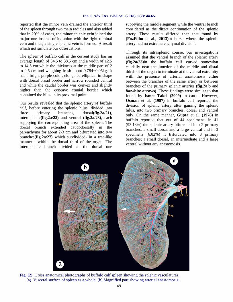
Our results revealed that the splenic artery of buffalocalf, before entering the splenic hilus, divided intothree primary branches, dorsal(fig.2a/21),intermediate(fig.2a/22) and ventral (fig.2a/23), eachsupplying the corresponding area of the spleen. Thedorsal branch extended caudodorsally in theparenchyma for about 2-3 cm and bifurcated into twobranches(fig.2a/27) which subdivided- in a tree-likemanner - within the dorsal third of the organ. Theintermediate branch divided as the dorsal one
supplying the middle segment while the ventral branchconsidered as the direct continuation of the splenicartery. These results differed than that found by(FozFilho et al., 2013)in horse where the splenicartery had no extra parenchymal division.
Through its intrasplenic course, our investigationsassumed that the ventral branch of the splenic artery(fig.2a/23)in the buffalo calf curved somewhatcaudally near the junction of the middle and distalthirds of the organ to terminate at the ventral extremitywith the presence of arterial anastomosis eitherbetween the branches of the same artery or betweenbranches of the primary splenic arteries (fig.2a,b and8a/white arrows). These findings were similar to thatfound by Ismet Takci (2009) in cattle. However,Osman et al. (1987) in buffalo calf reported thedivision of splenic artery after gaining the splenichilus, into two primary branches, dorsal and ventralonly. On the same manner, Gupta et al. (1978) inbuffalo reported that out of 44 specimens, in 41(93.18%) the splenic artery bifurcated into 2 primarybranches; a small dorsal and a large ventral and in 3specimens (6.82%) it trifurcated into 3 primarybranches; a small dorsal, an intermediate and a largeventral without any anastomosis.
Fig. (2). Gross anatomical photographs of buffalo calf spleen showing the splenic vasculatures.(a) Visceral surface of spleen as a whole. (b) Magnified part showing arterial anastomosis.
Int. J. Adv. Res. Biol. Sci. (2018). 5(2): 44-65
50
Our observations were in agreement with Osman et al.(1987) in buffalo calf, that alongthe course of theventral splenic branch, it gave off cranial and caudalbranches. The cranial branches (fig.2a/30) rangedfrom 6-10 in number extending in a cranioventraldirection to ramify within the corresponding part ofthe organ. They increased in length as traced ventrally.The caudal branches (fig.2a/32) ranged from 6-8 innumbers and supply the greater caudal part of thespleen. They extended mostly parallel to each otherobliquely in a caudoventral direction.
The splenic vein (fig.2a/20) in buffalo calf entered thehilus of the organ caudal to the artery (fig.2a and5b/19). The primary branches of the splenic vein(fig.2a and 5a,b/24,25,26) were accompanied to theartery, while their further ramifications did not followthe distribution pattern of the artery. Osman et al.(1987) also added that, a large number of arterial,venous and arterio-venous anastomoses had beenobserved in different parts of the organ revealing thatthe partial splenectomy was not recommended inbuffalo calves. A result which simulate ourobservations in the terminal arteries (fig.2a,b and8a/white arrows)in this study.
Osman et al. (1987)in buffalo calf had cited that,simulating the results of Happich (1961) in sheep;Wilkens and Munster (1976) in cattle; and Osman etal. (1985) in sheep and cattle, their work revealed thedivision of the splenic vessels inside the organ. On theother hand, Bolbol et al. (1985) in the buffalo andIsmet Takci (2009) in cattle mentioned that thesplenic vessels divided into cranial and caudalbranches 5-10 cm before the splenic hilus. A resultwhich had been revealed by the present work inbuffalo calves. On this basis, they concluded thepossibility of partial splenectomy in the buffalo calf.
The spleen of sheep in the current study has anaverage length of 12.5 to 14.5 cm and a width - at themiddle of both the cranial and caudal borders – of 5.5to 7 cm while the thickness at the middle part of 0.75to 1.22 cm and weighing fresh about 0.328±0.04kg. Ithas a purple blue color, triangular in shape with a widebroad dorsal border and a pointed ventral apex, cranialconvex border directed caudoventrally and nearlystraight caudal border with some corrugations. Thesplenic hilus appeared as small rounded area at thecranial border just below the proximal extremity by1.5-2 cm.
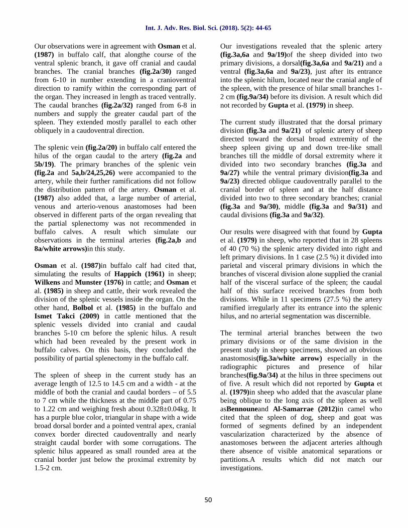
Our investigations revealed that the splenic artery(fig.3a,6a and 9a/19)of the sheep divided into twoprimary divisions, a dorsal(fig.3a,6a and 9a/21) and aventral (fig.3a,6a and 9a/23), just after its entranceinto the splenic hilum, located near the cranial angle ofthe spleen, with the presence of hilar small branches 1-2 cm (fig.9a/34) before its division. A result which didnot recorded by Gupta et al. (1979) in sheep.
The current study illustrated that the dorsal primarydivision (fig.3a and 9a/21) of splenic artery of sheepdirected toward the dorsal broad extremity of thesheep spleen giving up and down tree-like smallbranches till the middle of dorsal extremity where itdivided into two secondary branches (fig.3a and9a/27) while the ventral primary division(fig.3a and9a/23) directed oblique caudoventrally parallel to thecranial border of spleen and at the half distancedivided into two to three secondary branches; cranial(fig.3a and 9a/30), middle (fig.3a and 9a/31) andcaudal divisions (fig.3a and 9a/32).
Our results were disagreed with that found by Guptaet al. (1979) in sheep, who reported that in 28 spleensof 40 (70 %) the splenic artery divided into right andleft primary divisions. In 1 case (2.5 %) it divided intoparietal and visceral primary divisions in which thebranches of visceral division alone supplied the cranialhalf of the visceral surface of the spleen; the caudalhalf of this surface received branches from bothdivisions. While in 11 specimens (27.5 %) the arteryramified irregularly after its entrance into the splenichilus, and no arterial segmentation was discernible.
The terminal arterial branches between the twoprimary divisions or of the same division in thepresent study in sheep specimens, showed an obviousanastomosis(fig.3a/white arrow) especially in theradiographic pictures and presence of hilarbranches(fig.9a/34) at the hilus in three specimens outof five. A result which did not reported by Gupta etal. (1979)in sheep who added that the avascular planebeing oblique to the long axis of the spleen as wellasBennouneand Al-Samarrae (2012)in camel whocited that the spleen of dog, sheep and goat wasformed of segments defined by an independentvascularization characterized by the absence ofanastomoses between the adjacent arteries althoughthere absence of visible anatomical separations orpartitions.A results which did not match ourinvestigations.
Int. J. Adv. Res. Biol. Sci. (2018). 5(2): 44-65
51
Fig. (3). Gross anatomical photographs of sheep and goat spleens showing the splenic vasculatures.(a) Spleen of sheep.(b),(c),(d),(e),(f),(g)Spleens of goat.
Gupta et al. (1979) had cited that both Clausen(1958) and Gutierrez-Cubillos (1969) in sheepreported the presence of two to four arterial segmentsin the human spleen. However, in their study of thehuman spleen (Gupta et al., 1976) no specimenshowed more than three segments.They also addedthat the variation in the position of the arterialsegments in different species was perhaps associatedwith the position of the hilus and absence of thesplenic arterial segmentation was more common insheep than in goat and dog. In addition, all human andbuffalo spleens examined showed segmentation. Aresults which did not simulate our findings where thearterial anastomosis (fig.9a/white arrow) was veryclear in sheep and this resulted in the absence ofarterial segmentation.
The spleen of goat in the current study has an averagelength of 6.5 to 7.5 cm and a width of 3.5 to 5 cmwhile the thickness at the middle part of 0.85 to 1.5 cm
and weighing fresh about 0.195±0.02kg. It has a darkbluish brown color, rectangular in shape with roundedborders. The splenic hilus appeared as small roundedarea at the cranial border.
In agreement with Happich (1961) in sheep; Wilkensand Munster (1981) in cattle; Osman, El-Ayat andGeorge (1981) in sheep and cattle; El-Zomor (1985)in goat and calves; Dyce, Sack and Wensing (1987) inruminants; in addition to Osman et al. (1987) in thebuffalo calves, the splenic vessels passed undividedthrough a confined hilus and then divided inside thesplenic parenchyma. Otherwise, the splenic artery wasdivided extrasplenic 2-3 cm before reaching thesplenic hilus in 25% of the examined specimens whereit detached smaller branches at the region of hilus.This was in accordance with our results and thosegiven by Bolbol et al. (1985) in the buffalo and IsmetTakci (2009) in cattle.
Int. J. Adv. Res. Biol. Sci. (2018). 5(2): 44-65
52
In agreement with Gupta et al. (1978a,b) in thebuffalo and dog, El-Zomor (1985) in goat and Osmanet al. (1987) in the buffalo calves, the splenic artery ofgoat (fig.3e, 3g/19) was divided into two mainbranches, dorsal(fig.3d,3e and 3g/21) andventral(fig.3d,3e and 3g/23). While, Gupta et al.(1978c and 1979) in the goat and sheep respectively,named these two branches as right and left.
Our results revealed that the splenic artery of goat in11 specimens of 15 was divided before reaching thehilus(fig.3b,3d and 3c/19) by about 1-2 cm into twomain primary dorsal and ventral branches inclosing thesplenic vein(fig.3b/20) in-between.While it wasundivided in 4 specimens(fig.3g/19) entering the smallrounded splenic hilus on the visceral surface close tothe craniodorsal angle of the spleen. The arteryproceeded caudoventrally for about 2-3 cm, before itssplitting into a small dorsal branch (fig.3d,3e and3g/21) and a large ventral one(fig.3d,3e and 3g/23). Inaddition, Wally and Gad (1998) in goat reported thatonly in two specimens (25%), the division of thesplenic artery occurred extraparenchymal 2-3 cmbefore reaching the hilus.
In all our specimens in whom the splenic artery eitherdivided intra or extra splenic hilus, there were capsularbranches (fig.3e and 3f/34) arborizing at the roundedregion of the hilus. Otherwise, the intra parenchymalpart of splenic artery showed only in two specimens, ahilar branch (fig.6b/3 4) originating before its divisioninto primary dorsal and ventral branches, a resultwhich similar to that found by Ismet Takci (2009) incattle. While Gupta et al. (1978)in goat confirmedthat in 37specimens of 50 (74%), the splenic arterydivided into two main divisions only, right and left,but in 5 specimens (10%), it gave a hilar branch thendivided into right and left branches.
Gupta et al. (1978) observed thatin 8 specimens(16%) of goat spleens, the splenic artery, after itsentrance into the spleen, ramified irregularly. Thus noarterial segmentation was observed in thesespecimens. A result which was recorded only in 1 casein the current study revealing that the splenic arterydivided extra parenchymal into 4 branches(fig.3c/19),running 3 cm on the outer surface then directed intothe inside of the spleen which illustrated asdorsal(fig.3e/21), intermediate(fig.3e/22)and largeventral (fig.3e/23)divisions. The ventral primarydivision giving the cranial branch (fig.3e/30) earlierthen continued toward the caudoventral angle to divideinto middle(fig.3e/31) and caudal branches (fig.3e/32).
Our observations in the current study were similar tothat reported by Gupta et al. (1978)and Wally andGad (1998) in goatthat the dorsal branch(fig.3d/21) ofthe splenic artery was relatively small; it precededcaudodorsally parallel to the dorsal basal border of thespleen towards its caudal border, where it divided intotwo smaller branches(fig.3d/27) which nourished thecaudal angle. Moreover, along its course it released 3-6 dorsal and 4-7 ventral fine branches which weredistributed to the dorsal third of the spleen.
Our results observed that only in two cases of 15specimens, there was an artery originated at themidpoint of the ventral branch of splenic artery andbefore its division into two secondary branches(fig.6band 6c/22) that might be considered as a supportiveartery with the dorsal branch nourishing the proximalhalf of the goat spleen. In addition, another onespecimen showing the presence of only two divisionsof the primary ventral division, cranial (fig.3g/30) andcaudal branches(fig.3g/32) and absence of middlebranch which was compensated by small branchesfrom the cranial one. A result which not recorded byGupta et al. (1978) or Wally and Gad (1998)in goat.
The ventral branch was a large vessel and could beconsidered as the direct continuation of the splenicartery in goat. It preceded caudoventrally inside thesplenic parenchyma towards the ventral extremity.Along its course, it gave small tree-like branchesdirected cranial and caudal into the splenicparenchyma and after a distance of 1.5-3 cm becomedivided into two secondary branches each of themgiving about 6-8 cranial branches and 5-7 caudalbranches supplying the middle and ventral parts of thespleen(Wally and Gad, 1998). A results which werein accordance with our observations except that wefound the division of the ventral branch into threesmaller branches in the majority of cases and only onecase which recorded two branches of the ventraldivision(fig.3g/30,32).
There was numerous fine anastomoses observedbetween the dorsal and ventral branches(fig.10a/white arrows) of the splenic artery and, so, itwas difficult to divide the spleen of the goat into twoindependent arterial segments.A result which wasrecorded by Osman et al. (1987) in buffalo calves andWally and Gad (1998) in goat and similar to ourinvestigations but not recorded by Gupta et al.(1978c) in goat.
Int. J. Adv. Res. Biol. Sci. (2018). 5(2): 44-65
53
A segmental arrangement of the splenic vein has beendemonstrated in man (Fuld and Irwin, 1954) and indogs (Goldewski, Pelissier and Emberger, 1957).Such a finding was demonstrated in our examinedspecimens of "buffalo calve, sheep and goat" wherethe splenic vein follow the distribution pattern of theartery in some specimens (fig.5a,5b,6d,6f,8b,9b and
10b) and distributed irregularly in others (fig.6e/20)but without any venous anastomosis in these species.On the other hand, there was a venous connectionbetween the dorsal and middle segments in the spleenof camel (fig.1b and 7b/white arrows) which indicatethat there is no venous segmentation in camel but thearterial one is confirmed in the current study.
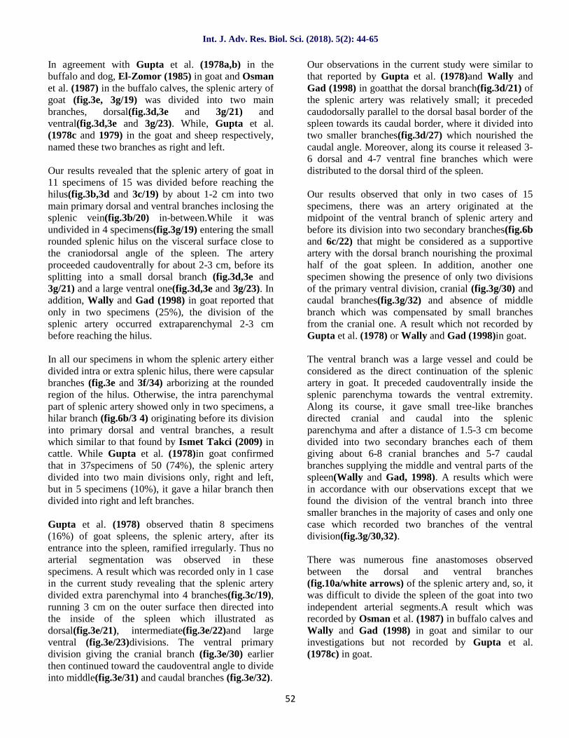
Fig. (4). Corrosion cast photographs of camel spleen showing the distribution of splenic vasculatures.(a) Dorsal splenic segment. (b) Intermediate, additional and ventral segments.
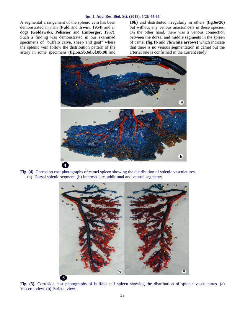
Fig. (5). Corrosion cast photographs of buffalo calf spleen showing the distribution of splenic vasculatures. (a)Visceral view. (b) Parietal view.
Int. J. Adv. Res. Biol. Sci. (2018). 5(2): 44-65
54
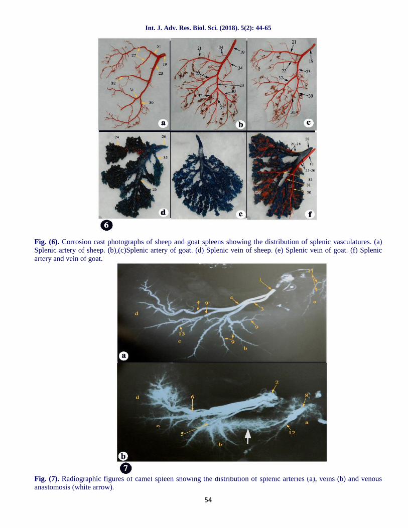
Fig. (6). Corrosion cast photographs of sheep and goat spleens showing the distribution of splenic vasculatures. (a)Splenic artery of sheep. (b),(c)Splenic artery of goat. (d) Splenic vein of sheep. (e) Splenic vein of goat. (f) Splenicartery and vein of goat.
Fig. (7). Radiographic figures of camel spleen showing the distribution of splenic arteries (a), veins (b) and venousanastomosis (white arrow).
Int. J. Adv. Res. Biol. Sci. (2018). 5(2): 44-65
55
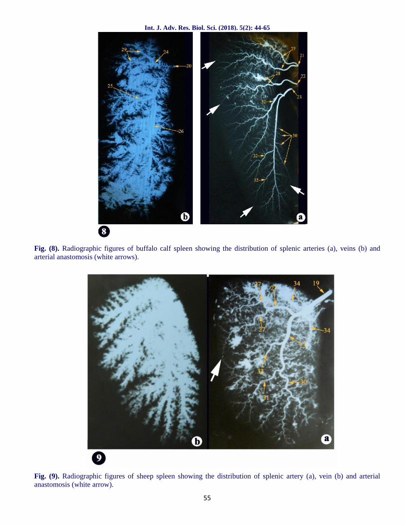
Fig. (8). Radiographic figures of buffalo calf spleen showing the distribution of splenic arteries (a), veins (b) andarterial anastomosis (white arrows).
Fig. (9). Radiographic figures of sheep spleen showing the distribution of splenic artery (a), vein (b) and arterialanastomosis (white arrow).
Int. J. Adv. Res. Biol. Sci. (2018). 5(2): 44-65
56
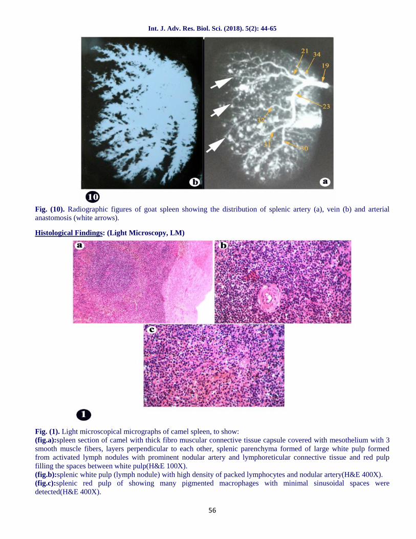
Fig. (10). Radiographic figures of goat spleen showing the distribution of splenic artery (a), vein (b) and arterialanastomosis (white arrows).
Histological Findings: (Light Microscopy, LM)
Fig. (1). Light microscopical micrographs of camel spleen, to show:(fig.a):spleen section of camel with thick fibro muscular connective tissue capsule covered with mesothelium with 3smooth muscle fibers, layers perpendicular to each other, splenic parenchyma formed of large white pulp formedfrom activated lymph nodules with prominent nodular artery and lymphoreticular connective tissue and red pulpfilling the spaces between white pulp(H&E 100X).(fig.b):splenic white pulp (lymph nodule) with high density of packed lymphocytes and nodular artery(H&E 400X).(fig.c):splenic red pulp of showing many pigmented macrophages with minimal sinusoidal spaces weredetected(H&E 400X).
Int. J. Adv. Res. Biol. Sci. (2018). 5(2): 44-65
57
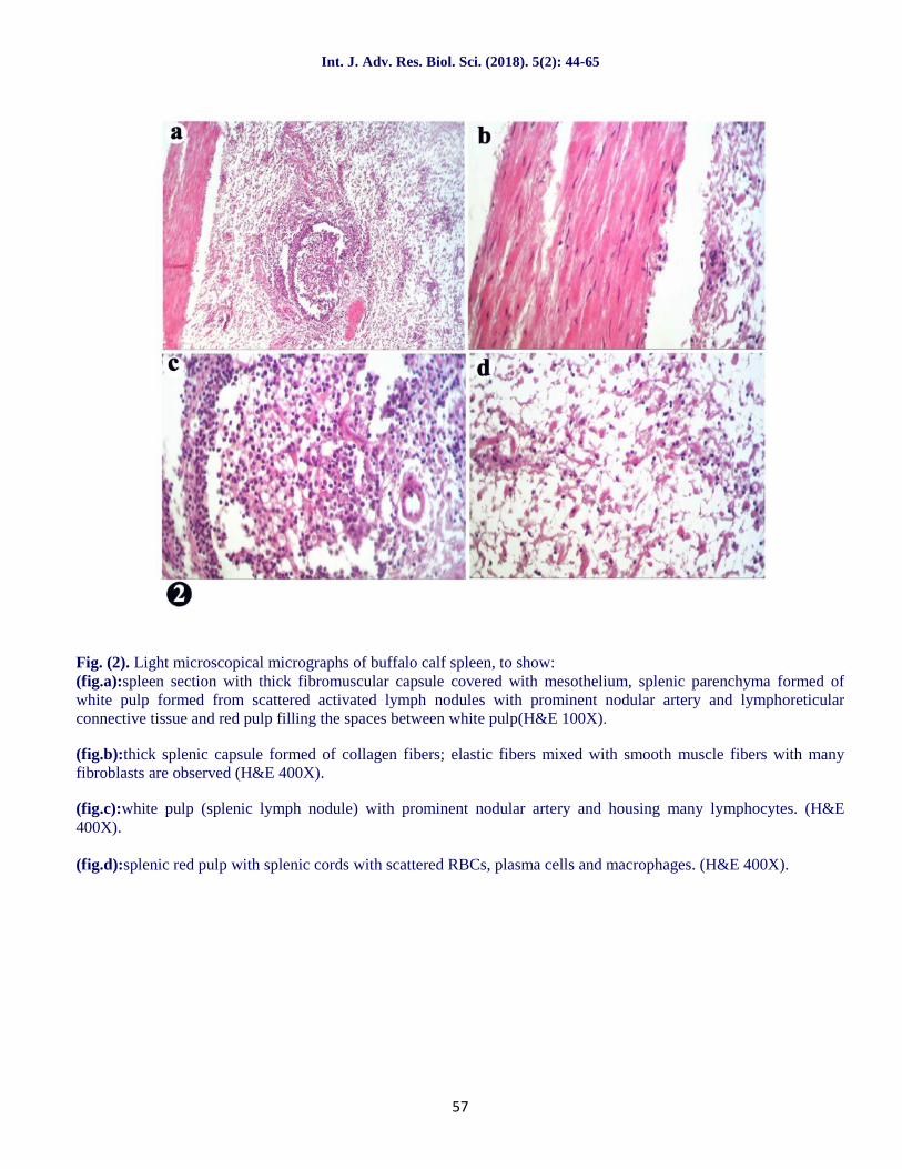
Fig. (2). Light microscopical micrographs of buffalo calf spleen, to show:(fig.a):spleen section with thick fibromuscular capsule covered with mesothelium, splenic parenchyma formed ofwhite pulp formed from scattered activated lymph nodules with prominent nodular artery and lymphoreticularconnective tissue and red pulp filling the spaces between white pulp(H&E 100X).
(fig.b):thick splenic capsule formed of collagen fibers; elastic fibers mixed with smooth muscle fibers with manyfibroblasts are observed (H&E 400X).
(fig.c):white pulp (splenic lymph nodule) with prominent nodular artery and housing many lymphocytes. (H&E400X).
(fig.d):splenic red pulp with splenic cords with scattered RBCs, plasma cells and macrophages. (H&E 400X).
Int. J. Adv. Res. Biol. Sci. (2018). 5(2): 44-65
58
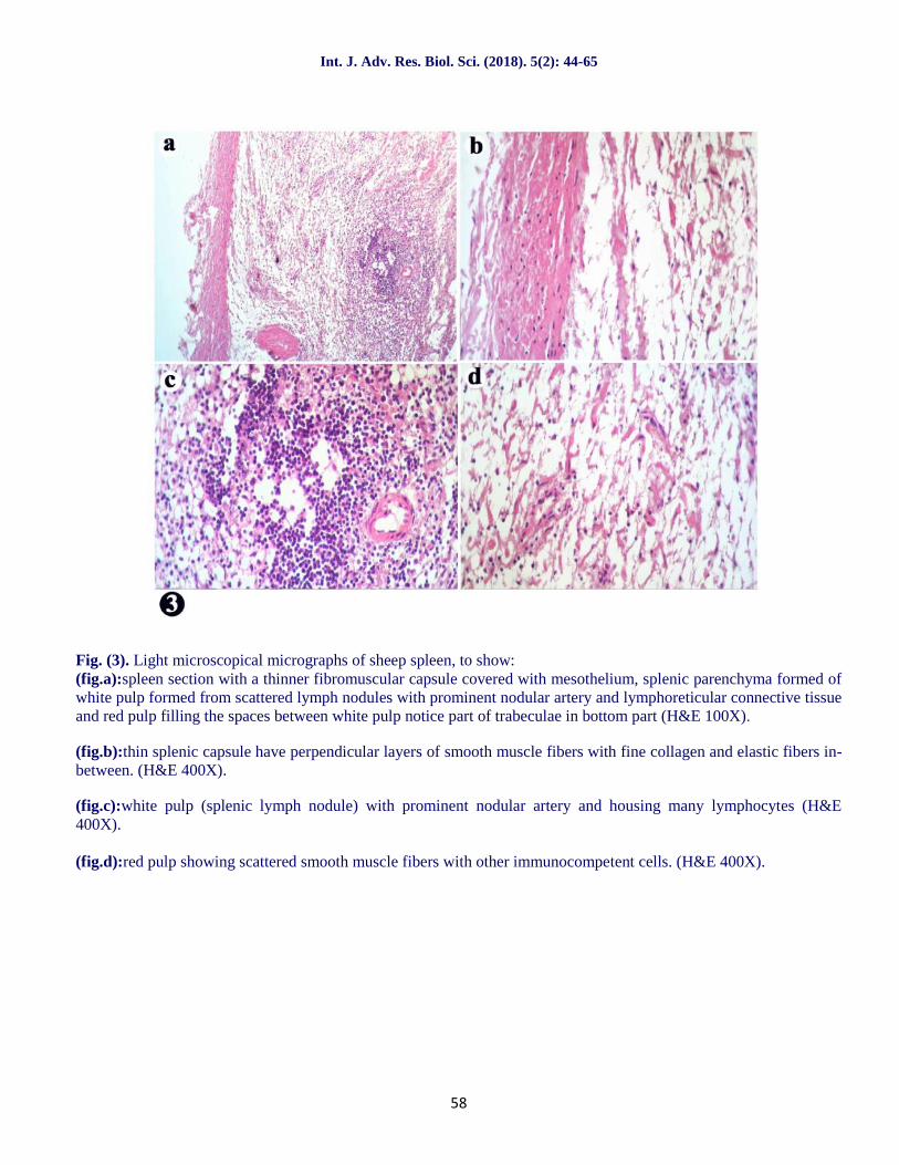
Fig. (3). Light microscopical micrographs of sheep spleen, to show:(fig.a):spleen section with a thinner fibromuscular capsule covered with mesothelium, splenic parenchyma formed ofwhite pulp formed from scattered lymph nodules with prominent nodular artery and lymphoreticular connective tissueand red pulp filling the spaces between white pulp notice part of trabeculae in bottom part (H&E 100X).
(fig.b):thin splenic capsule have perpendicular layers of smooth muscle fibers with fine collagen and elastic fibers in-between. (H&E 400X).
(fig.c):white pulp (splenic lymph nodule) with prominent nodular artery and housing many lymphocytes (H&E400X).
(fig.d):red pulp showing scattered smooth muscle fibers with other immunocompetent cells. (H&E 400X).
Int. J. Adv. Res. Biol. Sci. (2018). 5(2): 44-65
59
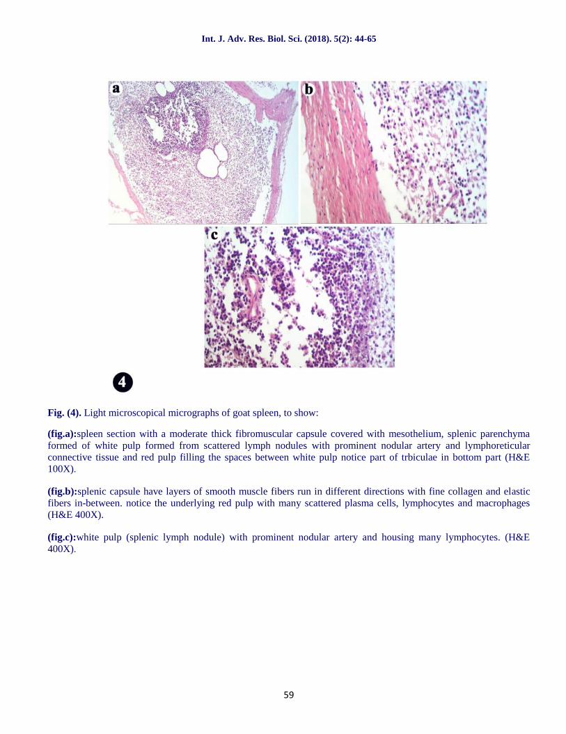
Fig. (4). Light microscopical micrographs of goat spleen, to show:
(fig.a):spleen section with a moderate thick fibromuscular capsule covered with mesothelium, splenic parenchymaformed of white pulp formed from scattered lymph nodules with prominent nodular artery and lymphoreticularconnective tissue and red pulp filling the spaces between white pulp notice part of trbiculae in bottom part (H&E100X).
(fig.b):splenic capsule have layers of smooth muscle fibers run in different directions with fine collagen and elasticfibers in-between. notice the underlying red pulp with many scattered plasma cells, lymphocytes and macrophages(H&E 400X).
(fig.c):white pulp (splenic lymph nodule) with prominent nodular artery and housing many lymphocytes. (H&E400X).
Int. J. Adv. Res. Biol. Sci. (2018). 5(2): 44-65
60
Histological Findings: (Scanning Electron Microscopy, SEM)
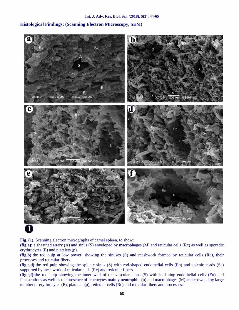
Fig. (1). Scanning electron micrographs of camel spleen, to show:(fig.a): a sheathed artery (A) and sinus (S) enveloped by macrophages (M) and reticular cells (Rc) as well as sporadicerythrocytes (E) and platelets (p).(fig.b):the red pulp at low power, showing the sinuses (S) and meshwork formed by reticular cells (Rc), theirprocesses and reticular fibers.(fig.c,d):the red pulp showing the splenic sinus (S) with rod-shaped endothelial cells (En) and splenic cords (Sc)supported by meshwork of reticular cells (Rc) and reticular fibers.(fig.e,f):the red pulp showing the inner wall of the vascular sinus (S) with its lining endothelial cells (En) andfenestrations as well as the presence of leucocytes mainly neutrophils (n) and macrophages (M) and crowded by largenumber of erythrocytes (E), platelets (p), reticular cells (Rc) and reticular fibers and processes.
Int. J. Adv. Res. Biol. Sci. (2018). 5(2): 44-65
61
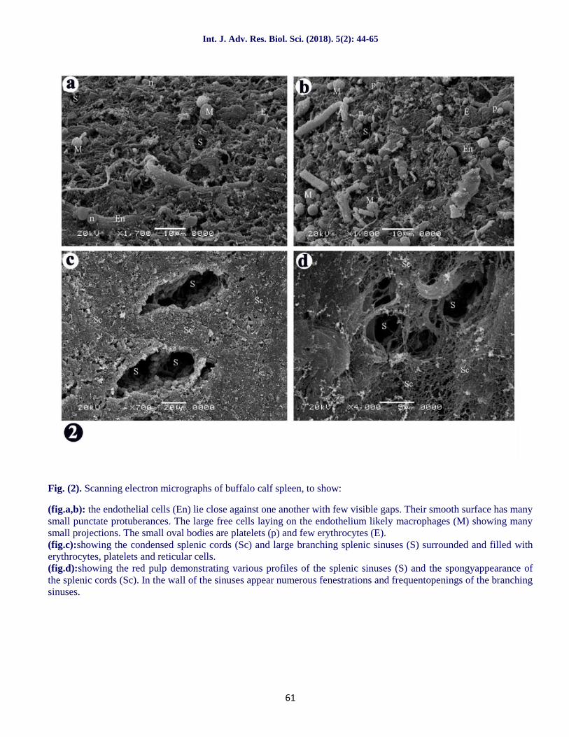
Fig. (2). Scanning electron micrographs of buffalo calf spleen, to show:
(fig.a,b): the endothelial cells (En) lie close against one another with few visible gaps. Their smooth surface has manysmall punctate protuberances. The large free cells laying on the endothelium likely macrophages (M) showing manysmall projections. The small oval bodies are platelets (p) and few erythrocytes (E).(fig.c):showing the condensed splenic cords (Sc) and large branching splenic sinuses (S) surrounded and filled witherythrocytes, platelets and reticular cells.(fig.d):showing the red pulp demonstrating various profiles of the splenic sinuses (S) and the spongyappearance ofthe splenic cords (Sc). In the wall of the sinuses appear numerous fenestrations and frequentopenings of the branchingsinuses.
Int. J. Adv. Res. Biol. Sci. (2018). 5(2): 44-65
62
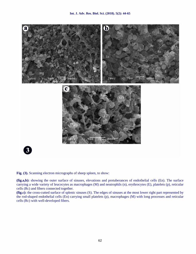
Fig. (3). Scanning electron micrographs of sheep spleen, to show:
(fig.a,b): showing the outer surface of sinuses, elevations and protuberances of endothelial cells (En). The surfacecarrying a wide variety of leucocytes as macrophages (M) and neutrophils (n), erythrocytes (E), platelets (p), reticularcells (Rc) and fibers connected together.(fig.c): the cross-cutted surface of splenic sinuses (S). The edges of sinuses at the most lower right part represented bythe rod-shaped endothelial cells (En) carrying small platelets (p), macrophages (M) with long processes and reticularcells (Rc) with well-developed fibers.
Int. J. Adv. Res. Biol. Sci. (2018). 5(2): 44-65
63
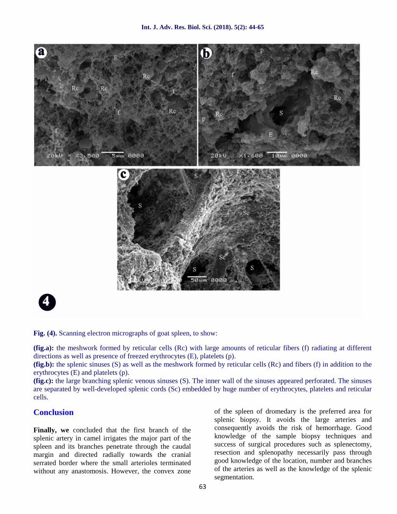
Fig. (4). Scanning electron micrographs of goat spleen, to show:
(fig.a): the meshwork formed by reticular cells (Rc) with large amounts of reticular fibers (f) radiating at differentdirections as well as presence of freezed erythrocytes (E), platelets (p).(fig.b): the splenic sinuses (S) as well as the meshwork formed by reticular cells (Rc) and fibers (f) in addition to theerythrocytes (E) and platelets (p).(fig.c): the large branching splenic venous sinuses (S). The inner wall of the sinuses appeared perforated. The sinusesare separated by well-developed splenic cords (Sc) embedded by huge number of erythrocytes, platelets and reticularcells.
Conclusion
Finally, we concluded that the first branch of thesplenic artery in camel irrigates the major part of thespleen and its branches penetrate through the caudalmargin and directed radially towards the cranialserrated border where the small arterioles terminatedwithout any anastomosis. However, the convex zone
of the spleen of dromedary is the preferred area forsplenic biopsy. It avoids the large arteries andconsequently avoids the risk of hemorrhage. Goodknowledge of the sample biopsy techniques andsuccess of surgical procedures such as splenectomy,resection and splenopathy necessarily pass throughgood knowledge of the location, number and branchesof the arteries as well as the knowledge of the splenicsegmentation.
Int. J. Adv. Res. Biol. Sci. (2018). 5(2): 44-65
64
According to the radiographic figures, the arterialanastomosis was very clear in all studied speciesexcept the camel while there was a clear venousconnection only in the camel between the dorsalsplenic segment and the intermediate segmentas wellas the presence of an additional segment between theintermediate and ventral segments which supplied by abranch from the caudal division of the major splenicartery, a result which characterizes the spleen ofcamel.
According to the figures of light microscope andscanning electron microscope, all the four speciesstudied were of sinusal type of spleen which wasillustrated by the numerous numbers of sinuses and theobvious occupation of the red pulp than the white oneand the amount of fibromuscular connective tissue.
References
Abu-Zaid, S.M.S., El-Khaligi, G.E.M. and El-Nahla, S.M.M. (1985): Some gross anatomicalstudies on the topography, arterial supply andvenous drainage of the spleen of the one humpedcamel (Camelus dromedaries) Alex. J. of Vet. Sci.,1, No. 2: 45-59.
Alam, M.S., Awal, M.A., Das, S.K. andIslam, M.N.(2005):Morphometry of spleen with specialemphasis on its arterial circulation of indigenoushorses in Bangladesh. Bangladesh, J. Vet. Med.,3(2): 166-168.
Banks, W. (1993):Applied Veterinary Histology. 3rd
ed. Mosby Year Book. p. 283-297.Bareedy, M., Anis, H., Ewais, M. andAmmer, S.
(1982): Some anatomical and histological studiesof the spleen of one humped camel(Camelusdromedarius) Egypt. J. Histol., 5(5): 5-83.
Bennoune, O. and AL-Samarrae, N.S.(2012):L’arbreartérielsplénique : application pourlamise en oeuvre de biopsie et de splenectomy chezle dromadaire (Camelus dromedarius)Revue Méd.Vét., 163, 10, 461-464.
Blue, J. andWeiss, L. (1981): Electron microscopy ofthe red pulp of dog spleen including vasculararrangements, pariatarial macrophages sheaths(ellipsoids) and the contractile, innervated reticularmeshwork, Am. J. Anat., 161: 189-218.
Brown, E. and Dellmann, H.D. (1976):Lymphaticsystem. In Textbook ofVeterinary Histology (Ed.H. D.Dellmann and E. Brown), pp.161-184.Philadelphia: Lea &Febiger.
Bolbol, A.E., Ali, A.M.A. and Ibrahim, I.A. (1985):Some radiographic studies on spleen of ruminants.First Int. Conf. App. Sci., Vol. II, 370-378.
Clausen, E. (1958): Cited by F. Goldby and R. J.Harrison (1961). In Recent Advances inAnatomy,2nd ed., p. 392. London: J. & A.Churchill Ltd.
Dellman, H.D. and Brown, E.M. (1981): Textbook ofVeterinary Histology. 2nd Ed.Philadelphia: Leaand Febiger. pp. 176-183.
Dyce, K.M., Sack, W.O. and Wensing, C.J.G.(1987): Text book of veterinary anatomy. W.B.Saunders, Philadelphia, London, Toronto,Montreal, Sydney, Tokya.
El-Zomor, S.T.S. (1985): Comparative studies onsplenectomy procedures in certain farm animals.M.V.Sc. Thesis, Cairo University.
El-Zomor, S.T.S. (1991): Studies on certain tendonsheaths and bursae in cattle and buffaloes. Ph.D.Thesis, Cairo University.
Flud, H. and Irwin, D.T. (1954): Clinical applicationof portal venography. Lancet, 1: 312-3313.
Golewski, M., Pelissier, M. and Emberger, J.M.(1957): Recent advances in anatomy. 2nd Edition,p. 392, London, J. and A. Churchill Ltd., Cited byGupta et al. (1976).
Gupta, C.D., Gupta, S.C., Arora, A.K.andSingh,P.J. (1976): Vascular segments in thehumanspleen. Journal ofAnatomy, 121, 613-616.
Gupta, S.C., Gupta, C.D., Arora, A.K.andGupta,S.B. (1978a): Vascular segments in thebuffalo(Bubalus bubalis) spleen. A study bycorrosion cast. AnatomischerAnzeiger 143, 493-495.
Gupta, S.C., Gupta, C.D.andGupta, S.B. (1978 b):Segmentation in the dog spleen - A studybycorrosion cast. Actaanatomica 101, 380-382.
Gupta, S.C., Gupta, C.D.andGupta, S.B. (1978c):Arterial segmentation in the goat (Caprahircus)spleen - A study by corrosion cast.Actaanatomica 102, 102-104.
Gutierrez-Cubillos, C. (1969): Segmentation of thespleen (segmentation esplenica). Revistaespanoladelasenfermedadesdelaparatodigestivo y lanutricion 29, 341-350.
Gupta, C.D., Gupta, S.C., Arora, A.K. andGupta,S.B. (1978): Vascular segments in the buffalo(Bubalus bubalis) spleen (a study by corrosioncast). Anat. Anz., 143, 493-495.
Gupta, S.B., Gupta, S.C. andGupta, C.D. (1979):Venous segments in the goat (Capra hircus) spleen.Acta. Anat., 105, 423-425.
Int. J. Adv. Res. Biol. Sci. (2018). 5(2): 44-65
65
Gupta, S.C., Gupta, C.D. and Gupta, S.B. (1979):Arterial segmentation inthe spleen of the sheep(Ovisaries). J. Anat., 129, 275-260.
FozFilho, R.P.P., De Martin, B.W., De Lima, A.R.and Miglino, M.A. (2013):Horse spleensegmentation technique as large animal model ofpreclinical trials.Anais da Academia Brasileira deCiências, 85(4).
Hagras, S.M. (1982): Some anatomical studies on thelung of buffalo in Egypt (Bos Bubalis L.). Ph.D.Thesis, Cairo University.
Happich, A. (1961):Blutgefassversorgung derverdauungsorgane in Bauchspeicheldrusebeimschaf. Diss. Vet. Med. Hannover.
IsmetTakci (2009): Splenic artery and its intrasplenictree in zavot breed cattle. Journal of animal andveterinary advances 8 (1): 16-18.
Maina, M.M., Usende, I.L., Igwenagu, E., Onyiche,T.E., Yusuf, Z.M. and Ntung, N.O. (2014):Gross,Histological and Histomorphometric Studies on theSpleen of One Humped Camel (Camelusdromedarius) Found in the Semi-Arid Region ofNorth Eastern Nigeria. J. Vet. Adv., 4(10): 703-711.
NominaAnatomicaVeterinaria (2012): 5th ed.Published by the International Committees onVeterinary Gross Anatomical Nomenclature.Frewein, J. and Habel, RE.
Onkar, D.P. and Govardhan, S.A. (2013):Comparative histology of human and dog spleen. J.morphological sci., 30(1): 16-20.
Osman, F.A., El-Ayat, M.A. and George, A.N.(1981): Comparative anatomical studies on the
intrasplenic distribution of splenic artery in certainanimals (ox, sheep, camel, pig and dog). EgyptVet. Med. J., Vol. XXIX, No. 29, 413-424.
Osman, F.A., El-Ayat, M.A. and El-Khaligi, G.M.(1987): Parenchymal distribution of the splenicvessels in buffalo calves. Vet. Med. J., 35, No. 2,175-181.
Pabst, R. (1993): Anatomische undphysiologischeVoraussetzungenzur Erhaltung derpostoperativenMilzfunktionnachmilzerhaltendenEingriffen. Chirurgische Gastro Enterologie. 9: 19-22.
Usende, I.L., Okafor, C.L., Aina, O.O., Onyiche,T.E., Durotoye, T.I., Omonuwa, A.O., Jarikre,T.A., Maina, M.M. andFalohun, O.O. (2014):Comparative Studies and Clinical Significance ofthe Spleens of Nigerian Indigenous Pig (Susscrofa)and Goat (Capra hircus). J. Vet. Adv., 4(7): 604-609. Doi:10.5455/jva.20140723040030.
Wally, Y.R. and Gad, M.R. (1998): Radiologicalstudies on the parenchymal distribution of thesplenic vessels in the goat. Beni-Suef, Vet. Med.Res. Vol. VIII, No. 1: 1-10.
Wilkens, H. and Munster, W. (1981): The circulatorysystem. In Nickel, Schummer und Seiferle;Lorbuch der Anatomie der Haustiere Band IIIVerlagPoulParey, Berlin and Hamburg.
Zidan, M., Kassem, A., Dougbag, A., Ghazzawi, E.,Aziz, M.A. and Pabst, R. (2000): The spleen ofthe one humped camel (Camelus dromedarius) hasa unique histological structure. J. Anat., 196: 425-432.
How to cite this article:Nawal, A. Noor and Maher, M.A. (2018). Gross Anatomical, Radiographic and Ultra-structural Identificationof Splenic Vasculature in some Ruminants (Camel, Buffalo Calf, Sheep and Goat). Int. J. Adv. Res. Biol. Sci.5(2): 44-65.DOI: http://dx.doi.org/10.22192/ijarbs.2018.05.02.006
Access this Article in OnlineWebsite:www.ijarbs.com
Subject:VeterinaryMedicineQuick Response
CodeDOI:10.22192/ijarbs.2018.05.02.006


