Embed Size (px)
Citation preview
1
Overview: Life at the Edge
The plasma membrane separates the living cell from
its surroundings
The plasma membrane exhibits selective
permeability, allowing some substances to cross it
more easily than others
© 2014 Pearson Education, Inc.
G.R. #1-3
CONCEPT 5.1: Cellular membranes are fluid mosaics of lipids and proteins
Phospholipids are the most abundant lipid in most
membranes
Phospholipids are amphipathic molecules,
containing hydrophobic and hydrophilic regions
A phospholipid bilayer can exist as a stable
boundary between two aqueous compartments
© 2014 Pearson Education, Inc.
G.R. #4-7
Figure 5.2
Glyco-
proteinGlycolipid
Fibers of extra-
cellular matrix (ECM)
Carbohydrate
Cholesterol
Microfilaments
of cytoskeletonPeripheral
proteins Integral
protein
EXTRACELLULAR
SIDE OF
MEMBRANE
CYTOPLASMIC SIDE
OF MEMBRANE
© 2014 Pearson Education, Inc.
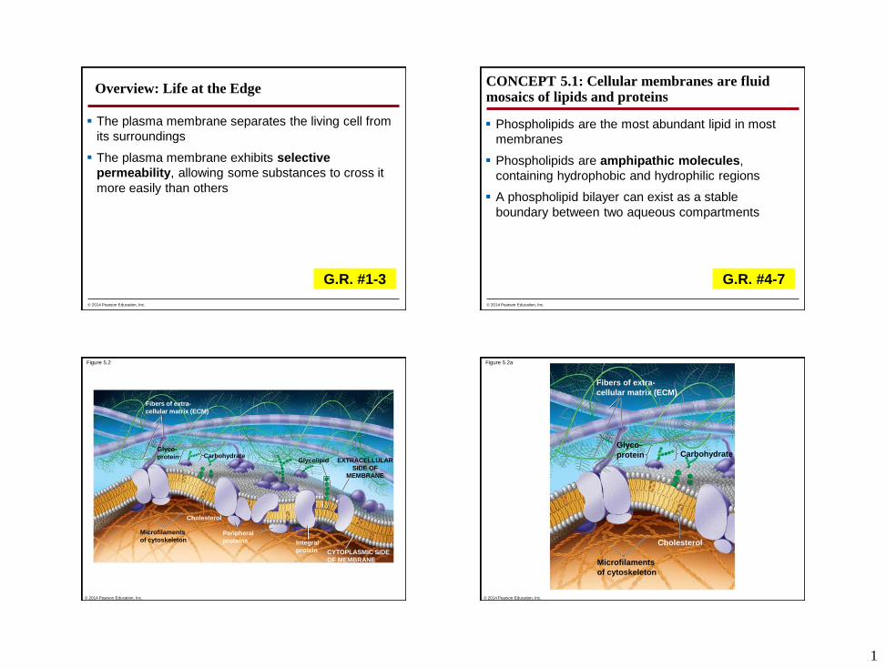
Figure 5.2a
Glyco-
protein
Fibers of extra-
cellular matrix (ECM)
Carbohydrate
Cholesterol
Microfilaments
of cytoskeleton
© 2014 Pearson Education, Inc.

2
Figure 5.2b
Glycolipid
Peripheral
proteinsIntegral
protein
EXTRACELLULARSIDE OF
MEMBRANE
CYTOPLASMIC SIDE
OF MEMBRANE
© 2014 Pearson Education, Inc.
G.R. #8
Most membrane proteins are also amphipathic and reside in the bilayer with their hydrophilic portions protruding
The fluid mosaic model states that the membrane is a mosaic of protein molecules bobbing in a fluid bilayer of phospholipids
Groups of certain proteins or certain lipids may associate in long-lasting, specialized patches
© 2014 Pearson Education, Inc.
G.R. #9
Figure 5.3
Hydrophobictail
Hydrophilichead
WATER
WATER
© 2014 Pearson Education, Inc.
G.R. #9
The Fluidity of Membranes
Most of the lipids and some proteins in a membrane
can shift about laterally
The lateral movement of phospholipids is rapid;
proteins move more slowly
Some proteins move in a directed manner; others
seem to be anchored in place
© 2014 Pearson Education, Inc.
G.R. #10
3
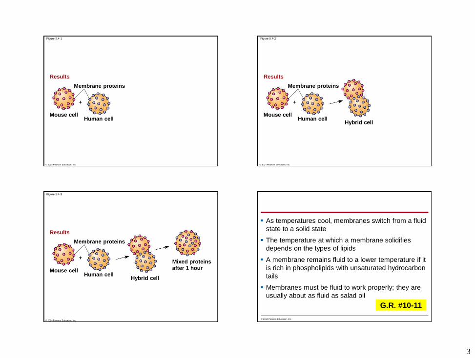
Figure 5.4-1
Results
Mouse cellHuman cell
Membrane proteins
© 2014 Pearson Education, Inc.
Figure 5.4-2
Results
Mouse cellHuman cell
Hybrid cell
Membrane proteins
© 2014 Pearson Education, Inc.
Figure 5.4-3
Results
Mouse cellHuman cell
Hybrid cell
Membrane proteins
Mixed proteinsafter 1 hour
© 2014 Pearson Education, Inc.
As temperatures cool, membranes switch from a fluid
state to a solid state
The temperature at which a membrane solidifies
depends on the types of lipids
A membrane remains fluid to a lower temperature if it
is rich in phospholipids with unsaturated hydrocarbon
tails
Membranes must be fluid to work properly; they are
usually about as fluid as salad oil
© 2014 Pearson Education, Inc.
G.R. #10-11
4
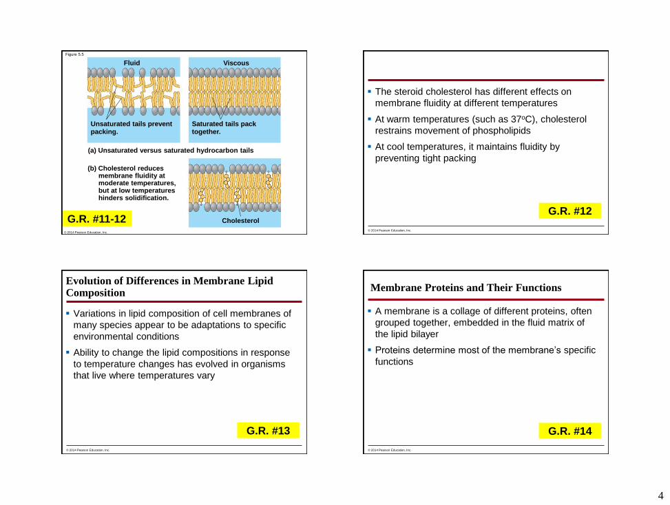
Figure 5.5
Fluid
Unsaturated tails preventpacking.
Cholesterol
Viscous
Saturated tails packtogether.
(a) Unsaturated versus saturated hydrocarbon tails
(b) Cholesterol reducesmembrane fluidity atmoderate temperatures,but at low temperatureshinders solidification.
© 2014 Pearson Education, Inc.
G.R. #11-12
The steroid cholesterol has different effects on
membrane fluidity at different temperatures
At warm temperatures (such as 37oC), cholesterol
restrains movement of phospholipids
At cool temperatures, it maintains fluidity by
preventing tight packing
© 2014 Pearson Education, Inc.
G.R. #12
Evolution of Differences in Membrane Lipid Composition
Variations in lipid composition of cell membranes of
many species appear to be adaptations to specific
environmental conditions
Ability to change the lipid compositions in response
to temperature changes has evolved in organisms
that live where temperatures vary
© 2014 Pearson Education, Inc.
G.R. #13
Membrane Proteins and Their Functions
A membrane is a collage of different proteins, often
grouped together, embedded in the fluid matrix of
the lipid bilayer
Proteins determine most of the membrane’s specific
functions
© 2014 Pearson Education, Inc.
G.R. #14
5
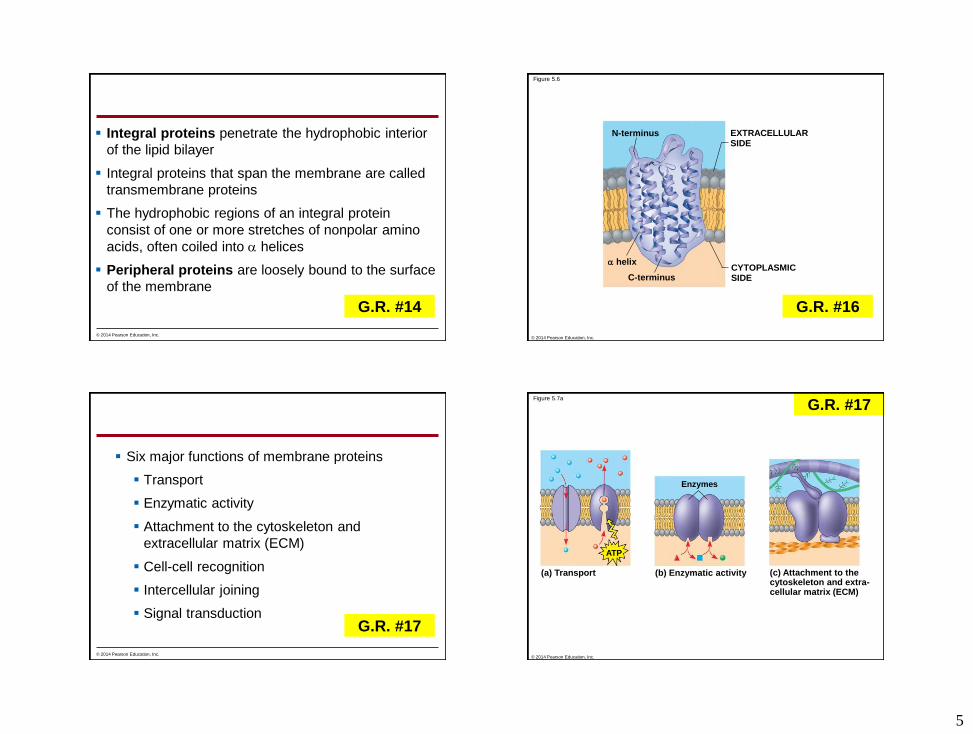
Integral proteins penetrate the hydrophobic interior
of the lipid bilayer
Integral proteins that span the membrane are called
transmembrane proteins
The hydrophobic regions of an integral protein
consist of one or more stretches of nonpolar amino
acids, often coiled into helices
Peripheral proteins are loosely bound to the surface
of the membrane
© 2014 Pearson Education, Inc.
G.R. #14
Figure 5.6
N-terminus
C-terminus
helixCYTOPLASMICSIDE
EXTRACELLULARSIDE
© 2014 Pearson Education, Inc.
G.R. #16
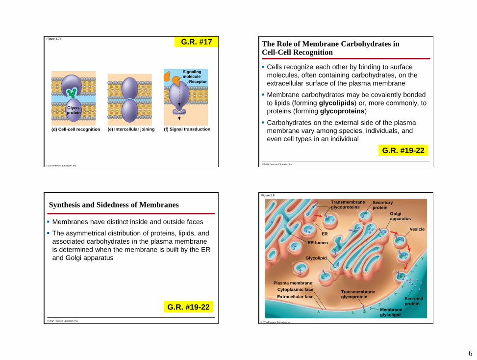
Six major functions of membrane proteins
Transport
Enzymatic activity
Attachment to the cytoskeleton and
extracellular matrix (ECM)
Cell-cell recognition
Intercellular joining
Signal transduction
© 2014 Pearson Education, Inc.
G.R. #17
Figure 5.7a
(a) Transport (b) Enzymatic activity
ATP
(c) Attachment to thecytoskeleton and extra-cellular matrix (ECM)
Enzymes
© 2014 Pearson Education, Inc.
G.R. #17
6
Figure 5.7b
Signalingmolecule
Receptor
(f) Signal transduction(e) Intercellular joining(d) Cell-cell recognition
Glyco-protein
© 2014 Pearson Education, Inc.
G.R. #17 The Role of Membrane Carbohydrates in Cell-Cell Recognition
Cells recognize each other by binding to surface
molecules, often containing carbohydrates, on the
extracellular surface of the plasma membrane
Membrane carbohydrates may be covalently bonded
to lipids (forming glycolipids) or, more commonly, to
proteins (forming glycoproteins)
Carbohydrates on the external side of the plasma
membrane vary among species, individuals, and
even cell types in an individual
© 2014 Pearson Education, Inc.
G.R. #19-22
Synthesis and Sidedness of Membranes
Membranes have distinct inside and outside faces
The asymmetrical distribution of proteins, lipids, and
associated carbohydrates in the plasma membrane
is determined when the membrane is built by the ER
and Golgi apparatus
© 2014 Pearson Education, Inc.
G.R. #19-22
Figure 5.8
Golgiapparatus
Vesicle
Cytoplasmic face
Plasma membrane:
ER
Secretoryprotein
Transmembraneglycoproteins
Transmembraneglycoprotein
ER lumen
Glycolipid
Extracellular face
Membraneglycolipid
Secretedprotein
© 2014 Pearson Education, Inc.

7
CONCEPT 5.2: Membrane structure results in selective permeability
A cell must regulate transport of substances across
cellular boundaries
Plasma membranes are selectively permeable,
regulating the cell’s molecular traffic
© 2014 Pearson Education, Inc.
G.R. #23
The Permeability of the Lipid Bilayer
Hydrophobic (nonpolar) molecules, such as
hydrocarbons, can dissolve in the lipid bilayer of the
membrane and cross it easily
Hydrophilic molecules, such as sugars, and polar
molecules, such as water, do not cross the
membrane easily
© 2014 Pearson Education, Inc.
Transport Proteins
Transport proteins allow passage of hydrophilic
substances across the membrane
Some transport proteins, called channel proteins,
have a hydrophilic channel that certain molecules or
ions can use as a tunnel
Channel proteins called aquaporins facilitate the
passage of water
© 2014 Pearson Education, Inc.
Other transport proteins, called carrier proteins, bind
to molecules and change shape to shuttle them
across the membrane
A transport protein is specific for the substance it
moves
© 2014 Pearson Education, Inc.
8
CONCEPT 5.3: Passive transport is diffusion of a substance across a membrane with no energy investment
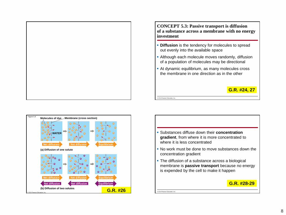
Diffusion is the tendency for molecules to spread
out evenly into the available space
Although each molecule moves randomly, diffusion
of a population of molecules may be directional
At dynamic equilibrium, as many molecules cross
the membrane in one direction as in the other
© 2014 Pearson Education, Inc.
G.R. #24, 27
Figure 5.9Molecules of dye
Net diffusion
WATER
(a) Diffusion of one solute
Net diffusion
Net diffusion
Net diffusion
Net diffusion
Net diffusion
Equilibrium
(b) Diffusion of two solutes
Equilibrium
Equilibrium
Membrane (cross section)
© 2014 Pearson Education, Inc.G.R. #26
Substances diffuse down their concentration
gradient, from where it is more concentrated to
where it is less concentrated
No work must be done to move substances down the
concentration gradient
The diffusion of a substance across a biological
membrane is passive transport because no energy
is expended by the cell to make it happen
© 2014 Pearson Education, Inc.
G.R. #28-29
9
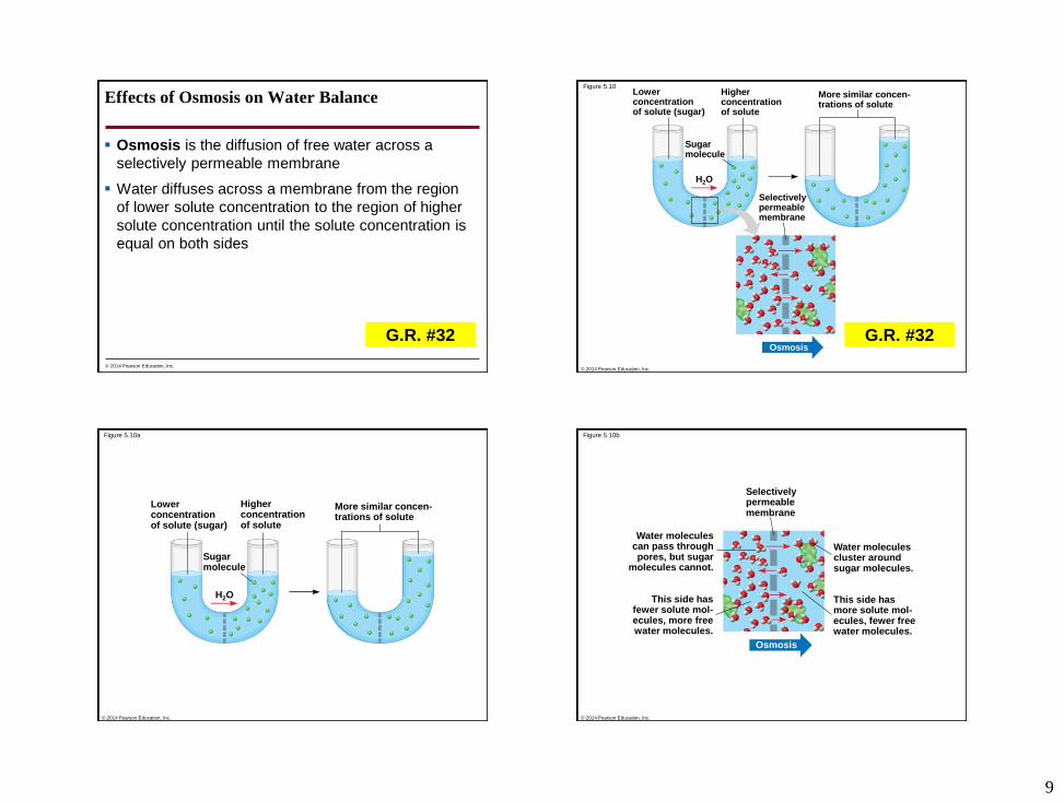
Effects of Osmosis on Water Balance
Osmosis is the diffusion of free water across a
selectively permeable membrane
Water diffuses across a membrane from the region
of lower solute concentration to the region of higher
solute concentration until the solute concentration is
equal on both sides
© 2014 Pearson Education, Inc.
G.R. #32
Figure 5.10
Sugarmolecule
Lowerconcentrationof solute (sugar)
Higherconcentrationof solute
H2O
Selectivelypermeablemembrane
More similar concen-trations of solute
Osmosis
© 2014 Pearson Education, Inc.
G.R. #32
Figure 5.10a
Sugarmolecule
Lowerconcentrationof solute (sugar)
Higherconcentrationof solute
H2O
More similar concen-trations of solute
© 2014 Pearson Education, Inc.
Figure 5.10b
Selectivelypermeablemembrane
Osmosis
Water moleculescan pass throughpores, but sugar
molecules cannot.
Water moleculescluster aroundsugar molecules.
This side hasfewer solute mol-ecules, more freewater molecules.
This side hasmore solute mol-ecules, fewer freewater molecules.
© 2014 Pearson Education, Inc.
10
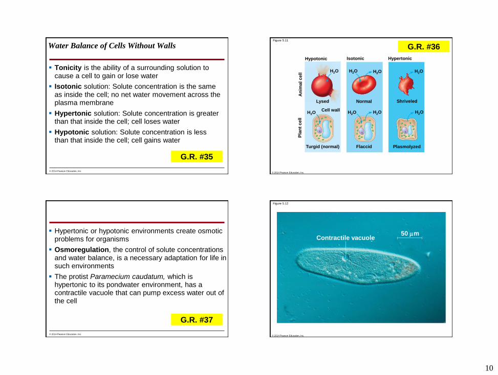
Water Balance of Cells Without Walls
Tonicity is the ability of a surrounding solution to cause a cell to gain or lose water
Isotonic solution: Solute concentration is the same as inside the cell; no net water movement across the plasma membrane
Hypertonic solution: Solute concentration is greater than that inside the cell; cell loses water
Hypotonic solution: Solute concentration is less than that inside the cell; cell gains water
© 2014 Pearson Education, Inc.
G.R. #35
Figure 5.11
Turgid (normal) Flaccid
Lysed
Cell wall
Normal
H2O H2O H2O
H2O H2O H2O
Plasmolyzed
Shriveled
H2O
H2O
HypertonicIsotonicHypotonic
An
ima
l c
ell
Pla
nt
cell
© 2014 Pearson Education, Inc.
G.R. #36
Hypertonic or hypotonic environments create osmotic problems for organisms
Osmoregulation, the control of solute concentrations and water balance, is a necessary adaptation for life in such environments
The protist Paramecium caudatum, which is hypertonic to its pondwater environment, has a contractile vacuole that can pump excess water out of the cell
© 2014 Pearson Education, Inc.
G.R. #37
Figure 5.12
Contractile vacuole50 m
© 2014 Pearson Education, Inc.
11
Water Balance of Cells with Walls
Cell walls help maintain water balance
A plant cell in a hypotonic solution swells until the
wall opposes uptake; the cell is now turgid (very
firm)
If a plant cell and its surroundings are isotonic, there
is no net movement of water into the cell; the cell
becomes flaccid (limp), and the plant may wilt
In a hypertonic environment, plant cells lose water;
eventually, the membrane pulls away from the wall, a
usually lethal effect called plasmolysis
© 2014 Pearson Education, Inc.
G.R. #38Figure 5.11
Turgid (normal) Flaccid
Lysed
Cell wall
Normal
H2O H2O H2O
H2O H2O H2O
Plasmolyzed
Shriveled
H2O
H2O
HypertonicIsotonicHypotonic
An
ima
l c
ell
Pla
nt
cell
© 2014 Pearson Education, Inc.
G.R. #39
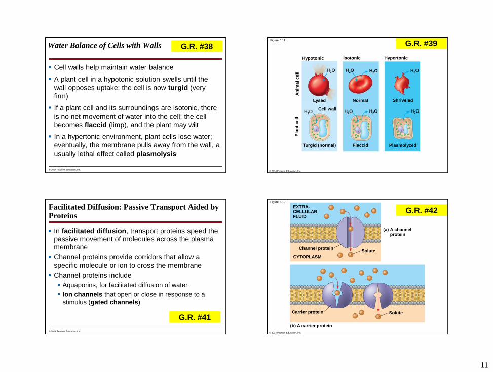
Facilitated Diffusion: Passive Transport Aided by Proteins
In facilitated diffusion, transport proteins speed the passive movement of molecules across the plasma membrane
Channel proteins provide corridors that allow a specific molecule or ion to cross the membrane
Channel proteins include
Aquaporins, for facilitated diffusion of water
Ion channels that open or close in response to a stimulus (gated channels)
© 2014 Pearson Education, Inc.
G.R. #41
Figure 5.13
Carrier protein
(b) A carrier protein
Channel protein
(a) A channelprotein
Solute
Solute
CYTOPLASM
EXTRA-CELLULARFLUID
© 2014 Pearson Education, Inc.
G.R. #42
12
Carrier proteins undergo a subtle change in shape
that translocates the solute-binding site across the
membrane
The shape change may be triggered by binding and
release of the transported molecule
No net energy input is required
© 2014 Pearson Education, Inc.
G.R. #42
CONCEPT 5.4: Active transport uses energy to move solutes against their gradients
Facilitated diffusion speeds transport of a solute by providing efficient passage through the membrane but does not alter the direction of transport
Some transport proteins, however, can move solutes against their concentration gradients
© 2014 Pearson Education, Inc.
G.R. #47
The Need for Energy in Active Transport
Active transport moves substances against their
concentration gradients
Active transport requires energy, usually in the form
of ATP
© 2014 Pearson Education, Inc.
G.R. #48, 50
13
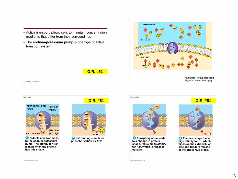
Active transport allows cells to maintain concentration
gradients that differ from their surroundings
The sodium-potassium pump is one type of active
transport system
© 2014 Pearson Education, Inc.
G.R. #51
Animation: Active Transport
Right click slide / Select play
Figure 5.14a
[K] high
EXTRACELLULAR
FLUID
CYTOPLASM
[Na] low
[K] low
[Na] high
21 Na binding stimulatesphosphorylation by ATP.
Cytoplasmic Na bindsto the sodium-potassiumpump. The affinity for Na
is high when the proteinhas this shape.
ADP
© 2014 Pearson Education, Inc.
G.R. #51
The new shape has ahigh affinity for K, whichbinds on the extracellularside and triggers releaseof the phosphate group.
Figure 5.14b
43 Phosphorylation leadsto a change in proteinshape, reducing its affinityfor Na, which is releasedoutside.
© 2014 Pearson Education, Inc.
G.R. #51
14
Figure 5.14c
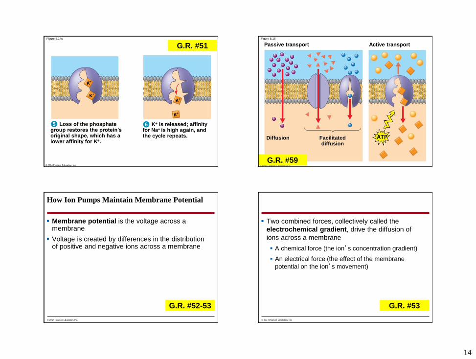
65 Loss of the phosphategroup restores the protein’soriginal shape, which has alower affinity for K.
K is released; affinityfor Na is high again, andthe cycle repeats.
© 2014 Pearson Education, Inc.
G.R. #51Figure 5.15
Diffusion Facilitateddiffusion
Passive transport Active transport
© 2014 Pearson Education, Inc.
G.R. #59
How Ion Pumps Maintain Membrane Potential
Membrane potential is the voltage across a membrane
Voltage is created by differences in the distribution of positive and negative ions across a membrane
© 2014 Pearson Education, Inc.
G.R. #52-53
Two combined forces, collectively called the
electrochemical gradient, drive the diffusion of
ions across a membrane
A chemical force (the ion’s concentration gradient)
An electrical force (the effect of the membrane
potential on the ion’s movement)
© 2014 Pearson Education, Inc.
G.R. #53

15
An electrogenic pump is a transport protein that
generates voltage across a membrane
The sodium-potassium pump is the major
electrogenic pump of animal cells
The main electrogenic pump of plants, fungi, and
bacteria is a proton pump
Electrogenic pumps help store energy that can be
used for cellular work
© 2014 Pearson Education, Inc.
G.R. #53
Figure 5.16
EXTRACELLULAR
FLUID
CYTOPLASM
Proton pump
© 2014 Pearson Education, Inc.
G.R. #54
Cotransport: Coupled Transport by a Membrane Protein
Cotransport occurs when active transport of a
solute indirectly drives transport of other solutes
Plant cells use the gradient of hydrogen ions
generated by proton pumps to drive active transport
of nutrients into the cell
© 2014 Pearson Education, Inc.
G.R. #55
Figure 5.17
Sucrose
Proton pump
Sucrose-H
cotransporterDiffusion of H
Sucrose
© 2014 Pearson Education, Inc.
G.R. #55-56
16
CONCEPT 5.5: Bulk transport across the plasma membrane occurs by exocytosis and endocytosis
Small solutes and water enter or leave the cell
through the lipid bilayer or by means of transport
proteins
Large molecules, such as polysaccharides and
proteins, cross the membrane in bulk by means of
vesicles
Bulk transport requires energy
© 2014 Pearson Education, Inc.
G.R. #60
Exocytosis
In exocytosis, transport vesicles migrate to the
membrane, fuse with it, and release their contents
Many secretory cells use exocytosis to export
products
© 2014 Pearson Education, Inc.
G.R. #61
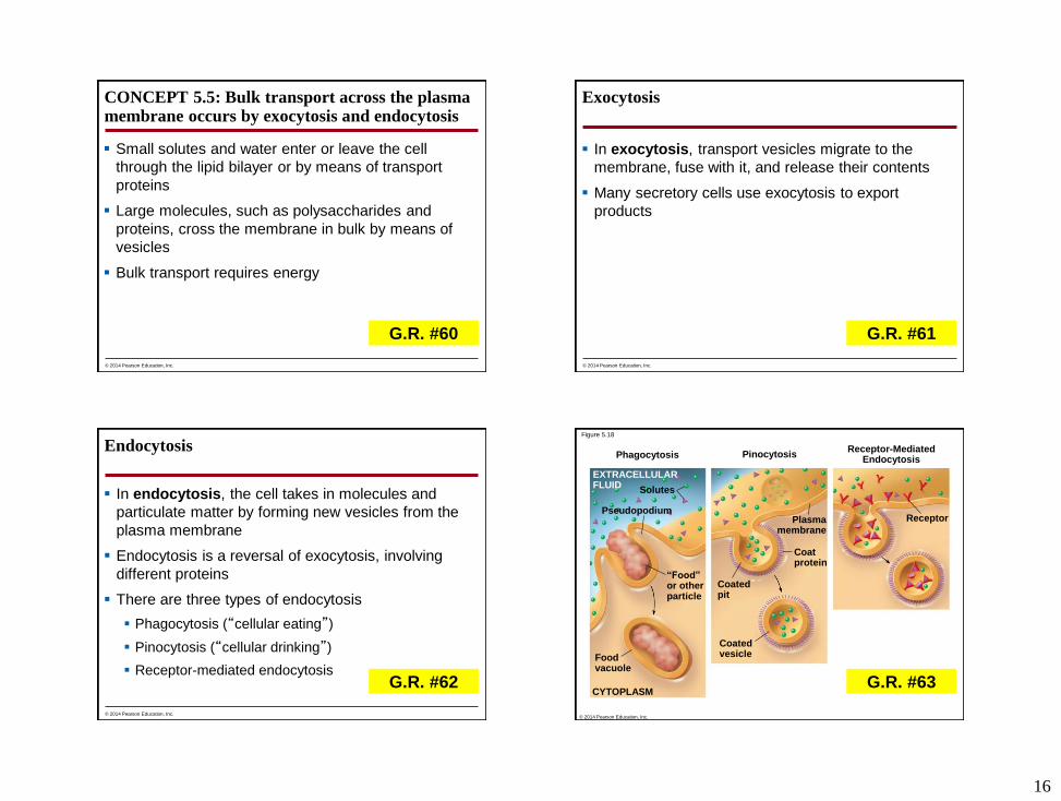
Endocytosis
In endocytosis, the cell takes in molecules and
particulate matter by forming new vesicles from the
plasma membrane
Endocytosis is a reversal of exocytosis, involving
different proteins
There are three types of endocytosis
Phagocytosis (“cellular eating”)
Pinocytosis (“cellular drinking”)
Receptor-mediated endocytosis
© 2014 Pearson Education, Inc.
G.R. #62
Figure 5.18
Phagocytosis PinocytosisReceptor-Mediated
Endocytosis
ReceptorPlasmamembrane
Coatprotein
Coatedpit
CoatedvesicleFood
vacuole
“Food”or otherparticle
CYTOPLASM
Pseudopodium
Solutes
EXTRACELLULARFLUID
© 2014 Pearson Education, Inc.
G.R. #63

17
Figure 5.18aPhagocytosis
Foodvacuole
“Food”or otherparticle
CYTOPLASM
Pseudopodium
Solutes
EXTRACELLULARFLUID
Pseudopodiumof amoeba
An amoeba engulfing a bacteriumvia phagocytosis (TEM)
Bacterium
Food vacuole 1
m
© 2014 Pearson Education, Inc.
G.R. #63
Figure 5.18bPinocytosis
Plasmamembrane
Coatprotein
Coatedpit
Coatedvesicle
Pinocytotic vesicles forming(TEMs)
0.2
5
m
© 2014 Pearson Education, Inc.
G.R. #63
Figure 5.18c
Top: A coated pit Bottom: A coatedvesicle forming during receptor-mediated endocytosis (TEMs)
0.2
5
m
Receptor-MediatedEndocytosis
ReceptorPlasmamembrane
Coatprotein
© 2014 Pearson Education, Inc.
G.R. #63