Embed Size (px)
Citation preview
Glucocorticoids enhance muscle endurance andameliorate Duchenne muscular dystrophy througha defined metabolic programAlexander Morrison-Nozika, Priti Ananda,b, Han Zhua, Qiming Duana,b, Mohamad Sabeha,c, Domenick A. Prosdocimoa,Madeleine E. Lemieuxd, Nikolai Nordsborge, Aaron P. Russellf, Calum A. MacRaeg, Anthony N. Gerberh,Mukesh K. Jaina,c, and Saptarsi M. Haldara,b,c,i,1
aCase Cardiovascular Research Institute, Department of Medicine, Case Western Reserve University School of Medicine, Cleveland, OH 44106; bGladstoneInstitutes, San Francisco, CA 94158; cHarrington Heart & Vascular Institute, University Hospitals Case Medical Center, Cleveland, OH 44106; dBioinfo,Plantagenet, Ontario, Canada K0B IL0; eDepartment of Nutrition, Exercise and Sports Sciences, University of Copenhagen, DK-200 Copenhagen, Denmark;fCentre for Physical Activity and Nutrition Research, School of Exercise and Nutrition Science, Deakin University, Burwood, VIC 3125, Australia;gCardiovascular Division, Brigham & Women’s Hospital, Harvard Medical School, Boston, MA 02115; hDepartment of Pulmonary Medicine, National JewishHealth and University of Colorado Denver School of Medicine, Denver, CO 80206; and iDepartment of Medicine and Cardiovascular Research Institute,University of California, San Francisco, CA 94158
Edited by David H. MacLennan, University of Toronto, Toronto, Canada, and approved October 19, 2015 (received for review July 1, 2015)
Classic physiology studies dating to the 1930s demonstrate thatmoderate or transient glucocorticoid (GC) exposure improvesmuscle performance. The ergogenic properties of GCs are furtherevidenced by their surreptitious use as doping agents by endur-ance athletes and poorly understood efficacy in Duchenne musculardystrophy (DMD), a genetic muscle-wasting disease. A definedmolecular basis underlying these performance-enhancing propertiesof GCs in skeletal muscle remains obscure. Here, we demonstratethat ergogenic effects of GCs are mediated by direct induction ofthe metabolic transcription factor KLF15, defining a downstreampathway distinct from that resulting in GC-related muscle atrophy.Furthermore, we establish that KLF15 deficiency exacerbates dys-trophic severity and muscle GC–KLF15 signaling mediates salutarytherapeutic effects in the mdx mouse model of DMD. Thus, al-though glucocorticoid receptor (GR)-mediated transactivation isoften associated with muscle atrophy and other adverse effectsof pharmacologic GC administration, our data define a distinctGR-induced gene regulatory pathway that contributes to ther-apeutic effects of GCs in DMD through proergogenic metabolicprogramming.
skeletal muscle metabolism | glucocorticoid | exercise |Duchenne muscular dystrophy | steroid hormone nuclear receptor
Synthetic derivatives of the glucocorticoid (GC) class of ste-roid hormones, which are ligands for the nuclear receptor
NR3C1 (also known as the glucocorticoid receptor; GR), arewidely used as antiinflammatory drugs (1). The vast majority ofliterature on muscle GR signaling has focused on muscle wast-ing, a side effect of excessive or sustained GC exposure that ismediated, in part, by direct GR-dependent transactivation ofgenes that drive myocyte atrophy (e.g., Foxo3a, Gdf8/Myostatin,Fbxo32/Atrogin1; also known as atrogenes) (1, 2). However,literature dating back to the 1930s, including the classic physio-logical studies from the laboratory of Dwight Ingle (3, 4), havedocumented that moderate or transient exposure to GCs canenhance muscle performance and produce ergogenic effects inanimals and humans (5–13). Consistent with this known physi-ological role of GCs in anticipatory metabolic adaptation, sur-reptitious GC ingestion is a well-known doping strategy used byelite endurance athletes, an act that has prompted disqualificationsand has led to the universal banning of these drugs by sports regu-latory agencies (14). Although mechanisms governing GC-mediatedmuscle atrophy have been extensively studied (1, 2), the molecularbasis for their ergogenic effects remains poorly understood.In addition to these ergogenic physiological effects, low-dose
GC therapy also improves muscle function, quality of life, andsurvival in patients with Duchenne muscular dystrophy (DMD)
(15–17), a progressive muscle-wasting disease caused by X-linkedinheritance of nonsense mutations in the gene encoding Dys-trophin (18). Although GCs have been widely used in thetreatment of DMD for nearly 25 years (15), the mechanism ofaction underlying their salutary effects in this condition remainsobscure. Several studies in patients and animal models havecalled into question whether the antiinflammatory properties ofGCs adequately explain their therapeutic effect in DMD (15,19). We hypothesized that the ergogenic properties of GCs andaspects of their therapeutic efficacy in DMD might be mediated,in part, by a defined GR-dependent metabolic transcriptionalpathway distinct from that resulting in GC-related muscle atro-phy. As GCs cause a myriad of systemic side effects that limittheir therapeutic index in DMD, elucidation of such a downstreamsignaling pathway could inform novel steroid-sparing treatmentstrategies, a major unmet need for patients suffering from thisdevastating and currently incurable disease (15, 20).
Significance
Classic physiological studies have documented the endurance-promoting effects of glucocorticoid (GC) hormones on skeletalmuscle. Pharmacologic GC therapy also improves muscle func-tion in patients with Duchenne muscular dystrophy (DMD), agenetic muscle-wasting disease. Despite these well-establishedphysiological and clinical observations, the molecular basisunderlying the beneficial effects of GCs in skeletal muscle re-mains obscure. This study shows that physiological effects ofGCs on muscle endurance and their therapeutic effect in DMDare mediated, in part, via activation of a potent metabolic genecalled Kruppel-like factor 15 (KLF15). Importantly, KLF15 doesnot drive GC-mediated muscle wasting. These data shed lighton the poorly understood ergogenic properties of GCs, findingsthat may inform steroid-sparing therapies for DMD and othermuscle diseases.
Author contributions: A.M.-N., P.A., H.Z., C.A.M., A.N.G., and S.M.H. designed research;A.M.-N., P.A., H.Z., Q.D., M.S., and D.A.P. performed research; N.N., A.P.R., C.A.M., M.K.J.,and S.M.H. contributed new reagents/analytic tools; A.M.-N., P.A., H.Z., M.E.L., A.N.G.,and S.M.H. analyzed data; and A.M.-N., A.N.G., and S.M.H. wrote the paper.
The authors declare no conflict of interest.
This article is a PNAS Direct Submission.
Data deposition: The data reported in this paper have been deposited in the Gene Ex-pression Omnibus (GEO) database, www.ncbi.nlm.nih.gov/geo (accession no. GSE74625).1To whom correspondence should be addressed. Email: [email protected].
This article contains supporting information online at www.pnas.org/lookup/suppl/doi:10.1073/pnas.1512968112/-/DCSupplemental.
E6780–E6789 | PNAS | Published online November 23, 2015 www.pnas.org/cgi/doi/10.1073/pnas.1512968112

A B C
D
E
F
G
H
Fig. 1. The GC–KLF15 axis dissociates ergogenic physiology frommuscle atrophy. (A) KLF15 expression in skeletal muscle of healthy human subjects pre- and postdexadministration. FKBP5 is a known direct GR target shown as a positive control (n = 5; *P < 0.05 for indicated comparisons). (B) Schematic for skeletal muscle(quadriceps) microarray study (n = 4 per group). (C) Venn diagram of dex-inducible and KLF15-regulated genes [2mg/kg i.p. × 1 dose, 8 h time point; fold change > 1.5and false discovery rate (FDR) < 0.05]. (D) Heat map of genes robustly regulated [family-wise error rate (FWER) < 0.05] by GC–KLF15 axis in mouse skeletal muscle.(E) Functional annotation [Database for Annotation, Visualization, and Integrated Discovery (DAVID)] of genes regulated by GC–KLF15 axis. (F) Body weight and(G) muscle (tibialis anterior) histology [wheat-germ agglutinin (WGA) stain] and cross-sectional area in a mouse model of chronic dex-induced atrophy (1 mg/kg·d s.c. ×10 d; n = 8; *P < 0.05 for indicated comparisons). (Scale bar, 50 μm.) (H) Schematic of dex-induced doping experiment (2 mg/kg i.p. × 1 dose) with time to exhaustionand distance run on a motorized treadmill 18 h post-dex administration (n = 7–9; *P < 0.05 for indicated comparisons). Data are shown as mean ± SEM.
Morrison-Nozik et al. PNAS | Published online November 23, 2015 | E6781
MED
ICALSC
IENCE
SPN
ASPL
US

The transcription factor KLF15, a direct GR-inducible targetin multiple cell types (21, 22), is an attractive candidate formediating important ergogenic effects of GCs in muscle. Studiesfrom our group using systemic KLF15-deficient mice have dem-onstrated KLF15 to be an important transcriptional regulator ofa gene program governing stress-dependent metabolic adapta-tion in muscle (23, 24). KLF15-deficient mice have impairedability to catabolize muscle branched-chain amino acids as fuel forgluconeogenic flux during fasting (25, 26). In addition, we havedemonstrated that during sustained aerobic exercise, KLF15-deficient mice fail to augment muscle lipid utilization andconsequently have impaired endurance exercise capacity (23). Ourdetailed characterization of KLF15-deficient mice has revealedthat these functional defects in muscle substrate flux occur withoutabnormalities in muscle development, gross histologic structure,fiber type distribution, mitochondrial number, or muscle mass (23).Although KLF15 is a direct GR target (21, 22, 27, 28), the
function of the GC–KLF15 axis in physiology and disease re-mains largely unknown. The observation that GCs induce KLF15in rodent myocytes and that adenoviral KLF15 overexpressionincreases expression of Atrogin-1 has led to the speculation thatKLF15 might promote muscle wasting via activation of an at-rophy-promoting transcriptional program (22). However, thesestudies used supraphysiologic levels of KLF15 overexpressionand have thus left unanswered whether the physiologic effects ofthe GC–KLF15 axis are harmful or beneficial in skeletal muscle.Here, we use unbiased transcriptomic profiling and physiologicanalysis of mice harboring KLF15 deficiency and skeletal mus-cle-specific KLF15 overexpression at physiologic levels to gain adeeper understanding of the GC–KLF15 axis in muscle function.We find that the GC–KLF15 axis does not regulate muscle at-rophy in vivo but rather activates a metabolic gene program thatmediates ergogenic effects in wild-type (WT) mice and amelio-rates dystrophic severity in the mdx mouse model of DMD. Thesestudies establish KLF15 as a critical metabolic effector of physi-ological GC signaling in vivo and suggest that GR-dependenttarget gene transactivation in skeletal muscle can, in certain con-texts, have a therapeutic role.
ResultsKLF15 Is Induced by GCs in Live Human Subjects. Although GCs caninduce KLF15 in rodent muscle tissue (22), it is not knownwhether GCs regulate KLF15 in human subjects in vivo. Healthyhuman volunteers underwent skeletal muscle biopsies before andafter ingesting the potent GR agonist dexamethasone (dex; 2 mgorally twice daily × 5 d). Quantitative RT-PCR (qRT-PCR)revealed a significant induction of KLF15 expression in humanskeletal muscle tissue in vivo (Fig. 1A; FKBP5 is a well-estab-lished GR target that serves as a positive control). Similarly, wefound that dex rapidly and robustly increased KLF15 expressionin primary human myotubes and mouse C2C12 myotubes in vitro(Fig. S1 A and B) and mouse quadriceps in vivo (Fig. S1C). Themammalian KLF15 locus contains two adjacent glucocorticoidresponse elements (GREs) in the first intron that are highlyconserved (schematized in Fig. S1D). We performed ChIP-qPCRin quadriceps tissue of adult mice and treated with or without dexand found dynamic enrichment of endogenous GR at theseintronic GREs in the Klf15 locus (Fig. S1E). These data confirmprior observations that KLF15 is a direct GR target in muscle anddemonstrate that GC-mediated induction of muscle KLF15 occursin live human subjects.
The GC–KLF15 Axis Regulates an Ergogenic Gene Program That Is NotInvolved in Muscle Atrophy. In an effort to definitively resolve thegene program regulated by the GC–KLF15 axis in vivo, weperformed unbiased transcriptomic profiling from skeletal muscletissue of WT and Klf15−/− (KO) mice treated with or without dex.WT and KO mice were given a single dose of dex (2 mg/kg i.p.)
versus vehicle, and quadriceps tissue was isolated for transcriptexpression profiling by cDNA microarrays (Fig. 1B). Dex induceda total of 897 genes, of which 7% were highly KLF15-dependent,as shown by Venn diagram (Fig. 1C) and heat map of repre-sentative genes (Fig. 1D; full list of differential gene expressionprovided in Excel format in Dataset S1). To our surprise, themicroarray study revealed that KLF15 was not required for therobust GC-meditated induction of the canonical atrogene pro-gram, findings that were confirmed independently by qRT-PCR(Fig. S1F). Rather, functional annotation of the GC–KLF15-regulated transcriptome revealed highly significant enrichmentfor genes critical for metabolism of several amino acids, fattyacids, and propanoate (Fig. 1 D and E, Fig. S1G, and Dataset S1).Based on these unbiased expression profiles, we hypothesized
that KLF15 was not involved in muscle wasting but, in contrast,was required for GC-mediated ergogenic effects in vivo. To testthis hypothesis, we first subjected mice to a standard model ofchronic dex-mediated muscle atrophy (dex 1 mg/kg given dailys.c. × 10 d) (29, 30). We found that both WT mice and KLF15KO mice had an identical degree of muscle atrophy, as assessedby changes in body weight, muscle weight, and myofiber cross-sectional area (Fig. 1 F and G and Fig. S1H). Thus, KLF15 is notrequired for GC-mediated muscle atrophy in vivo. To test therequirement of KLF15 in GC-mediated ergogenesis, WT andKO mice were given a single dose of dex and challenged to anendurance exercise trial 16 h after dex treatment (Fig. 1H).Consistent with the gene expression profiling, WT mice treatedwith dex were able to augment endurance exercise capacity by50%, whereas KO mice did not demonstrate any significant ergo-genic response to dex (Fig. 1H). Hence, these data demonstratethat KLF15 is dispensable for GC-mediated muscle atrophy butrequired for the ergogenic effects of GCs.
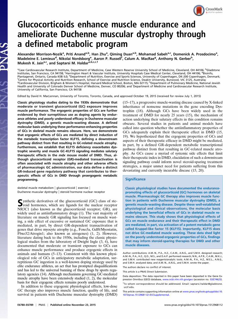
Skeletal Muscle-Specific KLF15 Induction Is Sufficient to Drive anErgogenic Gene Program in Vivo. As the data in Fig. 1 were gen-erated using mice harboring systemic KLF15 deficiency, we nextasked whether skeletal muscle-specific augmentation of KLF15expression was sufficient to activate an ergogenic metabolicprogram in vivo. We used the –4.8 kb MCK promoter/enhancerto generate transgenic mice expressing a full-length mouseKLF15 cDNA in a skeletal muscle-specific manner (Fig. 2A). Westudied a transgenic mouse line that expressed KLF15 levels at5–6 times higher than control exclusively in skeletal muscle withno induction in the heart or other tissues (muscle transgenic,MTg; Fig. 2B). This level of transgenic overexpression of KLF15is in the physiological range (23) and similar to the increase inKLF15 seen with nanomolar concentrations of dex (Fig. S1 Aand B). MTg mice showed no evidence of muscle atrophy asassessed by gravimetry (Fig. S2 A and B) or histology (Fig. 2 Cand D and Fig. S2C). In contrast, MTg mice had significantlyincreased endurance exercise capacity (Fig. 2 E and F). Metabolicexercise testing demonstrated that MTg mice had a decreasedrespiratory exchange ratio (Fig. 2G), signifying that muscle-specificKLF15 induction leads to a global shift in substrate preferencetoward amino acids and lipids. To confirm that these physiologicalobservations were associated with KLF15-dependent transcrip-tional control in skeletal muscle, we performed genome-wideexpression profiling in quadriceps of MTg mice using cDNAmicroarrays. These analyses revealed that skeletal muscle-spe-cific KLF15 overexpression in the physiological range resulted ina signature that was dominated by gene induction (90 inducedgenes out of 109 total differentially expressed genes; 83%). Arepresentative heat map of the top 50 genes induced in the MTgskeletal muscle is provided in Fig. S2D. As visualized in theglobal heat maps from MTg mice (Fig. 2H), the transcriptomicprofile of MTg mice reflected significant reversal of the meta-bolic gene expression abnormalities observed in KLF15 KOskeletal muscle. This highly significant overlap between genes
E6782 | www.pnas.org/cgi/doi/10.1073/pnas.1512968112 Morrison-Nozik et al.

induced in the MTg and genes reduced in KLF15 KO muscle wasstatistically confirmed by gene set enrichment analysis (GSEA;
Fig. 2I). Functional annotation of differentially expressed genesin MTg mice (Fig. 2J) revealed robust enrichment for the same
-4.8kb MCKpromoter
Full lengthmouse KLF15 cDNA
hGH polyA
0
2
4
6
8
NS
CntrlMTg
Quad Gastroc Heart Liver
NS
Klf1
5 re
altiv
e ex
pres
sion
Cntrl MTg
0
50
100
Cntrl MTg
Tim
e to
exh
aust
ion
(min
)
*
Res
pira
tory
exc
hang
e ra
tio CntrlMTg
0Cntrl MTg
NS
0
0.7
1.4
Cntrl MTg
Dis
tanc
e ru
n (k
m)
0
2.5
5.0
Late
ncy
to fa
ll (m
in)
Cntrl MTg
0.00
0.45
0.90
FWER p<0.001
Enr
ichm
ent s
core
MTg Cntrl Klf15-/-Klf15+/+
Category Term % List FDR %
GOTERM_CC_FAT GO:0005739~mitochondrion 39 <1e-11
GOTERM_BP_FAT GO:009081~BCAA metabolic process 6 <0.002KEGG_PATHWAY mmu00071: Fatty acid metabolism 7 <0.03
KEGG_PATHWAY mmu00640: Propanoate metabolism 7 <0.01KEGG_PATHWAY mmu00380: Tryptophan metabolism 5 4.2KEGG_PATHWAY mmu00310: Lysine degradation 5 4.5GOTERM_MF_FAT GO:0003995~acyl-CoA dehydrogenase activity 4 4.3INTERPRO IPR015590: Aldehyde dehydrogenase 4 4.5
*
Speed (m/min)
A
B
C D
E F G
HI
J
Myo
cyte
area
(m
2 x 1
00)
20
10
30
40
0 10 14 18 22 26 30 340.65
0.75
0.85
03 -3 03 -3
Genes regulated by GC-KLF15 axis
MTg Non-Tg
* *
* *
Fig. 2. Skeletal muscle-specific KLF15 overexpression is sufficient to drive an ergogenic metabolic program in vivo. (A) Schematic of transgenic construct. (B) qRT-PCR from indicated tissues demonstrating skeletal muscle-specific KLF15 overexpression in MTg mice versus control (littermate non-Tg mice; n = 5). (C) Repre-sentative histology from quadriceps (WGA staining) and (D) quantification of myocyte cross-sectional area (n = 3 independent mice per group). (Scale bar, 50 μm.)(E) Treadmill exercise (n = 6), (F) wire-hang assay (n = 7–10), and (G) metabolic exercise test (n = 7–10) in MTg and control mice. (H) Heat map of all genes inducedin MTg versus non-Tg muscle (Left panel; n = 4). Heat map for the same genes in WT versus KLF15 KO muscle (Right panel, WT levels colored as non-Tg in Leftpanel) illustrates the reversal in expression versus control in KLF15 KO relative to MTg. (I) GSEA of genes regulated by the GC–KLF15 axis in MTg versus non-Tgmuscle. (J) Functional annotation (DAVID) of genes regulated by KLF15 overexpression (*P < 0.05 for indicated comparisons). Data are shown as mean ± SEM.
Morrison-Nozik et al. PNAS | Published online November 23, 2015 | E6783
MED
ICALSC
IENCE
SPN
ASPL
US

pathways regulated by the GC–KLF15 axis (Fig. 1E), with sig-nificant induction of genes critical for amino acid, lipid, andpropanoate metabolism (Fig. 2J and Fig. S2E; full list of differentialgene expression provided in Excel format as Dataset S2). Con-sistent with our published (23) and current observations in KOmice (Fig. 1), the MTg microarrays did not demonstrate signifi-cant regulation of a gene program governing fiber type specifica-tion, mitochondrial biogenesis, autophagic flux, or angiogenesis.Again, expression profiles and direct qRT-PCR showed no in-duction of the canonical atrogene program in MTg mice (Fig. S2Eand Dataset S2). Thus, the data in Figs. 1 and 2 demonstrate thatKLF15 is not an atrogene but rather functions as a critical down-stream transcriptional effector of muscle GR signaling that regu-lates amino acid and lipid metabolic programs. Furthermore, thesedata demonstrate that KLF15 is both necessary and sufficient tomediate ergogenic effects of GCs in vivo.
The GC–KLF15 Transcriptional Axis Modulates Disease Phenotype inthe mdx Mouse Model of DMD. As the GC–KLF15 axis controls agene program governing muscle substrate utilization and ergo-genesis (Figs. 1 and 2), we asked whether this metabolic signalingpathway might, in part, mediate the salubrious effects of GCs inDMD. Landmark clinical studies have demonstrated that lowand intermittent dosing of GCs such as prednisolone can slowdisease progression and improve outcomes in DMD patients(15–17). Despite this established efficacy, a defined molecularbasis for the salutary effects of GCs in DMD remains poorlyunderstood (15, 20). Although GCs are known to exert antiinflam-matory effects via transrepression of NF-κB signaling (1), severalclinical and experimental studies cast significant doubt on thecontention that GCs ameliorate DMD via a primary antiinflam-matory mechanism (15, 19, 31–34). First, non-GC antiinflamma-tory or immunosuppressant drugs, such as cyclosporine-A orazathioprine, fail to show efficacy in DMD patients (32–34).Second, GC therapy provides benefit in DMD patients evenwhen administered via low or intermittent dosing schemes thatdo not provide a potent and sustained antiinflammatory effect(15). Third, GC therapy does not dramatically alter the inflam-matory cell infiltrate seen in dystrophic muscle (31, 34). Fourth,in the mdx mouse model of DMD (35), the therapeutic effects ofGCs are fully preserved in a Rag2−/− background, in which B andT lymphocytes are absent (19).In contrast, several studies in DMD patients and animal
models have documented abnormalities in muscle substrate andenergy metabolism (36–47), prompting the term “metabolic cri-sis” as a hallmark feature of this disease (41). Given the im-portant role for KLF15 in muscle metabolism, we hypothesizedthat DMD might be characterized by a state of relative KLF15deficiency and that GCs may mediate important therapeutic ef-fects in DMD, in part, via induction of KLF15. Skeletal musclebiopsies from DMD patients (Fig. 3A) and muscle tissue frommale mdx mice (Fig. 3B) had significantly decreased KLF15 ex-pression compared with control tissues. We next used GSEA tostatistically test whether the metabolic gene program regulatedby the GC–KLF15 axis was deficient in dystrophic muscle. TheseGSEAs confirmed in an unbiased and quantitative manner thatthe set of genes decreased in mdx skeletal muscle tissue (curatedfrom ref. 44) was highly enriched for the set of genes regulatedby the GC–KLF15 axis (Fig. 3C). These findings were also con-firmed by direct qRT-PCR analysis of representative genes (Fig.S3A). Hence, DMD is characterized by decreased expression ofKLF15 and its downstream metabolic targets.Prior studies from our group demonstrate that mice with KLF15
deficiency, which have been extensively characterized in a non-dystrophic genetic background, are viable into adulthood, arefertile, have normal muscle development and histology, andshow no evidence of muscle damage (23). We confirmed this lackof baseline histopathology in an independent cohort of mice
and found that male KLF15-deficient mice in a nondystrophicbackground showed no evidence of elevated plasma creatinephosphokinase (CPK) concentration or histologic muscledamage (Fig. S3 B and C). Although KLF15 deficiency alonedoes not cause dystrophic muscle pathology, we hypothesizedthat KLF15 deficiency might exacerbate dystrophic severity inthe mdx mouse model of DMD. To test this hypothesis, we bredKLF15-deficient mice into the mdx background by intercrossingKlf15+/−/Dmdmdx Y males with Klf15+/−/Dmdmdx Dmdmdx fe-males. Although pups of all three expected genotypes were bornin predicted Mendelian ratios, we observed significant prematuredeath of male Klf15−/− mdx mice postnatally with only ∼20%surviving to adulthood (Fig. 3D). We did not observe any statis-tically significant loss of Klf15−/−/Dmdmdx Dmdmdx female mice, afinding that may be related to gender-specific metabolic conse-quences of KLF15 deficiency or milder aspects of disease severityin Dmdmdx Dmdmdx versus Dmdmdx Y mice (48). Analysis of maleKlf15−/− mdx mice before death revealed severe dystrophic pa-thology in skeletal muscle tissue and massively elevated plasmaCPK concentration, an unbiased and quantitative clinical bio-marker of global muscle damage in DMD patients (15) (Fig. 3 Eand F). Importantly, single-mutant intercrosses of mice harboringeither the null Klf15 allele or the mdx allele alone do not producesuch mortality or muscle damage (23, 49). Our observations inthese compound mutant strains support the contention that thereis a genetic interaction between KLF15 deficiency and the dys-trophic pathology of the mdx mouse model.Given the low survival of male Klf15−/− mdx mice, we were
unable to perform longitudinal studies with appropriatelymatched littermate controls. Therefore, we focused our attentionon Klf15 haploinsufficient mice, which survive into adulthood(Fig. 3D) and reflect the partial loss of KLF15 expression seen inmuscle tissue from DMD patients and mdx mice (Fig. 3 A andB). We subjected male Klf15+/− mdx mice and Klf15+/+ mdx lit-termate controls to a therapeutic trial of prednisolone using alow and intermittent dosing regimen similar to that used clini-cally in DMD patients and in prior studies of mdx mice (Fig. 3G)(15, 19, 49). We confirmed that this regimen of chronic pred-nisolone produced the expected increase in muscle KLF15 and itsdownstream targets in Klf15+/+ mdx control mice, with bluntedexpression in Klf15+/− mdx mice (Fig. 3H and Fig. S3D). Klf15+/−
mdx mice had minimal baseline perturbations in muscle strengthand endurance but had significantly attenuated therapeutic re-sponses to prednisolone therapy, as demonstrated by decreasedendurance exercise capacity on treadmill (Fig. 3I) and wire-hangtests (Fig. 3J) and decreased grip strength (Fig. 3K). Klf15+/− mdxmice had histologic evidence of excessive muscle damage andplasma CPK elevation at baseline with no significant therapeuticresponse to prednisolone therapy (Fig. 3 L and M). Utrophinexpression, which is known to compensate for mutant Dystrophinin mice (50), was not affected by prednisolone therapy or KLF15deficiency (Fig. S3E). The data in Fig. 3 reveal that KLF15 de-ficiency, which does not cause significant muscle damage in anondystrophic background (Fig. S3 B and C) (23), exacerbatesfeatures of dystrophic severity in the mdx background. Further-more, the data in the Klf15 haploinsufficient mice demonstratethat partial deficiency of this gene attenuates the therapeuticeffect of GCs in the mdx mouse model.We next asked whether effects of KLF15 in the mdx model
were muscle-specific in vivo and whether KLF15 induction wassufficient to exert beneficial effects in the absence of exogenousGCs. To address both of these questions, we crossed KLF15MTg mice into the mdx background to produce male mice ofgenotype MTg+/− Dmdmdx and non-Tg Dmdmdx control litter-mates. The transgene increased total KLF15 expression in mdxskeletal muscle by fivefold, a level of induction that approximatesthe effect of GCs (Fig. 4A). To test whether the gene expressionprogram induced by the KLF15 transgene was statistically
E6784 | www.pnas.org/cgi/doi/10.1073/pnas.1512968112 Morrison-Nozik et al.

0
1
2
3
4
Grip
stre
ngth
(g) p
er g
ram
BW
- Pred + Pred
0
10
20
30
Late
ncy
to fa
ll (s
ec)
- Pred + Pred0
Tim
e to
exh
aust
ion
(min
)
- Pred + Pred
*
0
Dis
tanc
e ru
n (m
)
- Pred + Pred
*
0Cntrl DMD
Human muscle biopsy
Cntrl mdx
Mouse muscle
- Pred
+ Pred
0
50
100
Sur
viva
l (%
)
*
*
Klf15 +/+ / mdx
Klf15 +/- / mdx Klf15 -/- / mdx
Birth 3 weeks 8 weeks
Klf15 +/+ / mdx
Klf15 +/- / mdx
Age (weeks)
+ Prednisolone 5mpk gavage (3 doses/week)0 143 6
Start of phenotypic assessment
0
1
2
3
- Pred + Pred
Klf15
**
Klf15 +/+ / mdx Klf15 +/- / mdx
0.6
1.2
0
0.6
1.2
Age
Klf15 +/+
mdx
Klf15 -/-
mdx
20
40
60
80
200
400
600
800
Klf15 +/+ / mdx Klf15 +/- / mdx
0.0
0.3
0.6
Enr
ichm
ent s
core
0
3
650
150
250
Klf15 +/+
mdx Klf15 -/-
mdx P
lasm
a C
PK
(U/L
x 1
000)
A B C
D FE
G
H I J
K M
Rel
ativ
e ex
pres
sion
Rel
ativ
e ex
pres
sion
KLF15 Klf15
Rel
ativ
e ex
pres
sion
Genes regulated by GC-KLF15 axis
Cntrl mdx
FWER p<0.001
*
*
* *
*
*
*
** *
*
- Pred + Pred0
3
6
9*
Klf15 +/+ / mdx Klf15 +/- / mdx
Pla
sma
CP
K (U
/L x
100
0)
L * *
Fig. 3. KLF15 deficiency severely exacerbates DMD, and KLF15 is required for therapeutic efficacy of GCs. qRT-PCR of KLF15 from (A) vastus lateralis biopsiesof human subjects with DMD versus healthy controls (n = 10) and (B) quadriceps tissue ofmdx versus control mice (n = 5; *P < 0.05 for indicated comparisons).(C) GSEA demonstrating that genes reduced inmdx skeletal muscle tissue (curated from ref. 44) are highly enriched for those regulated by the GC–KLF15 axis.(D) Survival by genotype of male offspring generated from crosses between Klf15+/− mdx males and females. There is significant postnatal death of Klf15−/−
mdx males (P < 0.05; n = 157 total mice). (E) Plasma CPK concentration from mice of indicated genotypes (n = 6–7; *P < 0.05 for indicated comparison).(F) Representative H&E-stained section of hindlimb muscle tissue from mice of indicated genotypes before death (age P4–5; representative of n = 3). (Scalebar, 20 μm.) (G) Schematic of experimental design for GC therapeutic trial (± prednisolone 5 mg/kg·dose oral gavage given 3 times weekly). (H) Klf15 ex-pression in quadriceps (n = 4–7; *P < 0.05 for indicated comparisons). (I) Treadmill endurance exercise capacity (n = 6–15), (J) wire-hang endurance assay (n = 6–13),and (K) grip strength (n = 6–11) of indicated genotypes treated ± prednisolone (*P < 0.05 for indicated comparisons). (L) Plasma CPK concentration (n = 6–9; *P <0.05 for indicated comparisons). (M) Representative H&E-stained sections of quadriceps from mice of indicated genotypes and treatments (representative of n =3). (Scale bar, 100 μm). Data are shown as mean ± SEM.
Morrison-Nozik et al. PNAS | Published online November 23, 2015 | E6785
MED
ICALSC
IENCE
SPN
ASPL
US

enriched for genes down-regulated in mdx mice in an unbiasedmanner, we performed GSEA between our transgenic microarraydata (Fig. 2) and a curated profile of differentially expressed genesin mdx mouse skeletal muscle (44). GSEA revealed that themetabolic gene program induced in MTg mice was significantlyenriched for genes deficient in mdx skeletal muscle (Fig. 4B),findings that were confirmed by qRT-PCR of representativegenes (Fig. S4A). There was no differential expression of Utro-phin in MTg mdx mice (Fig. S4B). Phenotypic assessmentrevealed that MTg mdx mice had significantly increased endur-ance exercise capacity on a treadmill (Fig. 4C) and wire-hang test(Fig. 4D). In addition, MTg mdx mice had lower plasma CPKconcentration (Fig. 4E), a reduction in histologic muscle damage(Fig. 4F), and decreased Evans blue dye extravasation (Fig. S4C)compared with non-Tg mdx littermate controls. Taken together,the data in Figs. 3 and 4 demonstrate that DMD tissues have lowKLF15 expression and that genetic Klf15 deletion exacerbatesdystrophic pathology and attenuates the therapeutic response toGCs in mdx mice. Furthermore, genetic induction of KLF15 in askeletal muscle-specific and GC-independent manner can ame-liorate several pathologic features in mdx mice.
DiscussionThis study defines a molecular mechanism that mediates ergo-genic effects of GCs and contributes to their therapeutic efficacyin DMD (schematized in Fig. 4G). The GR directly transactivatesKLF15 and regulates a KLF15-dependent metabolic gene pro-gram in skeletal muscle that increases endurance exercise capacityin a nondystrophic background and attenuates features of diseaseseverity in the mdx mouse model of DMD. Classic physiologicalstudies from Dwight Ingle and colleagues documented that opti-mal muscle endurance required an intact adrenal cortex (3, 4). Infact, this ergogenic bioactivity formed the basis of a bioassay that
directly facilitated the eventual purification, characterization,and synthesis of cortisone from the adrenal cortex by the teamsled by Edward Kendall and Tadeus Reichstein (51). When consid-ered alongside these seminal observations, our current data highlightthe evolutionarily conserved role of GC signaling in anticipatorymetabolic adaptation to stress and establish the GC–KLF15 axis as acritical transcriptional effector of muscle physiology.Our in vivo gain- and loss-of-function data also support the
contention that KLF15 does not directly participate in skeletalmuscle atrophy. Although previous studies have demonstratedthat adenoviral overexpression of KLF15 can induce Atrogin-1and affect cell size in myocytes (22), unbiased gene expressionprofiling in adult KLF15-deficient mice reveals that KLF15 is notrequired for GC-mediated induction of canonical atrogenesand GC-mediated muscle wasting in vivo (Fig. 1). Furthermore,skeletal muscle-specific overexpression of KLF15 at levels withinthe physiological range does not induce atrogenes or producemuscle atrophy (Fig. 2). These data establish the principle that apathway mediating ergogenic metabolic effects of GCs can bedissociated from the molecular pathways that cause GC-mediatedmuscle wasting.We show that KLF15 expression is reduced in dystrophic hu-
man and murine muscle and that increasing or decreasingKLF15 levels affects disease phenotype in the mdx mouse modelof DMD. Although KLF15-deficient mice have been documentedto have abnormalities in adaptive substrate metabolism (23,52), this mouse strain has no evidence of abnormal myogenesis,myocyte fragility, or histologic muscle damage in a nondystrophicbackground (23), findings that were confirmed in the presentstudy. However, when introduced into themdx background, KLF15deficiency leads to exaggerated muscle damage, suggesting a causalrole for decreased muscle KLF15 in DMD disease pathogenesis.One caveat of our murine loss-of-function experiments is that
GlucocorticoidsGR
Atrogenes KLF15
Musclewasting
Ergogenic effects,Ameliorate DMD
Late
ncy
to fa
ll (s
ec)
0Tim
e to
exh
aust
ion
(min
)
Dis
tanc
e ru
n (m
)0
4
8
0
2
4
6 *Klf15
Non-Tgmdx
MTgmdx
12
16
Pla
sma
CP
K (U
/L x
103
)
0.00
0.35
0.70
FWER p<0.001
Enr
ichm
ent s
core
BA
DC
F
E
Rel
ativ
e ex
pres
sion
Genes induced in MTg
Cntrl mdx
* * * *
G
Non-Tg / mdxMTg / mdx
80
60
40
20
0
800
600
400
200
1000
0
30
20
10
Non-Tg / mdxMTg / mdx
Fig. 4. Muscle-specific KLF15 overexpression ameliorates DMD phenotype in mdx mice. (A) qRT-PCR of Klf15 in quadriceps from mice of indicated genotypes(n = 5). (B) GSEA demonstrating that genes reduced in mdx skeletal muscle tissue (curated from ref. 44) are highly enriched for those induced by the muscle-specific KLF15 transgene. (C) Treadmill exercise capacity (n = 9 MTg mdx, n = 5 non-Tg mdx) and (D) wire-hang test (n = 9 MTg mdx, n = 15 non-Tg mdx) inmice of indicated genotypes. (E) Plasma CPK concentration (n = 7 MTg mdx, n = 5 non-Tg mdx). (F) Representative H&E-stained quadriceps sections dem-onstrating histologic improvement of muscle damage in MTg mdx mice (representative of n = 3 per group; *P < 0.05 for indicated comparisons). (Scale bar,100 μm.) (G) Schematic depicting the role of the GC–KLF15 axis in skeletal muscle. Data are shown as mean ± SEM.
E6786 | www.pnas.org/cgi/doi/10.1073/pnas.1512968112 Morrison-Nozik et al.

KLF15 is systemically targeted, raising the possibility that KLF15deficiency in nonmuscle tissues may contribute to the observedphenotypes. However, our transgenic mouse studies support askeletal muscle-specific role for KLF15 in vivo. Muscle-specificKLF15 overexpression at levels that approximate the GC effectactivates a metabolic gene program, drives ergogenic physiology,and ameliorates dystrophic severity in mdx mice. Future studiesusing conditional deletion of KLF15 in the mdx background willbe useful to further annotate the muscle-specific contribution ofthis metabolic transcription factor in muscular dystrophy.Our published work detailing the consequences of KLF15
deficiency on muscle physiology (23) and the current study of theGC–KLF15 axis in vivo provide important insight into the pu-tative downstream mechanisms by which activation of this GRtarget can ameliorate DMD. Collectively, these studies show thatKLF15 does not directly regulate a transcriptional program gov-erning myogenesis, myofiber specification, mitochondrial bio-genesis, autophagic flux, or muscle mass. Rather, our physiologicaland transcriptomic studies in mice with KLF15 gain- or loss-of-function reveal a dominant role in regulation of metabolic sub-strate catabolism, particularly that of critical amino acid and lipidspecies. Deficiency of KLF15 may lead to accumulation of met-abolic intermediates that are toxic to muscle, produce excess re-active oxygen species (ROS), and render myocytes more susceptibleto dysfunction and death when combined with the “second hit” ofgenetic dystrophinopathy. Our finding that KLF15 and a signifi-cant number of its downstream targets are deficient in dystrophicmuscle is consistent with several studies that have documentedabnormalities in lipid and amino acid metabolism in DMD (36–47). The observation that muscle-specific overexpression ofKLF15 improves aspects of dystrophic pathology suggests thatrestoration of the KLF15-dependent metabolic program plays arole in ameliorating DMD. Although the clear common de-nominator between our gain- and loss-of-function models is regu-lation of a metabolic gene program, we recognize that KLF15-dependent regulation of other processes such as ROS homeostasis,neuromuscular junction plasticity, or sarcolemmal integrity may alsocontribute to the phenotypes that are observed in this study.Previous studies have used locus-specific ChIP to demonstrate
the principle that key metabolic genes are direct targets ofKLF15 (22, 23, 26, 27). We note that a number of KLF15-reg-ulated targets are also known to be direct targets of the GR (23,27, 53, 54), suggesting that these two transcription factors cancooperate. Indeed, a recent transcriptomic study from our groupin airway cells has established that GR and KLF15 participate inrobust and dynamic feed-forward transcriptional signaling atmetabolic targets (27), supporting our current finding that KLF15is a major molecular effector of the GR in vivo. We note thatcurrently available antibodies for KLF15 are not adequate forChIP-Seq analysis in skeletal muscle (23), precluding the genome-wide definition of the muscle KLF15 cistrome. Based on theimportance of the GC–KLF15 axis, we postulate that GR andKLF15 may enrich at common loci genome-wide and function-ally interact to fine tune GC-dependent transcriptional responses.In light of our current observations pertaining to GR, we note
that several other members of the nuclear receptor superfamilyand associated coregulators have been shown to be potent ef-fectors of adaptive muscle metabolism and ergogenic physiology(55–58). Activation of some of these transcriptional regulators,such as ERRγ (59), the androgen receptor (60), PPARδ (61),and the PGC-1 coactivators (62, 63), have also been shown toimprove pathology in animal models of DMD. These observationssupport the contention that gene regulatory pathways such as theGR–KLF15 axis ameliorate DMD via modulation of musclemetabolic programming and add to an emerging body of evi-dence that defines intimate functional relationships betweenKLF family members and nuclear receptors (23, 24, 64–66).
Our data also provide potentially important insights into oneof the most commonly prescribed classes of drugs. GCs havebeen classically associated with muscle wasting and weakness, aside effect of excessive or sustained exposure to pharmacologicGR agonists that is mechanistically linked to direct trans-activation of atrogenes (e.g., Myostatin, Foxo3a, Atrogin1,Trim63/Murf1) (2). This has fueled the pervasive view that thebeneficial therapeutic effects of GCs largely arise from theirtransrepressive function (e.g., inhibition of NFκB signaling),whereas GR-dependent gene transactivation is principally re-sponsible for side effects (1, 2), a view that has been the basis forlarge-scale drug development programs to improve GC-basedpharmacotherapy. The current study, however, establishes thattransient or moderate exposure to GCs improves muscle per-formance and ameliorates DMD via direct transactivation of apotent downstream metabolic effector (KLF15), establishingthat GR-dependent transactivation can mediate important ther-apeutic effects in certain settings. Furthermore, our transgenicexperiments provide proof-of-principle that muscle-specific ac-tivation of KLF15 can ameliorate DMD in a GC-independentmanner, suggesting a previously unidentified strategy for de-velopment of steroid-sparing therapeutics for DMD andother myopathic diseases.
Materials and MethodsMouse Models. All protocols concerning animal use were approved by theInstitutional Animal Care and Use Committee at Case Western Reserve Uni-versity and conducted in strict accordance with the NIH Guide for the Careand Use of Laboratory Animals (67). Mice were housed in a temperature andhumidity-controlled barrier facility with a 12-h light/dark cycle and ad libitumaccess to water and standard laboratory rodent chow. Dex studies in congenicWT mice were performed with age- and sex-matched littermate controls(12 wk old, male, pure C57BL/6 background). Klf15−/− mice in the C57BL/6background have been previously described (25). KLF15 MTg mice weregenerated by cloning a full-length mouse KLF15 cDNA downstream of theMCK (–4.8 kb) promoter/enhancer with a 3′ hGH-polyA sequence (68). The3.2 kb backbone (pBS II SK+) was released via digestion with XhoI and NotI.The linearized transgene was injected into pure C57BL/6 ES cells via the CaseWestern Reserve University Transgenic and Targeting Core. F0 offspring werescreened for transgene expression, and germ-line transmission was establishedin several lines. After screening several lines, we focused studies on a line withfivefold KLF15 overexpression that was highly skeletal muscle-specific andlacking any elevation of total KLF15 in the heart. The mdx mice in the C57BL/10-ScSn background and C57BL/10-ScSn controls were purchased from JacksonLaboratory (cat. no. 001801 and 000476). To generate KLF15-deficient miceharboring the mutant mdx allele, we first crossed Klf15−/− male mice withhomozygous mdx female mice to generate the F1 generation (i.e., maleKlf15+/− mice homozygous for the mdx allele and female Klf15+/− mice het-erozygous for the mdx allele). F1 intercrosses produced both male and femalemice that were Klf15+/− and homozygous for the mdx allele (F2 generation).All subsequent study mice were generated via intercrosses of littermate malesand females that were Klf15+/− and homozygous for the mdx allele. All datawere generated using F3–F6 generations and strict littermate controls tocontrol for the mixed background of the parental strains. To generateKLF15 MTg mice harboring the mdx allele, male MTg mice harboring a singlecopy of the KLF15 transgene were mated to female mice homozygous for themdx allele.We studied male offspring of this cross, which were homozygous forthe mdx allele and either carried the muscle transgene or were nontransgeniclittermate controls. Each cohort of study mice were bred from the originalparental strains, and all studies were performed using strict littermate controls.Genotyping for the mdx allele was performed using endpoint PCR as detailedon the Jackson Laboratories website (jaxmice.jax.org/strain/001801.html).
Statistical Analysis. All pooled results are expressed as means, and error barsdepict SEM. Statistical analysis to detect for genotype effect on energy fluxduring exercise was performed using ANOVA (69). Statistical analyses to probefor effects of GC administration or genotype were performed using two-wayANOVA followed by a Bonferroni posttest. Survival analysis of KLF15-deficientmice in the mdx background was performed on a total of 157 mice, and sta-tistical analysis was performed using a χ2 test with the expected number ofmice determined from predicted Mendelian inheritance ratios of the tar-geted Klf15 allele and the X-linked mutant mdx allele. For experiments
Morrison-Nozik et al. PNAS | Published online November 23, 2015 | E6787
MED
ICALSC
IENCE
SPN
ASPL
US

comparing means of two independent and normally distributed datasets,two-tailed Student’s t tests for unpaired data were used. Statistical signifi-cance was defined as P < 0.05. Statistical analysis of the human dex study,microarrays, and GSEA are described in SI Materials and Methods.
Human Skeletal Muscle Biopsies. Skeletal muscle samples were obtained fromfive healthy male subjects as previously described (70) in accordance with theDeclaration of Helsinki and was approved by the Ethics Committee andInstitutional Review Board of Copenhagen and Frederiksberg communities.
For further experimental details of human tissue studies, see SI Materialsand Methods.
ACKNOWLEDGMENTS. We thank Dr. Louise Lantier and the VanderbiltMouse Metabolic Phenotyping Center for assistance with mouse meta-bolic exercise studies and Sarah McMahon for assistance with Gene Ex-pression Omnibus datasets. This work was supported by The HartwellFoundation and NIH Grants R01 DK093821 and R01 HL127240 (to S.M.H.),T32 HL105338 (to A.M.-N.), R01 HL119195 (to M.K.J.), and R01 HL109557(to A.N.G.).
1. Patel R, Williams-Dautovich J, Cummins CL (2014) Minireview: New molecular medi-ators of glucocorticoid receptor activity in metabolic tissues. Mol Endocrinol 28(7):999–1011.
2. Schakman O, Gilson H, Kalista S, Thissen JP (2009) Mechanisms of muscle atrophyinduced by glucocorticoids. Horm Res 72(Suppl 1):36–41.
3. Heron WT, Hales WM, Ingle DJ (1934) Capacity of skeletal muscle in rats to maintainwork output. Am J Physiol 110(2):357–361.
4. Visscher MB (1992) Dwight Joyce Ingle: September 4, 1907-July 28, 1978. Biogr MemNatl Acad Sci 61:247–268.
5. Arlettaz A, et al. (2008) Effects of acute prednisolone intake on substrate utilizationduring submaximal exercise. Int J Sports Med 29(1):21–26.
6. Arlettaz A, et al. (2007) Effects of short-term prednisolone intake during submaximalexercise. Med Sci Sports Exerc 39(9):1672–1678.
7. Casuso RA, Melskens L, Bruhn T, Secher NH, Nordsborg NB (2014) Glucocorticoidsimprove high-intensity exercise performance in humans. Eur J Appl Physiol 114(2):419–424.
8. Eagle E, Britton S, Kline R (1932) The influence of cortico-adrenal extract on energyoutput. Am J Physiol 102(3):707–713.
9. Gorostiaga EM, Czerwinski SM, Hickson RC (1988) Acute glucocorticoid effects onglycogen utilization, O2 uptake, and endurance. J Appl Physiol (1985) 64(3):1098–1106.
10. Ingle D (1934) The time for the work capacity of the adrenalectomized rats treatedwith cortin. Am J Physiol 116:622–625.
11. Ingle DJ, Morley EH, Nezamis JE (1952) The work performance of normal rats givencontinuous intravenous injections of cortisone and of corticotropin. Endocrinology51(6):487–491.
12. Jakobi JM, Killinger DW, Wolfe BM, Mahon JL, Rice CL (2001) Quadriceps muscle func-tion and fatigue in women with Addison’s disease. Muscle Nerve 24(8):1040–1049.
13. Tharp GD (1975) The role of glucocorticoids in exercise. Med Sci Sports 7(1):6–11.14. Duclos M (2010) Evidence on ergogenic action of glucocorticoids as a doping agent
risk. Phys Sportsmed 38(3):121–127.15. Angelini C (2007) The role of corticosteroids in muscular dystrophy: A critical ap-
praisal. Muscle Nerve 36(4):424–435.16. Drachman DB, Toyka KV, Myer E (1974) Prednisone in Duchenne muscular dystrophy.
Lancet 2(7894):1409–1412.17. Mendell JR, et al. (1989) Randomized, double-blind six-month trial of prednisone in
Duchenne’s muscular dystrophy. N Engl J Med 320(24):1592–1597.18. Hoffman EP, Brown RH, Jr, Kunkel LM (1987) Dystrophin: The protein product of the
Duchenne muscular dystrophy locus. Cell 51(6):919–928.19. Golumbek PT, Keeling RM, Connolly AM (2007) Strength and corticosteroid re-
sponsiveness of mdx mice is unchanged by RAG2 gene knockout. Neuromuscul Disord17(5):376–384.
20. Malik V, Rodino-Klapac LR, Mendell JR (2012) Emerging drugs for Duchenne musculardystrophy. Expert Opin Emerg Drugs 17(2):261–277.
21. Masuno K, et al. (2011) Expression profiling identifies Klf15 as a glucocorticoid targetthat regulates airway hyperresponsiveness. Am J Respir Cell Mol Biol 45(3):642–649.
22. Shimizu N, et al. (2011) Crosstalk between glucocorticoid receptor and nutritionalsensor mTOR in skeletal muscle. Cell Metab 13(2):170–182.
23. Haldar SM, et al. (2012) Kruppel-like factor 15 regulates skeletal muscle lipid flux andexercise adaptation. Proc Natl Acad Sci USA 109(17):6739–6744.
24. Prosdocimo DA, et al. (2014) Kruppel-like factor 15 is a critical regulator of cardiaclipid metabolism. J Biol Chem 289(9):5914–5924.
25. Gray S, et al. (2002) The Krüppel-like factor KLF15 regulates the insulin-sensitiveglucose transporter GLUT4. J Biol Chem 277(37):34322–34328.
26. Jeyaraj D, et al. (2012) Klf15 orchestrates circadian nitrogen homeostasis. Cell Metab15(3):311–323.
27. Sasse SK, et al. (2013) The glucocorticoid receptor and KLF15 regulate gene expressiondynamics and integrate signals through feed-forward circuitry. Mol Cell Biol 33(11):2104–2115.
28. Asada M, et al. (2011) DNA binding-dependent glucocorticoid receptor activity pro-motes adipogenesis via Krüppel-like factor 15 gene expression. Lab Invest 91(2):203–215.
29. Clarke BA, et al. (2007) The E3 Ligase MuRF1 degrades myosin heavy chain protein indexamethasone-treated skeletal muscle. Cell Metab 6(5):376–385.
30. Gilson H, et al. (2007) Myostatin gene deletion prevents glucocorticoid-inducedmuscle atrophy. Endocrinology 148(1):452–460.
31. Fisher I, et al. (2005) Prednisolone-induced changes in dystrophic skeletal muscle.FASEB J 19(7):834–836.
32. Griggs RC, et al. (1993) Duchenne dystrophy: Randomized, controlled trial of pred-nisone (18 months) and azathioprine (12 months). Neurology 43(3 Pt 1):520–527.
33. Kirschner J, et al. (2010) Treatment of Duchenne muscular dystrophy with ciclosporinA: A randomised, double-blind, placebo-controlled multicentre trial. Lancet Neurol9(11):1053–1059.
34. Kissel JT, et al. (1993) Mononuclear cell analysis of muscle biopsies in prednisone- andazathioprine-treated Duchenne muscular dystrophy. Neurology 43(3 Pt 1):532–536.
35. Sicinski P, et al. (1989) The molecular basis of muscular dystrophy in the mdx mouse: Apoint mutation. Science 244(4912):1578–1580.
36. Haslett JN, et al. (2003) Gene expression profiling of Duchenne muscular dystrophyskeletal muscle. Neurogenetics 4(4):163–171.
37. Sharma U, Atri S, Sharma MC, Sarkar C, Jagannathan NR (2003) Skeletal muscle me-tabolism in Duchenne muscular dystrophy (DMD): An in-vitro proton NMR spectros-copy study. Magn Reson Imaging 21(2):145–153.
38. Barbiroli B, Funicello R, Ferlini A, Montagna P, Zaniol P (1992) Muscle energy me-tabolism in female DMD/BMD carriers: A 31P-MR spectroscopy study. Muscle Nerve15(3):344–348.
39. Lott DJ, et al. (2014) Assessment of intramuscular lipid and metabolites of the lowerleg using magnetic resonance spectroscopy in boys with Duchenne muscular dystro-phy. Neuromuscul Disord 24(7):574–582.
40. Nishio H, et al. (1990) Glucose, free fatty acid and ketone body metabolism in Du-chenne muscular dystrophy. Brain Dev 12(4):390–402.
41. Chen YW, Zhao P, Borup R, Hoffman EP (2000) Expression profiling in the musculardystrophies: Identification of novel aspects of molecular pathophysiology. J Cell Biol151(6):1321–1336.
42. Dunn JF, Tracey I, Radda GK (1993) Exercise metabolism in Duchenne muscular dys-trophy: A biochemical and [31P]-nuclear magnetic resonance study of mdx mice. ProcBiol Sci 251(1332):201–206.
43. Even PC, Decrouy A, Chinet A (1994) Defective regulation of energy metabolism inmdx-mouse skeletal muscles. Biochem J 304(Pt 2):649–654.
44. Kainulainen H, et al. (2015) Myostatin/activin blocking combined with exercise re-conditions skeletal muscle expression profile of mdx mice. Mol Cell Endocrinol 399:131–142.
45. Kuznetsov AV, et al. (1998) Impairedmitochondrial oxidative phosphorylation in skeletalmuscle of the dystrophin-deficient mdx mouse. Mol Cell Biochem 183(1-2):87–96.
46. Porter JD, et al. (2002) A chronic inflammatory response dominates the skeletal musclemolecular signature in dystrophin-deficient mdx mice. Hum Mol Genet 11(3):263–272.
47. Rayavarapu S, et al. (2013) Identification of disease specific pathways using in vivoSILAC proteomics in dystrophin deficient mdx mouse. Mol Cell Proteomics 12(5):1061–1073.
48. Grounds MD, Radley HG, Lynch GS, Nagaraju K, De Luca A (2008) Towards developingstandard operating procedures for pre-clinical testing in the mdx mouse model ofDuchenne muscular dystrophy. Neurobiol Dis 31(1):1–19.
49. Keeling RM, Golumbek PT, Streif EM, Connolly AM (2007) Weekly oral prednisoloneimproves survival and strength in male mdx mice. Muscle Nerve 35(1):43–48.
50. Grady RM, et al. (1997) Skeletal and cardiac myopathies in mice lacking utrophin anddystrophin: A model for Duchenne muscular dystrophy. Cell 90(4):729–738.
51. Glyn J (1998) The discovery and early use of cortisone. J R Soc Med 91(10):513–517.52. Gray S, et al. (2007) Regulation of gluconeogenesis by Krüppel-like factor 15. Cell
Metab 5(4):305–312.53. Phuc Le P, et al. (2005) Glucocorticoid receptor-dependent gene regulatory networks.
PLoS Genet 1(2):e16.54. Kuo T, et al. (2012) Genome-wide analysis of glucocorticoid receptor-binding sites in
myotubes identifies gene networks modulating insulin signaling. Proc Natl Acad SciUSA 109(28):11160–11165.
55. Narkar VA, et al. (2008) AMPK and PPARdelta agonists are exercise mimetics. Cell134(3):405–415.
56. Narkar VA, et al. (2011) Exercise and PGC-1α-independent synchronization of type Imuscle metabolism and vasculature by ERRγ. Cell Metab 13(3):283–293.
57. Handschin C, Spiegelman BM (2008) The role of exercise and PGC1alpha in in-flammation and chronic disease. Nature 454(7203):463–469.
58. Yamamoto H, et al. (2011) NCoR1 is a conserved physiological modulator of musclemass and oxidative function. Cell 147(4):827–839.
59. Matsakas A, Yadav V, Lorca S, Narkar V (2013) Muscle ERRγ mitigates Duchennemuscular dystrophy via metabolic and angiogenic reprogramming. FASEB J 27(10):4004–4016.
60. Cozzoli A, et al. (2013) GLPG0492, a novel selective androgen receptor modulator,improves muscle performance in the exercised-mdx mouse model of muscular dys-trophy. Pharmacol Res 72:9–24.
61. Miura P, et al. (2009) Pharmacological activation of PPARbeta/delta stimulates utro-phin A expression in skeletal muscle fibers and restores sarcolemmal integrity inmature mdx mice. Hum Mol Genet 18(23):4640–4649.
62. Handschin C, et al. (2007) PGC-1alpha regulates the neuromuscular junction programand ameliorates Duchenne muscular dystrophy. Genes Dev 21(7):770–783.
63. Chan MC, et al. (2014) Post-natal induction of PGC-1α protects against severe muscledystrophy independently of utrophin. Skelet Muscle 4(1):2.
64. Prosdocimo DA, et al. (2015) KLF15 and PPARα cooperate to regulate cardiomyocytelipid gene expression and oxidation. PPAR Res 2015:201625.
E6788 | www.pnas.org/cgi/doi/10.1073/pnas.1512968112 Morrison-Nozik et al.

65. Liao X, et al. (2015) Kruppel-like factor 4 is critical for transcriptional control of cardiacmitochondrial homeostasis. J Clin Invest 125(9):3461–3476.
66. Oishi Y, et al. (2008) SUMOylation of Krüppel-like transcription factor 5 acts as amolecular switch in transcriptional programs of lipid metabolism involving PPAR-delta.Nat Med 14(6):656–666.
67. National Research Council (2011) Guide for the Care and Use of Laboratory Animals(National Academies Press, Washington, DC) 8th Ed.
68. Akimoto T, et al. (2005) Exercise stimulates Pgc-1alpha transcription in skeletal musclethrough activation of the p38 MAPK pathway. J Biol Chem 280(20):19587–19593.
69. Laird NM, Ware JH (1982) Random-effects models for longitudinal data. Biometrics38(4):963–974.
70. Nordsborg N, Goodmann C, McKenna MJ, Bangsbo J (2005) Dexamethasone up-regulates skeletal muscle maximal Na+,K+ pump activity by muscle group specificmechanisms in humans. J Physiol 567(Pt 2):583–589.
71. Church JE, et al. (2014) Alterations in Notch signalling in skeletal muscles from mdxand dko dystrophic mice and patients with Duchenne muscular dystrophy. Exp Physiol99(4):675–687.
72. Ripperger JA, Schibler U (2006) Rhythmic CLOCK-BMAL1 binding to multiple E-boxmotifs drives circadian Dbp transcription and chromatin transitions. Nat Genet 38(3):369–374.
73. Bolstad BM, Irizarry RA, Astrand M, Speed TP (2003) A comparison of normalization
methods for high density oligonucleotide array data based on variance and bias.
Bioinformatics 19(2):185–193.74. Gentleman RC, et al. (2004) Bioconductor: Open software development for compu-
tational biology and bioinformatics. Genome Biol 5(10):R80.75. Smyth GK (2004) Linear models and empirical Bayes methods for assessing differential
expression in microarray experiments. Stat Appl Genet Mol Biol 3:Article3.76. Huang W, Sherman BT, Lempicki RA (2009) Systematic and integrative analysis of
large gene lists using DAVID bioinformatics resources. Nat Protoc 4(1):44–57.77. Irizarry RA, et al. (2003) Exploration, normalization, and summaries of high density
oligonucleotide array probe level data. Biostatistics 4(2):249–264.78. Wang YX, et al. (2004) Regulation of muscle fiber type and running endurance by
PPARdelta. PLoS Biol 2(10):e294.79. Connolly AM, Keeling RM, Mehta S, Pestronk A, Sanes JR (2001) Three mouse models of
muscular dystrophy: The natural history of strength and fatigue in dystrophin-, dystro-
phin/utrophin-, and laminin alpha2-deficient mice. Neuromuscul Disord 11(8):703–712.80. Calvo JA, et al. (2008) Muscle-specific expression of PPARgamma coactivator-1alpha
improves exercise performance and increases peak oxygen uptake. J Appl Physiol (1985)
104(5):1304–1312.
Morrison-Nozik et al. PNAS | Published online November 23, 2015 | E6789
MED
ICALSC
IENCE
SPN
ASPL
US