Embed Size (px)
Citation preview

Glomerular Disease Nomenclature
• Diffuse >50% glomeruli ---- Focal <50% glomeruli• Global: whole glomerular tuft ---- Segmental: part of glomerular tuft• Proliferative: increased number of cells in the glomerulus• Sclerosing: scarring• Necrotizing: areas of dead cells• Crescentic: accumulations of macrophages, fibroblasts, epithelial
cells and fibrin within Bowman’s space – represents rupture of both glomerular basement membrane and capillary wall

NEPHROTIC SYNDROME
• Proteinuria >3.5g/day (nephrotic range proteinuria)• Podocyte injury
• Hypoalbuminemia• Urinary loss• Increased catabolism
• Edema• Increased sodium absorption in the distal nephron• Increased capillary permeability (injured glycocalyx layer)
• Hyperlipidemia• Increased hepatic apolipoprotein synthesis (in response to low plasma oncotic
pressure) • Decreased activity of lipoprotein lipase and lecithin-cholesteral acyltransferase

NEPHROTIC SYNDROME
• Idiopathic• Minimal Change Disease (most common cause in children)• Membranous Glomerulopathy (most common cause in whites)• FSGS (most common cause in blacks)• Fibrillary Glomerulonephritis
• Secondary• Diabetes Mellitus (most common cause of nephrotic syndrome in adults)• Lupus nephritis• FSGS secondary – HIV, drugs, high body mass – (obesity/body builders)• Light chain deposit disease

NEPHRITIC SYNDROME
• Proteinuria (nephrotic or non-nephrotic range)• Hematuria (microscopic or macroscopic) – dysmorphic RBCs, +/- RBC
Casts• Pyuria

NEPHRITIC SYNDROME: pathophysiologic mechanisms• Type I: anti GBM disease – circulating antibodies against GBM• Type II: Immune complex - Immune complex formation and
complement activation in subendothelial space or mesangium• IgA• Post-infectious GN• Lupus nephritis• Cryoglobulinemia• Membranoproliferative glomerulonephritis
• Type III: pauci-immune • Circulating antibodies against the neutrophil cytoplasmic antigens (ANCA) →
antibody-induced leukocyte activation → necrotizing injury and inflammation of vascular and glomerular capillary walls
• Usually ANCA positive, but can be negative

Nephritic vs Nephrotic Syndrome: site of immune deposit or injury determines clinical presentation
• Subepithelial deposits → epithelial cell injury (podocyte injury) → nephrotic presentation
• Mesangial or subendothelialdeposits → glomerular inflammation → nephritic presentation

NEPHRITIC SYNDROME: role of complement
Hypocomplementemia is due to complement activation by the immune deposits at a rate greater than that at which new complement proteins can be synthesized
LOW• Immune complex GN (except
IgA)• Post-infectious GN• Lupus nephritis• Cryoglobulinemia• Membranoproliferative
glomerulonephritis
NORMAL• Anti-GBM• Pauci-immune GN• IgA nephropathy

NEPHRITIC SYNDROME- GBM disruption NEPHROTIC SYNDROME – podocyte injury
Thin Basement Membrane Nephropathy Minimal Change Disease
Acute Postinfectious Glomerulonephritis Focal Segmental Glomerulosclerosis
Rapidly progressive (crescentic) glomerulonephritis Membranous Glomerulopathy
Amyloidosis
Mesangioproliferative GlomerulonephritiS Light Chain Deposition Disease
Membranoproliferative glomerulonephritis (Type I & II)
Fibrillary Glomerulonphritis
Alport’s Syndrome Diabetic glomerulosclerosis
NEPHRITIC-NEPHROTIC SYNDROME
Mesangioproliferative Glomerulonephritis (SLE, IgA)
Focal or Diffuse Proliferative Glomerulonephritis (SLE, IgA)
Membranoproliferative Glomerulonephritis (Type I and II)

Case 1
• A 33-year-old man comes for a follow-up evaluation for persistent microscopic hematuria and proteinuria. He feels well and is otherwise asymptomatic. He has no history of edema or gross hematuria. There is no family history of kidney disease.
• On physical examination, temperature is normal, blood pressure is 130/76 mm Hg, pulse rate is 72/min, and respiration rate is 14/min. BMI is 29. The remainder of the examination, including cutaneous and neurologic examinations, is normal.

Case 1
CBC normalAlbumin 3.8LFTs normalCreatinine 1.2UA 2+ blood, 1+ protein, 15-20 dysmorphic red cellsUrine Protein:creatinine ratio 2g/g creatinine
Question for everyone: What else would you like to know?

Case 1
ANA negativeComplements normalAnti-ds DNA normalANCA negativeCryoglobulins negativeHepatitis B surface antigen negativeHepatitis C antibody negativeSPEP/UPEP negative
A renal biopsy is performed…

Case 1
Interns: Name 2 diagnoses which could look like this and which is more likely given his clinical presentation and lab values?

IgA Nephropathy
Kidney biopsy reveals diffuse mesangioproliferative lesions throughout all glomeruli with cellular proliferation. Immunofluorescence testing reveals significant IgA deposition and IgG, C3, and C4 deposition.

IgA Nephropathy
Question for 2nd years: What are his chances of progressing to ESRD within the next 10 years?A) 2%B) 15%C) 40%D) 80%

IgA Nephropathy• Clinical Manifestations
• Gross hematuria associated with pharyngitic or GI infection• persistent asymptomatic microscopic hematuria and proteinuria; or
the nephrotic syndrome• Approximately 5% to 10% of affected patients present with rapidly
progressive glomerulonephritis caused by diffuse proliferative glomerulonephritis or, rarely, a concomitant unrelated glomerulopathy.
• Prognosis• Although IgA nephropathy usually is a benign condition, approximately
15% of patients develop end-stage kidney disease within 10 years of diagnosis.
• Poor prognostic factors:• male sex• urine protein excretion greater than 1 mg/mg• hypoalbuminemia, hypertension• histologic evidence of diffuse disease with interstitial fibrosis

IgA NephropathyQuestion for 3nd years: How would you treat him?A) Monitor for 3-6 months on ACE-I or ARB alone without immunosuppressionB) AzathioprineC) CyclophosphamideD) PrednisoneE) Mycophenolate mofetil

IgA Nephropathy: treatment
• ACE-I/ARB: normal kidney function, normal blood pressure, and a urine protein-creatinine ratio less than 1 mg/mg
• If protein not <1g in 3-6 months, trial of steroids for 6 months
• Crescentic IgAN (crescents in more than 50% of glomeruli and with rapidly pregressive renal deterioration
• Steroids and cyclophosphamide – analogous to treatment of ANCA-vasculitis
KDIGO guidelines

Case 2
• A 45-year-old man with a 10-year history of HIV infection is evaluated in the hospital for an elevated serum creatinine level and abnormal urinalysis 5 days after admission for cytomegalovirus retinitis and latent syphilis. He has previously refused treatment with highly active antiretroviral therapy. Medications are ganciclovir, trimethoprim-sulfamethoxazole, metoprolol, intramuscular penicillin G benzathine, and low-molecular-weight heparin.
• On physical examination, temperature is normal, blood pressure is 150/88 mm Hg, pulse rate is 88/min, and respiration rate is 16/min. BMI is 22. Funduscopic examination reveals yellow-white, fluffy retinal lesions adjacent to retinal vessels. Cardiopulmonary examination is normal. Cutaneous and neurologic examinations are normal. There is trace bilateral lower-extremity edema.

Case 2Hgb 8.6Wbc 4.8Plt 168CD4 60VDRL positiveHepatitis C antibody positiveC3 71C4 7 (nl 13-38)Creatinine 1.9UA 3+ protein, 1+ blood, 15 dysmorphic erythrocytes, 2-5 leukocytes/hpf, occasional
rbc castsUrine Protein:creatinine ratio 2.3g/g creatinine
Renal ultrasound: the right kidney is 11.6 cm and the left kidney is 11.8 cm. The echotexture of the renal parenchyma is diffusely increased. There is no hydronephrosis, and no calculi or solid masses are seen.

Case 2
Question for interns: Name 3 glomerular disease that are associated with low complements?

NEPHRITIC SYNDROME: role of complement
Hypocomplementemia is due to complement activation by the immune deposits at a rate greater than that at which new complement proteins can be synthesized
LOW• Immune complex GN (except
IgA)• Post-infectious GN• Lupus nephritis• Cryoglobulinemia• Membranoproliferative
glomerulonephritis
NORMAL• Anti-GBM• Pauci-immune GN• IgA nephropathy

Case 2A biopsy is performed…
Question for 2nd years:Which of the following is the most likely diagnosis?A Acute interstitial nephritisB Collapsing focal segmental glomerulosclerosisC Immune complex–mediated glomerular nephritisD Pigment nephropathy

Case 2A renal biopsy is performed…Which of the following is the most likely diagnosis?A Acute interstitial nephritisB Collapsing focal segmental glomerulosclerosisC Immune complex–mediated glomerular nephritisD Pigment nephropathy

HIV-related renal disease
• Collapsing FSGS• Crystal-induced renal failure • ATN from protease inhibitors & sometimes nucleoside reverse
transcriptas inhibitors• TTP

HIV-related renal disease (cont.)
• PIGN• Membranous (w hep B/C)• MPGN or cryoglobulinemia (w hep C)• IgA nephropathy• Bactrim (AIN)• Amyloidosis due to chronic infections

Case 2: biopsy results
Question for 3rd years: What is the diagnosis?

Post-Infectious GN• Clinical manifestaions:
• Sudden onset edema, hematuria, renal failure• 2-3 weeks after infection• HTN may develop
• Diagnosis• Antistreptolysin O antibody• 90% have anti-DNAase B antibodies
• Prognosis• Diuresis usually begins in 1 week• Kidney function usually returns to baseline in about 1 month• Most patients have complete resolution• Some patients with severe glomerular damage have persistent proteinuria and HTN and
require long-term therapy• Recurrence rare – probably associated with elevated antistreptolysin O antibody titers
• Therapy• Treatment of the infection

Case 2
• The pathologist calls you and says that they got the slides mixed up. This is actually what the biopsy showed…
Question for Interns: What is this?

Membranoproliferative GN• Clinical manifestations
• Dysmorphic rbcs and rbc casts on UA, but not always• Proteinuria (<1.5g/day to nephrotic range)• Low C3, normal C4
• Classification• Type I: primary or secondary to SLE, mixed cryoglobulinemia, SLE,
PIGN, infective endocarditis• Immune deposits in mesangium and subendothelial space
• Type II• Diagnosed between 4 and 15 years of age• Drusen deposition in the retina and acquired partial lipodystrophy also
may be present. Kidney biopsy reveals dense ribbon-like deposits along the basement membrane, tubules, and Bowman’s capsule of the kidneys
• Also called dense deposit disease• Type III
• immune complexes are located on the subepithelial and subendothelialaspects of the GBM. This condition may occur as an inherited disorder.

Case 2
• Question for 2nd years: How would you treat his MPGN?A) ACE-I or ARB onlyB) Pegylated interferonC) Peylated interferon + steroidsD) Steroids aloneE) Plasmapheresis

MPGN: treatment
• Primary• Immunosuppression if nephrotic + progressive decline in renal function
• [Oral cyclophosphamide or mycophenolate mofetil] + steroids for no more than 6 months
• Secondary• HCV
• pegylated interferon + ribavirin for CKD 1-2• Pegylated interferon monotherapy with renal dosing for CKD 3-4• Cryoglobulinemia + nephrotic range proteinuria or progressive kidney disease
• [Plasmapheresis or rituximab or cyclophosphamide] + IV methylprednisolone + antiviral therapy
• HBV• Interferon-α or nucleoside analogues with renal dosing
KDIGO guidelines

Case 2
• What if this was also seen on light microscopy in this patient?
• Question for 3rd years: What is this and how would it change your treatment plan if at all?

Cryoglobulinemia: associated with MPGN
• There is a membranoproliferative pattern with increased cellularity and thickening of the glomerular capillary walls.
• The pathognomonic finding is PAS-positive microthrombi composed of precipitated cryoglobulins that are occluding some of the capillary loops (arrows).
• Treatment for Cryoglobulinemia + nephrotic range proteinuria or progressive kidney disease
• [Plasmapheresis or rituximab or cyclophosphamide] + IV methylprednisolone + antiviral therapy
KDIGO guidelines

Case 329-year-old woman comes for a routine examination. She has a 3-year history
of systemic lupus erythematosus. Over the past 3 weeks, her creatinine level has risen from 0.9 mg/dL to 1.4 mg/dL.
Laboratory Studies• Urine protein:creatinine 4.5g/g creatinine• C3 60 mg/dL (low)• C4 8 mg/dL (low)• Double-stranded DNA antibody 28 (high)• Antinuclear antibodies Positive
A renal biopsy is performed…
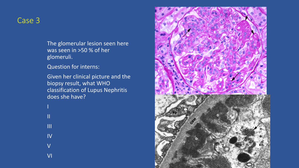
Case 3
The glomerular lesion seen here was seen in >50 % of her glomeruli.Question for interns:
Given her clinical picture and the biopsy result, what WHO classification of Lupus Nephritis does she have?III
IIIIVV
VI

Focal or Diffuse Proliferative Lupus Nephritis(Class III-IV)
• areas of cellular proliferation (long arrows) and by thickening of the glomerular capillary wall (due to immune deposits) that may be prominent enough to form a "wire-loop" (short arrows).
• Although proliferative changes can be focal (affecting less than 50 percent of glomeruli), disease of this severity is usually diffuse.

Focal or Diffuse Proliferative Lupus Nephritis (Class 3-4)
• massive subendothelial deposits (D) • and characteristic tubuloreticular
structures (arrow) in the endothelial cells (En).
• subendothelial deposits cause marked thickening of the glomerular capillary wall, leading to a wire loop appearance on light microscopy.

Case 3
Question for 2nd years: In addition to corticosteroid therapy, what are 2 medications that would be appropriate treatment for induction therapy in this patient?

Treatment: class II LN
• ACE-I/ARB• If proteinuria >3g: steroids or calcineurin inhibitors, as per minimal
change disease guidelines
KDIGO guidelines

Treatment: class III or class IV
• ACE-I/ARB• Pulse monthly steroids followed oral steroids + [IV Cytoxan OR MMF]
for 6 month induction• If worse during first 3 months, change to alternative recommended initially
therapy or repeat biopsy
• Resistant disease: Rituximab, IVIG, calcineurin inhibitors• Maintenance with MMF (1-2g/day in divided doses) or Azathiaprine
(1.5-2.5mg/kg/d) for at least 1 year before tapering after remission achieved
• If worse during taper, go up to previous level of immunosuppression
KDIGO guidelines
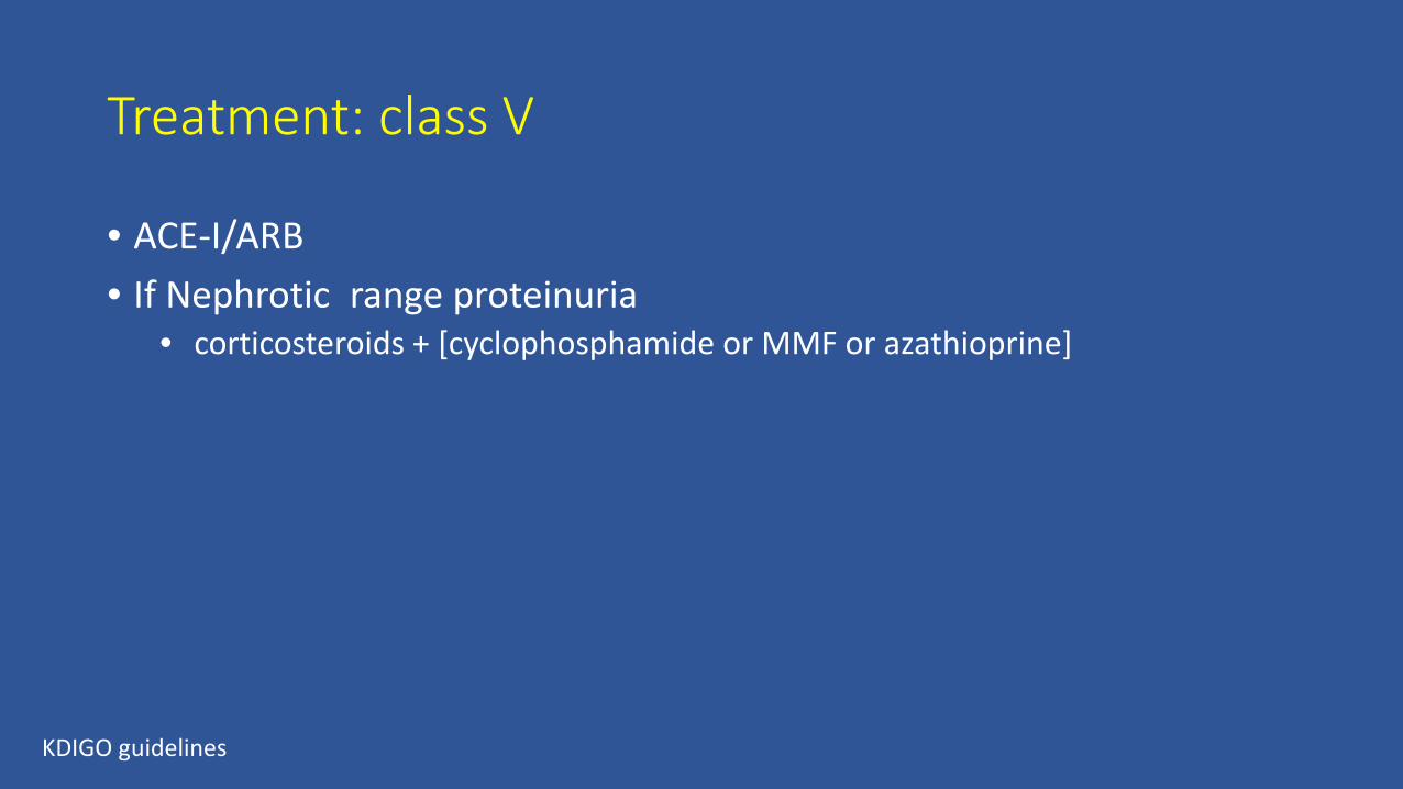
Treatment: class V
• ACE-I/ARB• If Nephrotic range proteinuria
• corticosteroids + [cyclophosphamide or MMF or azathioprine]
KDIGO guidelines

Treatment: Lupus with thrombotic microangiopathy• Antiphospholipid antibody syndrome – anticoagulation• TTP – plasma exchange
KDIGO guidelines

Case 3She is treated with prednisone, lisinopril, and mycophenolate mofetil for 6 months with proteinuria remission and her medications are changed to maintenance therapy. Creatinine 0.8, urine protein:creatinine 0.7g/g creatinine. Her complements normalize and anti-ds DNA is down to 8. She decides to become pregnant.
Question for 3nd years: Which of the following medications does she need to stop during her pregnancy?Prednisone 5mg qdMycophenolate mofetil 1000mg bidHydroxychloroquine 200mg bidLisinopril 40mg qd

Treatment: Lupus in Pregnancy
• Counsel delay in pregnancy until complete remission• No ACE-I/ARB; no MMF, no cyclophosphamide
• (switch MMF to azathioprine)
• Continue hydroxychloroquine• For relapse: steroids +/- azathioprine
• Do not taper until 3 months after delivery
• Low dose aspirin to prevent fetal loss
KDIGO guidelines

Case 4
45 year old woman with no past medical history presenting with fatigue and hemoptysis.Physical exam: BP: 130/82Lungs with bilateral rales diffuselyHeart: normal s1, s2, no murmurs, rubs or gallopsAbdomen: soft, nontenderExtremities: Trace lower extremity edemaSkin: no rash
Sodium: 140 (normal)Potassium: 4.0 (normal)Blood urea nitrogen 30 (high)Creatinine 2.6 (high)Bicarbonate 18 (low)Urinalysis: 1+ blood, 1+ proteinUrine microscopy: no WBCs, 40-60 RBCs, dysmorphic RBC’s and RBC castsUrine protein:creatinine ratio 2.0g/g creatinine

Case 4Labs:Complements normalANA, ANCA, anti-GBM Ab pending
A renal biopsy is performed…
Chest X-Ray

Question for Interns: What is the diagnosis?

Case 4
• Question for 2nd years: How would you treat this?

Treatment: Anti-GBM disease
• Induction:• Cyclophosphamide and steroids and plasmapheresis
• No Maintenance therapy
KDIGO guidelines

Case 4
• Question for 3nd years: What measures can be taken to prevent recurrence of anti-GBM disease in renal transplant?
a) Keep on maintenance prednisoneb) Keep on maintenance cyclophosphamidec) Do a plasmapheresis treatment just prior to the transplantd) No maintenance therapy but wait until anti-GBM antibodies are
negative for 6 months

Treatment: Anti-GBM disease
• Delay transplant until anti-GBM antibodies are undetectable for at least 6 months
KDIGO guidelines

Case 5A 70 year-old man presents with cough and leg
swelling. His hypertension recently been difficult to control.
PMH: HTN, DM2 for 2 years (no retinopathy or neuropathy), atrial fibrillation, sinusitis
PE: afebrile, BP 160/82, HR 85, RR 25, bibasilar rales on lung examirregular S1, S2 with 2/6 systolic murmur left lower sternal border, no gallops, no jugular venous distension, normal point of maximal impulseabdomen normal4+ lower extremity edema.Skin with rare scattered petechie

Case 5LABS: sodium 133, potassium 3.7, chloride 102,
bicarbonate 25, blood urea nitrogen 25, creatinine 2.0, albumin 1.8
urinalysis 1.015/5.0/1+ blood/4+ protein/several RBC casts.
Urine protein to creatinine ratio 3.5g/g.
RENAL ULTRASOUND: normal kidneys, simple 1.5cm cyst right kidney, no hydronephrosis.

Case 5A renal biopsy is perfomed…Question for Interns: What is this?
Immunofluorescence is negative

Pauci-Immune (ANCA) glomerulonephritis
• Light micrograph showing fresh segmental necrotizing lesions with bright red fibrin deposition
EM shows no capillary wall deposits. There is a glomerular basement membrane break, which is the unit lesion leading to exudation of plasma proteins which stimulates proliferation of parietal epithelial cells, leading to crescent formation

Case 5Question for 2nd years: How would you treat him?

Treatment: pauci-immune glomerulonephritis
• Induction:• Cyclophosphamide and steroids• Rituximab and steroids as alternative initial treatment• Stop Cytoxan after 3 months if no significant response
• Maintenance for at least 18 months:• Azathioprine 1-2mg/kg/d• MMF 1g bid• Methotrexate if GFR >60
KDIGO guidelines

Case 5Question for 3rd years: Would you do plasmapheresis?

Treatment: pauci-immune glomerulonephritis
• Induction:• Cyclophosphamide and steroids• Rituximab and steroids as alternative initial treatment• Plasmapheresis if need dialysis or rapidly worsening creatinine or pulmonary
hemorrhage• Stop Cytoxan after 3 months if no significant response
• Maintenance for at least 18 months:• Azathioprine 1-2mg/kg/d• MMF 1g bid• Methotrexate if GFR >60
KDIGO guidelines

Case 653yo M with no significant medical history except he is hepatitis C positive, presents with 2-3 months of hypertension and leg swelling.
PE: BP 140/80CTA BS1, S2Abd benign1+ LE edema bilaterallyNo rash
Labs:Na 140, k 4.2, creatinine 1.3urine protein:creatinine 4.3g/g creatinineUrinalysis 4+ blood 1+ bloodHep C Ab positiveANA positive; anti SSA positive
Renal ultrasound: R 12.7cm, L 11.6cm, 6mm calculus R kidney

Case 6
Anti-ds DNA negativeSPEP negativeANCA NegativeHep C PCR negative
Renal biopsy was perfomed

Case 6
Interns: What does the biopsy show?
IF is negative

Minimal Change Disease
• Epidemiology• Most common cause of idiopathic nephrotic syndrome in children• 10% of nephrotic syndrome in adults
• Etiology• Production of cytokines leading to podocyte dysfunction• Medications: NSAIDS, lithium pamidronate, interferons• Malignancy: Hodgkin Lymphoma, thymoma• Infection• Immunization

Case 6
2nd years: How would you treat him?

Minimal Change Disease: initial treatment
• Prednisone 1mg/kg qd (minimum 4 weeks, max 4 months) until complete remission, then taper slowly over 6 months after remission
• Alternative - Calcineurin inhibitors (unc DM, psychiatric conditions, severe osteoporosis)
KDIGO guidelines

Case 6
He was treated with prednisone 60mg qd and lisinopril1 month later:• Creatinine increased from 1.3->1.5• Urine protein:creatinine decreased 4.3->0.8g/g creatinine2 months later:• Creatinine increased to 1.7• Urine protein:creatinine increased to 1.9g/g creatinine
• Question for 3rd years: How would you treat steroid resistant minimal change disease?

Minimal Change Disease: treatment for steroid resistance or relapse
Relapse (40%) or Steroid resistant: • Oral cyclophosphamide 2-2.5mg/kg/d for 8 weeks• Cyclosporine 3-5mg/kg per day in divided doses, 12 months followed by slow
taper• Mycophenolate Mofetil + steroids• Rituximab
KDIGO guidelines

Case 6
He was treated with cyclosporine • Creatinine increased from 1.7 to 3.4• Urine protein:creatinine decreased from 1.9 to 0.5g/g creatinine
The cyclosporine was stopped• Creatinine decreased to 2.3• Urine protein:creatinine increased to 3.0
He went for a 2nd opinion to Mayo clinic and mycophenolate mofetil was started• Creatinine increased to 4.0• Urine protein:creatinine increased to 11g/g creatinine
He came to BUMC for a 3rd opinion and a repeat biopsy was done…

Case 6
Interns: What does the biopsy show?
IF is negative

FSGS
• Epidemiology• 25% Adults with idiopathic nephrotic syndrome• Most common cause in blacks
• Etiology• Genetic mutation to podocyte proteins• Circulating plasma factor (leads to recurrence after transplant)• Hyperfiltration injury
• HTN, DM2, decreased kidney mass (nephrectomy, congenitally small kidneys, CKD, sickle cell disease)
• Drugs• Pamidronate, interferon

FSGS: treatment
• Primary• Prednisone 1mg/kg qd (minimum 4 weeks, max 4 months) until complete
remission, then taper slowly over 6 months after remission• Alternative - Calcineurin inhibitors (unc DM, psychiatric conditions, severe
osteoporosis)• Relapse or Steroid resistant:
• Cyclosporine 3-5mg/kg per day in divided doses, 12 months followed by slow taper• Mycophenolate Mofetil + steroids
• Secondary• Stop insult• ACE-I/ARB
KDIGO guidelines

FSGS: prognosis
• 40-60% remission

Case 6
He was started on dialysis due to uremia and fluid overloadHe was given Rituxan
After 1 month, he was able to get off dialysis, but gradually his renal function deteriorated and he returned to dialysis
He was listed for a renal transplant and his daughter was a match.He continued to have normal urine output on dialysis and continued to have >10g/day proteinuria.

Case 6
Question for 2nd years: What is his chance of recurrence in the transplant?
5%15%30%80%

FSGS: prognosis after transplant
• 30% recurrence

Case 6
Question for 3rd years: After his transplant, how can we tell if he is getting recurrence in his transplant if his native kidneys are leaking >10g/d protein in the urine? Can you think of anything that could be done so that a recurrence could be detected promptly.

Case 6
He underwent bilateral nephrectomies in preparation for transplant.
After transplant, his creatinine decreased to 1.0 and he had no protein in his urine.
1 week after transplant, he began to get leg swelling and HTN. Creatinine still normal, but urine protein:creatinine was 4g/g creatinine.He has been on plasmapheresis with improvement in his proteinuria and Rituxan is planned.

Case 7
A 37 year old woman with no significant medical history presents to her PCP with dyspnea for 1 day and LE edema for 1 month. PE: BP 140/90, HR 110 BMI is 25, O2 sat 82% on room airCTA BNormal S1, S2Abdomen benign2+ LE edema bilaterallyNo rashThe doctor sends her to the ER due to the low oxygen saturation

Case 7
Labs:Na 135K 3.6Albumin 1.9Creatinine 1.2Urinalysis: negative for blood, 3+ protein; micro no rbcs or wbcsUrine protein:creatinine 6g/g creatinine
CXR clear

Case 7
She is treated for her pulmonary embolism with anticoagulation
Question for interns: What do you think is the cause of her renal disease?
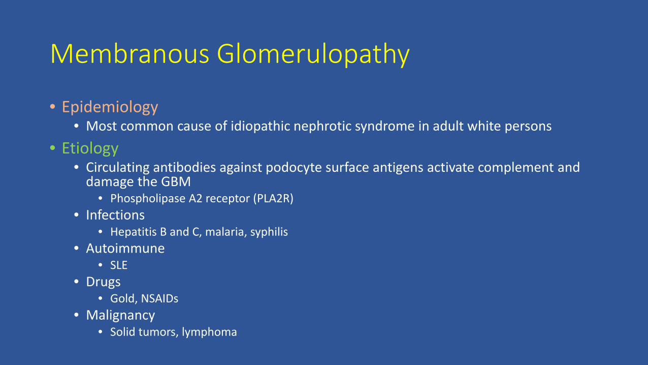
Membranous Glomerulopathy
• Epidemiology• Most common cause of idiopathic nephrotic syndrome in adult white persons
• Etiology• Circulating antibodies against podocyte surface antigens activate complement and
damage the GBM• Phospholipase A2 receptor (PLA2R)
• Infections• Hepatitis B and C, malaria, syphilis
• Autoimmune• SLE
• Drugs• Gold, NSAIDs
• Malignancy• Solid tumors, lymphoma

Case 7
Question for 2nd years: Would you do a renal biopsy at this time?

Membranous Glomerulopathy: treatment
• Primary• 6months ACE-I/ARB first ( 1/3 patients remit spontaneously in 6-12 months)• Immunosuppression if nephrotic + SCr rise by >30% AND GFR >25-30ml/min
• Secondary• Treatment of infection• Treatment of malignancy• Withdrawal of drugs
KDIGO guidelines

Case 7
She is treated with lisinopril and after 1 year her proteinuria is up to 7g/g creatinine. Creatinine is 1.2.Her anticoagulation is stopped and she undergoes a renal biopsy which confirms membranous glomerulopathy.An evaluation for secondary causes of membranous is negative.
Question for 3rd years: How would you treat her membranous at this time?

Membranous Glomerulopathy: treatment
• Primary• 6months ACE-I/ARB first ( 1/3 patients remit spontaneously in 6-12 months)• Immunosuppression if nephrotic + SCr rise by >30% AND GFR >25-30ml/min
• Alternating monthly cyclophosphamide (or chlorambucil) and oral/IV steroids for 6 months, then observe for 6 months
• Calcineurin inhibitors (cyclosporine of tacrolimus) for at least 6 months, followed by taper at 4-8 week intervals to 50% starting dosage and continued for at least 12 months
• Other: MMF, adrenocorticotropic hormone, rituximab
• Secondary• Treatment of infection• Treatment of malignancy• Withdrawal of drugs
KDIGO guidelines

THE PRIZE
NIGHT HIKE WITH ME BY THE LIGHT OF THE CRESCENT MOON IN THE PHOENIX MOUNTAIN PRESERVE IN CELEBRATION OF THE BEAUTY OF CRESCENTIC
GLOMERULONEPHRITIS BIOPSIES