Embed Size (px)
Citation preview

s
cols
SA
face reser-missions toife in theseEarth havecorrelated
d preservedr investi-ude higherdling. Thegeomicrobi-e and the
onments,ng a tracerd to verifygn of life
Icarus 174 (2005) 572–584www.elsevier.com/locate/icaru
Glacial ice cores:A model system for developing extraterrestrial decontamination proto
Brent C. Christner, Jill A. Mikucki, Christine M. Foreman, Jackie Denson, John C. Priscu∗
Department of Land Resources and Environmental Sciences, 334 Leon Johnson Hall, Montana State University, Bozeman, MT 59717, U
Received 15 February 2004; revised 8 September 2004
Abstract
Evidence gathered from spacecraft orbiting Mars has shown that water ice exists at both poles and may form a large subsurvoir at lower latitudes. The recent exploration of the martian surface by unmanned landers and surface rovers, and the plannedeventually return samples to Earth have raised concerns regarding both forward and back contamination. Methods to search for licy environments and adequate protocols to prevent contamination can be tested with earthly analogues. Studies of ice cores onestablished past climate changes and geological events, both globally and regionally, but only recently have these results beenwith the biological materials (i.e., plant fragments, seeds, pollen grains, fungal spores, and microorganisms) that are entrapped anwithin the ice. The inclusion of biology into ice coring research brings with it a whole new approach towards decontamination. Ougations on ice from the Vostok core (Antarctica) have shown that the outer portion of the cores have up to 3 and 2 orders of magnitbacterial density and dissolved organic carbon (DOC) than the inner portion of the cores, respectively, as a result of drilling and hanextreme gradients that exist between the outer and inner portion of these samples make contamination a very relevant aspect ofological investigations with ice cores, particularly when the actual numbers of ambient bacterial cells are low. To address this issuinherent concern it raises for the integrity of future investigations with ice core materials from terrestrial and extraterrestrial envirwe employed a procedure to monitor the decontamination process in which ice core surfaces are painted with a solution containimicroorganism, plasmid DNA, and fluorescent dye before sampling. Using this approach, a simple and direct method is proposethe authenticity of geomicrobiological results obtained from ice core materials. Our protocol has important implications for the desidetection experiments on Mars and the decontamination of samples that will eventually be returned to Earth. 2004 Elsevier Inc. All rights reserved.
Keywords:Exobiology; Ices; Mars; Experimental techniques
ex-anded-
n ofon
d as
uldon-lli,plo-of
llyd in
rded
1. Introduction
NASA and ESA currently have scheduled missions toamine the martian surface and ice caps for liquid waterlife, with the first sample return missions from Mars schuled to launch as early as 2014 (Hubbard et al., 2002; http://nssdc.gsfc.nasa.gov/planetary/mars_2003_05.html). A ma-jor concern in such missions is the forward contaminatioMars with microbes and biological molecules transportedspacecraft from Earth, which might then be misinterprete
* Corresponding author. Fax: +1-406-994-5863.E-mail address:[email protected](J.C. Priscu).
0019-1035/$ – see front matter 2004 Elsevier Inc. All rights reserved.doi:10.1016/j.icarus.2004.10.027
evidence for extraterrestrial life, but more importantly, coirreversibly alter the pristine nature of the martian envirment(Barengoltz, 2000; Rummel, 2000, 2001; Mancine2003). Assessing procedures used in Solar System exration and sample return missions is the responsibilityThe Planetary Protection Advisory Committee, originaestablished by NASA to directly address concerns raisethe United Nations Outer Space Treaty of 1967(Rummel,2001). The measures taken by NASA to control the forwacontamination of Mars during the Viking missions seem
appropriate at the time. However, nearly 30 years of delib-eration and an increased knowledge of the tenacity of lifehave made it necessary to reevaluate appropriate protocols
ite,
Microbial and molecular decontamination of glacial ice cores 573
Table 1Recent ice-based biological studies of glacial and subglacial environments
Origin of ice core or glacialsamples
Investigators Techniques employed Decontamination procedure
Vostok Station, Antarctica Abyzov (1993), Abyzov et al. (1998, 2001)Culture-based, physiological, andmicroscopy
Device that melts sample from theice core interior
Ellesmere Island, Canadian ArcticDancer et al. (1997) Culture-based Flame-sterilized ice axesHans Tausen ice cap, Greenland Willerslev et al. (1999) 16/18S rDNA-based identification Band saw removal of ice core
exteriors
Vostok Station, Antarctica Priscu et al. (1999) Geochemical, 16S rDNA-basedidentification, and microscopy
Washing procedure
Vostok Station, Antarctica Karl et al. (1999) Geochemical, physiological,microscopy, and flow cytometry
Washing procedure
Dye 3 and GISP2, Greenland Castello et al. (1999) RNA-based identification Aseptic subcoring of ice samplesand UV irradiance
Nonpolar and polar ice coresamples
Christner et al. (2000, 2001, 2003) Culture-based, 16S rDNA-basedidentification, physiological, andmicroscopy
Sampling device that melts samplefrom the ice core interior andwashing procedures
GISP2, Greenland Sheridan et al. (2003), Miteva et al.(2004)
Culture-based, 16S rDNA-basedidentification, and flow cytometry
Aseptic subcoring of cleaned icesamples
Ice made in the laboratorya Rogers et al. (2004) Culture and PCR based analysis Washing with sodium hypochlorethanol, HCl, NaOH, and H2O2,
UV-irradiance, and aseptic subcoringntam
ina-ap-rial
im-ableben
trialilar
de-istrynicro-bio-valutest-ughical
ngaltself
nd.g.,al
thenatebio-
bac-erialtioningre-ceonta-trialpre-
ma-ans in
oretiga-pro-plestotiveth-ex-d thesti-ter-tion
a “Sham” cores constructed to test various methods of ice core deco
for the protection of planets and satellites from contamtion by earthly microorganisms. The implementation ofpropriate procedures to decontaminate spacecraft mateand authenticate biological results is therefore of primeportance to these scientific endeavors. Developing suitapproaches to conduct these future studies can directlyefit from the experience gained from the study of terresglacial and subglacial environments, which confront simcontamination issues.
Ice cores from glacial ice on Earth have providedtailed paleorecords of temperature, precipitation, chemand gas composition of the lower atmosphere, volcaeruptions, solar variability, sea surface productivity, anthpogenic emissions, and a variety of other climate andgeochemical indicators. These data have proven to be inable in our understanding of climate processes and foring theories that can predict future climatic changes. Thothis research has integrated many disciplines, the biologmaterial (i.e., plant fragments, seeds, pollen grains, fuspores, and microorganisms) preserved within the ice ihas only recently received attention (Table 1). As a resultof this new information, we now believe that glaciers alarge subglacial lakes sealed below kilometers of ice (eSubglacial Lake Vostok) contain viable life and biologicmolecules that persist over extended periods of time(Priscuand Christner, 2004).
The foremost technical obstacle in ice core studies isimplementation of appropriate procedures to decontamisamples used in microbiological, molecular-based, and
geochemical analyses. Ice core drilling requires the use ofa drilling fluid (typically n-butyl acetate or kerosene and adensifier) to prevent lithostatic pressure from causing plas-ination.
s
-
-
tic collapse of the borehole at drilling depths>300 m of ice(Talalay and Gundestrup, 2002a, 2002b). These fluids arenot sterilized or pasteurized previous to use, contain notericides, and kerosene-based fluids contain known bactsubstrates. Ice cores are further handled during collecand archiving, with no special considerations for removor preventing additional biological contamination. Thefore, the major concern in a microbiological study with icore materials is that the sample was adequately decminated. While it has been argued that glaciated terresenvironments may serve as an appropriate analog forserving life that exists or did exists on Mars(Christner et al.,2000; Priscu and Christner, 2004), the information gained inthe development of strategies to decontaminate ice coreterials is directly applicable for evaluating biologically cleapproaches crucial to the search for life and biosignaturemartian ice samples.
The issue of microbiological decontamination of ice cmaterials has been addressed by a number of investors who have independently tested and implementedcedures in their laboratories to aseptically obtain sam(Table 1). While considerable effort has been put forthdevelop clean sampling technology, proving that a posiresult is not an artifact is nevertheless very difficult wiout experimental evidence to confirm the removal oftraneous cells and molecules. To address this issue aninherent concern it raises for the integrity of future invegations with ice materials from both terrestrial and extrarestrial sources, we employed quantitative decontamina
procedures in our laboratory to verify the authenticity ofresults obtained. Herein we report the efficacy of these pro-cedures on earthly ice and discuss their relevance to life
574 B.C. Christner et al. / Icarus 174 (2005) 572–584
Fig. 1. Epifluorescence images of apparently prokaryotic cells in successive samples collected after the exterior 0.47 cm was removed (A), and from interiore rad YBR-Gold,rial c
on
okapede
-ngeseinofnts
r tothelar
opedm-
oore
rho-
valthe
samples collected after 0.57 cm (B) and 0.82 cm (C) of the original cora DNA specific probe (A)–(C). Scanning electron micrographs of bacte
detection missions that will sample icy environmentsMars.
2. Ice core decontamination and sampling
In initial sampling trials with ice samples from the Vostice core, microscopic analysis established that rod-shcells (∼3 µm long and∼1 µm wide) were abundant on thcore exterior (Fig. 1A, 1D, and 1E), which apparently resulted from microbial contamination present in the drillifluid or were introduced during subsequent handling. Thcells were morphologically distinct from those observedmelted fractions from the core interior, the vast majoritywhich were cocci 0.7–1.4 µm in diameter. Direct cell cou
of collections from the ice core exterior verified that thenumbers of cells were up to 3 orders of magnitude higherthan those observed from the core interior (Table 2).ius was removed from Vostok core 1577. The cells were stained with Sontaminants on the outer portion of the core (D), (E).
Due to the extreme gradient of cells from the exteriothe interior of the ice core, it was essential to confirm thatsampling method was effective in removing external celluand molecular contamination. Based on strategies develto examine cellular contamination during drilling and sapling of permafrost samples (e.g.,Shi et al., 1997; Willerslevet al., 2003, 2004a, 2004b), we amended our protocol tinclude an intentional contamination of the ice core befsampling with the microorganismSerratia marcescens. Inaddition, a target DNA sequence and fluorescent dye (damine 6G) were also included in the tracer mixture (Ta-ble 3), which provided a means to also monitor the remoof macromolecular and molecular contamination duringsampling procedure.
After the tracer mixture was applied, the exterior por-tion (5 mm of the radius; 10 mm of the diameter) of the icecore was physically removed by scraping with a microtome

e
rface (mbs).
ice sheet;
Microbial and molecular decontamination of glacial ice cores 575
Table 2The numbers of cells per milliliter (cell ml−1) in melt water collected from the outer surface and innermost portion of samples from the Vostok ice cor
Core IDa Core ageb
(yrs.)Ice core exterior Ice core interior Ratio from
outside toinside
Cumulative average cell
ml−1 ± SDc%
errordCore radiusremoved(cm)
Cumulative average cell
ml−1 ± SD
%
errordTotal core radiusremoved(cm)
V179 7.12E+03 6.54E+03± 7.26E+01 1.1 0.52 1.01E+02± 2.17E+01 22 1.50 3.21E+01
V763 5.05E+04 4.77E+03± 1.86E+02 3.9 0.40 1.13E+02± 2.21E+01 19 0.78 4.21E+01
V1577 1.14E+05 3.02E+05± 3.55E+02 0.1 0.47 3.79E+02± 5.25E+01 14 0.82 7.98E+02
V2091 1.53E+05 1.83E+05± 1.81E+03 1.0 0.71 4.23E+01± 1.08E+01 25 1.26 4.33E+03
V2490 2.02E+05 2.34E+04± 3.99E+02 1.7 0.69 8.88E+01± 2.00E+01 23 1.01 2.64E+02
V2749 2.37E+05 4.81E+03± 1.08E+02 2.3 0.63 1.82E+02± 2.99E+01 16 1.30 6.47E+01
V3015 2.98E+05 2.36E+04± 8.21E+02 3.5 0.59 3.49E+01± 1.59E+01 45 0.93 6.78E+02
V3197 3.60E+05 2.84E+03± 1.28E+02 4.5 0.74 3.35E+01± 9.80E+00 29 1.16 8.49E+01
V3351 NA 1.17E+04± 3.61E+02 3.1 0.31 6.81E+01± 1.35E+01 20 0.37 1.72E+02
V3540e NA 2.41E+04± 4.90E+02 2.0 0.48 3.70E+02± 3.79E+01 10 0.67 6.49E+01
V3566e NA 2.94E+04± 5.67E+02 1.9 0.38 4.29E+02± 2.27E+01 5.3 0.56 6.84E+01
SD—standard deviation from the mean. NA—not available. See footnote e.a Ice core samples are designated by the depth of recovery from below the surface. For example, V179 was recovered from 179 m below the sub Based onPetit et al. (1999)time scale.c Numbers reflect the background level of cells on core exteriors due to contamination from drilling fluid, subsequent handling, and archiving.d The percent error is the standard deviation divided by the arithmetic mean.e Ice core samples not in stratigraphic order or of meteoric origin (i.e., derived by accretion of water from Lake Vostok onto the bottom of the
Jouzel et al., 1999).
Table 3Tracers used for monitoring ice core sampling
Tracer Specifics Concentration applied Rationale Detection limit
Cell Serratia marcescens 5× 10⁷ cells ∼105 cells ml−1 observed on coreexteriors
1 cfu ml−1
Macromolecule 4.3kb DNA plasmid:pTOPO-ITS-Naegleriaa
Mol. wt. ∼2.6× 10⁶
2 ng
(4.4× 10⁸ molecules)If ∼105 cells ml−1 on exterior,
105–106 16S rDNA copies arepresent
500 molecules ml−1 (round of PCR)−1
(theoretical)
Molecule Rhodamine 6G:Mol wt. 479.0
100 mg l−1 DOC values 2 log higher on coreexterior than core interior
0.001 mg l−1
cfu—colony forming units ofS. marcescenson tryptic-soy agar media.leotid o
anden-sheded
ture
alsoene-at it-ub-
r-
-was
nspro-
ted
allyf
a Plasmid DNA is pCRII-TOPO vector (Invitrogen) containing∼400 nucbe present in the sample.
blade to remove the contaminated exterior of the sampleexpose previously unhandled ice. In a biologically-cleanvironment, the scraped ice samples were thoroughly wawith 95% ethanol and then rinsed with sterile deionizH2O, to disinfect and remove another∼5 mm layer of theice core. Initial rinsing with 4◦C sterile deionized H2O re-sulted in extensive core fracturing, due to the temperadifference between the ice (−10◦C) and water (4◦C). Suchfractures can lead to complete sample disintegration andprovide a means of entry for surface contaminants to ptrate into the core interior. Ethanol had the advantage thremained liquid at−20◦C resulting in no ice core fracturing during rinsing, and the ethanol was easily diluted ssequently to nontoxic concentrations by rinsing with 4◦Csterile deionized H2O. As measurements of dissolved o
ganic carbon (DOC) are sensitive to the presence of ethanolsamples for these analyses were decontaminated using thscraping protocol described inAppendix A. The cleaned icee cloned DNA insert(Pélandakis et al., 2000)that would not be expected t
core was incubated at 22◦C until at least 15 mm of the original core radius had melted away, and the remaining iceallowed to melt completely in the dark at 4◦C. A detaileddescription of the sampling protocol appears inAppendix A;Fig. 2shows schematically the removal of external portioof the ice core during each step of the decontaminationcedure.
3. Assessment of the decontamination procedure
The outermost portion of each core was contaminawith a total of 5×10⁷ cells ofS. marcescens(Table 3), whichis a concentration equivalent to the cell numbers naturoccurring on core exteriors (Table 2). The effectiveness o
,eremoving viable cells ofS. marcescensfrom the ice surfaceis shown inFig. 3A, with 5.7 × 10⁴ and 1.6 × 10⁵ colonyforming units (cfu) ml−1 of S. marcescensrecovered from
/ Icaru
and
h-
pro-
of
ling
tes
µmristi-les;the
noton-
o re-deraturenc-larostnotdius
roach
576 B.C. Christner et al.
the melted ice scraped from the exterior of samples 1792749, respectively.
Physical scraping of the ice exterior followed by wasing with ethanol and deionized H2O removed 0.7–0.9 cmof the core radius and 25–35% of the ice mass. Thiscedure yielded a sample in which no viable cells ofS.marcescenswere recovered. Direct epifluorescent counts
or cell ml−1 to permit plotting zero data logarithmically. They-axis error bars6G (mg l−1), DOC (mg l−1), and kerosene (mg l−1) measured with the removmide-stained 1% agarose gel used to separate PCR amplified plasmid seq
s 174 (2005) 572–584
cells in washes and melt water recovered during samprevealed that the number of cells ml−1 plateaus after 1.3–1.5 cm of the core radius is removed (Fig. 3A), indicatingthat removal of at least 1.5 cm of core radius eliminasurface contamination.S. marcescenscells from logarithmicgrowth are rod-shaped cells that have a length of 2.5–3and a width of 1–1.5 µm. These test cells are charactecally much larger than those observed in ice core sampcells of this size were never observed in samples fromcore interior.
Although ethanol is an effective disinfectant, it doesdestroy nucleic acids. It was therefore necessary to demstrate that our washing and melting procedure was able tmove extraneous DNA, as the inability to do so would hinPCR-based approaches for characterizing the genetic n(i.e., phylogenetic identification using 16S rDNA sequeing) of microbial populations in the ice. The macromolecuplasmid DNA sequence was amplified from the outermcollected portion of the ice sample, but amplicons weregenerated in samples where at least 1 cm of the core rawas removed, even when a sensitive nested PCR appwas employed (Fig. 3C). Since∼108 copies of the plasmid
was placed on the external surface of each core (Table 3),iest
s
Fig. 2. Schematic illustrating the removal of ice core veneers during eachstep of the decontamination procedure.
which is equivalent to the number of 16S rRNA gene copanticipated in a population of∼107 cells (Klappenbach e
(A) (B)
(C)
Fig. 3. Removal of cells and molecules from the surface of Vostok ice cores 179 and 2749 during sampling. (A) Concentration of culturableSerratia marcescen(cfu ml−1) and the total number of directly counted cells in the fractionated wash and melt water collected during sampling. They-axis is denoted cfu ml−1
denote standard deviation from the mean. (B) Concentration of rhodamineal of core radius during sampling. (C) Negative image of the ethidium bro-
uence from collected samples. Amplification was conducted as detailed in the text.

econt
ina-thece isncearylls.ryNA-ted.
es tohly
on-ad-valalesfac-1;
falls-pecost
oreeredra-ostthe-
assionherd exentctedina-
oforeofple
sedbutsed
coreer,uredut
dis-two
ent
ns
re-mi-inceag-plem-
ga-entble,ro-icenti-ndpro-overtoki-ls in
-ice
rea-ved.thetly,vis-
andwntes.orec-ingene-
ac-
ent-eri-ntsice
Microbial and molecular d
al., 2001), it is reasonable to conclude that our decontamtion method was effective in removing DNA present oncore surface. While the use of a known plasmid sequena robust control for nucleic acid contamination, the preseof S. marcescenson the core exterior serves as a secondcheck for the persistence of DNA released from lysed ceThus, if the results from a PCR-amplified 16S rDNA librayielded sequences that were 100% identical to the 16S rDsequence of theS. marcescensstrain used in the tracer mixture, we would conclude that the sample was contaminaIt is important to acknowledge that PCR-based approachdetect DNA in low biomass and ancient samples are higsusceptible to contamination from the laboratory envirment and via molecular biology reagents. Therefore, indition to the methods described herein to verify the remoof DNA contamination from the sample, it is equally vitthat investigations of this nature follow verifiable guidelinto ensure that positive PCR amplifications are not artitual (e.g.,Cooper and Poinar, 2000; Hofreiter et al., 200Willerslev et al., 2004a).
The concentration of kerosene and rhodamine 6Gbelow detectable levels after∼1 cm of the core radius iremoved (Fig. 3B), implying that penetration of fluid contaminants was minimal. The gas chromatograph-mass strometry (GCMS) results for hydrocarbons on the outermsection of core 2749 was 3450 mg l−1, but fell below con-centrations detected in the blank after 1.27 cm of the cexterior was removed. Even though core 179 was recovwithout the use of drilling fluid, a hydrocarbon concenttion of 322 mg l−1 nevertheless existed on the outermportion of the ice. The mass spectra generated fromexterior of core 2749 (Fig. 4E) mirrors the spectral signature obtained from Jet-A kerosene (Fig. 4A), whereas thespectra generated from the exterior of core 179 has m71 ion peaks from hydrocarbons with a longer retenttime (Fig. 4C) and could be a result of substances otthan kerosene-based hydrocarbons. Diesel fuel is usetensively in drilling operations to run mechanical equipmand it is possible that the hydrocarbon signatures deteon core 179 may have resulted from secondary contamtion of ice cores during handling.
The concentration of DOC in the outermost portioncore 2749 was more than 3 times higher than that of c179 (Fig. 3B), which was expected due to the high levelkerosene observed on the surface of the core 2749 samFrom outside to inside, the DOC concentration decrea14- and 21-fold for core 179 and 2749, respectively,did not plateau at a single uniform value. As discusabove, the inability to detect kerosene after>1 cm of thecore surface was removed demonstrated that ice in theinterior was not contaminated by drilling fluid. Togeththese data indicate that the internal DOC levels measare not due to drilling fluid penetration into the core, b
are a reflection of the actual variability of DOC concentra-tions that exist within these samples. It is not surprising tofind such fluctuations in the level of DOC, as glacial iceamination of glacial ice cores 577
-
-
.
is a heterogeneous mixture containing a nonuniformpersal of impurities. The heterogeneous distribution ofand three-grain boundaries in an ice sample(Nye, 1992;Price, 2000)and the heterogeneous distribution of sediminclusions(Jouzel et al., 1999; Petit et al., 1999)can explainthe variation in DOC concentrations within adjacent portioof the same ice core.
4. Discussion and conclusions
The inclusion of biology into ice coring research hasquired developing effective approaches toward decontanation and methods to validate the results obtained. Sthe outer portion of these cores have up to 3 orders of mnitude higher bacterial density than the innermost sam(Table 2), the extreme gradients which exist make containation a very relevant aspect of microbiological investitions with ice cores, particularly when the actual ambinumbers of bacterial cells are low. We developed a reliamultifaceted protocol that can be used to monitor the micbiological, molecular and geochemical contamination ofcores. Importantly, this protocol provides us with a quatative tool to verify the authenticity of results obtained aeliminate samples that fail to pass the decontaminationcedure. The contrasting drilling techniques used to reccore 179 (borehole BH5) and 2749 (borehole 5G) at VosStation allow the effect of drilling fluid on the microbiologcal and geochemical results to be compared. The crystathe sample from 179 m are small (1–2 mm;Lipenkov et al.,1989), crystal sizes for depths>2083 m have not been published but are likely to be relatively larger at 2749 m, andfrom below 3539 m has a mean crystal size>20 cm to 1 m(Jouzel et al., 1999). Our protocol shows that samples afree of drilling fluid, microbial, and macromolecular contmination when at least 3 cm of the core diameter is remoHence, crystal structure appears to have little impact onpermeation of our experimental contaminants. Importanall of our studies were conducted on cores containing noible physical fractures (tested with cross-polarized lightmicroscopy). The sample from 762 m is from what is knoas the “brittle zone,” where gases begin to form clathraCoring this brittle ice is difficult and often yields core of poquality with visible fractures. We purposely selected a stion of this core that contained no visible fractures makour depth comparisons comparable. Presumably, the ptration of contaminants in ice samples with physical frtures will show different results than we present here.
Our data are consistent with previous studies documing the penetration of contaminants into ice core matals which include (i) electrical conductivity measuremeshowing that impurities diffuse about 10 mm into theover 10 years at−20◦C (Schwander et al., 1983), (ii) 1000-
fold lower lead concentrations after<2 cm of the core ra-dius was removed(Boutron and Patterson, 1986), and (iii)drilling fluid penetration analysis that indicates hydrophobic
mples 179
578 B.C. Christner et al. / Icarus 174 (2005) 572–584
Fig. 4. GCMS spectrum from organic phase extraction of (A) Jet-A fuel compared to spectrum from exterior and interior samples of Vostok ice sa
and 2749. (B) is a deionized water blank. (C) is the outermost scraped 1.0 cm of the exterior radius of core 179 and (D) is an internal sample of 179 afterfrom terior radiuoved e surface.
1.0 cm was removed and includes the ice present to a depth of 1.5 cmof core 2749 and (F) an internal sample of 2749 after 1.27 cm was rem
butyl acetate permeates<5 mm into the interior of glacialice cores(Gosink et al., 1994). Previous reports of microbiallife in the Vostok ice core byKarl et al. (1999), Priscu et al.
the original core surface. (E) The outermost scraped 1.27 cm of the exsand includes the ice present to a depth of 3.8 cm from the original cor
(1999), andChristner et al. (2001)used ice core decontami-nation procedures (Table 1) that either washed and removedapproximately 40% of the ice core (∼3 cm from the diame-

econt
thaesics,theau-
di-i-er-as-is-
n-)res
wasionex-atn
sria
ceen-toth-ntson.orenyan-ro-ncerel-
naldes
ru-ores
lassin-ncemi-theand3)
ato-l,
n-ntly
ially4 hreispo-rres-the
areion,ar-ad;
ole-a-
issuebehlyace.sid-
r-that
nce,n-and
lifel ul-
an-ableotics ofam-vedandtole-thatn becro-) of
ter-havema-;d-ex-
Microbial and molecular d
ter) or used a melting device (seeChristner et al., 2000) thatcollected sample from the ice core interior (>2 cm internalto the ice core exterior). The data we present here showsthe removal of∼3 cm from the diameter of Vostok ice coryields contaminated free ice in terms of dissolved organDNA, and microbial cells. Hence, our data corroboratemicrobial and geochemical results published by thesethors.
A number of approaches have been proposed torectly or indirectly detect extraterrestrial life, including mcroscopic observation of biogenically precipitated minals (McKay et al., 1996; Friedmann et al., 2001; ThomKeprta et al., 2001), investigating stable carbon isotopic dtributions(Mojzsis and Arrhenius, 1998), examining aminoacid homochirality(Bada, 2001), measuring elemental abudance of biogenic elements(Conrad and Nealson, 2001,and physiological analysis of biological redox signatu(Crawford et al., 2002). The failure of the Viking GCMSto detect any organic compounds in the martian soila strong incentive to explore a nonbiological explanatfor the positive result obtained from the labeled releaseperiment(Klein, 1978). It has since been documented thViking’s GCMS, which was claimed to have part per billiosensitivity(Biemann et al., 1977), could not detect organicin dry valley Antarctic soils that contained viable bacte(Levin and Straat, 1981)and required>3.0 × 10⁷ cells pergram of soil to achieve its detection threshold(Glavin et al.,2001). If fossilized or extant life is present in the martian icaps or frozen subsurface, it is expected to be of low dsity, which will require extremely sensitive instrumentsidentify diminutive signatures of life. As such, these meods will be particularly vulnerable to even trace amouof residual terrestrial microbial and organic contaminatiTo our knowledge, the cell densities in the Vostok ice c(Fig. 3A andTable 2) are among the lowest reported in anatural environment on Earth. Thus, studying microorgisms and biosignatures in Antarctic glacial ice not only pvides a physical analog of potential niches for the persisteor preservation of life on Mars, but also at concentrationsevant to those anticipated in the martian environment.
According to the recommendations of the U.S. NatioAcademy of Sciences Space Studies Board, spacecrafttined for the martian surface without life detection instmentation (category IVa missions) are permitted 300 spper square meter and 3× 10⁵ spores per vehicle(NationalResearch Council, 1992; DeVincenzi et al., 1996). Thesespacecraft are assembled within ultra clean facilities (C10–100,000) in which great lengths are taken to matain an environment that discourages microbial persisteand growth. Nevertheless, spores, viable nonsporulatingcroorganisms, and DNA persevere on spacecraft and inclean facilities where these instruments are constructedhoused(Venkateswaran et al., 2001; La Duc et al., 200.
Landers with life detection instrumentation (category IVbmissions) are expected to have a microbial burden com-mensurate with those of the 1976 Viking landers(Nationalamination of glacial ice cores 579
t
-
Research Council, 1992), which were exposed to dry hefor 30 h at 111.7◦C, but were not then accessible to micrbial assays after heat treatment(National Research Counci1974, 2000; DeVincenzi et al., 1996). While this stringenttreatment is likely to kill the vast majority of microorgaisms, it is interesting to note that an archaea was recedescribed that not only survives moist heat (a substantmore effective method of sterilization than dry heat) for 2at 121◦C, but is capable of cell division at this temperatu(Kashefi and Lovley, 2003). Even if such a heat treatmenteffective in sterilizing the spacecraft and its internal comnents, nucleic acids, proteins, amino acids, and other tetrial organic contamination can persist and may result inmisinterpretation of life detection experiments(Barengoltz,2000). Unless innovative technologies and proceduresdeveloped that can definitively remove such contaminatit is inevitable that probes and rovers landing on the mtian surface will carry a concealed and unwanted paylonamely earthly microorganisms and terrestrial organic mcules of biological origin. Despite this inevitable contamintion, the present Mars missions are scheduled and theof preventing the forward contamination of Mars maya moot point, as it may be already colonized with eartmicrobes seeded from previous explorations to the surfWhile the planned Mars sample return missions are conered by some to be too risky at this point(DiGregorio, 2001),if they are implemented, it will be vital to develop rigoous decontamination procedures and methods to proveterrestrial contamination has not been introduced. Hesimilar considerations for verifying the elimination of cotamination in studies of samples from terrestrial glacialsubglacial environments are apropos to discussions ofdetection experiments in situ and in the samples that wiltimately be returned to Earth.
The use of a tracer that consists of viable microorgisms and biological macromolecules may not be acceptin all extraterrestrial circumstances, and alternative abimixtures that mimic the physical and chemical propertiecells and macromolecules should be investigated. For exple, we have observed in fractionated melt water remoprogressively during ice core sampling that disinfectionremoval of<1.5 cm of the core radius may be sufficientkill extraneous cells, but it is still possible for macromocules to persist. Therefore, we can only be confidenta sample has been adequately decontaminated if it cademonstrated that all three components (i.e., cells, mamolecules, and the smaller fluorescent organic moleculeour tracer mixture are not detectable. Nonbiological wasoluble chemical tracers and fluorescent microspheresbeen used to monitor microbial contamination in deeprine sediment cores and permafrost(House et al., 2003Juck et al., 2004), and similar mixtures are worth consiering as potential tracer compounds for life detection
periments in extraterrestrial environments and sample returnmissions. To definitively rule out the possibility of contami-nation, it will be essential to sequentially test every sample,
/ Icaru
an-ple.lied
ngd inteddeis-
rth-;l.,ndio-
lingthereasirearear-
ntedin
3;
-
tore-uchit isingal.,rief;scan008
ern
sub-ing
andureich-icyter-
larly
ghex-anx-
con-on-notght.
ids,am-for
a-ThethisPP-250,
-ndtified-ul-rol-
00nal,
ofmed,esis-ingednc.,ter.127)
olu-
fthe
580 B.C. Christner et al.
outside to inside, to demonstrate that terrestrial microorgisms and molecules have not infiltrated the inner samThis could be confirmed by the absence of a surface-apptracer in the inner sample.
Sampling of the martian subsurface will require drilliand core recovery strategies analogous to those useterrestrial studies, although substantially more complicalogistically. Examples of such anticipated efforts incluthe Robotic Shallow Mission and Deep Sub-surface Msion, which will attempt to core depths of∼300 m and∼3 km, respectively(Mancinelli, 2003). Evidence for H2Oice in permafrost at high latitudes and in both the noern and southern ice caps of Mars(Boynton et al., 2002Mitrofanov et al., 2003; Titus et al., 2003; Bibring et a2004) implicate the polar regions as the most likely aconvenient environments to detect extant life or the bchemical preservation of past life. In contrast to sampthe deep subsurface, biological investigations of ice inpolar regions (such as the high latitude layered ice athat the Phoenix 2008 mission will analyze) will not requa major invasive drilling effort, as these environmentsrelatively accessible from the surface or sediment-rich mgins of the ice caps. A number of reports have documemicrobial survival for hundreds of thousands of yearsglacial ice (Abyzov et al., 1998; Christner et al., 200Sheridan et al., 2003; Miteva et al., 2004)and for millions ofyears in permafrost(Shi et al., 1997), so it is not unreasonable to suppose microbial longevity over 105–107 years, theestimated time the martian ice caps have existed(Herkenhoffand Plaut, 2000). However, the most favorable placesearch for existing martian life in the near surface is ingions that have experienced recent or periodic melting, sas the environments contiguous to the ice caps, wherepredicted that the discharge of liquid water occurs durhigh obliquity (Pathare and Paige, 1998; Jakowski et2003). There is also evidence that liquid water exists for bperiods in mid to low latitudes(Malin and Edgett, 2000Christensen, 2003; Arvidson et al., 2004), but it seems leslikely that surface environments at these lower latitudessustain a permanent environment for life. The proposed 2Phoenix NASA mission will land between latitudes 65◦ and75◦ N, and will operate for up to 150 sols during the northsummer (http://phoenix.lpl.arizona.edu). The key objectiveof the mission is to examine samples of surface andsurface soil and ice, returning a wealth of data concernnear-surface ices, the potential habitability of the soil,the history of climate as written into the icy soils. To ensenvironmental protection of the polar environment, whhas a higher probability of habitability, it is especially important that this mission and other future missions to theregions of Mars include methods to prevent and detectrestrial contamination.
The contamination concerns raised herein are particu
relevant to any discussion of life detection in samples fromthe ice caps or permafrost in the martian polar regions. Whileour view may seem alarmist, the issue of terrestrial conta-s 174 (2005) 572–584
mination is an important and legitimate matter. Althouimplementing a verifiably clean sampling strategy fortraterrestrial life detection experiments will entail addingadditional technological barrier to an already difficult, epensive, and precarious mission, it will be necessary tovince the scientific and lay communities that evidence csistent with a martian life signature is unequivocal andjust the result of contamination caused by a lack of foresi
Acknowledgments
We are grateful to Kathy Sheehan for providing plasmDNA and primers, Kathy Welch, Mary Voytek, Ed Adamand Tucker Stevens for assisting with ice core cutting, spling, and processing, Phil Butterfield and the CenterBiofilm Engineering at MSU for help with the DOC mesurements, and Joe Sears for support with the GCMS.comments of 2 anonymous reviewers greatly improvedmanuscript. This study was funded by NSF grants O0085400, OPP-0440943, MCB-0237335 and OPP-0096awarded to J.C.P.
Appendix A
A.1. Bacterial strain, plasmid DNA, and fluorescent dye
Serratia marcescens(MSU strain) was cultured in trypticsoy broth or on 1.5% agar solidified media (Difco Inc.) aused as the indicator strain. Cell suspensions were quanby directly counting cells with a Nikon Optiphot epifluorescent microscope equipped with a DM510 filter. Cture stocks were preserved in a solution of 15% glyceand stored at−80◦C. The plasmid DNA construct consists of the pCRII-TOPO vector (Invitrogen) with a 4bp insert amplified from the 5.8S rRNA gene and intertranscribed spacers ofNaegleria fowleri(Pélandakis et al.2000), a eukaryotic pathogen which is the primary causeamoebic meningoencephalitis. The plasmid was transforinto competentEscherichia coliJM 109 cells (Promega)plasmid-containing colonies were selected based on rtance to ampicillin, and plasmid DNA was prepared usthe QIAprep Spin Miniprep Kit (Qiagen) and quantitatusing Pico Green nucleic acid stain (Molecular Probes, Icat. no. P-7589) on a Biotek FL600 microplate fluoromeRhodamine 6G was purchased from Sigma (cat. no. R 4and kept as a stock solution of 1000 mg l−1.
The above components were combined in a 1.1 ml stion containing 4.5 × 10⁷ cells ml−1 of S. marcescens, 2 ngof plasmid DNA, and 100 mg l−1 (final concentration) orhodamine 6G. The rationale and detection limits forconstituents are summarized inTable 3. This solution was
spread evenly on all ice core surfaces with a sterile latex-gloved hand. The visible presence of rhodamine on ice sur-faces ensured that the entire core was contaminated.
econt
bs)ca)
assbs),bs)bs)bs)
5.32corelativeereple
of
01;
ewfuelom-illa-ical,2,4-, thevententas a
Na-teder-dedsityg
ra-
in acol-thennchtelydof
iceow
osi-delh
3-cednderra-thiswased
s
ro-sec-
tionus-
h ace-col-
vialste-ingszen
oldfiednllere
ldid-re-
wasndenar-dotope
on,cat.lineted
-llsuo-nalds
Microbial and molecular d
A.2. Ice core source
Core section 179 (178.00–179.00 m below surface, moriginated from a borehole at Vostok Station (Antarctidesignated BH5. The BH5 borehole is shallow, and wtherefore drilled without the aid of drilling fluid. Section763 (762.00–762.32 mbs), 1577 (1576.00–1576.33 m2091 (2090.30–2090.60 mbs), 2490 (2489.00–2489.31 m2749 (2748.43–2748.94 mbs), 3015 (3014.71–3015.00 m3197 (3196.28–3196.51 mbs), 3351 (3350.05–3350.50 m3540 (3539.62–3539.99 mbs), and 3566 (3564.99–356mbs) were recovered from the deep 5G borehole. Icesamples are referred to and designated by the depth reto the surface (i.e., V179 represents the ice sample recovfrom 178.00–179.00 mbs). The age of each ice core samappears inTable 2. The physical and chemical propertiesthe Vostok ice core have been studied extensively (e.g.,Petitet al., 1999; Priscu et al., 1999; Cuffey and Vimeux, 20Caillon et al., 2003).
The drilling fluid used by the Russian-based drilling crin the 5G borehole is commercial grade kerosene or jet(trade names: Jet-A, TC-1, JP-8), which has a complex cbination of C9 to C16 hydrocarbons produced by the disttion of crude oil and includes other trace components typof high end jet fuel such as toluene, ethylbenzene, and 1trimethylbenzene. For core depths between 1–2700 mfuel mixture was supplemented with the halogenated solCFC-11. Following the Montreal Protocol and subsequban on CFCs, the freon derivative Foran 141b was useddensifier (V. Lipenkov, personal communication).
Ice cores were obtained from the United Statestional Ice Core Laboratory (Denver, CO) and airfreighovernight to Montana State University under close supvision. The temperature during shipping never excee−15◦C. Ice cores are archived at Montana State Univerin a walk in freezer at−20◦C, sealed in polyethylene tubinand stored in cardboard core tubes.
A.3. Ice core sampling
The outermost layer of ice (at least 5 mm of the coredius, 10 mm of the core diameter;Fig. 2) was scraped witha 95% ethanol-rinsed stainless steel microtome blade−10◦C cold room and the removed ice scrapings werelected for analysis as described below. Ice samples weretransferred to a Class 100 Purifier Horizontal Clean Be(Labconco Corporation, model 36125), housed separawithin a −20◦C walk in freezer, which was precleanewith a solution of 95% ethanol and exposed to 20 minultraviolet (UV-C) irradiation. Sections of the scrapedcores were held with sterile tongs within the laminar flhood and completely rinsed with 500 ml of−20◦C 0.2 µm-filtered 95% ethanol, and then rinsed with 500 ml of 4◦C,
sterile, organic-free, deionized H2O (EASYpure™UV/UF,Barnstead Thermolyne Corp.). The deionized H2O wash wascollected for analysis. The remaining ice cores were placedamination of glacial ice cores 581
,,,
d
in sterile polypropylene containers and transferred to a ptive pressure glove box (Analytical Balance Chamber, mo830-ABC, Plas-Labs, Inc.) at 22◦C that was precleaned wita solution of 0.25% sodium hypochlorite and evacuatedtimes with 0.2 µm-filtered zero air. Ice samples were plain sterile 16 cm stainless steel strainers and incubated uthese conditions until at least 15 mm of the original coredius had melted away. The melt water generated duringprocedure was collected for analysis. The remaining icesealed within a sterile polypropylene container and allowto melt completely in the dark at 4◦C, which took as long a72 h.
Samples for dissolved organic carbon (DOC) and kesene analysis were decontaminated separately from thetions of cores used for biological analysis. The outer porof the ice core surface was removed at 1–2 mm intervalsing a sterile razor blade inside a−20◦C walk-in freezer. Forkerosene measurements, razor blades were cleaned wittone and dried thoroughly. Successive scrapings werelected in acid washed combusted glass Shimadzu TOCto monitor the DOC content from the outside to the inrior of the ice cores. For analysis of kerosene, ice shavwere collected in precombusted glass vials and kept froat−20◦C until extraction.
A.4. Viable and direct cell counts
The number of culturable cells ofS. marcescenswas de-termined by counting the colonies that formed on 10-fserial dilutions of sample spread plated on agar-soliditryptic-soy media, following visible growth after incubatioat 25◦C for 48 h. Culturable refers to the ability of a ceto form a colony on agar-solidified media, designated has the number of colony-forming units per ml (cfu ml−1) ofsample.
Sodium borate-buffered formalin and the SYBR Go(Molecular Probes, Inc., cat. no. S-11494) nucleic acstaining solutions were passed through 0.2 µm filters tomove extraneous particles and cells. A 10 ml samplefixed in a final concentration of 5% buffered formalin astained with SYBR Gold for 15 min. The sample was thfiltered on a 5 mm spot of a 25 mm–0.2 µm black polycbonate filter (Poretics, cat. No. K02BP02500) using ablot apparatus. Filters were mounted on glass microscslides with the addition of 2 drops of an anti-fade solutiwhich consisted of 0.1% p-phenylenediamine (Sigma,no. P-1519) in a 1:1 solution of phosphate-buffered saand glycerin. All filtering and manipulations were conducwithin a BioGard vertical laminar flow hood (Baker Company, model B6000-1) with a germicidal UV-C lamp. Ceon the filters were counted using a Nikon Optiphot epiflrescent microscope, equipped with a DM510 filter, at fimagnification of 1000X. The total number of cocci and ro
in 60 fields of view was determined, and the number ofcells ml−1 was calculated by computing the cumulative aver-age cell field−1 (field of view area at 1000X is 16741 µm2).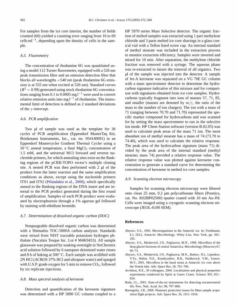
/ Icaru
lds69m-
us-nmthats-rvestra-t-ons
30it;an
g a
nu-nk-g
thetioners
in-undalu-wed
inedrdsph-plemixsesthed
frac-lenecon-rdess
andideasewople
umndro-ari-dro-85,
s ofspe-nedtive
wasmost0 tonse.) di-thyl. Thecon-the
redtics,Pd.mi-
nn,–
he67,
v,v,
ove
rties821–
strial
582 B.C. Christner et al.
For samples from the ice core interior, the number of fiecounted (60) yielded a counting error ranging from 10 tocells ml−1, depending upon the density of cells in the saple.
A.5. Fluorometry
The concentration of rhodamine 6G was quantitateding a model 112 Turner fluorometer, equipped with a 526peak transmission filter and an emission detection filterblocks all wavelengths<540 nm (peak rhodamine 6G emision is at 555 nm when excited at 526 nm). Standard cu(R2 > 0.99) generated using stock rhodamine 6G concentions ranging from 0.1 to 0.0005 mg l−1 were used to converrelative emission units into mg l−1 of rhodamine. The instrumental limit of detection is defined as 2 standard deviatiof they-intercept.
A.6. PCR amplification
Two µl of sample was used as the template forcycles of PCR amplification (Eppendorf MasterTaq KBrinkmann Instruments, Inc., cat. no. 954140091) inEppendorf Mastercycler Gradient Thermal Cycler usin50◦C anneal temperature, a final MgCl2 concentration of1.5 mM, and the universal M13 forward and reversecleotide primers, for which annealing sites exist on the flaing regions of the pCRII-TOPO vector’s multiple cloninsite. A nested PCR was then performed with 2 µl ofproduct from the latter reaction and the same amplificaconditions as above, except using the nucleotide primITS1 and ITS2(Pélandakis et al., 2000), which specificallyanneal to the flanking regions of the DNA insert and areternal to the PCR product generated during the first roof amplification. Samples of each PCR product were evated by electrophoresis through a 1% agarose gel folloby staining with ethidium bromide.
A.7. Determination of dissolved organic carbon (DOC)
Nonpurgeable dissolved organic carbon was determwith a Shimadzu TOC-5000A carbon analyzer. Standawere mixed from NIST traceable potassium hydrogenthalate (Nacalais Tesque Inc. Lot # M4K9453). All samglassware was prepared by soaking overnight in NoChroacid solution followed by 6 nanopure deionized water rinand 8 h of baking at 500◦C. Each sample was acidified wi2N HCl (ACROS 37% HCl and ultrapure water) and spargwith U.S.P. grade oxygen for 5 min to remove CO2, followedby six replicate injections.
A.8. Mass spectral analysis of kerosene
Detection and quantification of the kerosene signaturewas determined with a HP 5890 GC column coupled to a
s 174 (2005) 572–584
HP 5970 series Mass Selective detector. The organiction of melted samples was extracted using 1 part methychloride and 3 parts melted ice core shavings in a glassical vial with a Teflon lined screw cap. An internal standaof methyl stearate was included in the extraction procto monitor extraction efficiency. Samples were invertedmixed for 10 min. After separation, the methylene chlorfraction was removed with a syringe. The aqueous phwas re-extracted to insure the removal of all organics. Tµl of the sample was injected into the detector. A samof Jet-A kerosene was separated on a VG 70E GC colwith a mass spectrometer detector to determine the hycarbon signature indicative of this mixture and for compson with signatures obtained from ice core samples. Hycarbons typically fragment into ions of masses 57, 71,and smaller (masses are denoted bym/z, the ratio of themass to the number of ion charges). The ion with a mas71 (ranging between 70.70 and 71.70) represented thecific marker compound for hydrocarbons and was scanfor by setting the mass spectrometer to run in the selecion mode. HP Chem Station software (version B.02.05)used to calculate peak areas of the mass 71 ion. Theabundant ion of methyl stearate has a mass of 74 (73.774.40), which was used to calculate the relative respoThe peak area of the hydrocarbon signature (mass 71vided by the peak area of the internal standard (mestearate; mass 74) provided a relative response valuerelative response value was plotted against kerosenecentration to generate a standard curve for determiningconcentration of kerosene in melted ice core samples.
A.9. Scanning electron microscopy
Samples for scanning electron microscopy were filteonto clean 25 mm, 0.2 µm polycarbonate filters (Porecat. No. K02BP02500) sputter coated with 10 nm Au–Cells were imaged using a cryogenic scanning electroncroscope (JEOL-6100 SEM).
References
Abyzov, S.S., 1993. Microorganisms in the Antarctic ice. In: FriedmaE.I. (Ed.), Antarctic Microbiology. Wiley–Liss, New York, pp. 265295.
Abyzov, S.S., Mitskevich, I.N., Poglazova, M.N., 1998. Microflora of tdeep glacier horizons of central Antarctica. Microbiology (Moscow)66–73.
Abyzov, S.S., Mitskevich, I.N., Poglazova, M.N., Barkov, N.I., LipenkoV.Ya., Bobin, N.E., Koudryashov, B.B., Pashkevich, V.M., IvanoM.V., 2001. Microflora in the basal strata at Antarctic ice core abthe Vostok lake. Adv. Space Res. 28, 701–706.
Arvidson, R.E., 30 colleagues, 2004. Localization and physical propeexperiments conducted by Spirit at Gusev Crater. Science 305,824.
Bada, J.L., 2001. State-of-the-art instruments for detecting extraterre
life. Proc. Natl. Acad. Sci. 98, 797–800.Barengoltz, J.B., 2000. Planetary protection issues for Mars sample acqui-sition flight projects. Adv. Space Res. 26, 1911–1916.

econt
r ice
nd in. 82,
ntarc-225.ear, 81–
nkov,e
Bac-saic
melt-
, V.,able
001.ice.
003..
de-
ll.
y inh to
m-tion.
ion opl.
ane-316.
In-
s ofical
De-Sci.
d-In:s.,
of the
ient
onings
the
.T.,bit-
tica.
.W.,FP-edi-ctic.
s,rc-
for
ndb:cids
34,
iallation
for
ex-35,
page
eath
ek,96.ian
hys-and
e300,
obio-Res.
teril-5-
suesPress,
ationgton., N.,sity
ionsandus-
geneleri.
past29–
roc.
Microbial and molecular d
Bibring, J.-P., 12 colleagues, the OMEGA Team, 2004. Perennial wateidentified in the south polar cap of Mars. Nature 428, 627–630.
Biemann, K., 11 colleagues, 1977. The search for organic substances aorganic volatile compounds in the surface of Mars. J. Geophys. Res4641–4658.
Boutron, C.F., Patterson, C.C., 1986. Lead concentration changes in Atic ice during the Wisconsin/Holocene transition. Nature 323, 222–
Boynton, W.V., 24 colleagues, 2002. Distribution of hydrogen in the nsurface of Mars: evidence for subsurface ice deposits. Science 29785.
Caillon, N., Severinghaus, J.P., Jouzel, J., Barnola, J.M., Kang, J., LipeV.Y., 2003. Timing of atmospheric CO2 and Antarctic temperaturchanges across termination III. Science 299, 1728–1731.
Castello, J.D., Rogers, S.O., Starmer, W.T., Catranis, C.M., Ma, L.,hand, G.D., Zhao, Y., Smith, J.E., 1999. Detection of tomato motobamovirus RNA in ancient glacial ice. Polar Biol. 22, 207–212.
Christensen, P.R., 2003. Formation of recent martian gullies throughing of extensive water-rich snow deposits. Nature 422, 45–48.
Christner, B.C., Mosley-Thompson, E., Thompson, L.G., ZagorodnovSandman, K., Reeve, J.N., 2000. Recovery and identification of vibacteria immured in glacial ice. Icarus 144, 479–485.
Christner, B.C., Mosley-Thompson, E., Thompson, L.G., Reeve, J.N., 2Isolation of bacteria and, 16S rDNAs from Lake Vostok accretionEnviron. Microbiol. 3, 570–577.
Christner, B.C., Mosley-Thompson, E., Thompson, L.G., Reeve, J.N., 2Bacterial recovery from ancient ice. Environ. Microbiol. 5, 433–436
Conrad, P.G., Nealson, K.H., 2001. A non-earthcentric approach to lifetection. Astrobiology 1, 15–24.
Cooper, A., Poinar, H.N., 2000. Ancient DNA: do it right or not at aScience 289, 1139.
Crawford, R.L., 12 colleagues, 2002. Measurement of microbial activitsoil by colorimetric observation of in situ dye reduction: an approacdetection of extraterrestrial life. BMC Microbiol. 2, 22.
Cuffey, K.M., Vimeux, F., 2001. Covariation of carbon dioxide and teperature from the Vostok ice core after deuterium-excess correcNature 412, 523–527.
Dancer, S.J., Shears, P., Platt, D.J., 1997. Isolation and characterizatcoliforms from glacial ice and water in Canada’s High Arctic. J. ApMicrobiol. 82, 597–609.
DeVincenzi, D.L., Stabekis, P., Barengoltz, J., 1996. Refinement of pltary protection policy for Mars missions. Adv. Space Res. 18, 311–
DiGregorio, B.E., 2001. The dilemma of Mars sample return. Chem.nov. 31, 18–27.
Friedmann, E.I., Wierzhos, J., Ascaso, C., Winklhofer, M., 2001. Chainmagnetite crystals in the meteorite ALH84001: evidence of biologorigin. Proc. Natl. Acad. Sci. 98, 2176–2181.
Glavin, D.P., Schubert, M., Botta, O., Kminek, G., Bada, J.L., 2001.tecting pyrolysis products from bacteria on Mars. Earth Planet.Lett. 185, 1–5.
Gosink, T.A., Kelley, J.J., Tumeo, M.A., Koci, B., Stanford, K., Zagoronov, V., Ehlert, G., 1994. Fluids for use in deep ice core drilling.Proc. Ice Drilling Technology, Tokyo, 1993. Mem. Natl. Inst. Polar ReSpecial Issue 49, 335–346.
Herkenhoff, K.E., Plaut, J.J., 2000. Surface ages and resurfacing ratespolar layered deposits and dunes on Mars. Icarus 144, 243–253.
Hofreiter, M., Serre, D., Poinar, H.N., Kuch, M., Paabo, S., 2001. AncDNA. Nat. Rev. Gen. 2, 353–359.
House, C.H., Cragg, B., Teske, A., 2003. Drilling contamination testsODP Leg, 201 using chemical and particulate tracers. In: Proceedof the Ocean Drilling Program. Initial Reports. In press.
Hubbard, G.S., Naderi, F.M., Garvin, J.B., 2002. Following the water,new program for Mars exploration. Acta. Astronaut. 51, 337–350.
Jakosky, B.M., Nealson, K.H., Bakermans, C., Ley, R.E., Mellon, M2003. Subfreezing activity of microorganisms and the potential ha
ability of Mars’ polar regions. Astrobiology 3, 343–350.Jouzel, J., Petit, J.R., Souchez, R., Barkov, N.I., Lipenkov, V.Y., Raynaud,D., Stievenard, M., Vassiliev, N.I., Verbeke, V., Vimeux, F., 1999. More
amination of glacial ice cores 583
-
f
than, 200 meters of lake ice above subglacial Lake Vostok, AntarcScience 286, 2138–2141.
Juck, D.F., Whissell, G., Steven, B., Pollard, W., McKay, C.P., Greer, CWhyte, L.G., 2004. Utilization of fluorescent microspheres and a Gmarked strain for detecting microbial contamination in permafrost sment and ground ice drill core samples from the Canadian High ArAppl. Environ. Microbiol. In press.
Karl, D.M., Bird, D.F., Björkman, K., Houlihan, T., Shackelford, R., TupaL., 1999. Microorganisms in the accreted ice of Lake Vostok, Antatica. Science 286, 2144–2147.
Kashefi, K., Lovley, D.R., 2003. Extending the upper temperature limitlife. Science 301, 934.
Klappenbach, J.A., Saxman, P.R., Cole, J.T., Schmidt, T.M., 2001. rrthe ribosomal RNA operon copy number database. Nucleic ARes. 29, 181–184.
Klein, H.P., 1978. The Viking biological experiments on Mars. Icarus666–674.
La Duc, M.T., Nicholson, W., Kern, R., Venkateswaran, K., 2003. Microbcharacterization of the Mars Odyssey spacecraft and its encapsufacility. Environ. Microbiol. 5, 977–985.
Levin, G.V., Straat, P.A., 1981. Antarctic soil no. 726 and implicationsthe Viking labeled release experiment. J. Theor. Biol. 91, 41–45.
Lipenkov, V.Y., Barkov, N.I., Duval, P., Pimienta, P., 1989. Crystalline tture of the, 2083 m ice core at Vostok Station, Antarctica. J. Glaciol.392–398.
Malin, M.C., Edgett, K.S., 2000. Evidence for recent groundwater seeand surface runoff on Mars. Science 288, 2330–2335.
Mancinelli, R.L., 2003. Planetary protection and the search for life benthe surface of Mars. Adv. Space Res. 31, 103–107.
McKay, D.S., Gibson Jr., E.K., Thomas-Keprta, K.L., Vali, H., RomanC.S., Clemett, S.J., Chiller, X.D.F., Maechling, C.R., Zare, R.N., 19Search for past life on Mars: possible relic biogenic activity in martmeteorite ALH84001. Science 273, 924–990.
Miteva, V.I., Sheridan, P.P., Brenchley, J.E., 2004. Phylogenetic and piological diversity of microorganisms isolated from a deep Greenlglacier ice core. Appl. Environ. Microbiol. 70, 202–213.
Mitrofanov, I.G., 11 colleagues, 2003. CO2 snow depth and subsurfacwater-ice abundance in the northern hemisphere of Mars. Science2081–2084.
Mojzsis, S.J., Arrhenius, G., 1998. Phosphates and carbon on Mars: exlogical implications and sample return considerations. J. Geophys.Planets 103, 28495–28511.
National Research Council, 1974. Viking ’75 program lander capsule sization plan. Viking ’75 Project, M75-155-0-5 (Spacecraft A) and M7155-0-6 (Spacecraft B).
National Research Council, 1992. Biological Contamination of Mars, Isand Recommendations. Space Studies Board, National AcademyWashington.
National Research Council, 2000. Preventing the Forward Contaminof Europa. Space Studies Board, National Academy Press, Washin
Nye, J.F., 1992. Water veins and lenses in polycrystalline ice. In: MaenoHondoh, T. (Eds.), Physics and Chemistry of Ice. Hokkaido UniverPress, Sapporo, Japan, pp. 200–205.
Pathare, A.V., Paige, D.A., 1998. Recent liquid water in the polar regof Mars. In: First International Conference on Mars Polar ScienceExploration, LPI Contribution 953. Lunar and Planetary Institute, Hoton, pp. 31–32.
Pélandakis, M., Serre, S., Pernin, P., 2000. Analysis of the 5.8S rRNAand internal transcribed spacers in Naegleria spp. and in N. fowJ. Euk. Microbiol. 47, 116–121.
Petit, J.R., 18 colleagues, 1999. Climate and atmospheric history of the420,000 years from the Vostok ice core, Antarctica. Nature 399, 4436.
Price, P.B., 2000. A habitat for psychrophiles in deep Antarctic ice. PNatl. Acad. Sci. 97, 1247–1251.
Priscu, J.C., Christner, B.C., 2004. Earth’s icy biosphere. In: Bull, A.T.(Ed.), Microbial Diversity and Bioprospecting. American Society forMicrobiology Press, Washington, DC, pp. 130–145.

/ Icaru
bove
.-G.,de-cu-
s for
gy:98,
mentca-
is ofland
rac-NA
corear
gy,
L.,F.,ystals98,
dis-
, C.,bly
er, P.,tl.
etic791–
leicl. 19,
C.,
584 B.C. Christner et al.
Priscu, J.C., 11 colleagues, 1999. Geomicrobiology of subglacial ice aLake Vostok, Antarctica. Science 286, 2141–2144.
Rogers, S.O., Theraisnathan, V., Ma, L.J., Zhao, Y., Zhang, G., Shin, SCastello, J.D., Starmer, W.T., 2004. Comparisons of protocols forcontamination of environmental ice samples for biological and molelar examinations. Appl. Environ. Microbiol. 70, 2540–2544.
Rummel, J.D., 2000. Implementing planetary protection requirementsample return missions. Adv. Space Res. 26, 1893–1899.
Rummel, J.D., 2001. Planetary exploration in the time of astrobioloprotecting against biological contamination. Proc. Natl. Acad. Sci.2128–2131.
Schwander, J., Neftel, A., Oeschger, H., Stauffer, B., 1983. Measureof direct current conductivity on ice samples for climatological applitions. J. Phys. Chem. 87, 4157–4160.
Sheridan, P.P., Miteva, V.I., Brenchley, J.E., 2003. Phylogenetic analysanaerobic psychrophilic enrichment cultures obtained from a Greenglacier ice core. Appl. Environ. Microbiol. 69, 2153–2160.
Shi, T., Reeves, R.H., Gilichinsky, D.A., Friedmann, E.I., 1997. Chaterization of viable bacteria from Siberian permafrost by, 16S rDsequencing. Microbial Ecol. 33, 169–179.
Talalay, P.G., Gundestrup, N.S., 2002a. Hole fluids for deep icedrilling. In: Proc. Ice Drilling Technology, 2000. Mem. Natl. Inst. Pol
Res., Special Issue 56, 148–170.Talalay, P.G., Gundestrup, N.S., 2002b. Hydrostatic pressure and fluid den-
s 174 (2005) 572–584
sity profile in deep ice bore-holes. In: Proc. Ice Drilling Technolo2000. Mem. Natl. Inst. Polar Res., Special Issue 56, 171–181.
Thomas-Keprta, K.L., Clemett, S.J., Bazylinski, D.A., Kirschvink, J.McKay, D.S., Wentworth, S.J., Vali, H., Gibson Jr., E.K., McKay, M.Romanek, C.S., 2001. Truncated hexa-octahedral magnetite crin ALH84001: presumptive biosignatures. Proc. Natl. Acad. Sci.2164–2169.
Titus, T.N., Kieffer, H.H., Christensen, P.R., 2003. Exposed water icecovered near the south pole of Mars. Science 299, 1048–1051.
Venkateswaran, K., Satomi, M., Chung, S., Kern, R., Koukol, R., BasicWhite, D., 2001. Molecular microbial diversity of a spacecraft assemfacility. Syst. Appl. Microbiol. 24, 311–320.
Willerslev, E.A., Hansen, J., Christensen, B., Steffensen, J.P., Arctand1999. Diversity of Holocene life forms in fossil glacier ice. Proc. NaAcad. Sci. 96, 8017–8021.
Willerslev, E.A., 10 colleagues, 2003. Diverse plant and animal genrecords from Holocene and Pleistocene sediments. Science 300,795.
Willerslev, E.A., Hansen, A.J., Poinar, H.N., 2004a. Isolation of nucacids and cultures from fossil ice and permafrost. Trends Ecol. Evo141–147.
Willerslev, E.A., Hansen, A.J., Rønn, R., Brand, T.B., Barnes, I., Wiuf,
Gilichinsky, D., Mitchell, D., Cooper, A., 2004b. Long-term persistenceof bacterial DNA. Curr. Biol. 14, 9–10.