Embed Size (px)
Citation preview

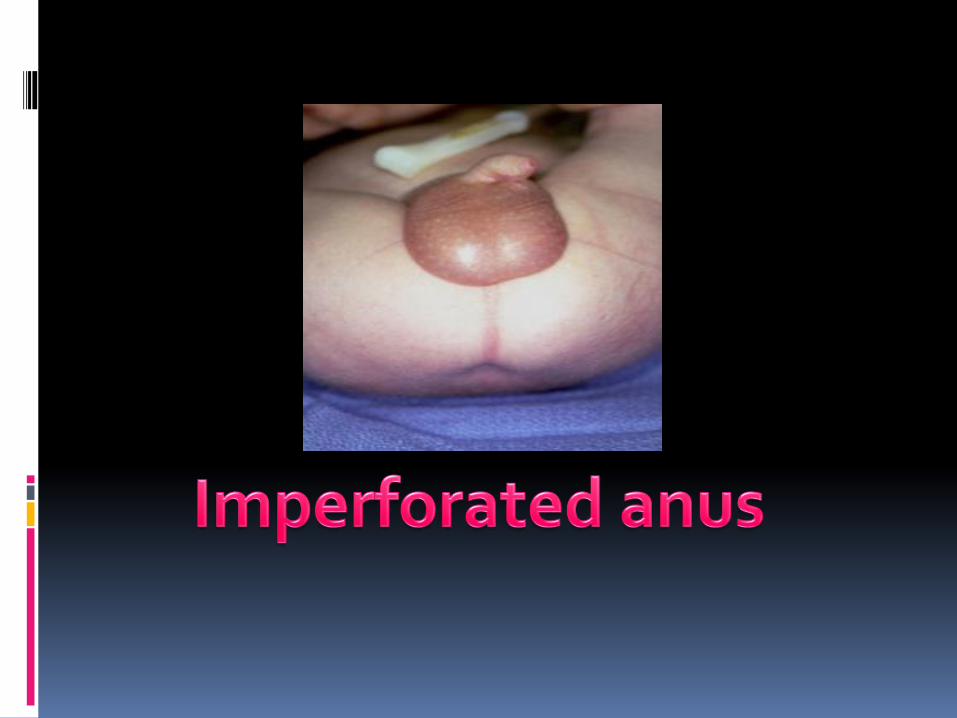
GI EMBRYOLOGY 2
Fareed Khdair , MDAssistant Professor
Chief, Section of Pediatric Gastroenterology, Hepatology, and Nutrition
University of Jordan – School of Medicine
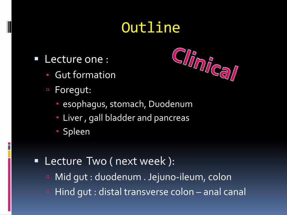
Outline
▪ Lecture one :
• Gut formation
Foregut:
esophagus, stomach, Duodenum
Liver , gall bladder and pancreas
Spleen
▪ Lecture Two ( next week ):
Mid gut : duodenum . Jejuno-ileum, colon
Hind gut : distal transverse colon – anal canal
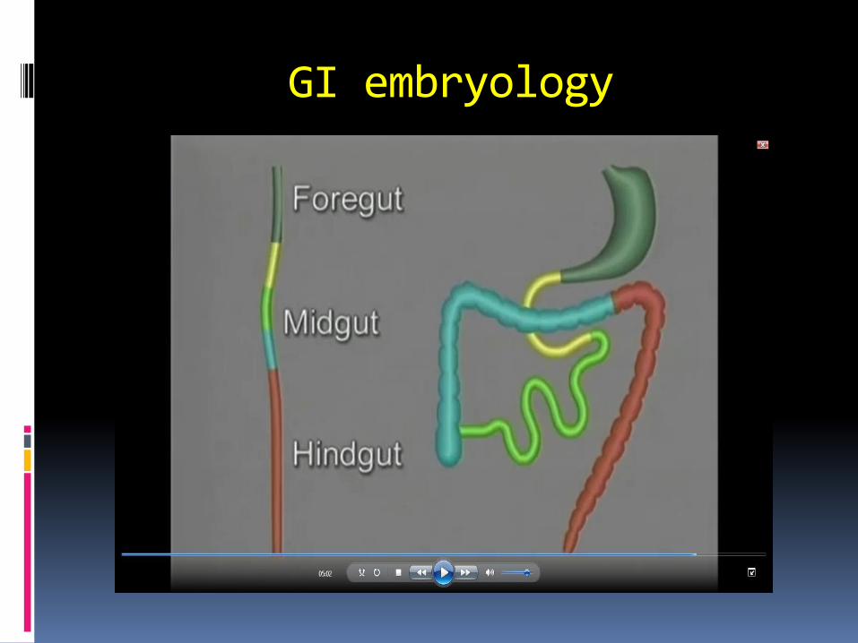
GI embryology
References
▪ Lecture slides
▪ Langman medical embryology
Chap 15
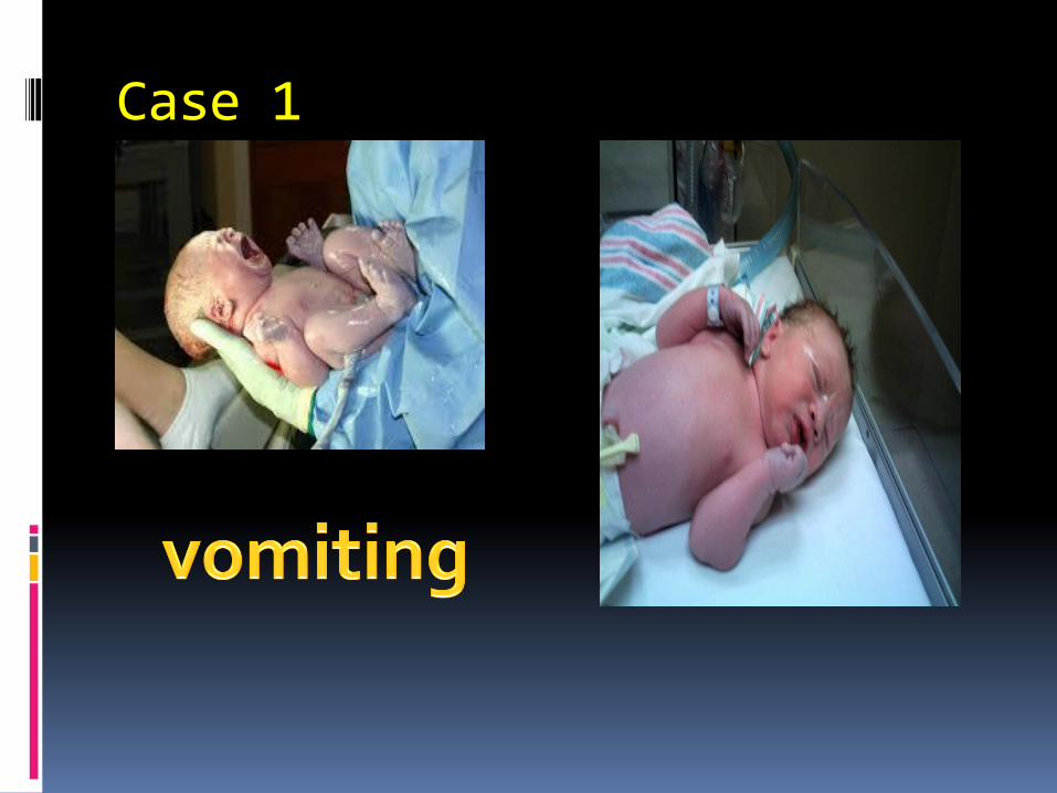
Case 1
Mid Gut
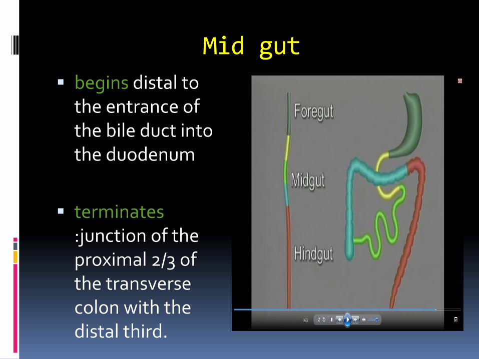
Mid gut
▪ begins distal to the entrance of the bile duct into the duodenum
▪ terminates:junction of the proximal 2/3 of the transverse colon with the distal third.
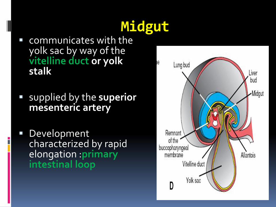
Midgut▪ communicates with the
yolk sac by way of the vitelline duct or yolk stalk
▪ supplied by the superior mesenteric artery
▪ Development characterized by rapid elongation :primary intestinal loop
Growth phases of mid gut
1. Herniation – physiologic 6th week
2. Rotation 90 degrees
3. Retraction 10 th week
4. Further rotation 180 degrees

Mid gut growth
▪ The cephalic limb of the loop develops into: the distal part of the duodenum, the jejunum, and part of the ileum.
▪ The caudal limb becomes the lower portion of the ileum, the cecum, the appendix, the ascending colon, and the proximal two-thirds of the transverse colon.
Mid gut
▪ WAPWON.COM_Embryological_Rotation_of_the_Midgut.mp4
Physiological Herniation
▪ At 6 Weeks
Due to :
▪ rapid growth of the liver.
▪ Rapid growth of intestinal loops
▪ the abdominal cavity becomes too small
they enter the extraembryonic cavity in the umbilical cord

ROTATION OF THE MIDGUT▪ rotates around axis of
superior mesenteric artery
▪ Counterclockwise
▪ approximately 270◦ when complete
Rotation occurs :
▪ during herniation (about 90◦)
▪ during return of the intestinal loops into the abdominal cavity (remaining 180◦)
during mid gut rotation
Small intestine :
▪ elongation
▪ jejunum and ileum :coiled loops
large intestine :
▪ Elongation
▪ No coiling
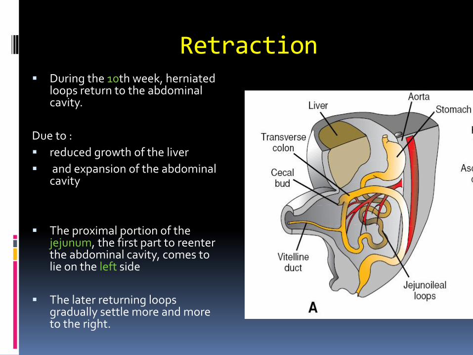
Retraction▪ During the 10th week, herniated
loops return to the abdominal cavity.
Due to :
▪ reduced growth of the liver
▪ and expansion of the abdominal cavity
▪ The proximal portion of the jejunum, the first part to reenter the abdominal cavity, comes to lie on the left side
▪ The later returning loops gradually settle more and more to the right.
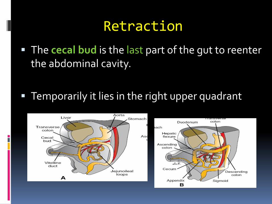
Retraction
▪ The cecal bud is the last part of the gut to reenter the abdominal cavity.
▪ Temporarily it lies in the right upper quadrant
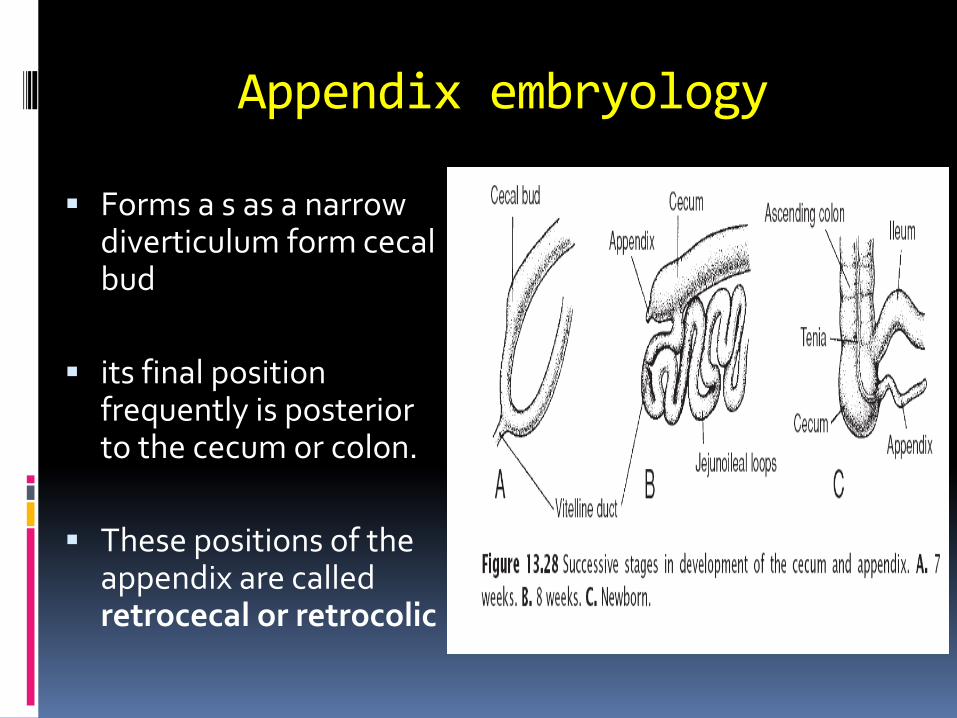
Appendix embryology
▪ Forms a s as a narrow diverticulum form cecalbud
▪ its final position frequently is posterior to the cecum or colon.
▪ These positions of the appendix are called retrocecal or retrocolic

Mesenteries of intestinal loops
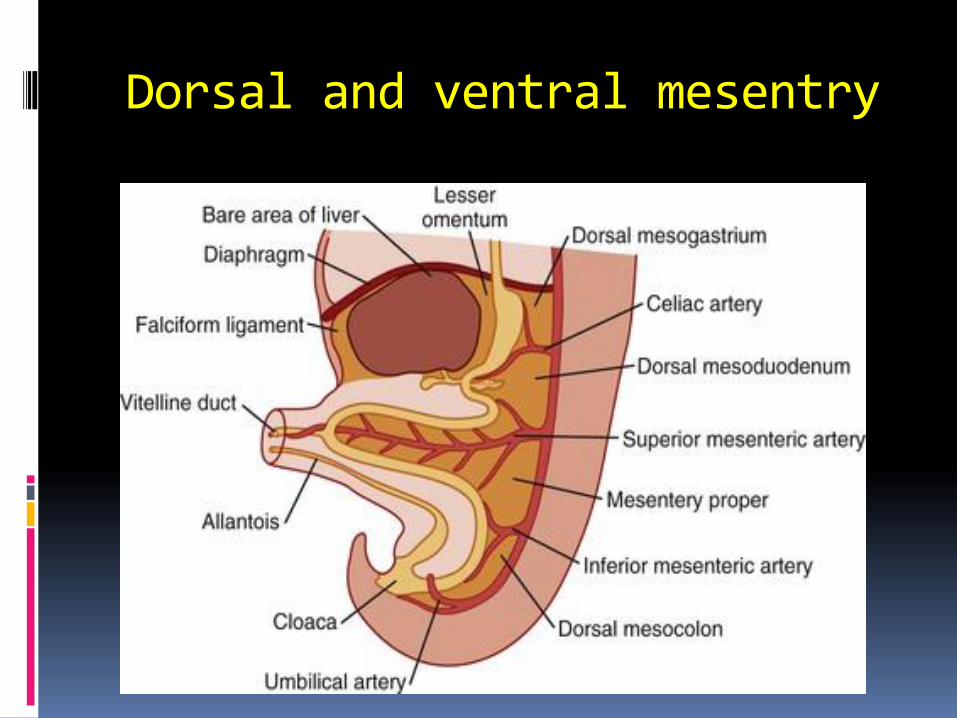
Dorsal and ventral mesentry
Mesentery proper
▪ The mesentery of the primary intestinal loop.
▪ changes with rotation and coiling of the bowel.
1- caudal limb of the loop moves to the right side of the abdominal cavity.
2- the dorsal mesentery twists around the origin of the superior mesenteric artery
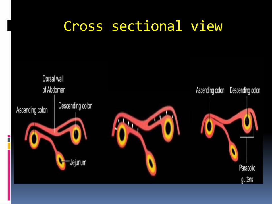
Cross sectional view
Retro Vs intraperitoneal
▪ After fusion of these layers: the ascending and descending colons are
permanently anchored in a retroperitoneal position
▪ The appendix, lower end of the cecum, and sigmoid colon: retain their free mesenteries
( intraperitoneal structures)
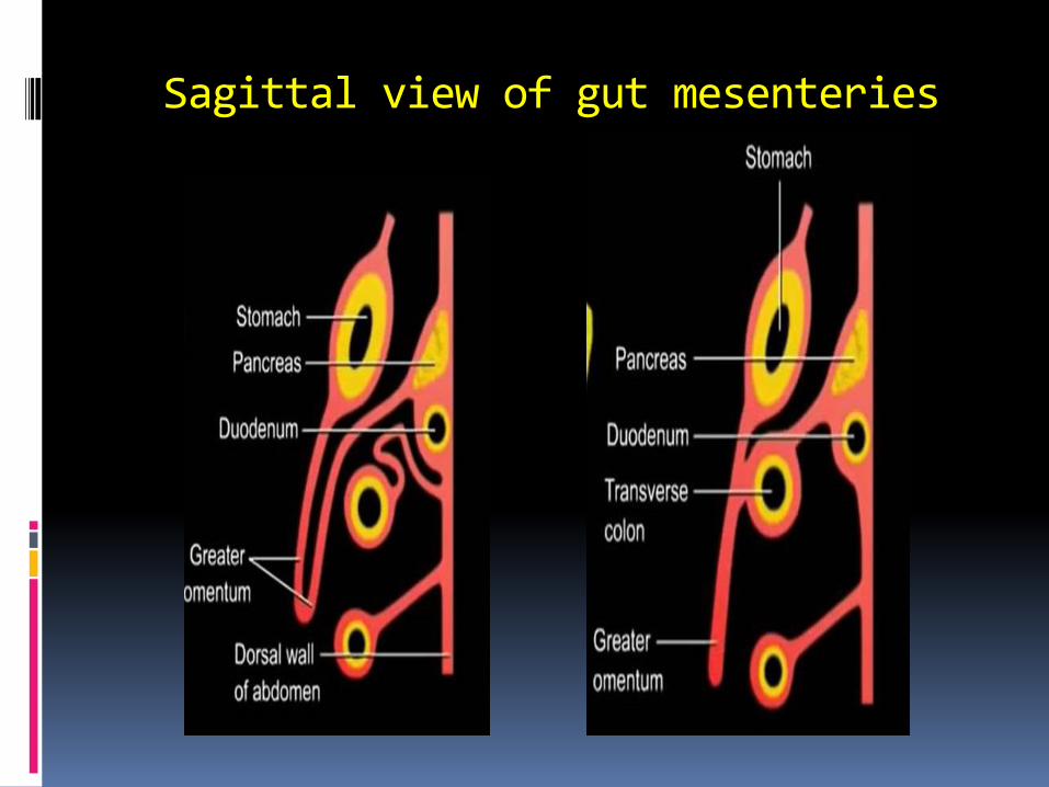
Sagittal view of gut mesenteries
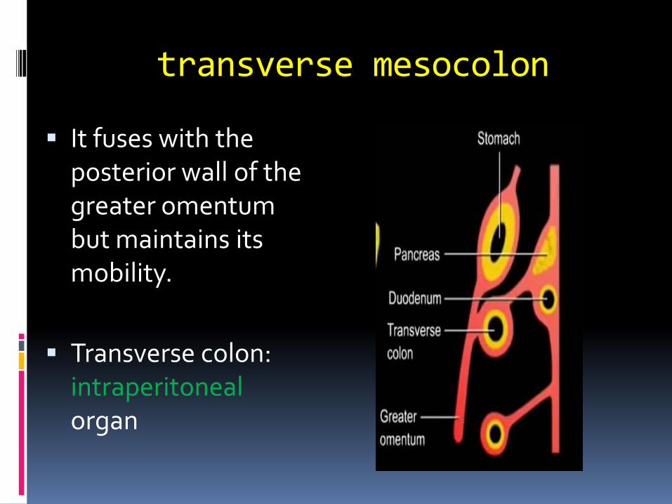
transverse mesocolon
▪ It fuses with the posterior wall of the greater omentum but maintains its mobility.
▪ Transverse colon: intraperitoneal organ
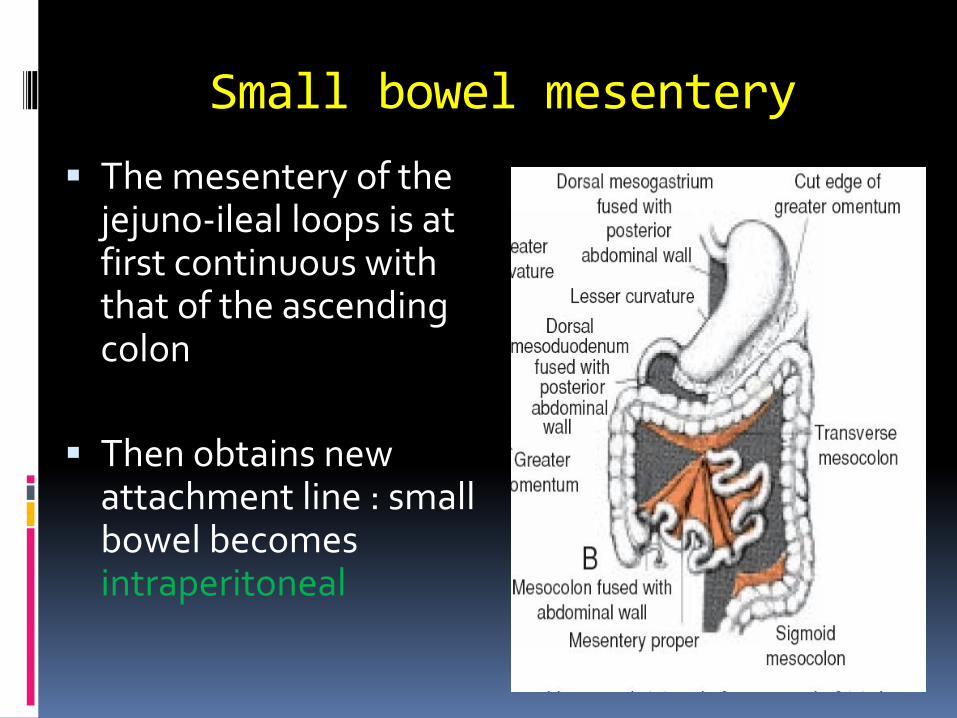
Small bowel mesentery
▪ The mesentery of the jejuno-ileal loops is at first continuous with that of the ascending colon
▪ Then obtains new attachment line : small bowel becomes intraperitoneal
Congenital anomalies of mid gut
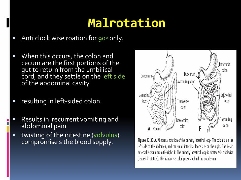
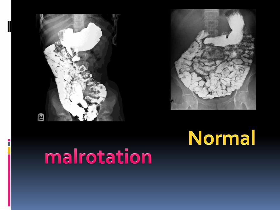
Malrotation▪ Anti clock wise roation for 90◦ only.
▪ When this occurs, the colon and cecum are the first portions of the gut to return from the umbilical cord, and they settle on the left side of the abdominal cavity
▪ resulting in left-sided colon.
▪ Results in recurrent vomiting and abdominal pain
▪ twisting of the intestine (volvulus) compromise s the blood supply.

Reversed rotation
▪ primary loop rotates 90◦ clockwise
▪ In this abnormality the transverse colon passes behind the duodenum and lies behind the superior mesenteric artery.
▪ Symptoms usually occur early in life
Gut Atresias and Stenoses
▪ Atresias and stenoses may occur anywhere along the intestine
▪ Most occur in the duodenum, fewest occur in the colon, and equal numbers occur in the jejunum and ileum (1/1500 births).
▪ Atresias in the upper duodenum are probably due to a lack of recanalization
Body Wall Defects
Omphalocele
Gastroschisis
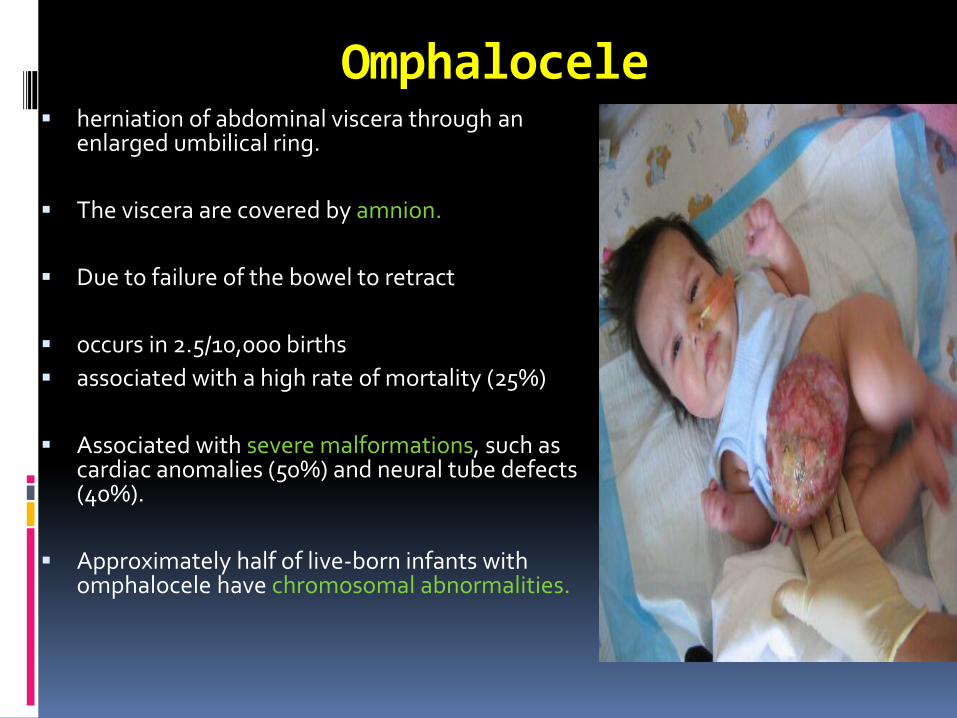
Omphalocele▪ herniation of abdominal viscera through an
enlarged umbilical ring.
▪ The viscera are covered by amnion.
▪ Due to failure of the bowel to retract
▪ occurs in 2.5/10,000 births
▪ associated with a high rate of mortality (25%)
▪ Associated with severe malformations, such as cardiac anomalies (50%) and neural tube defects (40%).
▪ Approximately half of live-born infants with omphalocele have chromosomal abnormalities.
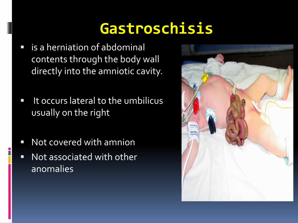
Gastroschisis▪ is a herniation of abdominal
contents through the body wall directly into the amniotic cavity.
▪ It occurs lateral to the umbilicus usually on the right
▪ Not covered with amnion
▪ Not associated with other anomalies
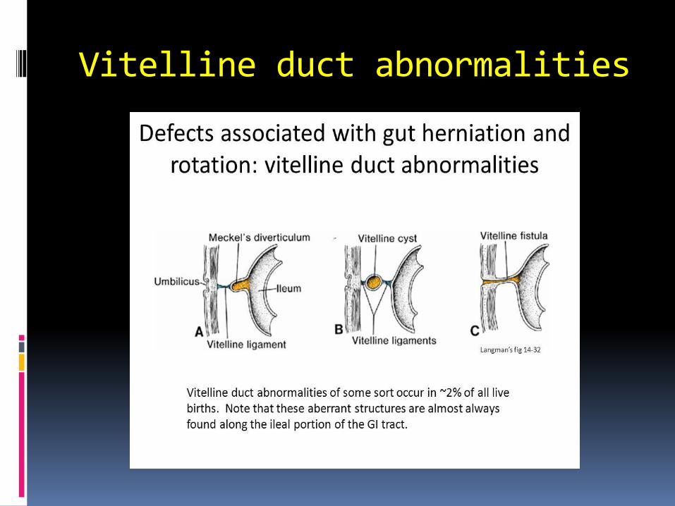
Vitelline duct abnormalities

Hind Gut
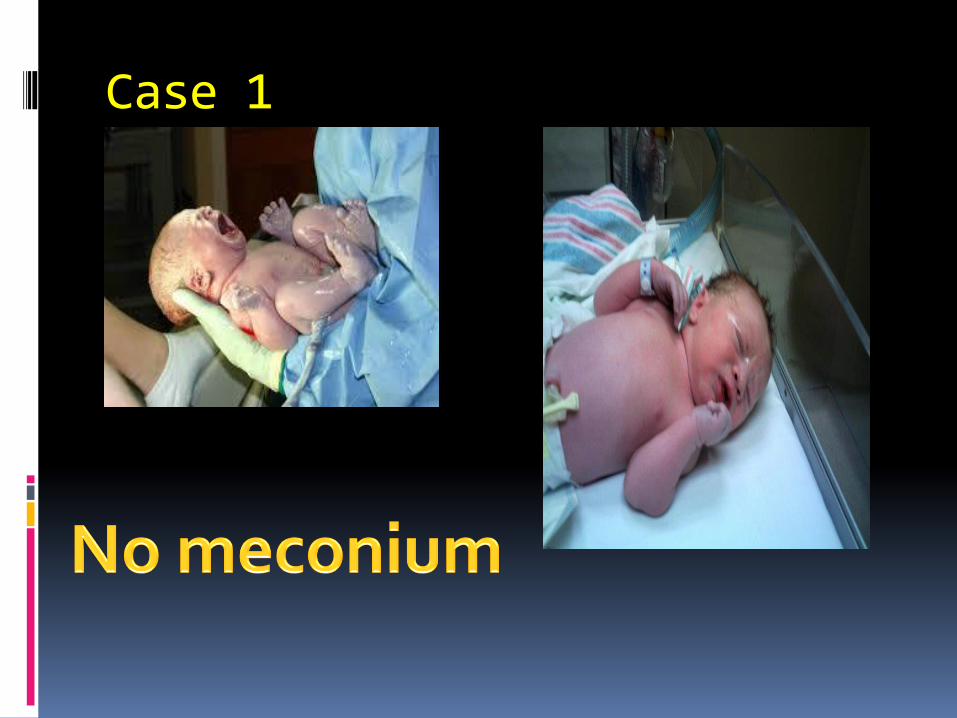
Case 1
Hindgut
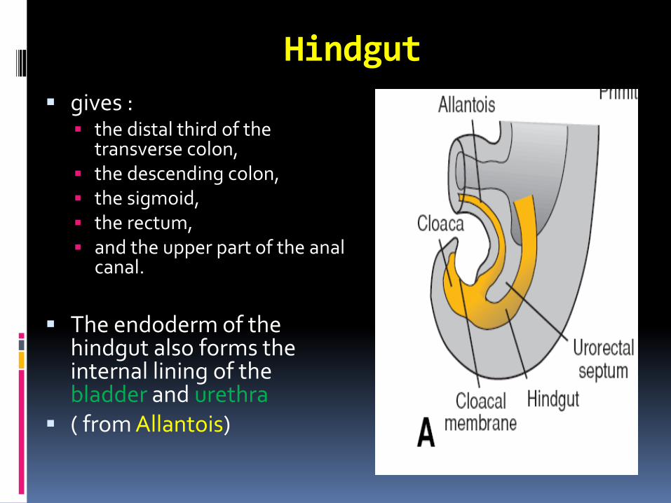
▪ gives :▪ the distal third of the
transverse colon,▪ the descending colon,▪ the sigmoid,▪ the rectum,▪ and the upper part of the anal
canal.
▪ The endoderm of the hindgut also forms the internal lining of the bladder and urethra
▪ ( from Allantois)
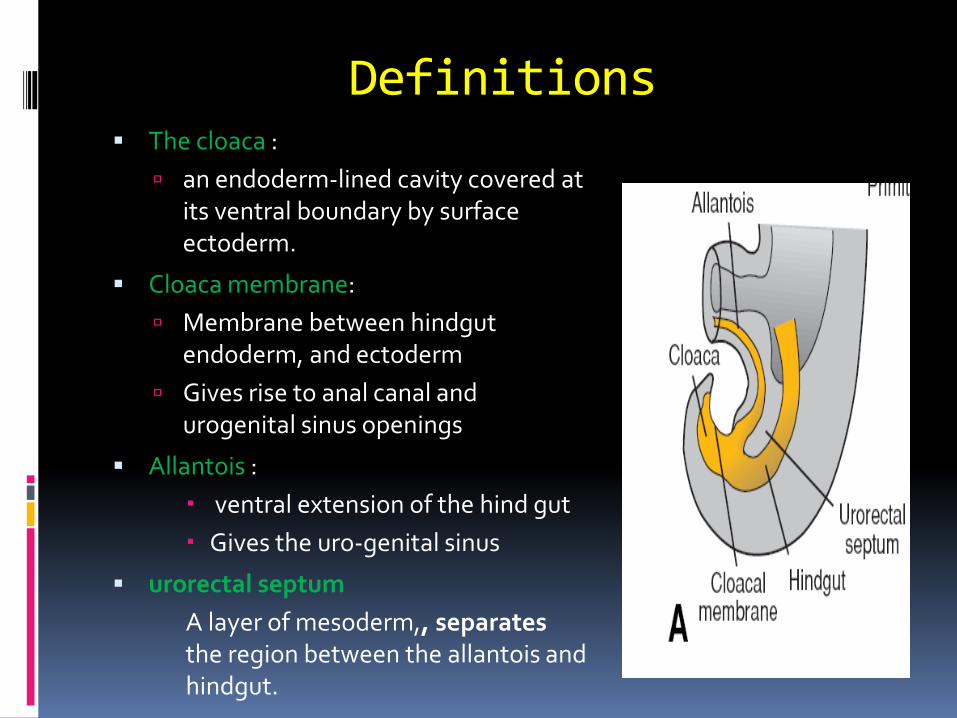
Definitions▪ The cloaca :
an endoderm-lined cavity covered at its ventral boundary by surface ectoderm.
▪ Cloaca membrane:
Membrane between hindgut endoderm, and ectoderm
Gives rise to anal canal and urogenital sinus openings
▪ Allantois :
ventral extension of the hind gut
Gives the uro-genital sinus
▪ urorectal septum
A layer of mesoderm,, separates the region between the allantois and hindgut.
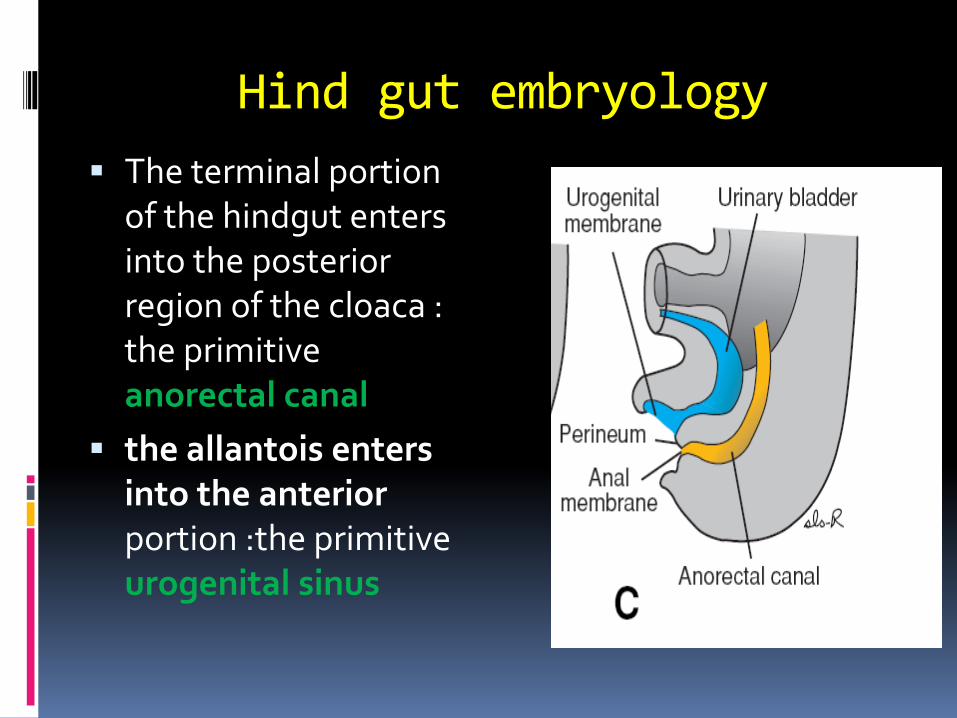
Hind gut embryology
▪ The terminal portion of the hindgut enters into the posterior region of the cloaca : the primitive anorectal canal
▪ the allantois enters into the anterior portion :the primitive urogenital sinus
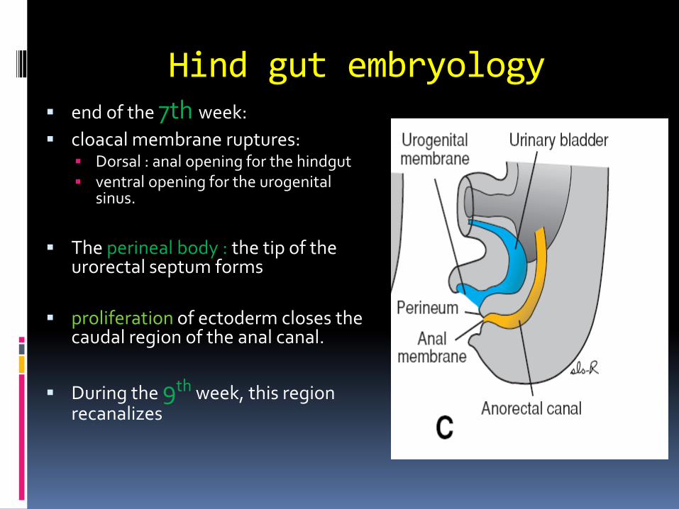
Hind gut embryology▪ end of the 7th week:
▪ cloacal membrane ruptures: ▪ Dorsal : anal opening for the hindgut ▪ ventral opening for the urogenital
sinus.
▪ The perineal body : the tip of the urorectal septum forms
▪ proliferation of ectoderm closes the caudal region of the anal canal.
▪ During the 9th week, this region recanalizes
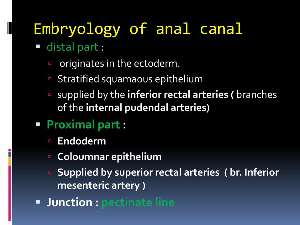
Embryology of anal canal▪ distal part :
originates in the ectoderm.
Stratified squamaous epithelium
supplied by the inferior rectal arteries ( branches of the internal pudendal arteries)
▪ Proximal part :
Endoderm
Coloumnar epithelium
Supplied by superior rectal arteries ( br. Inferior mesenteric artery )
▪ Junction : pectinate line
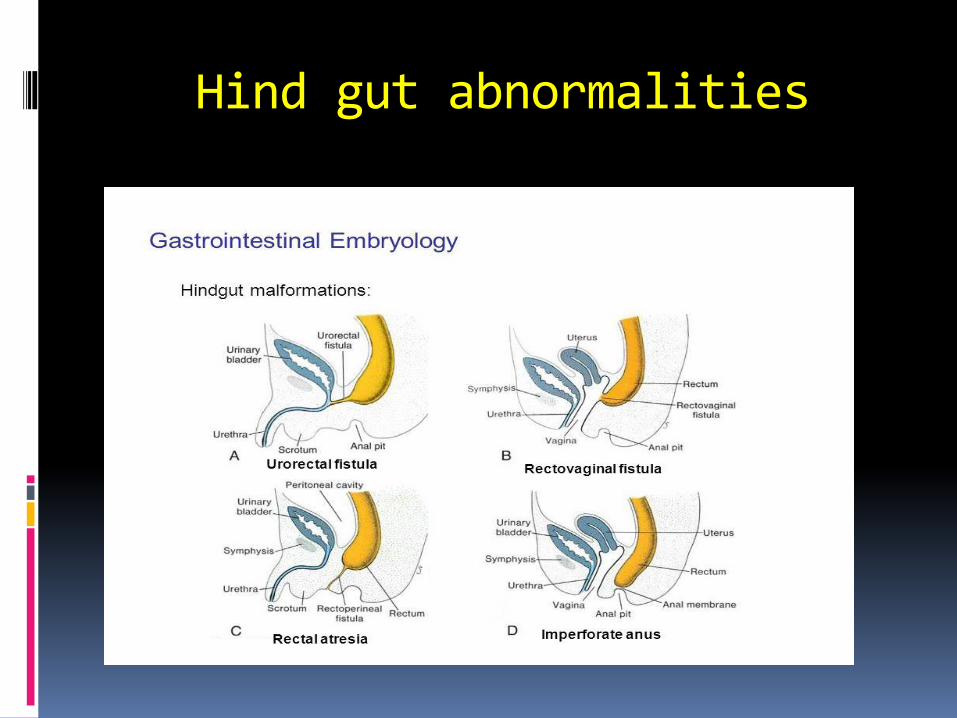
Hind gut abnormalities
Summary
▪ WAPWON.COM_10-_The_development_of_the_gastrointestinal_tract.mp4


The End

QUESTIONS?