Embed Size (px)
Citation preview

Genomics and proteomics approaches to understand virulence of Entamoeba histolytica.
C. López-Camarillo1*, E. Azuara-Liceaga1, A. Zamorano2, O. Hernández de la Cruz1, I. López Rosas1 and L. Marchat2
1Posgrado en Ciencias Genómicas, Universidad Autónoma de la Ciudad de México. San Lorenzo 290, Col del Valle. 03100. México DF. 2Programa Institucional de Biomedicina Molecular, Doctorado en Biotecnología en Red, ENMyH-IPN., Guillermo Massieu Helguera 239, Ticoman. 07320. México DF.
*Corresponding author. Email: [email protected]
Entamoeba histolytica is the protozoan parasite responsible for human amoebiasis. This infection has a world-wide distribution affecting more than 50 million people each year, mainly in developing countries. E. histolytica causes fulminating intestinal dysentery, and bloody diarrhea. In some cases, virulent trophozoites can cross the intestinal epithelia to reach different organs of the human body, usually the liver (but also the lungs, brain or spleen) to provoke liver abscesses, which can result in 70,000 -100,000 deaths a year. Virulence variability of trophozoites has been related to genome plasticity and in vivo changes in gene expression induced during invasion. Some virulence factors have been identified as key factors in pathogenesis, however most molecular mechanisms relevant for infection establishment are still poorly understood.
Genome sequence represents an invaluable tool to dissect the molecular mechanisms modulating virulence and gene expression in parasites. The fully sequenced genome of E. histolytica provides new insights into parasite biology and genome evolution and it also opens the omics research era in this major human pathogen. Microarrays made from synthetic oligonucleotides based on annotated genes have been successfully applied in E. histolytica to obtain transcriptional profiling in some in vivo and in vitro conditions. In addition, proteomic profiles have been obtained from virulent and non-virulent trophozoites providing novel exciting novel data about factors potentially involved in virulence. Here, we reviewed the actual efforts based in genomics and proteomics approaches to understand the pathogenesis of this early branch eukaryotic parasite.
Keywords Entamoeba histolytica; amoebiasis; virulence; genomics; proteomics; gene regulation.
1. Introduction
The protozoan parasite Entamoeba histolytica has a world-wide prevalence but it is often endemic in developing tropical countries, such as India, Mexico, Central and South America because of poor hygienic and sanitation conditions. About 10% of the world’s population is infected with E. histolytica, which represents more than 50 million people each year. Amoebiasis is the third most common cause of death from parasitic infections, after Schistosomiasis and Malaria [1]. E. histolytica is usually transmitted by the fecal-oral route, through contaminated food and water. Infection begins with the ingestion of the cyst form of the parasite that is able to survive in the environment due to its protective cell wall. Cyst undergoes excystation in the intestine and produces the proliferative trophozoite forms that cause amoebic dysentery or amoebic colitis. If the parasite reaches the bloodstream, it can spread through the body and invade other organs, mainly the liver (but also the lungs, brain or spleen), to provoke abscesses that can result in 70,000 -100,000 deaths a year. On the other hand, about 90 percent of infections remain asymptomatic [2]. The virulence variability of trophozoites has been related to genome plasticity and in vivo changes in gene expression induced during host invasion. Some virulence factors have been identified as key factors in pathogenesis, however most molecular mechanisms relevant for infection establishment are still poorly understood. The elucidation of molecular mechanisms modulating virulence and gene expression is of major interest for researchers working with E. histolytica in order to understand parasite biology and be able to identify biochemical targets or vaccine candidates that could help to control amoebiasis. Here, we reviewed the actual efforts based in genomics and proteomics approaches to understand the pathogenesis of this early branch eukaryotic parasite. Particularly, we focused on the recent sequencing of the parasite genome that has revealed a variety of metabolic adaptations and the existence of gene families associated with virulence. We also described data obtained from transcription and proteomic profiling that altogether contributed to the knowledge about factors potentially involved in virulence.
511©FORMATEX 2011
Science against microbial pathogens: communicating current research and technological advances A. Méndez-Vilas (Ed.)_______________________________________________________________________________
2. Entamoeba histolytica genome
The draft sequence of the complete genome of E. histolytica was published in 2005 making it one of the first protist genomes to be sequenced by The Institute for Genomic Research (http://www.tigr.org/) and the Sanger Institute (http://www.sanger.ac.uk/). A whole-genome shotgun method was used to sequence parasite genome as a better choice for sequencing [3]. This protist has an A/T-rich genome which have made difficult its sequencing and assembly. E. histolytica genome is 20.8 Mb in size and is carried on 14–17 chromosomes. The length variation of homologous chromosomes is thought to be caused by expansion and contraction of the sub-telomeric region, which might be formed by tRNA-containing arrays that contain between one and five tRNA types per repeat [3, 4]. The genome analysis was carried out on a 12.5-fold coverage genome assembly consisting of 23,751,783 base pairs (bp) distributed among 888 scaffolds. Initially 9,938 genes were predicted and automatically annotated. But recently E. histolytica genome was re-assembled and re-annotated using a combination of manual and automated methods. The new genome assembly consists of 20 Mb of sequence organized into 1,496 scaffolds. This new assembly showed a higher fragmentation and a reduction in genome size with respect of data published earlier. The new assembly contains less predicted protein coding genes. The main reason for gene number reduction is the elimination of genes within repetitive regions, artifactual tandem duplications, and the removal of genes smaller than 300 bp without any supporting evidence [5]. In summary the E. histolytica genome size is 20.8 Mb with identification of approximately 8300 predicted genes, each averaging 1260.9 pb in size, and corresponding to 49.7% of the genome. The shortest gene is 147 pb and the longest gene is 15,210 pb. Introns are contained in 24.4 % of the predicted genes. The intergenic regions mean length is about 708.7 bp with a 20.5% GC content [5]. E. histolytica genome is also characterized by its repetition and redundancy [6]. Redundancy can be seen in the predicted proteins, which can be organized into protein families. A total of 897 protein families were identified from the 8,201 predicted polypeptides. Among the families, 247 families have no homology to any known Pfam or TIGRfam domain [5]. Analysis of the genome reveals redundancy in genes encoding virulence factors. For example, E. histolytica genome contains a total of 86 genes coding for putative peptidases [4, 6]. These peptidases comprise 50 cysteine peptidases; some of them have a predicted N-terminal transmembrane anchor, which might allow them to be localized on the Entamoeba cell surface. Additionally, four aspartic, 10 serine and 22 metallo peptidase genes were also identified in Entamoeba genome [6]. Furthermore, 16 genes coding for putative saposin-like proteins were identified. The proteins encoded by these genes could be related to amoebapores [4]. Finally there are over 75 genes encoding leucine-rich tandem repeats of the type found in BspA-like proteins. These proteins generally have a surface location and may be involved in cell–cell interactions [5]. Vesicular trafficking is important in pathogenesis through phagocytosis and delivery of proteases and amoebapores to cell surface. Rab and Arf protein family expansions reflect the increased complexity and number of vesicle fusion and recycling steps that have been associated with this process. E. Histolytica has families with more than 50 members, such as small GTP binding proteins, which control a number of processes involving the actin cytoskeleton [3]. Signal-transduction-related proteins like protein kinases (270 gene encoding proteins), protein phosphatases (100 gene encoding proteins), which dephosphorylate proteins, numerous putative seven-transmembrane receptors, trimeric G proteins, Ras-family proteins were also identified in Entamoeba genome [3]. This represents the most varied set of signal-transduction-related proteins yet described in a single-celled eukaryote. Another important feature of Entamoeba genome is the lateral gene transfer (LTG) primarily from Bacteroidetes phyla (Cytophaga–Flavobacterium–Bacteroides). Initially 96 genes were identified using phylogenetic analyses [3], but only 41 remain as strongly supported [4]. Most of these genes encode proteins involved in carbohydrate and protein metabolism increasing the range of substrates available for energy generation. Most of the proteins encoded by these genes are specific to the parasite and may be used as potential drug targets for the design of new anti-amebic drugs. Finally, Entamoeba genome and DNA microarrays expression data is organized in AmoebaDB database (http://amoebadb.org/amoeba/). This database belongs to the National Institute of Allergy and Infectious Diseases (NIAID) funded EuPathDB (http://EuPathDB.org) Bioinformatics Resource Center family of integrated databases. AmoebaDB contains the genomes of E. dispar, E. invadens besides E. histolityca. This database contains functional genomic databases for the scientific community studying eukaryotic pathogens [8].
3. Genomic expression profiling in E. histolytica
3.1 Genomics basics
Genomics is a recent biology discipline focused in the study of genomes. Genome science is focused in the study of the structure, content, and evolution of genomes [9]. The field includes the initial intensive efforts to determine the entire nucleotides sequence of uni- and multicelular organisms, as well as the development of sophisticated equipments and bioinformatic tools. Functional genomics attempts to understand the relationships between complete genome sequences of organisms and gene (and protein) functions. To gain insights into cell function, genomics exploits the genome-wide
512 ©FORMATEX 2011
Science against microbial pathogens: communicating current research and technological advances A. Méndez-Vilas (Ed.)______________________________________________________________________________
approach involving high-throughput methods rather than a more traditional “gene-by-gene” method. Notably, the availability of genome sequences from parasites of medical importance for human, such as E. histolytica, provides opportunities to study genomic expression profiles in pathogens under diverse conditions.
3.2 DNA microarrays
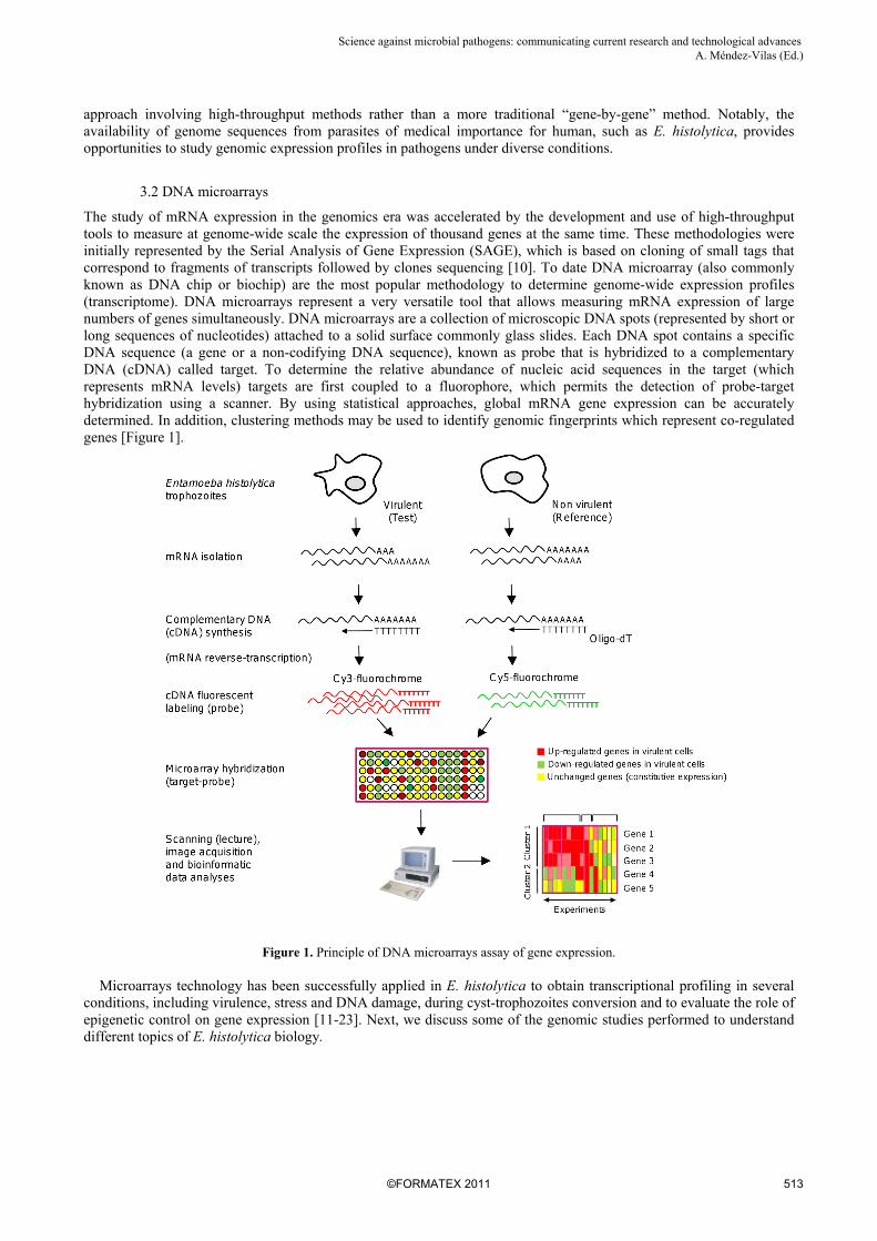
The study of mRNA expression in the genomics era was accelerated by the development and use of high-throughput tools to measure at genome-wide scale the expression of thousand genes at the same time. These methodologies were initially represented by the Serial Analysis of Gene Expression (SAGE), which is based on cloning of small tags that correspond to fragments of transcripts followed by clones sequencing [10]. To date DNA microarray (also commonly known as DNA chip or biochip) are the most popular methodology to determine genome-wide expression profiles (transcriptome). DNA microarrays represent a very versatile tool that allows measuring mRNA expression of large numbers of genes simultaneously. DNA microarrays are a collection of microscopic DNA spots (represented by short or long sequences of nucleotides) attached to a solid surface commonly glass slides. Each DNA spot contains a specific DNA sequence (a gene or a non-codifying DNA sequence), known as probe that is hybridized to a complementary DNA (cDNA) called target. To determine the relative abundance of nucleic acid sequences in the target (which represents mRNA levels) targets are first coupled to a fluorophore, which permits the detection of probe-target hybridization using a scanner. By using statistical approaches, global mRNA gene expression can be accurately determined. In addition, clustering methods may be used to identify genomic fingerprints which represent co-regulated genes [Figure 1].
Figure 1. Principle of DNA microarrays assay of gene expression. Microarrays technology has been successfully applied in E. histolytica to obtain transcriptional profiling in several conditions, including virulence, stress and DNA damage, during cyst-trophozoites conversion and to evaluate the role of epigenetic control on gene expression [11-23]. Next, we discuss some of the genomic studies performed to understand different topics of E. histolytica biology.
513©FORMATEX 2011
Science against microbial pathogens: communicating current research and technological advances A. Méndez-Vilas (Ed.)_______________________________________________________________________________

3.3 Genome-wide expression profiling in response to L-cysteine deprivation
L-cysteine is a sulfur-containing amino acid which plays an essential role in different cellular processes including stability, structure, regulation of catalytic activity, and posttranslational modification of proteins. Due to the ability of its thiol group to undergo redox reactions, it plays an important role in antioxidative defense and is used for biosynthesis of glutathione, the major endogenous antioxidant produced by the cells, participating directly in the neutralization of free radicals and reactive oxygen compounds. In E. histolytica, L-cysteine is required for growth, attachment, survival, and protection from oxidative stress [24] and L-cysteine deprivation leads to drastic changes in metabolic pathways, including energy, amino acid, and phospholipid metabolism [25]. To better understand the role of L-cysteine in E. histolytica, Husain et al. [26] performed DNA microarrays analysis of gene expression at different times after L-cysteine deprivation, and reported that 290 genes with functions in metabolism, signaling, DNA/RNA regulation, electron transport, stress response, membrane transport, vesicular trafficking/secretion, and cytoskeleton were ≥ 3 fold differentially expressed at one or more time points upon L-cysteine deprivation; 129 genes were up-regulated and 167 genes were down-regulated, while 6 genes showed both up- and down-regulation depending upon the time. This study also confirmed that most of the L-cysteine deprivation-mediated metabolomic changes in amino acid, central energy, and phospholipid metabolism are not associated with changes in the expression of the corresponding genes. This general lack of correlation between metabolome, proteome, and transcriptome appears to be a general characteristic in various organisms including E. histolytica, indicating that they have complex mechanisms of gene expression regulation.
3.4 Effects of DNA methylation inhibitors on global gene expression
Methylation is an enzymatic modification of DNA that epigenetically contributes to transcriptional regulation of gene expression. DNA methylation can silence genes by blocking the interaction of transcription factors to their regulatory sequences; it can also attract methyl-binding protein, which recruits histone deacetylases and histone methyltransferases resulting in an inactive chromatin structure. In E. histolytica, a DNA methyltransferase (Ehmeth) has been characterized [27], and an amoebic protein which preferentially binds to methylated DNA has been identified (EhMLBP) [28]. Interestingly, when E. histolytica strain HM1:IMSS is grown with 5-azacytidine (5-AzaC), a potent inhibitor of DNA methyltransferase, the in vitro and in vivo virulence is significantly reduced. To identify the genome-wide effects of DNA methylation in E. histolytica, Ali et al. [29] used a short oligonucleotides microarray representing 9,435 genes and compared the expression profile of HM-1:IMSS strain parasites with those treated with 23 µM 5-AzaC for up to one week. Overall, 2.1% of genes tested were transcriptionally modulated under these conditions. 68 genes were up-regulated and 131 genes down-regulated. Sodium-bisulfite treatment and genes sequencing indicated that there were at least two subsets of genes with genomic DNA methylation in E. histolytica: genes that were endogenously silenced by genomic DNA methylation and for which 5-AzaC treatment induced transcriptional de-repression, and genes that have genomic DNA methylation, but were not endogenously silenced by methylation. Interestingly, authors showed that a cysteine proteinase and a lysozyme, both having known roles in amebic pathogenesis, were down-regulated by 5-AzaC treatment. Decreased expression of these genes in the 5-AzaC treated E. histolytica may account in part for the reduced cytolytic activities in parasite.
3.5 Genomic studies of a Myb domain-containing protein involved in expression regulation of stage specific genes
Myb family transcription factors are important in regulating cell proliferation, differentiation, stage conversion and cell cycle progression and are widely found in eukaryotes. Conversion between cyst and trophozoite stage is essential to disease transmission and pathogenesis in many protozoa. E. histolytica differentiates into infectious cysts to survive outside of the host. Previous work has identified the transcriptome of E. histolytica cysts and determined two subsets of developmentally regulated genes, including 672 cyst-specific and 767 trophozoite-specific genes, respectively [30]. However, the molecular basis of encystations regulation is not known. The search for genes involved in the regulation of stage conversion allowed the identification of the first E. histolytica developmentally regulated transcription factor called EhMyb-dr. Overexpression of the nuclear EhMyb-dr resulted in a set of regulated genes that significantly overlapped with the expression profile of amoebic cysts, suggesting a role for EhMyb-dr in the regulation of stage-specific genes expression and its participation in E. histolytica development. Data from expression profiling allowed the identification of 117 genes that were up-regulated by EhMyb-dr overexpression, 40 overlapped with cyst-specific genes; of the 88 genes that were down-regulated by EhMyb-dr overexpression, 30 overlapped with trophozoite-specific genes. Some genes of interest in these lists include the chitin synthase, the developmentally regulated protein and chitinase. Moreover, bioinformatic methods allowed the identification of transcriptional regulatory networks and CCCCCC DNA binding motifs to which amoebic nuclear proteins bind in a sequence-specific manner in a subset of cyst-specific genes. This DNA microarrays-based study provided evidence for the involvement of EhMyb-dr in the differentiation of E. histolytica trophozoites into cysts [31].
514 ©FORMATEX 2011
Science against microbial pathogens: communicating current research and technological advances A. Méndez-Vilas (Ed.)______________________________________________________________________________

3.6 Genomic analysis of URE3-BP transcription factor
The transcription factor named upstream regulatory element 3-binding protein (URE3-BP) is a calcium-responding regulator of E. histolytica Gal/GalNAc lectin and ferredoxin genes, both implicated in virulence. To investigate the impact of URE3-BP regulation on virulence, the occurrence of URE3 consensus motif in promoter of genes specifically induced during in vivo infection, was measured by microarrays [32]. Amoeba expressing the dominant positive mutant form of URE3-BP was more virulent in two different animal models of amebiasis. An increase in liver abscess size was observed when trophozoites expressing the dominant positive mutant URE3-BP were injected into the gerbil liver. In the mouse model of amebic colitis, a competitive advantage at the host tissue interface was conferred upon amebae expressing the dominant positive mutant URE3-BP protein. URE3-BP therefore promoted the expression of the E.histolytica virulence phenotype. Distinct patterns of E. histolytica gene expression have been observed under a variety of experimental conditions. A comparison of the transcripts expressed during in vitro culture trophozoite and one day after infection in the mouse model of amoebiasis identified changes in parasite gene expression. Consistent with a role for URE3-BP in virulence, a statistically significant enrichment of the URE3 motif was observed in promoters of in vivo. However, further studies are required to understand the particular contribution of URE3-BP-regulated proteins to this complex process and their contribution to trophozoite virulence in different host environments [32]. However, it is clear that URE3-BP plays an important role in the virulence processes.
3.7 Genome-wide analysis of E. histolytica DNA damage response
The maintenance of DNA integrity is essential for cells viability and the life of organisms. The genetic material is constantly under attack from external environmental factors and endogenous metabolic products that can alter its chemical structure and nucleotides sequence. External and internal DNA damaging agents can induce changes in genome by producing single-strand breaks, double strand breaks (DSBs), inter- and intra-strand cross-links in the form of cyclobutane pyrimidine dimers and (6-4)-photoproducts, oxidation and alkylation of bases, or formation of bulky chemical adducts. Exogenous agents are represented by ionizing (IR) and ultraviolet (UV) irradiation, or chemicals, whereas cellular endogenous physiological events include restoration of collapsed replication forks in the course of DNA synthesis, telomere maintenance, or programmed events, such as V(D)J recombination and meiotic exchange [33, 34]. In order to determine changes in global mRNA expression profile in response to DNA damage in E. histolytica, Weber et al. (2008) performed a DNA microarrays study [16]. They used UV (150 J/m2) irradiated trophozoites. DNA damage and double strand-breaks were previously confirmed in irradiated cells by TUNEL and comet assays and evaluation of the EhH2AX histone phosphorylation status [35]. The overall expression analysis identified E. histolytica genes that may contribute to adaptation and survival of trophozoites in response to DNA damage response. Data showed that 11.6% (350 ORFs) and 17.2% (522 ORFs) of genes were modulated at 5 min and 3 h after UV irradiation, respectively. Surprisingly, most genes were less than 2-fold modulated evidencing a weak transcriptional activation. Genes encoding RAD52-epistasis group DNA repair genes (RAD54, MRE11, RAD50, and RAD52) were slightly regulated in trophozoites submitted to UV irradiation. Functional classification of regulated genes by Gene Ontology showed that they were involved in potential DNA damage response pathways, including cell cycle, signal transduction, and oxidative stress response. However, most genes were involved in pathways including RNA binding and processing, cell structure, protein synthesis and degradation, energy metabolism, adhesion and vesicle trafficking. Several hypothetical genes were also regulated [16]. Most of these genes have not been previously associated with DNA damage response in other organisms. These data provided insights about the potential roles of the E. histolytica RAD52 epistasis group genes and novel ORFs in DNA damage response in this ancient eukaryotic parasite.
3.8 Genomic-wide approach to understand E. histolytica virulence in vivo
A genomic transcriptional analysis of E. histolytica was performed in trophozoites isolated from the colon of six infected mice and from in vitro culture cells [36]. In this study, authors designed an Affymetrix platform gene expression array that included probe sets for 9435 ORFs. Interestingly, 523 transcripts (5.2% of all E. histolytica genes) were significantly modulated in trophozoites obtained from mice intestine on days 1 and 29 after infection, which represents the early and late responses to parasite colonization. Genes modulated correspond to cell signalling proteins, including transmembrane kinases, ras and rho family GTPases, and calcium binding proteins. A decrease in mRNA levels for genes involved in glycolysis and an increase in lipases were consistent with changes in energy metabolism. As expected, a decrease in oxygen detoxification pathways was also observed in the anaerobic colonic lumen. Other known virulence factors that showed significant changes include a 20–35-fold increase in a cysteine proteinase four-like gene, and a 2–3-fold decrease in two members of the Gal/GalNAc lectin light subunit family. Quantitative real-time reverse transcriptase PCR confirmation for 11/12 genes was performed. This study represents the first genome-wide analysis of the transcriptome of E. histolytica during infection in an animal model [36].
515©FORMATEX 2011
Science against microbial pathogens: communicating current research and technological advances A. Méndez-Vilas (Ed.)_______________________________________________________________________________

4. Proteomic approaches to study amoebiasis
4.1 Proteomics basics
Proteomics is the study of whole protein set obtained from a given tissue or cell type. These analyses are usually performed by two-dimensional gel electrophoresis (2-DE) followed by protein identification by mass spectroscopy (MS) and database queries. Although 2-DE dates back to 1970s [37] characterization by MS became a reality in the last 10 years with soft ionization methods, such as matrix-assisted laser-desorption ionization (MALDI) [38] and electrospray ionization (ESI) [39]. The success of this technique would not be possible without the development and availability of databases. Identification of proteins by MS uses two different approaches: in the peptide mass fingerprinting (PMF), protein spot of interest is in-gel digested with a specific enzyme [40-43] and in the second approach, peptides after in-gel digestion are fragmented in the mass spectrometer yielding partial amino acid sequences from peptides (sequence tags). PMF is usually performed by MALDI-TOF [44, 45]. In turn, 2-DE includes a proteins separation by isoelectric focusing (IEF) in an immobilized pH gradient (IPG) gel according to their isoelectric point [46], followed by a separation according to molecular weight through SDS-polyacrylamide electrophoresis. Finally, protein pattern is revealed through gel staining by Coomassie, silver or fluorescence and the pattern of spots can be analyzed quali- and quantitatively by imaging programs.
4.2 Proteomics profiles in the study of amoebiasis
Variations in the severity of E. histolytica infections are influenced by host immunity, host nutritional status, gut microbiota, and genetic divergence between isolates. These alternative effects are related to distinct protein expression profiles where proteomic studies have been a relevant tool to identify target molecules and propose their interactions. Despite the effort required, the significance of proteomic results is well acknowledged. Nowadays it is accepted that mRNA expression and protein levels do not necessarily correlate [47, 48]. Thus, post-translational modifications and their physiological role can be investigated by MS [49]. Studies of 2-DE for E. histolytica trophozoites have been developed and modified, so that pH ranges now permit to evidence more significant landmarks [50]. Because degradation of extracellular matrix by amoebic proteases, production of toxic factors, activation of host immune system cells, as well as killing and phagocytosis of human cells are features of amoebiasis, the characterization of proteins participating in these processes is of particular interest. In addition, host cells exhibit an innate immune response, which activates NF-kB and secretes cytokines to recruit neutrophils and macrophages to the invasion site [51, 52]. During tissue inflammation, phagocytosis is an important mechanism for clearance of apoptotic cells, and a variety of molecules on the surface of the phagocytic cells participate in the adhesion of apoptotic bodies, the engulfment process, as well as their later destruction. In terms of apoptotic cells, CD14, CD68, avb3 integrin, MER (tyrosine kinase from monocytes and tissue of epithelial and reproductive origin) and PSR (phosphatidylserine receptor) promote the adhesion of dying cell to phagocyte [53, 54]. In the Jurkat human T lymphocytes cell line, activation of Caspase-3 and the exposition of phosphatidylserine from inner to the outer leaflet of the plasma membrane, as well as DNA fragmentation, take place after amoebic contact. Moreover, blocking of the formation of amoebic liver abscesses via treatment with pan-caspase inhibitor showed that apoptosis of host cells is critical for parasite virulence [55]. In particular, E. histolytica trophozoites can induce cell death by Caspase-8 independent pathways, but they can also destroy Caspase-8 deficient cells treated with Caspase-9.
4.3 Proteomic analysis of phagosomes in E. histolytica
Adhesion of E. histolytica to colonic mucins and host epithelial cell is essentially carried out through the Gal-GalNAc lectin. The Gal-GalNAc lectin is constituted by a heavy subunit (Hgl) and a covalent attached glycosyl phosphatidyl inositol (GPI) anchored light chain (Lgl) [56]. Proteomic analysis has already identified groups of novel proteins involved in phagosome biogenesis [57]. Particularly, 85 phagosome proteins participating in surface recognition, cytoskeleton, vesicular trafficking, and degradation were detected in primary phagosomes, but kinetics of maturation and biogenesis remained unclear [58]. Other proteomics studies with purified phagosomes or affinity-purified Gal/GalNAc binding proteins using reversed-phase capillary liquid chromatography and ion trap tandem MS allowed the identification of proteins involved in phagosome biogenesis [59-61]. Some examples include proteins acting on phagosome recruitment derived from endoplasmic reticulum and mitochondrion-like organelle, as well as proteins involved in vesicular trafficking and recruitment of hydrolytic enzymes. Likewise, via protein isolation using magnetic beads and sequencing by tandem MS, peptides from all of the lectin subunits (Hgl, Igl, and Lgl) were identified in GalNAc-BSA bead purified proteins. Other proteins were also isolated, including cytoskeletal-associated proteins such as actin and myosin, a talin homologue, homologues of Rab11-B and calreticulin, and E. histolytica cysteine proteinase 2. Interestingly, the most abundant protein isolated with GalNAc beads incubated with amebic lysates was myosin. A categorization led by MS of phagosome-associated proteins proposed groups, such as lectins and surface proteins,
516 ©FORMATEX 2011
Science against microbial pathogens: communicating current research and technological advances A. Méndez-Vilas (Ed.)______________________________________________________________________________
vesicular trafficking regulators and other small GTPases, hydrolytic enzymes and degradative proteins, and calcium and proton pumps. Predominantly, 90% of these peptides were assigned to 85 proteins. The identification of molecules responsible for a specific phenotype can be addressed by differential expression, so that protein profile in a particular context can be contrasted with another. These studies have been conducted in E. histolytica, some of them have explored the protein composition of purified phagosomes. Several proteins involved in uptake-processes were recognized. Furthermore, in the same study, approximately 1,500 protein spots were detected by silver-staining and 10 landmark spots of the respective Coomassie-stained gels were identified by MALDI-TOF or protein sequencing. To construct a 2-DE reference map for E. histolytica, and after successful solubilisation, 100 proteins were subjected to analysis by MALDI-TOF mass spectroscopy. The resultant peptide mass fingerprints were analyzed by contrasting with data available in E. histolytica genome and NCBI databases. As a result, these proteins could be clustered into the following categories: a) cytoskeleton proteins; b) glycolysis; c) surface-associated proteins; d) RNA/DNA metabolism; e) ubiquitin-proteosome pathway; f) vesicular trafficking and signal transduction [62].
4.4 Proteomic analysis of trophozoites with diverse virulence degrees
In terms of virulence traits, the comparison of two genetically closely related E. histolytica cell lines with substantial difference in virulence were studied by 2-DE [63]. As a result of this investigation, only a very limited number of differentially synthesized proteins were detected. Preponderantly, the proteins found to be differentially synthesized are involved in cytoskeletal organization. In addition, three differentially up-regulated antioxidants were exclusively found in the pathogenic cell line, Fe-hydrogenase 2, peroxiredoxin and superoxide dismutase (SOD). Notably, only for two differentially regulated proteins, differential expression was also seen at the RNA level. Therefore, this implies that the regulation of these proteins often occurs at the posttranscriptional level. A similar study took advantage of the ability to compare the proteome of E.histolytica to those of E. dispar, in order to investigate virulence factors of E. histolytica [64]. This report used E. histolytica HM-1:IMSS and E. dispar SAW760 to identify proteins that are differentially expressed between these two species. Results evidenced the relevance of the alcohol dehydrogenase 3 (EhADH3). Specially, E. histolytica possesses a higher amount of NADP-dependent alcohol dehydrogenase than E. dispar and EhADH3 can be detected in the surface of E. histolytica. Based on the same methodology, it has been reported that increased grainin levels may contribute to reduce virulence phenotype. This finding was the result of a comparative analysis of E. histolytica Rahman and E. histolytica HM-1:IMSS. Particularly, grainin 2 was expressed at significantly higher levels in E. dispar when compared to E. histolytica. Another protein expressed at higher levels in E. histolytica contained a LIM domain, which is a cysteine and histidine rich domain composed of two zinc fingers which has been related to differentiation, cytoskeleton reorganization, and regulation of transcription [65, 66]. Likewise a comparative proteomics of E. histolytica HM-1:IMSS and E. histolytica Rahman to identify virulence factors showed differential levels of two molecules linked to resistance to host oxidative defenses, peroxiredoxin, and SOD, and three proteins of unknown function, including grainins 1 and 2 [67].
5. Conclusions
The power of genomics and proteomics approaches lies in their ability to evidence gene expression networks and therefore molecular mechanisms and biochemical pathways that are related to different contexts of virulence in E. histolytica. Particularly, they allowed the determination of changes in gene expression level (transcripts and proteins) the study of protein- protein interactions, and the identification of post-translational epigenetic regulation, which could help to understand the pathogenesis of this parasite.
Acknowledgments This work was supported by UACM (México), CONACyT (project number 79293, México), COFAA-IPN (Mexico), SIP-IPN (Mexico) and European Community grants.
References
[1] WHO. Entamoeba taxonomy. Bull. World Health Organ. 2007;75:291-294 [2] Haque R, Huston CD, Hughes M, Houpt E, Petri WA Jr. Amebiasis. N Engl J Med. 2003;348:1565-1573. [3] Loftus B, Anderson I, Davies R, Alsmark UC, Samuelson J, Amedeo P, Roncaglia P, Berriman M, Hirt RP, Mann BJ, Nozaki T,
Suh B, et al. The genome of the protist parasite Entamoeba histolytica. Nature. 2005; 433:865–868. [4] Clark CG, Alsmark UC, Tazreiter M, Saito-Nakano Y, Ali V, et al. Structure and content of the Entamoeba histolytica genome.
Adv Parasitol. 2007;65: 51–190. [5] Lorenzi HA, Puiu D, Miller JR, Brinkac LM, Amedeo P, Hall N, Caler EV. New assembly, reannotation and analysis of the
Entamoeba histolytica genome reveal new genomic features and protein content information. PLoS Negl Trop Dis. 2010;4(6):e716.
[6] Stanley SL Jr. The Entamoeba histolytica genome: something old, something new, something borrowed and sex too? Trends Parasitol. 2005;21(10):451-453.
517©FORMATEX 2011
Science against microbial pathogens: communicating current research and technological advances A. Méndez-Vilas (Ed.)_______________________________________________________________________________
[7] Tillack M, Biller L, Irmer H, Freitas M, Gomes MA, Tannich E, Bruchhaus I. The Entamoeba histolytica genome: primary structure and expression of proteolytic enzymes. BMC Genomics. 2007;8:170.
[8] Aurrecoechea C, Barreto A, Brestelli J, Brunk BP, Caler EV, Fischer S, Gajria B, Gao X, Gingle A, Grant G, Harb OS, Heiges M, Iodice J, Kissinger JC, Kraemer ET, Li W, Nayak V, Pennington C, Pinney DF, Pitts B, Roos DS, Srinivasamoorthy G, Stoeckert CJ Jr, Treatman C, Wang H. AmoebaDB and MicrosporidiaDB: functional genomic resources for Amoebozoa and Microsporidia species. Nucleic Acids Res. 2011;39(Database issue):D612-9.
[9] Gregg Gibson and Spencer V. Muse. (2004). A primer to genome science. Second edition. ISBN 0-87893-232-1. [10] Velculescu VE, Zhang L, Vogelstein B, Kinzler KW. Serial analysis of gene expression. Science. 1995; 270 (5235):484–487. [11] Debnath A, Tashker JS, Sajid M, McKerrow JH. Transcriptional and secretory responses of Entamoeba histolytica to mucins,
epithelial cells and bacteria. Int J Parasitol. 2007;37:897-906. [12] Debnath A, Das P, Sajid M, McKerrow JH. Identification of genomic responses to collagen binding by trophozoites of
Entamoeba histolytica. J Infect Dis. 2004;190:448-457. [13] Gilchrist CA, Houpt E, Trapaidze N, Fei Z, Crasta O, Asgharpour A, Evans C, Martino-Catt S, Baba DJ, Stroup S, Hamano S,
Ehrenkaufer G, Okada M, Singh U, Nozaki T, Mann BJ, Petri WA Jr. Impact of intestinal colonization and invasion on the Entamoeba histolytica transcriptome. Mol Biochem Parasitol. 2006;147:163-176.
[14] MacFarlane RC, Singh U. Identification of differentially expressed genes in virulent and nonvirulent Entamoeba species: potential implications for amebic pathogenesis. Infect Immun. 2006;74:340-351.
[15] Weber C, Guigon G, Bouchier C, Frangeul L, Moreira S, Sismeiro O, Gouyette C, Mirelman D, Coppee JY, Guillen N. Stress by heat shock induces massive down regulation of genes and allows differential allelic expression of the Gal/GalNAc lectin in Entamoeba histolytica. Eukaryot Cell. 2006;5:871-875.
[16] Weber C, Marchat LA, Guillen N, López-Camarillo C. Effects of DNA damage induced by UV irradiation on gene expression in the protozoan parasite Entamoeba histolytica. Mol Biochem Parasitol. 2009;164(2):165-169.
[17] Ehrenkaufer GM, Haque R, Hackney JA, Eichinger DJ, Singh U. Identification of developmentally regulated genes in Entamoeba histolytica: insights into mechanisms of stage conversión in a protozoan parasite. Cell Microbiol. 2007a;9:1426-1434.
[18] Ehrenkaufer GM, Eichinger DJ, Singh U. Trichostatin A effects on gene expression in the protozoan parasite Entamoeba histolytica. BMC Genomics. 2007b;8: 216.
[19] Ali IK, Ehrenkaufer GM, Hackney JA, Singh U. Growth of the protozoan parasite Entamoeba histolytica in 5-azacytidine has limited effects on parasite gene expression. BMC Genomics. 2007;8:7-11.
[20] Davis PH, Schulze J, Stanley SL Jr. Transcriptomic comparison of two Entamoeba histolytica strains with defined virulence phenotypes identifies new virulence factor candidates and key differences in the expression patterns of cysteine proteases, lectin light chains, and calmodulin. Mol Biochem Parasitol. 2007;151:118-128.
[21] Vicente-Joao B, Ehrenkaufer GM, Saraiva LM, Teixeira M, Singh U. Entamoeba histolytica modulates a complex repertoire of novel genes in response to oxidative and nitrosative stress: implications for amebic pathogenesis. Cell Microbiol. 2008;11:51-69.
[22] Santi-Rocca J,Weber C,Guigon G,Sismeiro O,Coppee JY,Guillen N.The lysine- and glutamic acid-rich protein KERP1 plays a role in Entamoeba histolytica liver abscess pathogenesis. Cell Microbiol. 2008;10: 202-217.
[23] Biller L, Davis PH, Tillack M, Matthiesen J, Lotter H, Stanley SL Jr, Tannich E, Bruchhaus I. Differences in the transcriptome signatures of two genetically related Entamoeba histolytica cell lines derived from the same isolate with different pathogenic properties. BMC Genomics. 2010;11:63.
[24] Gillin FD, Diamond LS. Entamoeba histolytica and Giardia lamblia: Effects of cysteine and oxygen tension on trophozoite attachment to glass and survival in culture media. Exp Parasitol. 52;1:9-17.
[25] Husain A, Sato D, Jeelani G, Mi-ichi F, Ali V, Suematsu M, Soga T, Nozaki T. Metabolome analysis revealed increase in S-methylcysteine and phosphatidylisopropanolamine synthesis upon L-cysteine deprivation in the anaerobic protozoan parasite Entamoeba histolytica. J Biol Chem. 2010;285(50):39160-39170.
[26] Husain A, Jeelani G, Sato D, Nozaki T. Global Analysis of Gene Expression in Response to L-Cysteine Deprivation in the Anaerobic Protozoan Parasite Entamoeba histolytica. BMC Genomics. 2011;12:275.
[27] Banerjee S, Fisher O, Lohia A, Ankri S. Entamoeba histolytica DNA methyltransferase (Ehmeth) is a nuclear matrix protein that binds EhMRS2, a DNA that includes a scaffold/matrix attachment region (S/MAR). Mol Biochem Parasitol. 2005, 1:91-97.
[28] Lavi T, Siman-Tov R, Ankri S. EhMLBP is an essential constituent of the Entamoeba histolytica epigenetic machinery and a potential drug target. Mol Microbiol. 2008;69:55-66.
[29] Ali IKM, Ehrenkaufer GM, Hackney JA, Singh U. Growth of the protozoan parasite Entamoeba histolytica in 5-azacytidine has limited effects on parasite gene expression. BMC Genomics. 2007;8:7.
[30] Ehrenkaufer GM, Haque R, Hackney JA, Eichinger DJ, Singh U. Identification of developmentally regulated genes in Entamoeba histolytica: insights into mechanisms of stage conversion in a protozoan parasite. Cell Microbiol. 2007b;9:1426–1444.
[31] Ehrenkaufer GM, Hackney JA, Singh U. A developmentally regulated Myb domain protein regulates expression of a subset of stage-specific genes in Entamoeba histolytica. Cell Microbiol. 2009;11(6):898–910.
[32] Gilchrist CA, Moore ES, Zhang Y, Bousquet CB, Lannigan JA, Mann BJ, Petri WA. Regulation of Virulence of Entamoeba histolytica by the URE3-BP Transcription Factor. MBio. 2010;18:1(1). pii: e00057-10.
[33] Gellert M, Hesse JE, Hiom K, Melek M, Modesti M, Paull TT, Ramsden DA, van Gent DC. V(D)J recombination: links to transposition and double-strand break repair. Cold Spring Harb Symp Quant Biol. 1999;64:161-167.
[34] Neale MJ, Keeney S. Clarifying the mechanics of DNA strand exchange in meiotic recombination. Nature. 2006;442:153–158. [35] López-Casamichana M, Orozco E, Marchat LA, López-Camarillo C. Transcriptional profile of the homologous recombination
machinery and characterization of the EhRAD51 recombinase in response to DNA damage in Entamoeba histolytica. BMC Molecular Biology. 2008;9(1):35.
518 ©FORMATEX 2011
Science against microbial pathogens: communicating current research and technological advances A. Méndez-Vilas (Ed.)______________________________________________________________________________

[36] Gilchrist CA, Houpt E, Trapaidze N, Fei Z, Crasta O, Asgharpour A, Evans C, Martino-Catt S, Baba DJ, Stroup S, Hamano S, Ehrenkaufer G, Okada M, Singh U, Nozaki T, Mann BJ, Petri WA Jr. Impact of intestinal colonization and invasion on the Entamoeba histolytica transcriptome. Mol Biochem Parasitol. 2006;147(2):163-176.
[37] O’Farrell PH. High resolution two-dimensional electrophoresis of proteins. J Biol Chem. 1975;250(10):4007-4021. [38] Karas M, Bachmann D, Bahr U, Hillenkamp F. Matrix-assisted ultraviolet laser desorption of non-volatile compounds. Int J
Mass Spectrom and Ion Proc. 1987;78:53-68. [39] Fenn JB, Mann M, Meng CK, Wong SF, Whitehouse CM. Electrospray ionization for mass spectrometry of large biomolecules.
Science. 1989;246(4926):64-71. [40] Henzel WJ, Billeci TM, Stults JT, et al. Identifying proteins from two-dimensional gels by molecular mass searching of peptide
fragments in protein sequence databases. Proc Natl Acad Sci USA. 1993;90(11):5011-5015. [41] Mann M, Højrup P, Roepstorff P. Use of mass spectrometric molecular weight information to identify proteins in sequence
databases. Biol Mass Spectrom. 1993;22(6):338-345. [42] James P, Quadroni M, Carafoli E, Gonnet G. Protein identification by mass profile fingerprinting. Biochem Biophys Res
Commun. 1993;195(1):58-64. [43] Mann M, Wilm M. Error-tolerant identification of peptides in sequence databases by peptide sequence tags. Anal Chem.
1994;66(24):4390-4399. [44] Eng JK, McCormack AL, Yates JR. An approach to correlate tandem mass spectral data of peptides with amino acid sequences
in a protein database. J Am Soc Mass Spectrom. 1994;5(11):976-989. [45] Bjellqvist B, Ek K, Righetti PG, et al. Isoelectric focusing in immobilized pH gradients: principle, methodology and some
applications. J Biochem Biophys Methods. 1982;6(4):317-339. [46] Görg A, Weiss W, Dunn MJ. Current two-dimensional electrophoresis technology for proteomics. Proteomics.
2004;4(12):3665-3685. [47] Anderson L, Seilhamer J. A comparison of selected mRNA and protein abundances in human liver. Electrophoresis. 1997;18(3-
4):533-537. [48] Gygi SP, Rochon Y, Franza BR, Aebersold R. Correlation between protein and mRNA abundance in yeast. Mol Cell Biol.
1999;19(3):1720-1730. [49] Görg A, Boguth G, Obermaier C, Weiss W. Two-dimensional electrophoresis of proteins in an immobilized pH 4-12 gradient.
Electrophoresis. 1998;19(8-9):1516-1519. [50] Leitsch D, Radauer C, Paschinger K, et al. Entamoeba histolytica: analysis of the trophozoite proteome by two-dimensional
polyacrylamide gel electrophoresis. Exp Parasitol. 2005;110(3):191-195. [51] Seydel KB, Li E, Zhang Z, Stanley SL Jr. Epithelial cell-initiated inflammation plays a crucial role in early tissue damage in
amebic infection of human intestine. Gastroenterology. 1998;115(6):1446-1453. [52] Seydel KB, Stanley SL Jr. Entamoeba histolytica induces host cell death in amebic liver abscess by a non-Fas-dependent, non-
tumor necrosis factor alpha-dependent pathway of apoptosis. Infect Immun. 1998;66(6):2980-2983. [53] Marion S, Guillén N. Genomic and proteomic approaches highlight phagocytosis of living and apoptotic human cells by the
parasite Entamoeba histolytica. Int J Parasitol. 2006;36(2):131-139. [54] Fadok VA, Chimini G. The phagocytosis of apoptotic cells. Semin Immunol. 2001;13(6):365-372. [55] Yan L, Stanley SL Jr. Blockade of caspases inhibits amebic liver abscess formation in a mouse model of disease. Infect Immun.
2001;69(12):7911-7914. [56]. Cheng XJ, Hughes MA, Huston CD, et al. Intermediate subunit of the Gal/GalNAc lectin of Entamoeba histolytica is a member
of a gene family containing multiple CXXC sequence motifs. Infect Immun. 2001;69(9):5892-5898. [57] Okada M, Huston CD, Mann BJ, et al. Proteomic analysis of phagocytosis in the enteric protozoan parasite Entamoeba
histolytica. Eukaryotic Cell. 2005;4(4):827-831. [58] Hamon Y, Chambenoit O, Chimini G. ABCA1 and the engulfment of apoptotic cells. Biochem Biophys Acta. 2002;1585(2-
3):64-71. [59] Okada M, Huston CD, Oue M, et al. Kinetics and strain variation of phagosome proteins of Entamoeba histolytica by proteomic
analysis. Mol Biochem Parasitol. 2006;145(2):171-183. [60] Marion S, Laurent C, Guillén N. Signalization and cytoskeleton activity through myosin IB during the early steps of
phagocytosis in Entamoeba histolytica: a proteomic approach. Cell Microbiol. 2005;7(10):1504-1518. [61] McCoy JJ, Mann BJ. Proteomic analysis of Gal/GalNAc lectin-associated proteins in Entamoeba histolytica. Exp Parasitol.
2005;110(3):220-225. [62] Tolstrup J, Krause E, Tannich E, Bruchhaus I. Proteomic analysis of Entamoeba histolytica. Parasitology. 2007;134(Pt 2):289-
298. [63] Biller L, Schmidt H, Krause E, et al. Comparison of two genetically related Entamoeba histolytica cell lines derived from the
same isolate with different pathogenic properties. Proteomics. 2009;9(17):4107-4120. [64] Davis PH, Chen M, Zhang X, et al. Proteomic comparison of Entamoeba histolytica and Entamoeba dispar and the role of E.
histolytica alcohol dehydrogenase 3 in virulence. PLoS Negl Trop Dis. 2009;3(4):e415. [65] Bruchhaus I, Roeder T, Lotter H, Schwerdtfeger M, Tannich E. Differential gene expression in Entamoeba histolytica isolated
from amoebic liver abscess. Mol Microbiol. 2002;44(4):1063-1072. [66] Kadrmas JL, Beckerle MC. The LIM domain: from the cytoskeleton to the nucleus. Nat Rev Mol Cell Biol. 2004;5(11):920-931. [67] Davis PH, Zhang X, Guo J, Townsend RR, Stanley SL Jr. Comparative proteomic analysis of two Entamoeba histolytica strains
with different virulence phenotypes identifies peroxiredoxin as an important component of amoebic virulence. Mol Microbiol. 2006;61(6):1523-1532.
519©FORMATEX 2011
Science against microbial pathogens: communicating current research and technological advances A. Méndez-Vilas (Ed.)_______________________________________________________________________________