Embed Size (px)
Citation preview
OPEN ACCESS | www.microbialcell.com 70 Microbial Cell | March 2014 | Vol. 1 No. 3
www.microbialcell.com
Review
ABSTRACT Telomeres are specialized DNA-protein structures at the ends of
eukaryotic chromosomes. Telomeres are essential for chromosomal stability
and integrity, as they prevent chromosome ends from being recognized as
double strand breaks. In rapidly proliferating cells, telomeric DNA is synthe-
sized by the enzyme telomerase, which copies a short template sequence
within its own RNA moiety, thus helping to solve the “end-replication prob-
lem”, in which information is lost at the ends of chromosomes with each DNA
replication cycle. The basic mechanisms of telomere length, structure and
function maintenance are conserved among eukaryotes. Studies in the yeast
Saccharomyces cerevisiae have been instrumental in deciphering the basic
aspects of telomere biology. In the last decade, technical advances, such as
the availability of mutant collections, have allowed carrying out systematic
genome-wide screens for mutants affecting various aspects of telomere biol-
ogy. In this review we summarize these efforts, and the insights that this Sys-
tems Biology approach has produced so far.
Genome-wide studies of telomere biology
in budding yeast
Yaniv Harari and Martin Kupiec* Department of Molecular Microbiology and Biotechnology, Tel Aviv University, Ramat Aviv 69978, Israel.
* Corresponding Author: Martin Kupiec, Tel Aviv University; Ramat Aviv 69978, Israel; Tel: +972-3-640-9031; Fax: +972-3-640-9407;
E-mail: [email protected]
INTRODUCTION
The ends of the eukaryotic chromosomes are protected by
special nucleoprotein structures called telomeres. Telo-
meres play pivotal roles in maintaining the stability of the
genome: they serve to distinguish between the natural
chromosomal ends, which should not be repaired, and
double stranded DNA breaks (DSBs), which need to be re-
paired urgently to prevent loss of genomic information.
DSBs occur often, due to external insults, or to the normal
metabolism of the cell [1]. Telomeres protect the chromo-
somal ends by virtue of the special chromatin configuration
conferred by telomeric proteins, as well as by particular
three-dimensional folding of the DNA [2, 3]. In addition,
telomeres provide a solution to the “end-replication prob-
lem”: due to the need for primers, the regular DNA poly-
merases are unable to fully replicate the chromosomal
ends [4, 5]; this problem is exacerbated by resection of the
ends by nucleases, which leads to loss of information from
the chromosomal ends in each cell division. Eventually
most somatic cells senesce and stop dividing [6-8].
Unicellular organisms and embryonic mammalian cells,
which are highly proliferative, solve the end-replication
problem by expressing telomerase [9, 10], a specialized
reverse transcriptase able to extend the telomeres by cop-
ying telomeric sequences from an internal RNA template.
Telomerase is not expressed in most somatic cells; striking-
ly, it suffices to express a functional and active telomerase
to overcome cellular senescence in these cells [11]. Cancer
cells are also highly proliferative, and therefore require
functional telomeres: in about 80% of tumors, the te-
lomerase gene is expressed [12]; in the rest, an alternative
mechanism, based on homologous recombination, allows
telomere length extension (alternative lengthening of te-
lomeres or ALT; reviewed in [13]). Moreover, replenishing
telomeres is one of the few essential steps that a normal
mammalian fibroblast must take in order to become can-
cerous [14]. Mutations that affect telomere function result
in human diseases, such as Idiopathic Pulmonary Fibrosis,
Dyskeratosis Congenita, and others [15-17]. Thus, our un-
derstanding of the biology of telomeres has significant
medical implications, in particular for the fields of aging
and cancer.
Although differences exist in the composition and or-
ganization of telomeres in yeast and mammals, many basic
rules are universal. In 2009 Elizabeth Blackburn, Carol
Greider and Jack Szostak received the Nobel Prize in Medi-
cine for their work on telomeres and telomerase. Much of
this work was carried out in model organisms, including the
yeast Saccharomyces cerevisiae. Other yeasts, particularly
K. lactis and S. pombe, have also contributed extensively to
doi: 10.15698/mic2014.01.132
Received originally: 20.01.2014;
in revised form: 14.02.2014,
Accepted 16.02.2014
Published 01.03.2014.
Keywords: yeast, telomeres, genome
stability, cancer, aging, systems
biology.

Y. Harari and Kupiec. (2014) Genome-wide studies of telomere biology
OPEN ACCESS | www.microbialcell.com 71 Microbial Cell | March 2014 | Vol. 1 No. 3
our understanding of telomere biology. In this review we
will concentrate particularly on S. cerevisiae.
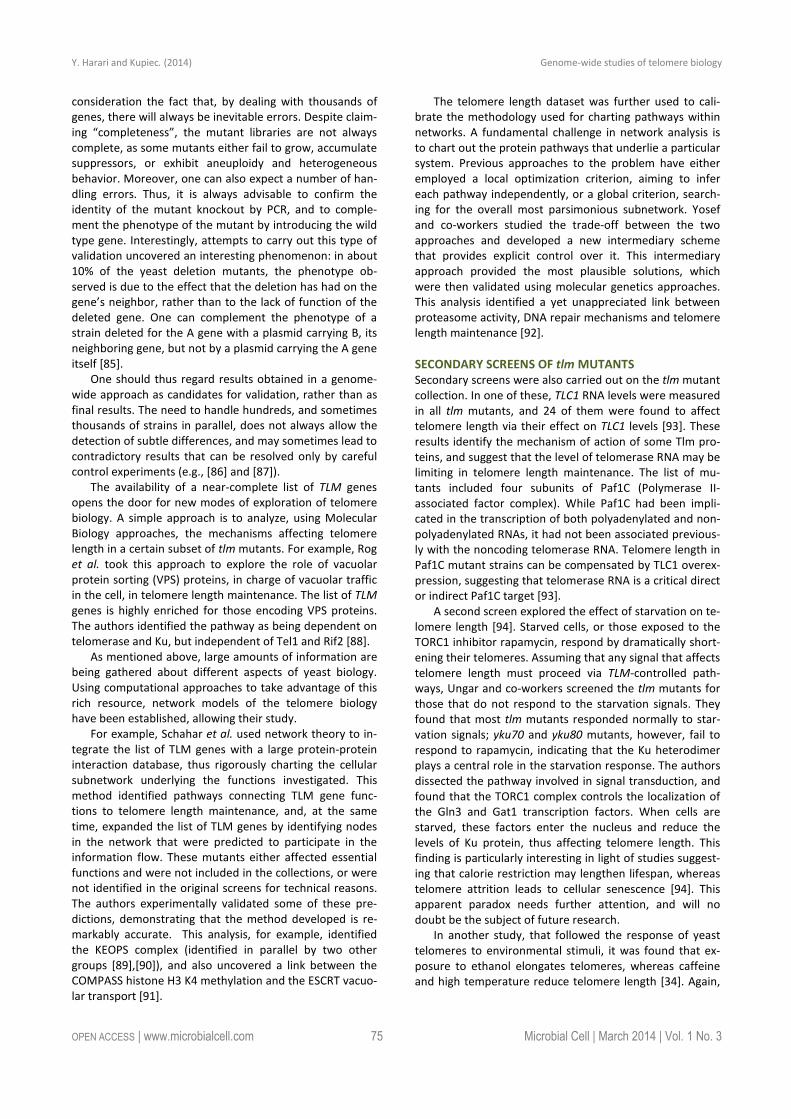
COMPOSITION OF YEAST TELOMERES
The basic structure of the yeast telomere is depicted in
Figure 1. The RNA template of telomerase (encoded by the
TLC1 gene) bears the template sequence
CACACACCCACACCAC [18]. However, the telomeric se-
quence in S. cerevisiae is not regular, and can be described
as T(G1-3) [19, 20]. Thus, only very short stretches are cop-
ied in each round of telomerase activity from the RNA
template [21]. This contrasts with the sequence regularity
observed in other organisms, such as K. lactis [22].
A number of proteins bind yeast telomeres:
Rap1: Rap1, an essential DNA binding protein, recognizes
and binds to the yeast telomeric DNA repeats, and serves
to determine telomere length by a “counting” mechanism
that monitors Rap1 concentration (Figure 1B) [23-25]. Rap1
also contributes to telomere capping and prevents repair
of the telomeres as regular double-stranded breaks, as well
as playing roles in localizing telomeres to the nuclear pe-
riphery [26-28]. In addition to its telomeric role, Rap1 also
works as a general cellular transcriptional activator and
repressor that binds to upstream promoter regions at a
large number of genes and interacts with various regulato-
ry proteins [29, 30].
Rif1 and Rif2: Two proteins, Rif1 and Rif2, bind the C-
terminal region of Rap1 (Figure 1B) [31, 32]. Cells mutated
for RIF1 or RIF2 exhibit long telomeres, indicating that the
function of these proteins is to negatively regulate the
elongation of telomeres [31, 32]. Rif1 and Rif2 binding con-
fers to Rap1-bound telomeric DNA a higher order structure
by interconnecting different Rap1 units. The functional
importance of this structure, and its role in telomere biolo-
gy are still unclear [33]. Despite the coordinated binding,
Rif1 and Rif2 seem to work separately of each other. As
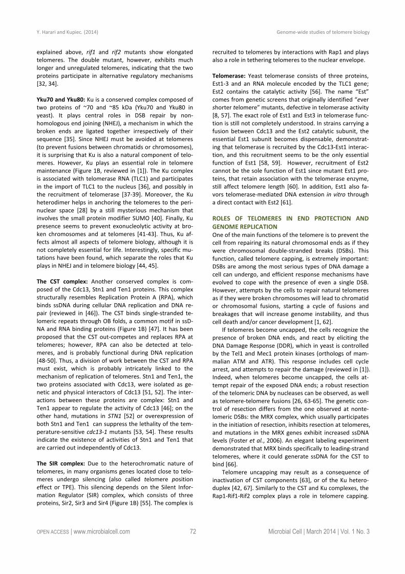
FIGURE 1: Structure of the yeast telomere. (A) Schematic representation of a yeast telomere. All yeast chromosomes carry subtelomeric
repeats called X elements, and between 0 and 4 copies of another sub-telomeric element, the Y’ sequences. Telomeric repeats are composed
of variations of the T(G1-3) formula. The TG-rich strand (with its 3’ OH) is longer than the complementary strand (TG overhangs). (B) Sche-
matic representation of the telomeric chromatin, with representative proteins. Rap1 binds the telomeric repeats, and Rif1, Rif2 and the SIR
proteins bind to Rap1. The Ku heterodimer binds to telomeric dsDNA and the CST complex binds the terminal ssDNA end. Telomerase is re-
cruited to telomeres present in an “extensible” configuration by interactions with the CST.
Y. Harari and Kupiec. (2014) Genome-wide studies of telomere biology
OPEN ACCESS | www.microbialcell.com 72 Microbial Cell | March 2014 | Vol. 1 No. 3
explained above, rif1 and rif2 mutants show elongated
telomeres. The double mutant, however, exhibits much
longer and unregulated telomeres, indicating that the two
proteins participate in alternative regulatory mechanisms
[32, 34].
Yku70 and Yku80: Ku is a conserved complex composed of
two proteins of ~70 and ~85 kDa (Yku70 and Yku80 in
yeast). It plays central roles in DSB repair by non-
homologous end joining (NHEJ), a mechanism in which the
broken ends are ligated together irrespectively of their
sequence [35]. Since NHEJ must be avoided at telomeres
(to prevent fusions between chromatids or chromosomes),
it is surprising that Ku is also a natural component of telo-
meres. However, Ku plays an essential role in telomere
maintenance (Figure 1B, reviewed in [1]). The Ku complex
is associated with telomerase RNA (TLC1) and participates
in the import of TLC1 to the nucleus [36], and possibly in
the recruitment of telomerase [37-39]. Moreover, the Ku
heterodimer helps in anchoring the telomeres to the peri-
nuclear space [28] by a still mysterious mechanism that
involves the small protein modifier SUMO [40]. Finally, Ku
presence seems to prevent exonucleolytic activity at bro-
ken chromosomes and at telomeres [41-43]. Thus, Ku af-
fects almost all aspects of telomere biology, although it is
not completely essential for life. Interestingly, specific mu-
tations have been found, which separate the roles that Ku
plays in NHEJ and in telomere biology [44, 45].
The CST complex: Another conserved complex is com-
posed of the Cdc13, Stn1 and Ten1 proteins. This complex
structurally resembles Replication Protein A (RPA), which
binds ssDNA during cellular DNA replication and DNA re-
pair (reviewed in [46]). The CST binds single-stranded te-
lomeric repeats through OB folds, a common motif in ssD-
NA and RNA binding proteins (Figure 1B) [47]. It has been
proposed that the CST out-competes and replaces RPA at
telomeres; however, RPA can also be detected at telo-
meres, and is probably functional during DNA replication
[48-50]. Thus, a division of work between the CST and RPA
must exist, which is probably intricately linked to the
mechanism of replication of telomeres. Stn1 and Ten1, the
two proteins associated with Cdc13, were isolated as ge-
netic and physical interactors of Cdc13 [51, 52]. The inter-
actions between these proteins are complex: Stn1 and
Ten1 appear to regulate the activity of Cdc13 [46]; on the
other hand, mutations in STN1 [52] or overexpression of
both Stn1 and Ten1 can suppress the lethality of the tem-
perature-sensitive cdc13-1 mutants [53, 54]. These results
indicate the existence of activities of Stn1 and Ten1 that
are carried out independently of Cdc13.
The SIR complex: Due to the heterochromatic nature of
telomeres, in many organisms genes located close to telo-
meres undergo silencing (also called telomere position
effect or TPE). This silencing depends on the Silent Infor-
mation Regulator (SIR) complex, which consists of three
proteins, Sir2, Sir3 and Sir4 (Figure 1B) [55]. The complex is
recruited to telomeres by interactions with Rap1 and plays
also a role in tethering telomeres to the nuclear envelope.
Telomerase: Yeast telomerase consists of three proteins,
Est1-3 and an RNA molecule encoded by the TLC1 gene;
Est2 contains the catalytic activity [56]. The name “Est”
comes from genetic screens that originally identified “ever
shorter telomere” mutants, defective in telomerase activity
[8, 57]. The exact role of Est1 and Est3 in telomerase func-
tion is still not completely understood. In strains carrying a
fusion between Cdc13 and the Est2 catalytic subunit, the
essential Est1 subunit becomes dispensable, demonstrat-
ing that telomerase is recruited by the Cdc13-Est1 interac-
tion, and this recruitment seems to be the only essential
function of Est1 [58, 59]. However, recruitment of Est2
cannot be the sole function of Est1 since mutant Est1 pro-
teins, that retain association with the telomerase enzyme,
still affect telomere length [60]. In addition, Est1 also fa-
vors telomerase-mediated DNA extension in vitro through
a direct contact with Est2 [61].
ROLES OF TELOMERES IN END PROTECTION AND
GENOME REPLICATION
One of the main functions of the telomere is to prevent the
cell from repairing its natural chromosomal ends as if they
were chromosomal double-stranded breaks (DSBs). This
function, called telomere capping, is extremely important:
DSBs are among the most serious types of DNA damage a
cell can undergo, and efficient response mechanisms have
evolved to cope with the presence of even a single DSB.
However, attempts by the cells to repair natural telomeres
as if they were broken chromosomes will lead to chromatid
or chromosomal fusions, starting a cycle of fusions and
breakages that will increase genome instability, and thus
cell death and/or cancer development [1, 62].
If telomeres become uncapped, the cells recognize the
presence of broken DNA ends, and react by eliciting the
DNA Damage Response (DDR), which in yeast is controlled
by the Tel1 and Mec1 protein kinases (orthologs of mam-
malian ATM and ATR). This response includes cell cycle
arrest, and attempts to repair the damage (reviewed in [1]).
Indeed, when telomeres become uncapped, the cells at-
tempt repair of the exposed DNA ends; a robust resection
of the telomeric DNA by nucleases can be observed, as well
as telomere-telomere fusions [26, 63-65]. The genetic con-
trol of resection differs from the one observed at nonte-
lomeric DSBs: the MRX complex, which usually participates
in the initiation of resection, inhibits resection at telomeres,
and mutations in the MRX genes exhibit increased ssDNA
levels (Foster et al., 2006). An elegant labeling experiment
demonstrated that MRX binds specifically to leading-strand
telomeres, where it could generate ssDNA for the CST to
bind [66].
Telomere uncapping may result as a consequence of
inactivation of CST components [63], or of the Ku hetero-
duplex [42, 67]. Similarly to the CST and Ku complexes, the
Rap1-Rif1-Rif2 complex plays a role in telomere capping.
Y. Harari and Kupiec. (2014) Genome-wide studies of telomere biology
OPEN ACCESS | www.microbialcell.com 73 Microbial Cell | March 2014 | Vol. 1 No. 3
However, Rap1 inactivation leads to cell cycle arrest at G1
instead of the Mec1-dependent G2 arrest [68].
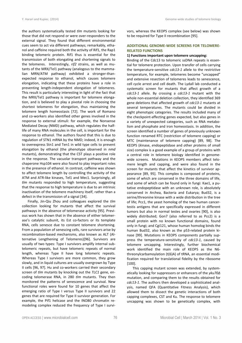
As explained above, telomerase plays an important role
in solving the “end-replication problem” [4, 5]. Telomerase
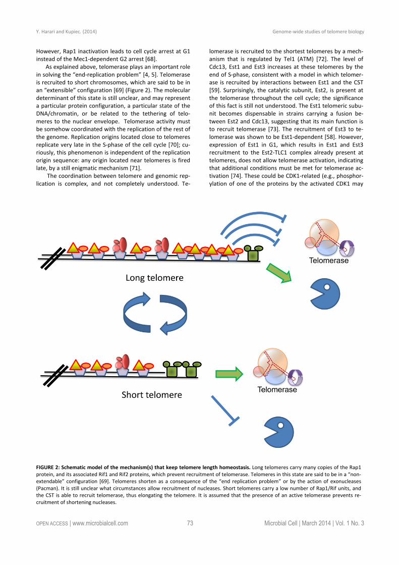
is recruited to short chromosomes, which are said to be in
an “extensible” configuration [69] (Figure 2). The molecular
determinant of this state is still unclear, and may represent
a particular protein configuration, a particular state of the
DNA/chromatin, or be related to the tethering of telo-
meres to the nuclear envelope. Telomerase activity must
be somehow coordinated with the replication of the rest of
the genome. Replication origins located close to telomeres
replicate very late in the S-phase of the cell cycle [70]; cu-
riously, this phenomenon is independent of the replication
origin sequence: any origin located near telomeres is fired
late, by a still enigmatic mechanism [71].
The coordination between telomere and genomic rep-
lication is complex, and not completely understood. Te-
lomerase is recruited to the shortest telomeres by a mech-
anism that is regulated by Tel1 (ATM) [72]. The level of
Cdc13, Est1 and Est3 increases at these telomeres by the
end of S-phase, consistent with a model in which telomer-
ase is recruited by interactions between Est1 and the CST
[59]. Surprisingly, the catalytic subunit, Est2, is present at
the telomerase throughout the cell cycle; the significance
of this fact is still not understood. The Est1 telomeric subu-
nit becomes dispensable in strains carrying a fusion be-
tween Est2 and Cdc13, suggesting that its main function is
to recruit telomerase [73]. The recruitment of Est3 to te-
lomerase was shown to be Est1-dependent [58]. However,
expression of Est1 in G1, which results in Est1 and Est3
recruitment to the Est2-TLC1 complex already present at
telomeres, does not allow telomerase activation, indicating
that additional conditions must be met for telomerase ac-
tivation [74]. These could be CDK1-related (e.g., phosphor-
ylation of one of the proteins by the activated CDK1 may
FIGURE 2: Schematic model of the mechanism(s) that keep telomere length homeostasis. Long telomeres carry many copies of the Rap1
protein, and its associated Rif1 and Rif2 proteins, which prevent recruitment of telomerase. Telomeres in this state are said to be in a “non-
extendable” configuration [69]. Telomeres shorten as a consequence of the “end replication problem” or by the action of exonucleases
(Pacman). It is still unclear what circumstances allow recruitment of nucleases. Short telomeres carry a low number of Rap1/Rif units, and
the CST is able to recruit telomerase, thus elongating the telomere. It is assumed that the presence of an active telomerase prevents re-
cruitment of shortening nucleases.
Y. Harari and Kupiec. (2014) Genome-wide studies of telomere biology
OPEN ACCESS | www.microbialcell.com 74 Microbial Cell | March 2014 | Vol. 1 No. 3
be a pre-requisite), or the telomeric DNA may need to be
in a particular molecular configuration for telomerase to
act [69]. Interestingly, the Rif1 and Rif2 proteins and the Ku
heteroduplex may also restrict G1 activation [42, 75, 76].
THE SYSTEMS BIOLOGY REVOLUTION
In the last fifteen years, the efforts to get more systematic
and complete information about the interactions between
molecules resulted in the development of methodologies,
such as the DNA microarray, which provide data about the
status of the whole genome. This technical achievement
lead to a change in attitude, which allowed turning the
traditional reductionist approach of Molecular Biology up-
side-down, to attempt a whole-encompassing view of the
cell. One could thus describe genetic or protein interac-
tions in the same way an astronomer would describe rela-
tionships between stars in the sky. By analyzing large
amounts of data one could discern the existence of order
in the distribution of the bodies analyzed (e.g., galaxies in
the sky or protein complexes in a cell), and attempt to de-
cipher the rules behind their behavior. In addition, there
are “emerging properties”, which cannot be seen by look-
ing at each gene/protein separately and require a “bird’s
eye view” to be discerned (as one can see a pattern in the
distribution of highways in a map, that are hard to see
while traveling a highway by car). A flurry of genome-wide
methodologies was launched, in which a systematic ap-
proach was taken to try to map all the genes (genomics),
RNA molecules (transcriptomics), proteins (proteomics),
and metabolites (metabolomics) in a given organism. This
“omics” revolution required dealing with increasing
amounts of data, and new algorithms and computational
approaches had to be developed. The easiness with which
yeast cells can be grown and manipulated, combined with
the already significant knowledge of yeast life-style, genes
and proteins, made yeast (mainly Saccharomyces cere-
visiae) a natural choice of organism in which to ask ge-
nome-wide questions. Technically, the new discipline of
Systems Biology was driven by the sophisticated genetics
of yeast, which allowed the construction of mutant collec-
tions, fusion protein collections, and many other tools.
These, in turn, quickly became the basis for additional new
genome-wide technologies [77]. The Systems Biology revo-
lution is still ongoing, and a communal effort is being made
to map, for example, all the genetic and physical interac-
tions in the yeast cells. Interestingly, as time passes, the
field as a whole is shifting from general questions about
genome architecture and function, to more focused ques-
tions related to particular aspects of Biology, which are
analyzed with a mixture of Systems and Molecular Biology.
Here we summarize the efforts directed at studying the
biology of telomeres at a system-level.
GENOME-WIDE STUDIES OF TELOMERE BIOLOGY
Telomere Biology is greatly benefitting from the new all-
encompassing approach, which greatly enlarged our
knowledge about the genes involved in telomere biology
and their regulation. The yeast genome has close to 6000
recognized genes. A collection of 4700 mutants was con-
structed by systematically deleting each individual non-
essential gene in yeast (non-essential yeast mutant collec-
tion, [78]). This collection was later complemented by two
additional mutants libraries encompassing all the essential
genes (yeast has ~1300 essential genes): either hypo-
morphic [79] or temperature-sensitive alleles [80] of each
of the essential genes were created. In addition to allowing
systematic crosses of all possible yeast mutants with a par-
ticular mutation/gene construct/reporter [81], these mu-
tant collections allow researchers to carry out systematic
mutant screens even if the phenotype of interest is not
selectable. Once a biochemical assay (or a microscopic
phenotype) is available, it can be applied in a systematic
fashion to each member of the deletion collections, search-
ing systematically for mutant strains defective for the par-
ticular assay. Even complex assays that take hours to carry
out, or microscopic phenotypes that require complex and
slow measurements can be performed in the whole mu-
tant collections in batches, so that the whole collection can
be screened in a matter of months.
Three publications reported the systematic screening
of the mutant collections, looking for those mutants that
affect telomere length (telomere length maintenance or
tlm mutants). DNA was extracted from each individual
yeast strain and telomere length was measured by South-
ern blot, using as probes telomeric repeats that hybridize
to the terminal restriction fragment [82-84]. Together,
these papers identified ~400 genes affecting telomere
length (either shorter or longer than the wild type). To
understand the strength of this approach, it suffices to
mention that only 30 or so genes were known to affect
telomere length at the time the screens were carried out
[82]. This list of genes underscores the central role played
by telomere biology in the yeast life cycle, as ~7% of the
genome affects telomere length. Moreover, it also demon-
strates the complexity of the challenge: telomere length is
determined by mechanisms that elongate (telomerase) or
shorten (nucleases) telomeres (each of which may be posi-
tively and negatively regulated). Mutation in any of the
TLM genes changes the final telomere size; this means that
each of the 400 genes participates in determining the equi-
librium between the two types of activity. It is remarkable
to observe that in each genetic background (e.g., S288c or
W303) wild type cells exhibit always telomeres of the same
size; thus, in the tug-of-war between elongating and short-
ening mechanisms, the equilibrium is always attained at
the same telomere length. Thus, a very strong homeostatic
mechanism is at play to ensure the exact telomere size for
each cell (Figure 2). The genes uncovered in these screens,
as expected, include those affecting DNA and chromatin
metabolism, but almost all functions in the cell are also
represented, including RNA and protein synthesis, traffic
and modification, metabolic pathways, mitochondrial func-
tions, etc. The challenge ahead, of course, is to determine
how all these genes impinge on the telomere length de-
termination.
Up to now we have extoled the advantages of the ge-
nome-wide approach. One should, however, also take into
Y. Harari and Kupiec. (2014) Genome-wide studies of telomere biology
OPEN ACCESS | www.microbialcell.com 75 Microbial Cell | March 2014 | Vol. 1 No. 3
consideration the fact that, by dealing with thousands of
genes, there will always be inevitable errors. Despite claim-
ing “completeness”, the mutant libraries are not always
complete, as some mutants either fail to grow, accumulate
suppressors, or exhibit aneuploidy and heterogeneous
behavior. Moreover, one can also expect a number of han-
dling errors. Thus, it is always advisable to confirm the
identity of the mutant knockout by PCR, and to comple-
ment the phenotype of the mutant by introducing the wild
type gene. Interestingly, attempts to carry out this type of
validation uncovered an interesting phenomenon: in about
10% of the yeast deletion mutants, the phenotype ob-
served is due to the effect that the deletion has had on the
gene’s neighbor, rather than to the lack of function of the
deleted gene. One can complement the phenotype of a
strain deleted for the A gene with a plasmid carrying B, its
neighboring gene, but not by a plasmid carrying the A gene
itself [85].
One should thus regard results obtained in a genome-
wide approach as candidates for validation, rather than as
final results. The need to handle hundreds, and sometimes
thousands of strains in parallel, does not always allow the
detection of subtle differences, and may sometimes lead to
contradictory results that can be resolved only by careful
control experiments (e.g., [86] and [87]).
The availability of a near-complete list of TLM genes
opens the door for new modes of exploration of telomere
biology. A simple approach is to analyze, using Molecular
Biology approaches, the mechanisms affecting telomere
length in a certain subset of tlm mutants. For example, Rog
et al. took this approach to explore the role of vacuolar
protein sorting (VPS) proteins, in charge of vacuolar traffic
in the cell, in telomere length maintenance. The list of TLM
genes is highly enriched for those encoding VPS proteins.
The authors identified the pathway as being dependent on
telomerase and Ku, but independent of Tel1 and Rif2 [88].
As mentioned above, large amounts of information are
being gathered about different aspects of yeast biology.
Using computational approaches to take advantage of this
rich resource, network models of the telomere biology
have been established, allowing their study.
For example, Schahar et al. used network theory to in-
tegrate the list of TLM genes with a large protein-protein
interaction database, thus rigorously charting the cellular
subnetwork underlying the functions investigated. This
method identified pathways connecting TLM gene func-
tions to telomere length maintenance, and, at the same
time, expanded the list of TLM genes by identifying nodes
in the network that were predicted to participate in the
information flow. These mutants either affected essential
functions and were not included in the collections, or were
not identified in the original screens for technical reasons.
The authors experimentally validated some of these pre-
dictions, demonstrating that the method developed is re-
markably accurate. This analysis, for example, identified
the KEOPS complex (identified in parallel by two other
groups [89],[90]), and also uncovered a link between the
COMPASS histone H3 K4 methylation and the ESCRT vacuo-
lar transport [91].
The telomere length dataset was further used to cali-
brate the methodology used for charting pathways within
networks. A fundamental challenge in network analysis is
to chart out the protein pathways that underlie a particular
system. Previous approaches to the problem have either
employed a local optimization criterion, aiming to infer
each pathway independently, or a global criterion, search-
ing for the overall most parsimonious subnetwork. Yosef
and co-workers studied the trade-off between the two
approaches and developed a new intermediary scheme
that provides explicit control over it. This intermediary
approach provided the most plausible solutions, which
were then validated using molecular genetics approaches.
This analysis identified a yet unappreciated link between
proteasome activity, DNA repair mechanisms and telomere
length maintenance [92].
SECONDARY SCREENS OF tlm MUTANTS
Secondary screens were also carried out on the tlm mutant
collection. In one of these, TLC1 RNA levels were measured
in all tlm mutants, and 24 of them were found to affect
telomere length via their effect on TLC1 levels [93]. These
results identify the mechanism of action of some Tlm pro-
teins, and suggest that the level of telomerase RNA may be
limiting in telomere length maintenance. The list of mu-
tants included four subunits of Paf1C (Polymerase II-
associated factor complex). While Paf1C had been impli-
cated in the transcription of both polyadenylated and non-
polyadenylated RNAs, it had not been associated previous-
ly with the noncoding telomerase RNA. Telomere length in
Paf1C mutant strains can be compensated by TLC1 overex-
pression, suggesting that telomerase RNA is a critical direct
or indirect Paf1C target [93].
A second screen explored the effect of starvation on te-
lomere length [94]. Starved cells, or those exposed to the
TORC1 inhibitor rapamycin, respond by dramatically short-
ening their telomeres. Assuming that any signal that affects
telomere length must proceed via TLM-controlled path-
ways, Ungar and co-workers screened the tlm mutants for
those that do not respond to the starvation signals. They
found that most tlm mutants responded normally to star-
vation signals; yku70 and yku80 mutants, however, fail to
respond to rapamycin, indicating that the Ku heterodimer
plays a central role in the starvation response. The authors
dissected the pathway involved in signal transduction, and
found that the TORC1 complex controls the localization of
the Gln3 and Gat1 transcription factors. When cells are
starved, these factors enter the nucleus and reduce the
levels of Ku protein, thus affecting telomere length. This
finding is particularly interesting in light of studies suggest-
ing that calorie restriction may lengthen lifespan, whereas
telomere attrition leads to cellular senescence [94]. This
apparent paradox needs further attention, and will no
doubt be the subject of future research.
In another study, that followed the response of yeast
telomeres to environmental stimuli, it was found that ex-
posure to ethanol elongates telomeres, whereas caffeine
and high temperature reduce telomere length [34]. Again,
Y. Harari and Kupiec. (2014) Genome-wide studies of telomere biology
OPEN ACCESS | www.microbialcell.com 76 Microbial Cell | March 2014 | Vol. 1 No. 3
the authors systematically tested tlm mutants looking for
those that did not respond or were over-responders to the
external signal. They found that different environmental
cues seem to act via different pathways; remarkably, etha-
nol and caffeine required both the activity of Rif1, the Rap1
binding telomeric protein. Rif1 thus is essential for the
transmission of both elongating and shortening signals to
the telomeres. Interestingly, rif2 strains, as well as mu-
tants of the MRX/Tel1 pathway (analogous to the mamma-
lian MRN/ATM pathway) exhibited a stronger-than-
expected response to ethanol, which causes telomere
elongation, indicating that these proteins have a role in
preventing length-independent elongation of telomeres.
This result is particularly interesting in light of the fact that
the MRX/Tel1 pathway is important for telomere elonga-
tion, and is believed to play a pivotal role in choosing the
shortest telomeres for elongation, thus maintaining the
telomere length homeostasis [72]. The work of Romano
and co-workers also identified other genes involved in the
response to external stimuli: for example, the Nonsense
Mediated Decay (NMD) pathway, which regulates the half-
life of many RNA molecules in the cell, is important for the
response to ethanol. The authors found that this is due to
regulation of STN1 mRNA by the NMD; indeed, it is enough
to overexpress Stn1 and Ten1 in wild type cells to prevent
elongation by ethanol (the phenotype observed in nmd
mutants), demonstrating that the CST plays a positive role
in the response. The vacuolar transport pathway and the
chaperone Hsp104 were also found to play important roles
in the presence of ethanol. In contrast, caffeine was shown
to affect telomere length by controlling the activity of the
ATM and ATR-like kinases, Tel1 and Mec1. Surprisingly, all
tlm mutants responded to high temperature, suggesting
that the response to high temperature is due to an intrinsic
inactivation of the telomere machinery itself, rather than a
defect in the transmission of a signal [34].
Finally, Jin-Qiu Zhou and colleagues explored the tlm
collection looking for mutants that affect the survival
pathways in the absence of telomerase activity [95]. Previ-
ous work has shown that in the absence of either telomer-
ase’s catalytic subunit, its Est co-factors or its template
RNA, cells senesce due to constant telomere shortening.
From a population of senescing cells, rare survivors arise by
recombination-based mechanisms, also known as ALT (Al-
ternative Lengthening of Telomeres)[96]. Survivors are
usually of two types: Type I survivors amplify internal sub-
telomeric repeats, but have telomeric repeats of normal
length, whereas Type II have long telomeric repeats.
Whereas Type I survivors are more common, they grow
slowly, and in liquid cultures are usually overgrown by Type
II cells [96, 97]. Hu and co-workers carried their secondary
screen of tlm mutants by knocking out the TLC1 gene, en-
coding telomerase RNA, in 280 tlm mutants. They then
monitored the patterns of senescence and survival. New
functional roles were found for 10 genes that affect the
emerging ratio of Type I versus Type II survivors and 22
genes that are required for Type II survivor generation. For
example, the Pif1 helicase and the INO80 chromatin re-
modeling complex reduced the frequency of Type I survi-
vors, whereas the KEOPS complex (see below) was shown
to be required for Type II recombination [95].
ADDITIONAL GENOME-WIDE SCREENS FOR TELOMERE-
RELATED FUNCTIONS
1) Functions important upon telomere uncapping:
Binding of the Cdc13 to telomeric ssDNA repeats is essen-
tial for telomere protection. Upon transfer of cells carrying
the temperature-sensitive cdc13-1 allele to the restrictive
temperature, for example, telomeres become “uncapped”
and extensive resection of telomeres leads to senescence,
cell cycle arrest and cell death. The Lydall lab conducted a
systematic screen for mutants that affect growth of a
cdc13-1 allele. By crossing a cdc13-1 mutant with the
whole non-essential deletion collection, they identified 369
gene deletions that affected growth of cdc13-1 mutants at
several temperatures. The mutants could be divided in
eight phenotypic categories. The results included many of
the checkpoint-affecting genes expected, but also genes in
a variety of unexpected categories, such as RNA metabo-
lism and phosphate and iron homeostasis. In addition, the
screen identified a number of genes of previously unknown
function renamed RTC (restriction of telomere capping) or
MTC (maintenance of telomere capping) [89, 98]. The
KEOPS (kinase, endopeptidase and other proteins of small
size) complex is a good example of a group of proteins with
a central role in telomere biology identified in genome-
wide screens. Mutations in KEOPS members affect telo-
mere length and capping, and were also found in the
screen for mutants that affect the kinetics of survivor ap-
pearance [89, 95]. This complex is composed of proteins,
some of which are conserved in the three domains of life,
and some of which can be found only in fungi: Kae1, a pu-
tative endopeptidase with an unknown role, is absolutely
conserved in Archea, Bacteria and Eukarya; Bud32, is a
serine/threonine kinase with a wide distribution in the tree
of life; Pcc1, the yeast homolog of the two human cancer-
testis antigens that are specifically expressed in different
tumors but also in normal testes and ovaries [90], is also
widely distributed; Gon7 (also referred to as Pcc2) is a
small protein with no known functional domains, found
only in fungi; and Cgi121, whose human homolog binds the
human Bud32, also known as the p53-related protein ki-
nase [99]. Mutations in KEOPS components partially sup-
press the temperature-sensitivity of cdc13-1, caused by
telomere uncapping. Interestingly, further biochemical
work identified the main role of KEOPS as the N6-
threonylcarbamoylation (t(6)A) of tRNA, an essential modi-
fication required for translational fidelity by the ribosome
[100].
This capping mutant screen was extended, by system-
atically looking for suppressors or enhancers of the yku70∆
mutation, and comparing them to the results obtained for
cdc13-1. The authors then developed a sophisticated anal-
ysis, named QFA (Quantitative Fitness Analysis), which
allowed them to dissect the genetic interactions of both
capping complexes, CST and Ku. The response to telomere
uncapping was shown to be genetically complex, with
Y. Harari and Kupiec. (2014) Genome-wide studies of telomere biology
OPEN ACCESS | www.microbialcell.com 77 Microbial Cell | March 2014 | Vol. 1 No. 3
many genes involved in a variety of processes affecting the
outcome. Many genes had differential effects: for example,
inactivating the Nonsense Mediated Decay (NMD) pathway,
which affects half-life of mRNAs in the cells, led to suppres-
sion of cdc13-1 but enhanced the phenotype of yku70∆.
The authors showed that this effect is Stn1 dependent,
again stressing the pivotal role of the CST in regulating
telomere biology [101].
2) Genes important for telomere three-dimensional con-
figuration:
An additional genome-wide screen looked for mutants
affecting the telomere three-dimensional configuration. In
mammalian cells telomeric DNA forms a loop that allows
the ssDNA end to invade telomeric dsDNA forming a Hol-
liday Junction, in a structure that protects the ssDNA end
and is called a T-loop [2]. Due to the small size of yeast
telomeres, it has been impossible to visualize T-loops in
yeast, as has been done in mammals. Instead, functional
assays have been used. For example, a construct carrying a
TATA-less galactose-inducible UAS (upstream activating
sequence) downstream of the URA3 gene is able to tran-
scribe the URA3 gene only at telomeres, where it could
fold back on itself, but not at other locations in the genome
[3]. The Luke group took advantage of this fact to carry out
a genome-wide screen for mutants affecting telomere fold-
back, looking for those that were unable to express the
URA3 construct and could thus grow on medium contain-
ing galactose and 5-FOA (which is lethal when URA3 is ex-
pressed). This screen identified 112 genes that disrupt
looping when mutated [3]. Among various biological pro-
cesses uncovered, lysine deacetylation was found to be
essential for the fold-back through Rif2-dependent re-
cruitment of the Rpd3L complex to telomeres. Absence of
Rpd3 function generates increased susceptibility to nucleo-
lytic degradation and the initiation of premature senes-
cence, suggesting a protective role for Rpd3 deacetylation
activity [3]. Thus, this work identifies a major mechanism
of end protection, together with the way it is recruited to
telomeres.
3) Genes affecting the Tel1 (ATM) pathway:
A screen for enhancers of the MMS sensitivity of tel1∆ mu-
tants uncovered a small number (13) of genes. These in-
cluded Yku70, members of the 9-1-1 pathway, the CCR4-
NOT deadenylase complex, nuclear pore components and
several histone deacetylases. Most of these mutants
caused the MMS sensitivity due to their effects on telo-
meres [102]. Interestingly, this report also suggests that
shortened telomeres confer sensitivity to DNA damaging
agents, such as methyl methanesulfonate (MMS).
4) Genes affecting senescence:
The tlm collection was screened by Hu and co-workers,
looking for mutants affecting survivor appearance in the
absence of telomerase activity [95](see above). A genome-
wide screen [87] examined, in a systematic fashion, the
kinetics of senescence, by crossing the est1∆ mutation
(lacking an essential telomerase recruitment factor) to the
whole nonessential mutant collection. As expected, the
vast majority of gene deletions showed no strong effects
on the entry into/exit from senescence. However, ~200
gene deletions (among them the well-characterized rad52∆
mutant) accelerated entry into senescence, and such cells
often could not recover growth. A smaller number of
strains (among them rif1∆) accelerated both entry into
senescence and subsequent recovery [87]. Interestingly,
the mutants that accelerated senescence tended to also
have negative genetic interactions with yku70∆, but were
neutral when combined with cdc13-1, as determined in
the genome-wide screen described above [101].
Although the Systems Biology revolution is only at its
infancy, we can summarize at this stage that the genome-
wide studies have vastly extended our view of telomere
biology. The number of cellular processes affecting various
aspects of telomere integrity, replication, length regulation
and structure is remarkable. Most of the genes uncovered
in these screens are evolutionarily conserved, and likely to
act similarly in other organisms, including humans. A better
understanding of the mechanisms regulating telomere
biology will have significant medical implications, especially
in the fields of aging and cancer.
ACKNOWLEDGMENTS
Work in the Kupiec lab is supported by the Israel Ministry of
Science and Technology, the Israel Cancer Research Fund and
the Israel Cancer Foundation.
CONFLICT OF INTEREST
The authors declare no conflict of interest.
COPYRIGHT
© 2014 Harari and Kupiec. This is an open-access article
released under the terms of the Creative Commons Attrib-
ution (CC BY) license, which allows the unrestricted use,
distribution, and reproduction in any medium, provided
the original author and source are acknowledged.
Please cite this article as: Yaniv Harari and Martin Kupiec (2014).
Genome-wide studies of telomere biology in budding yeast. Mi-
crobial Cell 1(3): 70-80. doi: 10.15698/mic2014.01.132
REFERENCES
1. Dewar JM, Lydall D (2012). Similarities and differences between
"uncapped" telomeres and DNA double-strand breaks. Chromosoma
121(2): 117-30.
2. Griffith JD, Comeau L, Rosenfield S, Stansel RM, Bianchi A, Moss H,
de Lange T (1999). Mammalian telomeres end in a large duplex loop.
Cell 97(4): 503-14.
Y. Harari and Kupiec. (2014) Genome-wide studies of telomere biology
OPEN ACCESS | www.microbialcell.com 78 Microbial Cell | March 2014 | Vol. 1 No. 3
3. Poschke H, Dees M, Chang M, Amberkar S, Kaderali L, Rothstein R,
Luke B (2012). Rif2 promotes a telomere fold-back structure through
Rpd3L recruitment in budding yeast. PLoS Genet 8(9): e1002960.
4. Olovnikov AM (1971). [Principle of marginotomy in template
synthesis of polynucleotides]. Doklady Akademii nauk SSSR 201(6):
1496-9.
5. Watson JD (1972). Origin of concatemeric T7 DNA. Nature: New
biology 239(94): 197-201.
6. Harley CB, Futcher AB, Greider CW (1990). Telomeres shorten
during ageing of human fibroblasts. Nature 345(6274): 458-60.
7. Hayflick L (1979). The cell biology of aging. The Journal of
investigative dermatology 73(1): 8-14.
8. Lundblad V, Szostak JW (1989). A mutant with a defect in telomere
elongation leads to senescence in yeast. Cell 57(4): 633-43.
9. Greider CW, Blackburn EH (1987). The telomere terminal
transferase of Tetrahymena is a ribonucleoprotein enzyme with two
kinds of primer specificity. Cell 51(6): 887-98.
10. de Lange T (2009). How telomeres solve the end-protection
problem. Science 326(5955): 948-52.
11. Bodnar AG, Ouellette M, Frolkis M, Holt SE, Chiu CP, Morin GB,
Harley CB, Shay JW, Lichtsteiner S, Wright WE (1998). Extension of
life-span by introduction of telomerase into normal human cells.
Science 279(5349): 349-52.
12. DeMasters BK, Markham N, Lillehei KO, Shroyer KR (1997).
Differential telomerase expression in human primary intracranial
tumors. Am J Clin Pathol 107(5): 548-54.
13. Conomos D, Pickett HA, Reddel RR (2013). Alternative lengthening
of telomeres: remodeling the telomere architecture. Front Oncol
3(27).
14. Hahn WC, Counter CM, Lundberg AS, Beijersbergen RL, Brooks
MW, Weinberg RA (1999). Creation of human tumour cells with
defined genetic elements. Nature 400(6743): 464-8.
15. Gramatges MM, Bertuch AA (2013). Short telomeres: from
dyskeratosis congenita to sporadic aplastic anemia and malignancy.
Translational research : the journal of laboratory and clinical
medicine.
16. Armanios M (2012). Telomerase and idiopathic pulmonary fibrosis.
Mutation research 730(1-2): 52-8.
17. Calado RT, Young NS (2009). Telomere diseases. The New England
journal of medicine 361(24): 2353-65.
18. Lin J, Smith DL, Blackburn EH (2004). Mutant telomere sequences
lead to impaired chromosome separation and a unique checkpoint
response. Mol Biol Cell 15(4): 1623-34.
19. Shampay J, Szostak JW, Blackburn EH (1984). DNA sequences of
telomeres maintained in yeast. Nature 310(5973): 154-7.
20. McEachern MJ, Blackburn EH (1994). A conserved sequence motif
within the exceptionally diverse telomeric sequences of budding
yeasts. Proc Natl Acad Sci U S A 91(8): 3453-7.
21. Forstemann K, Lingner J (2001). Molecular basis for telomere
repeat divergence in budding yeast. Molecular and cellular biology
21(21): 7277-86.
22. McEachern MJ, Blackburn EH (1995). Runaway telomere
elongation caused by telomerase RNA gene mutations. Nature
376(6539): 403-9.
23. Marcand S, Wotton D, Gilson E, Shore D (1997). Rap1p and
telomere length regulation in yeast. Ciba Foundation symposium
211(76-93); discussion -103.
24. Krauskopf A, Blackburn EH (1998). Rap1 protein regulates
telomere turnover in yeast. Proc Natl Acad Sci U S A 95(21): 12486-91.
25. Krauskopf A, Blackburn EH (1996). Control of telomere growth by
interactions of RAP1 with the most distal telomeric repeats. Nature
383(6598): 354-7.
26. Pardo B, Marcand S (2005). Rap1 prevents telomere fusions by
nonhomologous end joining. Embo J 24(17): 3117-27.
27. Negrini S, Ribaud V, Bianchi A, Shore D (2007). DNA breaks are
masked by multiple Rap1 binding in yeast: implications for telomere
capping and telomerase regulation. Genes & development 21(3): 292-
302.
28. Laroche T, Martin SG, Gotta M, Gorham HC, Pryde FE, Louis EJ,
Gasser SM (1998). Mutation of yeast Ku genes disrupts the subnuclear
organization of telomeres. Current biology : CB 8(11): 653-6.
29. Tornow J, Zeng X, Gao W, Santangelo GM (1993). GCR1, a
transcriptional activator in Saccharomyces cerevisiae, complexes with
RAP1 and can function without its DNA binding domain. Embo J 12(6):
2431-7.
30. Zhao Y, McIntosh KB, Rudra D, Schawalder S, Shore D, Warner JR
(2006). Fine-structure analysis of ribosomal protein gene transcription.
Molecular and cellular biology 26(13): 4853-62.
31. Hardy CF, Sussel L, Shore D (1992). A RAP1-interacting protein
involved in transcriptional silencing and telomere length regulation.
Genes & development 6(5): 801-14.
32. Wotton D, Shore D (1997). A novel Rap1p-interacting factor, Rif2p,
cooperates with Rif1p to regulate telomere length in Saccharomyces
cerevisiae. Genes & development 11(6): 748-60.
33. Shi T, Bunker RD, Mattarocci S, Ribeyre C, Faty M, Gut H, Scrima A,
Rass U, Rubin SM, Shore D, Thoma NH (2013). Rif1 and Rif2 shape
telomere function and architecture through multivalent Rap1
interactions. Cell 153(6): 1340-53.
34. Romano GH, Harari Y, Yehuda T, Podhorzer A, Rubinstein L, Shamir
R, Gottlieb A, Silberberg Y, Pe'er D, Ruppin E, Sharan R, Kupiec M
(2013). Environmental stresses disrupt telomere length homeostasis.
PLoS Genet 9(9): e1003721.
35. Pastwa E, Blasiak J (2003). Non-homologous DNA end joining. Acta
biochimica Polonica 50(4): 891-908.
36. Rathmell WK, Chu G (1994). Involvement of the Ku autoantigen in
the cellular response to DNA double-strand breaks. Proc Natl Acad Sci
U S A 91(16): 7623-7.
37. Taccioli GE, Gottlieb TM, Blunt T, Priestley A, Demengeot J, Mizuta
R, Lehmann AR, Alt FW, Jackson SP, Jeggo PA (1994). Ku80: product of
the XRCC5 gene and its role in DNA repair and V(D)J recombination.
Science 265(5177): 1442-5.
38. Gravel S, Larrivee M, Labrecque P, Wellinger RJ (1998). Yeast Ku as
a regulator of chromosomal DNA end structure. Science 280(5364):
741-4.
39. Roy R, Meier B, McAinsh AD, Feldmann HM, Jackson SP (2004).
Separation-of-function mutants of yeast Ku80 reveal a Yku80p-Sir4p
interaction involved in telomeric silencing. J Biol Chem 279(1): 86-94.
40. Marvin ME, Becker MM, Noel P, Hardy S, Bertuch AA, Louis EJ
(2009). The association of yKu with subtelomeric core X sequences
prevents recombination involving telomeric sequences. Genetics
183(2): 453-67, 1SI-13SI.
41. Mimitou EP, Symington LS (2010). Ku prevents Exo1 and Sgs1-
dependent resection of DNA ends in the absence of a functional MRX
complex or Sae2. Embo J 29(19): 3358-69.
Y. Harari and Kupiec. (2014) Genome-wide studies of telomere biology
OPEN ACCESS | www.microbialcell.com 79 Microbial Cell | March 2014 | Vol. 1 No. 3
42. Bonetti D, Clerici M, Anbalagan S, Martina M, Lucchini G, Longhese
MP (2010). Shelterin-like proteins and Yku inhibit nucleolytic
processing of Saccharomyces cerevisiae telomeres. PLoS Genet 6(5):
e1000966.
43. Bonetti D, Clerici M, Manfrini N, Lucchini G, Longhese MP (2010).
The MRX complex plays multiple functions in resection of Yku- and
Rif2-protected DNA ends. PloS one 5(11): e14142.
44. Ribes-Zamora A, Mihalek I, Lichtarge O, Bertuch AA (2007). Distinct
faces of the Ku heterodimer mediate DNA repair and telomeric
functions. Nat Struct Mol Biol 14(4): 301-7.
45. Lopez CR, Ribes-Zamora A, Indiviglio SM, Williams CL, Haricharan S,
Bertuch AA (2011). Ku must load directly onto the chromosome end in
order to mediate its telomeric functions. PLoS Genet 7(8): e1002233.
46. Churikov D, Corda Y, Luciano P, Geli V (2013). Cdc13 at a
crossroads of telomerase action. Front Oncol 3(39).
47. Sun J, Yang Y, Wan K, Mao N, Yu TY, Lin YC, DeZwaan DC, Freeman
BC, Lin JJ, Lue NF, Lei M (2011). Structural bases of dimerization of
yeast telomere protein Cdc13 and its interaction with the catalytic
subunit of DNA polymerase alpha. Cell research 21(2): 258-74.
48. Grandin N, Charbonneau M (2013). RPA provides checkpoint-
independent cell cycle arrest and prevents recombination at
uncapped telomeres of Saccharomyces cerevisiae. DNA Repair (Amst)
12(3): 212-26.
49. Luciano P, Coulon S, Faure V, Corda Y, Bos J, Brill SJ, Gilson E,
Simon MN, Geli V (2012). RPA facilitates telomerase activity at
chromosome ends in budding and fission yeasts. Embo J 31(8): 2034-
46.
50. Schramke V, Luciano P, Brevet V, Guillot S, Corda Y, Longhese MP,
Gilson E, Geli V (2004). RPA regulates telomerase action by providing
Est1p access to chromosome ends. Nature genetics 36(1): 46-54.
51. Grandin N, Damon C, Charbonneau M (2001). Ten1 functions in
telomere end protection and length regulation in association with
Stn1 and Cdc13. Embo J 20(5): 1173-83.
52. Grandin N, Reed SI, Charbonneau M (1997). Stn1, a new
Saccharomyces cerevisiae protein, is implicated in telomere size
regulation in association with Cdc13. Genes & development 11(4):
512-27.
53. Petreaca RC, Chiu HC, Eckelhoefer HA, Chuang C, Xu L, Nugent CI
(2006). Chromosome end protection plasticity revealed by Stn1p and
Ten1p bypass of Cdc13p. Nature cell biology 8(7): 748-55.
54. Sun J, Yu EY, Yang Y, Confer LA, Sun SH, Wan K, Lue NF, Lei M
(2009). Stn1-Ten1 is an Rpa2-Rpa3-like complex at telomeres. Genes
& development 23(24): 2900-14.
55. Rusche LN, Kirchmaier AL, Rine J (2003). The establishment,
inheritance, and function of silenced chromatin in Saccharomyces
cerevisiae. Annual review of biochemistry 72:481-516.
56. Hughes TR, Evans SK, Weilbaecher RG, Lundblad V (2000). The Est3
protein is a subunit of yeast telomerase. Current biology : CB 10(13):
809-12.
57. Lendvay TS, Morris DK, Sah J, Balasubramanian B, Lundblad V
(1996). Senescence mutants of Saccharomyces cerevisiae with a
defect in telomere replication identify three additional EST genes.
Genetics 144(4): 1399-412.
58. Tuzon CT, Wu Y, Chan A, Zakian VA (2011). The Saccharomyces
cerevisiae telomerase subunit Est3 binds telomeres in a cell cycle- and
Est1-dependent manner and interacts directly with Est1 in vitro. PLoS
Genet 7(5): e1002060.
59. Taggart AK, Teng SC, Zakian VA (2002). Est1p as a cell cycle-
regulated activator of telomere-bound telomerase. Science
297(5583): 1023-6.
60. Evans SK, Lundblad V (2002). The Est1 subunit of Saccharomyces
cerevisiae telomerase makes multiple contributions to telomere
length maintenance. Genetics 162(3): 1101-15.
61. DeZwaan DC, Freeman BC (2009). The conserved Est1 protein
stimulates telomerase DNA extension activity. Proc Natl Acad Sci U S
A 106(41): 17337-42.
62. Aylon Y, Kupiec M (2003). The checkpoint protein Rad24 of
Saccharomyces cerevisiae is involved in processing double-strand
break ends and in recombination partner choice. Molecular and
cellular biology 23(18): 6585-96.
63. Garvik B, Carson M, Hartwell L (1995). Single-stranded DNA arising
at telomeres in cdc13 mutants may constitute a specific signal for the
RAD9 checkpoint. Molecular and cellular biology 15(11): 6128-38.
64. Mieczkowski PA, Mieczkowska JO, Dominska M, Petes TD (2003).
Genetic regulation of telomere-telomere fusions in the yeast
Saccharomyces cerevisae. Proc Natl Acad Sci U S A 100(19): 10854-9.
65. Marcand S, Pardo B, Gratias A, Cahun S, Callebaut I (2008).
Multiple pathways inhibit NHEJ at telomeres. Genes & development
22(9): 1153-8.
66. Faure V, Coulon S, Hardy J, Geli V (2010). Cdc13 and telomerase
bind through different mechanisms at the lagging- and leading-strand
telomeres. Molecular cell 38(6): 842-52.
67. Maringele L, Lydall D (2002). EXO1-dependent single-stranded
DNA at telomeres activates subsets of DNA damage and spindle
checkpoint pathways in budding yeast yku70Delta mutants. Genes &
development 16(15): 1919-33.
68. Vodenicharov MD, Laterreur N, Wellinger RJ (2010). Telomere
capping in non-dividing yeast cells requires Yku and Rap1. Embo J
29(17): 3007-19.
69. Teixeira MT, Arneric M, Sperisen P, Lingner J (2004). Telomere
length homeostasis is achieved via a switch between telomerase-
extendible and -nonextendible states. Cell 117(3): 323-35.
70. Raghuraman MK, Winzeler EA, Collingwood D, Hunt S, Wodicka L,
Conway A, Lockhart DJ, Davis RW, Brewer BJ, Fangman WL (2001).
Replication dynamics of the yeast genome. Science 294(5540): 115-21.
71. Ferguson BM, Fangman WL (1992). A position effect on the time of
replication origin activation in yeast. Cell 68(2): 333-9.
72. Chang M, Arneric M, Lingner J (2007). Telomerase repeat addition
processivity is increased at critically short telomeres in a Tel1-
dependent manner in Saccharomyces cerevisiae. Genes &
development 21(19): 2485-94.
73. Evans SK, Lundblad V (1999). Est1 and Cdc13 as comediators of
telomerase access. Science 286(5437): 117-20.
74. Osterhage JL, Talley JM, Friedman KL (2006). Proteasome-
dependent degradation of Est1p regulates the cell cycle-restricted
assembly of telomerase in Saccharomyces cerevisiae. Nat Struct Mol
Biol 13(8): 720-8.
75. Gallardo F, Laterreur N, Cusanelli E, Ouenzar F, Querido E,
Wellinger RJ, Chartrand P (2011). Live cell imaging of telomerase RNA
dynamics reveals cell cycle-dependent clustering of telomerase at
elongating telomeres. Molecular cell 44(5): 819-27.
76. Clerici M, Mantiero D, Guerini I, Lucchini G, Longhese MP (2008).
The Yku70-Yku80 complex contributes to regulate double-strand
break processing and checkpoint activation during the cell cycle.
EMBO reports 9(8): 810-8.
Y. Harari and Kupiec. (2014) Genome-wide studies of telomere biology
OPEN ACCESS | www.microbialcell.com 80 Microbial Cell | March 2014 | Vol. 1 No. 3
77. Castrillo JI, Oliver SG (2011). Yeast systems biology: the challenge
of eukaryotic complexity. Methods Mol Biol 759:3-28.
78. Winzeler EA, Shoemaker DD, Astromoff A, Liang H, Anderson K,
Andre B, Bangham R, Benito R, Boeke JD, Bussey H, Chu AM, Connelly
C, Davis K, Dietrich F, Dow SW, El Bakkoury M, Foury F, Friend SH,
Gentalen E, Giaever G, Hegemann JH, Jones T, Laub M, Liao H,
Liebundguth N, Lockhart DJ, Lucau-Danila A, Lussier M, M'Rabet N,
Menard P, et al. (1999). Functional characterization of the S.
cerevisiae genome by gene deletion and parallel analysis. Science
285(5429): 901-6.
79. Breslow DK, Cameron DM, Collins SR, Schuldiner M, Stewart-
Ornstein J, Newman HW, Braun S, Madhani HD, Krogan NJ, Weissman
JS (2008). A comprehensive strategy enabling high-resolution
functional analysis of the yeast genome. Nature methods 5(8): 711-8.
80. Ben-Aroya S, Coombes C, Kwok T, O'Donnell KA, Boeke JD, Hieter P
(2008). Toward a comprehensive temperature-sensitive mutant
repository of the essential genes of Saccharomyces cerevisiae.
Molecular cell 30(2): 248-58.
81. Costanzo M, Baryshnikova A, Bellay J, Kim Y, Spear ED, Sevier CS,
Ding H, Koh JL, Toufighi K, Mostafavi S, Prinz J, St Onge RP, VanderSluis
B, Makhnevych T, Vizeacoumar FJ, Alizadeh S, Bahr S, Brost RL, Chen Y,
Cokol M, Deshpande R, Li Z, Lin ZY, Liang W, Marback M, Paw J, San
Luis BJ, Shuteriqi E, Tong AH, van Dyk N, et al. (2010). The genetic
landscape of a cell. Science 327(5964): 425-31.
82. Askree SH, Yehuda T, Smolikov S, Gurevich R, Hawk J, Coker C,
Krauskopf A, Kupiec M, McEachern MJ (2004). A genome-wide screen
for Saccharomyces cerevisiae deletion mutants that affect telomere
length. Proc Natl Acad Sci U S A 101(23): 8658-63.
83. Gatbonton T, Imbesi M, Nelson M, Akey JM, Ruderfer DM,
Kruglyak L, Simon JA, Bedalov A (2006). Telomere length as a
quantitative trait: genome-wide survey and genetic mapping of
telomere length-control genes in yeast. PLoS Genet 2(3): e35.
84. Ungar L, Yosef N, Sela Y, Sharan R, Ruppin E, Kupiec M (2009). A
genome-wide screen for essential yeast genes that affect telomere
length maintenance. Nucleic Acids Res.
85. Ben-Shitrit T, Yosef N, Shemesh K, Sharan R, Ruppin E, Kupiec M
(2012). Systematic identification of gene annotation errors in the
widely used yeast mutation collections. Nature methods 9(4): 373-8.
86. Ballew BJ, Lundblad V (2013). Multiple genetic pathways regulate
replicative senescence in telomerase-deficient yeast. Aging cell 12(4):
719-27.
87. Chang HY, Lawless C, Addinall SG, Oexle S, Taschuk M, Wipat A,
Wilkinson DJ, Lydall D (2011). Genome-wide analysis to identify
pathways affecting telomere-initiated senescence in budding yeast.
G3 (Bethesda) 1(3): 197-208.
88. Rog O, Smolikov S, Krauskopf A, Kupiec M (2005). The yeast VPS
genes affect telomere length regulation. Current genetics 47(1): 18-28.
89. Downey M, Houlsworth R, Maringele L, Rollie A, Brehme M, Galicia
S, Guillard S, Partington M, Zubko MK, Krogan NJ, Emili A, Greenblatt
JF, Harrington L, Lydall D, Durocher D (2006). A genome-wide screen
identifies the evolutionarily conserved KEOPS complex as a telomere
regulator. Cell 124(6): 1155-68.
90. Kisseleva-Romanova E, Lopreiato R, Baudin-Baillieu A, Rousselle JC,
Ilan L, Hofmann K, Namane A, Mann C, Libri D (2006). Yeast homolog
of a cancer-testis antigen defines a new transcription complex. Embo J
25(15): 3576-85.
91. Shachar R, Ungar L, Kupiec M, Ruppin E, Sharan R (2008). A
systems-level approach to mapping the telomere length maintenance
gene circuitry. Mol Syst Biol 4:172.
92. Yosef N, Ungar L, Zalckvar E, Kimchi A, Kupiec M, Ruppin E, Sharan
R (2009). Toward accurate reconstruction of functional protein
networks. Mol Syst Biol 5:248.
93. Mozdy AD, Podell ER, Cech TR (2008). Multiple yeast genes,
including Paf1 complex genes, affect telomere length via telomerase
RNA abundance. Molecular and cellular biology 28(12): 4152-61.
94. Ungar L, Harari Y, Toren A, Kupiec M (2011). Tor complex 1
controls telomere length by affecting the level of Ku. Current biology :
CB 21(24): 2115-20.
95. Hu Y, Tang HB, Liu NN, Tong XJ, Dang W, Duan YM, Fu XH, Zhang Y,
Peng J, Meng FL, Zhou JQ (2013). Telomerase-null survivor screening
identifies novel telomere recombination regulators. PLoS Genet 9(1):
e1003208.
96. Teng SC, Zakian VA (1999). Telomere-telomere recombination is
an efficient bypass pathway for telomere maintenance in
Saccharomyces cerevisiae. Molecular and cellular biology 19(12):
8083-93.
97. Grandin N, Charbonneau M (2007). Control of the yeast telomeric
senescence survival pathways of recombination by the Mec1 and
Mec3 DNA damage sensors and RPA. Nucleic acids research 35(3):
822-38.
98. Addinall SG, Downey M, Yu M, Zubko MK, Dewar J, Leake A,
Hallinan J, Shaw O, James K, Wilkinson DJ, Wipat A, Durocher D, Lydall
D (2008). A genomewide suppressor and enhancer analysis of cdc13-1
reveals varied cellular processes influencing telomere capping in
Saccharomyces cerevisiae. Genetics 180(4): 2251-66.
99. Miyoshi A, Kito K, Aramoto T, Abe Y, Kobayashi N, Ueda N (2003).
Identification of CGI-121, a novel PRPK (p53-related protein kinase)-
binding protein. Biochem Biophys Res Commun 303(2): 399-405.
100. Wan LC, Mao DY, Neculai D, Strecker J, Chiovitti D, Kurinov I,
Poda G, Thevakumaran N, Yuan F, Szilard RK, Lissina E, Nislow C,
Caudy AA, Durocher D, Sicheri F (2013). Reconstitution and
characterization of eukaryotic N6-threonylcarbamoylation of tRNA
using a minimal enzyme system. Nucleic Acids Res 41(12): 6332-46.
101. Addinall SG, Holstein EM, Lawless C, Yu M, Chapman K, Banks AP,
Ngo HP, Maringele L, Taschuk M, Young A, Ciesiolka A, Lister AL, Wipat
A, Wilkinson DJ, Lydall D (2011). Quantitative fitness analysis shows
that NMD proteins and many other protein complexes suppress or
enhance distinct telomere cap defects. PLoS Genet 7(4): e1001362.
102. Piening BD, Huang D, Paulovich AG (2013). Novel connections
between DNA replication, telomere homeostasis, and the DNA
damage response revealed by a genome-wide screen for TEL1/ATM
interactions in Saccharomyces cerevisiae. Genetics 193(4): 1117-33.