Embed Size (px)
Citation preview
Cell Stem Cell
Article
Genetic Predisposition Directs Breast CancerPhenotype by Dictating Progenitor Cell FateTheresa A. Proia,1,2,9 Patricia J. Keller,1,2,9 Piyush B. Gupta,4,10 Ina Klebba,1,2 Ainsley D. Jones,1,2 Maja Sedic,1,2
Hannah Gilmore,5,6 Nadine Tung,6,7 Stephen P. Naber,3 Stuart Schnitt,5,6 Eric S. Lander,4,8 and Charlotte Kuperwasser1,2,*1Department of Anatomy & Cellular Biology, Sackler School of Biomedical Research, Tufts University School of Medicine, 136 Harrison Ave,
Boston, MA 02111, USA2Molecular Oncology Research Institute3Department of Pathology
Tufts Medical Center, Boston, MA 02111, USA4Department of Biology, MIT and Broad Institute of MIT and Harvard, Cambridge, MA 02139, USA5Department of Pathology6Department of Medicine7Department of Surgical Oncology
Harvard Medical School, Beth Israel Deaconess Medical Center, Boston MA 02115, USA8Department of Systems Biology, Harvard Medical School, Boston, MA 02115, USA9These authors contributed equally to this work10Present address: Whitehead Institute for Biomedical Research, 9 Cambridge Center, Cambridge MA 02139, USA*Correspondence: [email protected]
DOI 10.1016/j.stem.2010.12.007
SUMMARY
Women with inherited mutations in the BRCA1 genehave increased risk of developing breast cancerbut also exhibit a predisposition for the develop-ment of aggressive basal-like breast tumors. Wereport here that breast epithelial cells derived frompatients harboring deleterious mutations in BRCA1(BRCA1mut/+) give rise to tumorswith increased basaldifferentiation relative to cells from BRCA1+/+
patients. Molecular analysis of disease-free breasttissues from BRCA1mut/+ patients revealed defectsin progenitor cell lineage commitment even beforecancer incidence. Moreover, we discovered that thetranscriptional repressor Slug is an important func-tional suppressor of human breast progenitor celllineage commitment and differentiation and that itis aberrantly expressed in BRCA1mut/+ tissues. Slugexpression is necessary for increased basal-likephenotypes prior to and after neoplastic transforma-tion. These findings demonstrate that the geneticbackground of patient populations, in addition toaffecting incidence rates, significantly impactsprogenitor cell fate commitment and, therefore,tumor phenotype.
INTRODUCTION
Tumor suppressor genes, such as BRCA1, repress malignant
transformation by ensuring the fidelity of DNA replication and
chromosomal segregation in response to potentially deleterious
events. The increased risk of breast cancer development in indi-
viduals with inherited mutations in BRCA1 has been attributed to
compromised DNA damage repair activity (Welcsh and King,
C
2001). However, it has been unclear why mutations in BRCA1
are also preferentially associated with an increased propensity
for developing a specific subtype of breast cancers, basal-like
tumors, with a distinct molecular phenotype and a poor prog-
nosis (Foulkes et al., 2004; Arnes et al., 2005). Recent evidence
has indicated that BRCA1 might function to regulate mammary
epithelial cell morphogenesis and differentiation (Furuta et al.,
2005; Liu et al., 2008; Kubista et al., 2002). Whether these
functions of BRCA1 directly relate to the increased development
of basal-like breast cancer, however, is not known.
Human breast tissue contains two major specialized epithelial
cell types: luminal cells with secretory functions surrounding the
inner breast duct lumen and basal/myoepithelial cells with
contractile functions that interface between luminal cells and
the basement membrane. Corresponding to these cell states,
human breast cancers are broadly classified into luminal-like or
basal-like tumors based on their gene expression patterns
(Peppercorn et al., 2008). Accordingly, it has been proposed
that tumors with ‘‘luminal’’ characteristics may result from the
transformation of cellswithin the luminal lineage,whereas tumors
exhibiting ‘‘basal-like’’ differentiation may arise from basal cells.
However, there is also awealth of evidence indicating that breast
tumors exhibiting luminal or basal-like differentiation have
distinct constellations of genetic aberrations, which may also
influence tumor phenotype. For example, luminal tumors
frequently express elevated levels of cyclin D1 (CCND1) and
sustain mutations in phosphoinositide 3-kinase (PI3K) (Gauthier
et al., 2007; Loi et al., 2009; Saal et al., 2005; Campbell et al.,
2004), whereas dysregulated expression of ras isoforms, muta-
tions in p53, loss of PTEN expression, and loss or silencing of
BRCA1 are more commonly associated with basal-like tumors
(Gluz et al., 2009; Rakha et al., 2008; Miyakis et al., 1998). More-
over, as mentioned above, inherited mutations in BRCA1
(BRCA1mut/+) strongly predispose for the formation of basal-like
tumors (Foulkes, 2003; Foulkes et al., 2004; Arnes et al., 2005).
In principle, the predisposition for basal-like tumors inBRCA1-
mutation carriers could result either from the differentiation state
ell Stem Cell 8, 149–163, February 4, 2011 ª2011 Elsevier Inc. 149
Cell Stem Cell
BRCA1 and Slug in Epithelial Progenitor Cell Fate
of the precursor cells or from the genetic alterations acquired
during tumor formation. In this study, we examined the biology
of disease-free breast tissues from patients harboring delete-
rious mutations in BRCA1. In doing so, we found a relationship
between genetic alterations in perturbing mammary progenitor
differentiation and their influence on tumor phenotype.
RESULTS
Creation of Human Breast Cancers In Vivo ExhibitingHeterogeneous DifferentiationTo examine the connection between the role of BRCA1 in breast
progenitor cell differentiation with the susceptibility of BRCA1-
mutation carriers to developing basal-like breast cancers, we
used a recently described method for creating human breast
tissues in vivo (Proia and Kuperwasser, 2006; Wu et al., 2009).
This method involves three distinct temporal steps: (1) clearing
of the murine mammary fat pad, (2) reconstitution of the
mammary fat pad with human stromal cells, and (3) introduction
of lentiviral-infected organoids comixed with activated fibro-
blasts into the humanized fat pad. Because this system does
not require any cell culture, the likelihood of genetic alterations
or the selection of variant phenotypes during in vitro expansion
is minimized.
In an attempt to generate tumors from patient-derived breast
epithelial cells, we modified step (3) above by introducing onco-
genes into dissociated single cell suspensions of epithelial cells
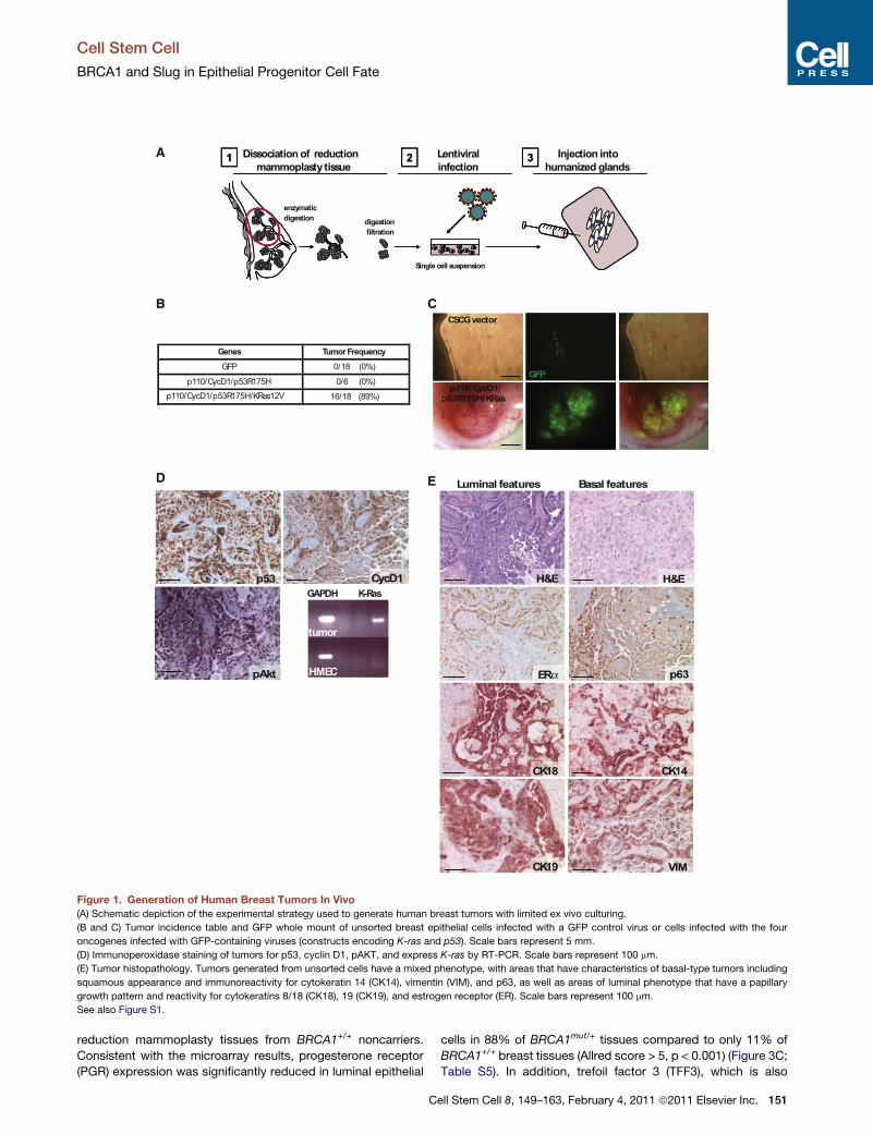
before introducing them into humanized stroma (Figure 1A). We
chose a set of oncogenes reflective of both the luminal and basal
tumor classes to reduce the potential for genetic bias toward
either tumor subtype. We infected uncultured breast epithelial
cell suspensions obtained from dissociated reduction mammo-
plasty tissues with lentiviruses harboring genes for a mutated
form of p53 (p53R175H), wild-type cyclin D1 (CCND1), a consti-
tutively activated form of P13K (PI3KCA), and an oncogenic form
of K-ras (RasG12V). Breast tumors developed when all four
genes were introduced simultaneously into the breast epithelial
cells (Figures 1B and 1C).
Tumor formation with this procedure was observed with
reduction mammoplasty tissues obtained from multiple patient
samples. Expression of the introduced genes in the generated
breast tumors was verified by immunostaining (for p53, cyclin
D1, and p-Akt) and RT-PCR (for K-ras) (Figure 1D). Hematoxylin
and eosin (H&E) stains of tumor sections revealed that the
tumors were heterogeneous invasive carcinomas with regions
of mixed squamous and papillary features (Figure 1E; Figure S1
available online). Immunostaining showed that cancer cells in
squamous metaplastic regions expressed markers indicative of
basal differentiation (cytokeratin 14 [CK14], p63, and vimentin
[VIM]) and those within papillary regions expressed luminal
markers (estrogen receptor [ER], CK8/18, and CK19) (Figure 1E).
We next applied this transformation protocol to mammary
epithelial cells obtained from prophylactic mastectomy tissues
from patients harboring deleterious mutations in BRCA1
(BRCA1mut/+) (Table S1, Figure S1). We observed that the iden-
tical set of oncogenes was sufficient to transform epithelial cells
obtained from BRCA1mut/+ patients (Figure 2A). Although the
introduced oncogenes were expressed to the same extent in
wild-type and BRCA1 tumor tissues, immunostaining of tissue
150 Cell Stem Cell 8, 149–163, February 4, 2011 ª2011 Elsevier Inc.
sections revealed strong expression of the basal epithelial
markers CK14, p63, and vimentin in BRCA1mut/+ tumor cells
(Figures 2B and 2C). In addition, although tumors arising from
BRCA1+/+ epithelium exhibited regions that were CK8/18 and
ER positive, tumors arising from BRCA1mut/+ cells showed
a statistically significant reduction in both CK8/18 and ER
expression and increased CK14 expression, which is typical of
basal-like tumors (Figure 2C).
To evaluate more comprehensively whether the tumors gener-
ated from BRCA1mut/+ epithelium exhibited increased basal-like
features, we performed global gene expression analyses (Table
S2). Hierarchical clustering indicated that tumors arising from
either BRCA1+/+ or BRCA1mut/+ epithelium could be segregated
from one another based on global transcriptional profiles (Fig-
ure 2D). Gene set enrichment analysis (GSEA) revealed that
BRCA1mut/+ tumors exhibited a significant upregulation of genes
associated with breast epithelial basal/myoepithelial cell
differentiation compared to the tumors arising from BRCA1+/+
cells (Figure 2E: basal gene set I, p < 0.024; basal gene set II,
p < 10�4; Table S3). In addition, GSEA indicated specific upregu-
lation of genes in the human breast cancer ‘‘basal-like’’ centroid,
which identifies the human basal-like tumor phenotype (Hu et al.,
2006) in BRCA1mut/+ tumors (basal centroid, Figure 2E, p <
0.033; Table S3) relative to BRCA1+/+ tumors. Collectively, these
results indicate that compared toBRCA1+/+ tumors,BRCA1mut/+
tumors generated with identical transforming oncogenes ex-
hibited increased basal-like differentiation.
Lineage Differentiation Defects in Breast Tissuesfrom BRCA1-Mutation CarriersBecause the BRCA1+/+ and BRCA1mut/+ tumors were generated
with identical oncogenes, these results suggest that the predis-
position ofBRCA1mut/+ patients for developing basal-like tumors
may result from cellular distinctions present prior to neoplastic
transformation. We therefore purified breast epithelial cells
from BRCA1+/+ and BRCA1mut/+ disease-free breast tissues
and assessed the differentiation state of normal precursors in
age-matched breast tissue samples. BRCA1+/+ and BRCA1mut/+
breast epithelial cells expressed similar levels of BRCA1 tran-
script and protein (Figure S2). However, gene-expression
profiling indicated that many genes were differentially expressed
between BRCA1+/+ and BRCA1mut/+ epithelial cells (Figure 3A;
Table S4; Figure S2). Examination of gene ontology functional
processes indicated that a number of genes associated with
DNA transcription (repressor and activator), DNA binding, estab-
lishment and/or maintenance of chromatin architecture,
and chromatin assembly or disassembly were differentially
expressed in BRCA1mut/+ epithelium relative to BRCA1+/+
epithelium (Figure 3B).
Examination of genes associated with epithelial differentiation
revealed that luminal genes and various hormone-related
genes including progesterone and estrogen beta receptors
(PGR, ESR2) (Table S4) were downregulated in BRCA1mut/ +
cells, while genes associated with progenitor or basal cells
were upregulated (Figure 3A; Table S4). We confirmed these
differences in differentiation by using semiquantitative immuno-
histochemistry (Allred scoring metric, see Experimental Pro-
cedures) applied to disease-free prophylactic mastectomy
tissues obtained from BRCA1mut/+ carriers and age-matched
A
B C
D E
GFP
CSCG vector
p110/CycD1/
p53R175H/KRas
0/18 (0%)GFP
p110/CycD1/p53R175H/KRas12Vp110/CycD1/p53R175H
Genes
16/18 (89%)
0/6 (0%)
Tumor Frequency
GAPDH K-Ras
tumor
HMEC
p53 CycD1
pAkt
H&E
p63
Luminal features Basal features
Dissociation of reduction
mammoplasty tissue
Lentiviral
infection
Injection into
humanized glands
Single cell suspension
1
enzymatic
digestiondigestion
2 31 2 3
H&E
ERα
CK18 CK14
CK19 VIM
fi
Figure 1. Generation of Human Breast Tumors In Vivo
(A) Schematic depiction of the experimental strategy used to generate human breast tumors with limited ex vivo culturing.
(B and C) Tumor incidence table and GFP whole mount of unsorted breast epithelial cells infected with a GFP control virus or cells infected with the four
oncogenes infected with GFP-containing viruses (constructs encoding K-ras and p53). Scale bars represent 5 mm.
(D) Immunoperoxidase staining of tumors for p53, cyclin D1, pAKT, and express K-ras by RT-PCR. Scale bars represent 100 mm.
(E) Tumor histopathology. Tumors generated from unsorted cells have a mixed phenotype, with areas that have characteristics of basal-type tumors including
squamous appearance and immunoreactivity for cytokeratin 14 (CK14), vimentin (VIM), and p63, as well as areas of luminal phenotype that have a papillary
growth pattern and reactivity for cytokeratins 8/18 (CK18), 19 (CK19), and estrogen receptor (ER). Scale bars represent 100 mm.
See also Figure S1.
Cell Stem Cell
BRCA1 and Slug in Epithelial Progenitor Cell Fate
reduction mammoplasty tissues from BRCA1+/+ noncarriers.
Consistent with the microarray results, progesterone receptor
(PGR) expression was significantly reduced in luminal epithelial
C
cells in 88% of BRCA1mut/+ tissues compared to only 11% of
BRCA1+/+ breast tissues (Allred score > 5, p < 0.001) (Figure 3C;
Table S5). In addition, trefoil factor 3 (TFF3), which is also
ell Stem Cell 8, 149–163, February 4, 2011 ª2011 Elsevier Inc. 151
BRCA1mut/+
BRCA1+/+
-0.1
0
0.1
0.2
0.3
0.4
0.5
0.6
0.7
0.8
0.9Basal Gene Set I (p <0.03)Basal Gene Set II (p < 0.0001)Basal Centroid (p < 0.02)
Gene rank
Enr
ichm
ent s
core
A
D
B
H&E PanCK
CK14ER
p63 VIM
GFP
CSCG vector
p110/CycD1/
p53R175H/KRas
-2 -1 210
Tumor tissues
0 5000 10000 15000
0
1.52
2.5
3
3.5
4
BRCA1
TumorsBRCA1
Tumorsmut/++/+ 0
51015202530354045
CK18 CK14
Per
cent
tum
or a
rea
BRCA1
TumorsBRCA1
Tumorsmut/++/+
0.51
p = 0.02
E
p53 CycD1
C
05
10152025303540
Cyclin D1 P530
5
10
15
20
25
30
Cyc
lin D
1exp
ress
ion p53 expression
BRCA1mut/+
BRCA1+/+
p= 0.194p= 0.918
Tumors Tumors
ER
exp
ress
ion
K-R
as m
RN
A e
xpre
ssio
n
BRCA1mut/+
TumorsBRCA1
+/+
Tumors
0
1
2
3
4
5
6
78
p =0.01
p = 0.03
ZDHHC7ABCF1CLUB4GALT5ZFP106RAB6IP2KLK7C3ICMTAZGP1GALNAC4S-6STMRPS30UBTFITGB5PSG4CDKN1COACT2GTF2IRD1AHRAGR2CRYZHNRPLCNOT1PECAM1CD59AZGP1C4BPAVPS13DSFRS14LHFPL2B4GALT5NPAS2KIAA1641NQO2MAP3K5NACLOCKDUSP14TEGTCLUSHBSASH1FLJ35801RARRES1HSD17B7BRD3PHLDA1STC1ABHD2C19orf2CXCL10MYH4RBP1THBS2RIMS2AQP9ESDARF3C1orf46MYOM2HLA-GFABP1THBS1NAMARCKSPTPN11U2AF2PCDHB3DIO2CDH11GABARAPL3MYOTCNTN5GPR171HOXB3SLC6A14NAPPP2R2BGSTT1LOC286434GPX7ULBP1ELA1APOEGPM6BLOC442535EFHD1LGALS7NKX2-8LMAN1LCOL2A1ACTA2GPM6BZNF207PTHNAGGCX
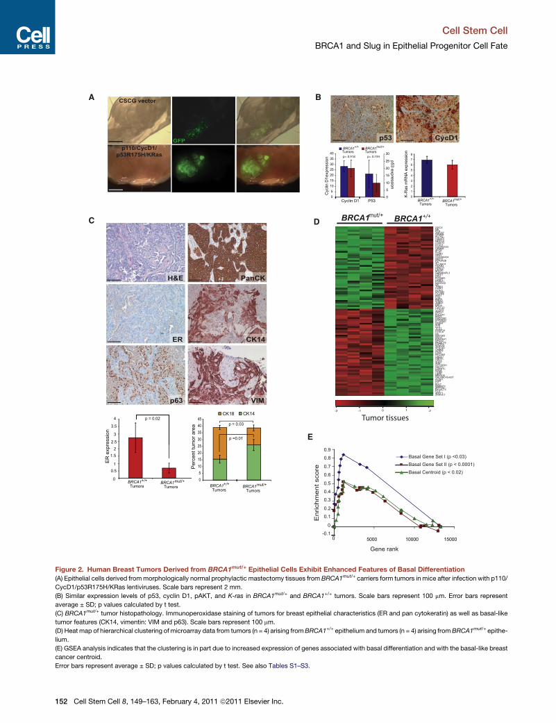
Figure 2. Human Breast Tumors Derived from BRCA1mut/+ Epithelial Cells Exhibit Enhanced Features of Basal Differentiation
(A) Epithelial cells derived frommorphologically normal prophylactic mastectomy tissues from BRCA1mut/+ carriers form tumors in mice after infection with p110/
CycD1/p53R175H/KRas lentiviruses. Scale bars represent 2 mm.
(B) Similar expression levels of p53, cyclin D1, pAKT, and K-ras in BRCA1mut/+ and BRCA1+/+ tumors. Scale bars represent 100 mm. Error bars represent
average ± SD; p values calculated by t test.
(C) BRCA1mut/+ tumor histopathology. Immunoperoxidase staining of tumors for breast epithelial characteristics (ER and pan cytokeratin) as well as basal-like
tumor features (CK14, vimentin: VIM and p63). Scale bars represent 100 mm.
(D) Heat map of hierarchical clustering of microarray data from tumors (n = 4) arising fromBRCA1+/+ epithelium and tumors (n = 4) arising fromBRCA1mut/+ epithe-
lium.
(E) GSEA analysis indicates that the clustering is in part due to increased expression of genes associated with basal differentiation and with the basal-like breast
cancer centroid.
Error bars represent average ± SD; p values calculated by t test. See also Tables S1–S3.
Cell Stem Cell
BRCA1 and Slug in Epithelial Progenitor Cell Fate
152 Cell Stem Cell 8, 149–163, February 4, 2011 ª2011 Elsevier Inc.
Cell Stem Cell
BRCA1 and Slug in Epithelial Progenitor Cell Fate
associated withmature luminal differentiation, was nearly absent
in 88% of BRCA1mut/+ tissues compared to only 36% of
BRCA1+/+ tissues (Allred score < 4, p < 0.0398; Figure 3C; Table
S5). In contrast, 88% of BRCA1mut/+ tissue samples exhibited
moderate-to-high expression of the basal marker vimentin
compared to 16% of BRCA1+/+ tissues (Figure 3C; Table S5;
p < 0.086).
We next used flow cytometry to assess the proportion of
lineage-committed and progenitor epithelial cells in breast
tissues. Cells expressing CD24 or high levels of EpCAM (ESA)
enrich for cells of the luminal lineage, whereas cells expressing
high levels of CD49f enrich for cells of the myoepithelial (ME)/
basal lineage (Villadsen et al., 2007; Shipitsin et al., 2007). Anal-
ysis of reduction mammoplasty breast tissues from BRCA1+/+
patients identified four populations of epithelial cells: EpCAMhi/
CD49f� mature luminal cells, EpCAMhi/CD49f+ luminal progen-
itor cells, EpCAMlow/CD49f+ basal/myoepithelial (ME) cells,
and EpCAM�/CD49f+ basal progenitor cells (Figure 3D; Fig-
ure S2; Keller et al., 2010; Lim et al., 2009; Eirew et al., 2008).
Analysis of prophylactic mastectomy tissues fromBRCA1mut/+
(<50 year) tissues also identified four populations of epithelial
cells but revealed a statistically significant increase in the
proportion of EpCAM�/CD49f+ basal progenitor cells (p < 0.04;
Figure 3D) and an appreciable but not statistically significant
decrease in the number of EpCAMhi/CD49f+ luminal progenitor
cells. These results indicate that BRCA1mut/+ tissues exhibit
luminal and basal epithelial cell differentiation defects prior to
any evidence of cancer.
Characterization of Progenitor Cellsfrom BRCA1-Mutation CarriersWe next evaluated progenitor activity of mammary epithelial
cells obtained directly from breast tissues. We employed mam-
mosphere (Dontu et al., 2003) and adherent colony-formation
(Stingl et al., 2001) assays to assess breast progenitor activity,
and we evaluated whether they arose from luminal-committed,
basal/ME-committed, or bipotent progenitors by staining for
the differentiation markers CK14 and CK8/18. We found no
significant differences in the formation of primary mammo-
spheres, suggesting that the total number of stem/progenitor
cells may not differ between BRCA1+/+ and BRCA1mut/+ tissues
(Figure S3). In addition, there was no significant difference in the
distribution of CK8/18+ and CK14+ cells within both BRCA1+/+
and BRCA1mut/+ mammospheres (Figure S3).
Under adherent conditions, we found that human breast
epithelial cells generated spherical colonies that grew in suspen-
sion as well as adherent colonies that grew on plastic (Figure 3E;
Figures S3). Although we did not observe a statistically signifi-
cant difference in the number of adherent progenitor colonies
arising from BRCA1mut/+ cells, we did observe that spherical
luminal colonies derived from BRCA1mut/+ cells expressed
significantly higher levels of the basal maker CK14 in comparison
to colonies from BRCA1+/+ cells that were more uniformly CK8/
18 positive (Figure 3E).
We also assessed the in vivo outgrowth competency of
progenitor cells from BRCA1mut/+ and BRCA1+/+ cells. By using
the humanized cleared fat pad system, we found that BRCA1+/+
cells generated mature bilayered ductal/acinar outgrowths,
which contained an inner luminal layer of epithelial cells that
C
stained predominantly for CK8/18 and 19, and an outer myoepi-
thelial layer that stained for the basal/MEmarker CK14 and SMA.
In contrast, BRCA1mut/+ cells gave rise primarily to immature
ductal/acinar outgrowths that exhibited a significant increase
in bipotent luminal cells that expressed both CK19 and CK14
and to a lesser degree CK8/18 and CK14 (Figure 3F; Figure S3).
Taken together, these results reveal that luminal progenitor cells
from BRCA1mut/+ tissue exhibit defects in full maturation and
differentiation and retain features of basal differentiation.
Luminal Cells Give Rise to Tumorsin BRCA1-Mutation CarriersWe next wanted to determine whether the increased basal
differentiation observed after neoplastic transformation of
BRCA1mut/+ cells was due to the increased numbers of EpCAM�
basal cells or to the increased basal differentiation state of
luminal progenitor cells. Accordingly, we enriched for either
luminal (EpCAM+) or basal/ME (CD10+) cells (Figure 4A) prior to
lentiviral infection and injection into the mammary fat pad.
Each subpopulation was isolated from breast tissues to
>90% purity, as gauged by immunofluorescence (Figure 4B).
The CD10+ subpopulation was enriched for basal/ME CK14+
cells, but CK14+ cells were depleted from the CD10�/EpCAM+
fraction (Figure 4C). Conversely, CK8/18+ luminal cells were
enriched in the CD10�/EpCAM+ fraction compared to the
CD10+ and parental unsorted populations (Figure 4C).
Basal/ME-enriched (CD10+), luminal/progenitor-enriched
(CD10�/EpCAM+), and marker-depleted (CD10�/EpCAM�) cellswere infected with the p53R175H, CCND1, PI3KCA, and
RasG12V oncogenes and injected into humanized murine
mammary glands. The luminal-enriched CD10�/EpCAM+ frac-
tion consistently formed tumors with growth kinetics, frequen-
cies, and histopathology similar to tumors arising from unsorted
cells from either BRCA1mut/+- or BRCA1+/+-derived tissues
(Figures 4D and 4E). Thus, basal/ME (CD10+) or depleted
(CD10�/EpCAM�) cells from either BRCA1mut/+ or BRCA1+/+
breast epithelial cell populations were not preferentially trans-
formed with this combination of oncogenes. Rather, these
results indicate that the target cell for transformation probably
resides within the luminal EpCAM+/CD10� population. Collec-
tively, these results imply that the increased basal phenotype
of BRCA1-associated tumors results from the pre-existing
increased basal differentiation state of the luminal progenitor
population.
Slug Suppresses Breast Progenitor CellLineage CommitmentTo investigate the mechanism by which BRCA1mutation affects
progenitor cell differentiation, we classified the breast epithelial
gene-expression signature described above based on signaling
pathways that were differentially expressed in BRCA1mut/+ cells.
Remarkably, the most significantly represented signaling path-
ways identified in BRCA1mut/+ breast epithelial signature were
the Wnt, Notch, and melanogenesis pathways (Figure S4).
Notably, the transcriptional repressor Slug, which is an estab-
lished regulator of melanocyte development, is a downstream
target of both Wnt and Notch signaling (Niessen et al., 2008;
DiMeo et al., 2009). This connection prompted us to examine
Slug expression in breast epithelial tissues and cells harboring
ell Stem Cell 8, 149–163, February 4, 2011 ª2011 Elsevier Inc. 153
A
1.30E-062.20E-07
1.50E-141.00E-09
3.90E-083.00E-06
9.60E-11
Number of genes
0 20 40 60 80 100
Nuclear hormone receptor, ligand-binding
Steroid hormone receptor activity
Repressor
Chromatin Modification
Chromatin
Chromatin Assembly or disassembly
Transcription coactivator activity
Activator
Transcription factor binding
Maintenance of chromatin architecture
Sequence specific DNA binding
Transcription Regulation
2.00E-126.30E-14
3.00E-06
2.90E-161.30E-57
B
CBRCA1
+/+BRCA1
mut/+
E
Pe
rc
en
t o
f c
ell
s
0.0
0.5
1.0
1.5
2.0
2.5
3.0
Av
era
ge
C
K1
4 s
tain
sc
ore
BRCA1+/+
Ep
CA
M
CD49f
BRCA1+/+ BRCA1
mut/+
PGR
TFF3
VIM
Patient breast tissue-2 -1 210
BRCA1+/+
BRCA1mut/+
Brightfield
p < 0.04
BRCA1+/+
n = 10 (<50 yrs)BRCA1
mut/+
n = 7 (<50yrs)D
EpCAM+
CD49f-
EpCAMhi
CD49f+
EpCAM-
CD49f+
EpCAM-
CD49f-
EpCAMlo
CD49f+
ADRB1SEPX1CD1C
CTNND1INSL6
ATP2A3LOC94431RBMY2FP
KCNK7CIZ1
OACT2DCTN1
MST1GABBR2
IL1RAPL2SMOJAG1
PRSS7MTMR4PDLIM4
RDH5SCN9A
KIAA1614LARS2CDC27
CD3EAPMAP4K5
AMD1STK4
NALOC346329
NANOGCARD10
NPAS1USP7
GUCY2FCOL4A3RANBP9
ADH7LOC388796
Patient breast epithelium
0
5
10
15
20
25
30
35
mut/+BRCA1
p<0.03
BRCA1+/+
BRCA1mut/+
OtherMature
0%
20%
40%
60%
80%
100%
*
Immature
0%
20%
40%
60%
80%
100%p=0.004
F
BRCA1+/+ BRCA1
0%
20%
40%
60%
80%
100%
0%
20%
40%
60%
80%
100%
BRCA1+/+ mut/+
BRCA1
CK14/CK18CK14/CK19
CK14 CK18 CK14 CK18CK14 CK19CK19 CK14
mut/+BRCA1+/+
BRCA1mut/+
Cell Stem Cell
BRCA1 and Slug in Epithelial Progenitor Cell Fate
154 Cell Stem Cell 8, 149–163, February 4, 2011 ª2011 Elsevier Inc.
Cell Stem Cell
BRCA1 and Slug in Epithelial Progenitor Cell Fate
mutations in BRCA1. We did not find differences in SLUGmRNA
expression, consistent with the microarray data, but we
did observe abundant Slug protein in 87% of disease-free
BRCA1mut/+ prophylactic mastectomy tissues, while its expres-
sion was reduced in tissues from reduction mammoplasty
BRCA1+/+ tissues (Allred score > 1, p < 0.01) (Figure 5A).
Because Slug is a transcriptional repressor, we next investi-
gated whether Slug expression might be affecting breast
progenitor lineage commitment and differentiation. Because
serum addition can promote cellular differentiation of immortal-
ized human mammary epithelial cells (HMECs), which are
a model for bipotent breast progenitor cells (Keller et al., 2010;
Zhao et al., 2010), we treated HMECs from patient-derived
BRCA1+/+ and BRCA1mut/+ tissues with serum and assessed
epithelial differentiation. Treatment of BRCA1+/+ HMECs with
serum resulted in luminal differentiation, as measured by an
increase EpCAM+/CD24+ cells as well as increased CD24
expression and increased CK8/18 expression (Figure 5B, data
not shown). However, addition of serum to BRCA1mut/+ HMEC
cells failed to induce complete differentiation, consistent with
defects in luminal lineage competency (Figures 5B and 5C).
Luminal differentiation was accompanied by a reduction in
Slug protein level in both BRCA1mut/+ and BRCA1+/+ cells,
although the overall reduction was somewhat reduced in
BRCA1mut/+ cells (Figure 5C).
To investigate whether Slug directly represses breast epithelial
lineage commitment and differentiation, we used lentiviral-medi-
ated short hairpin inhibition of Slug expression in primary
prophylactic mastectomy cells isolated from three different
patients with deleterious BRCA1 mutations. Slug knockdown
led to a reduction in the proportion of EpCAM�/CD49f+ progen-itor cells and a concomitant increase in the proportion of
EpCAM+, CD44lo, andCD24+ luminal cells (Figure 5D; Figure S5).
Furthermore, expression of the basal marker vimentin was
greatly reduced, while expression of the luminal marker CD24
was increased (Figure 5C; Figure S5). We also examined
the effects of Slug inhibition in immortalized HMECs derived
from BRCA1mut/+ patients. As with primary cells, inhibition of
Slug resulted in a decrease in the numbers of EpCAM�/CD49f+
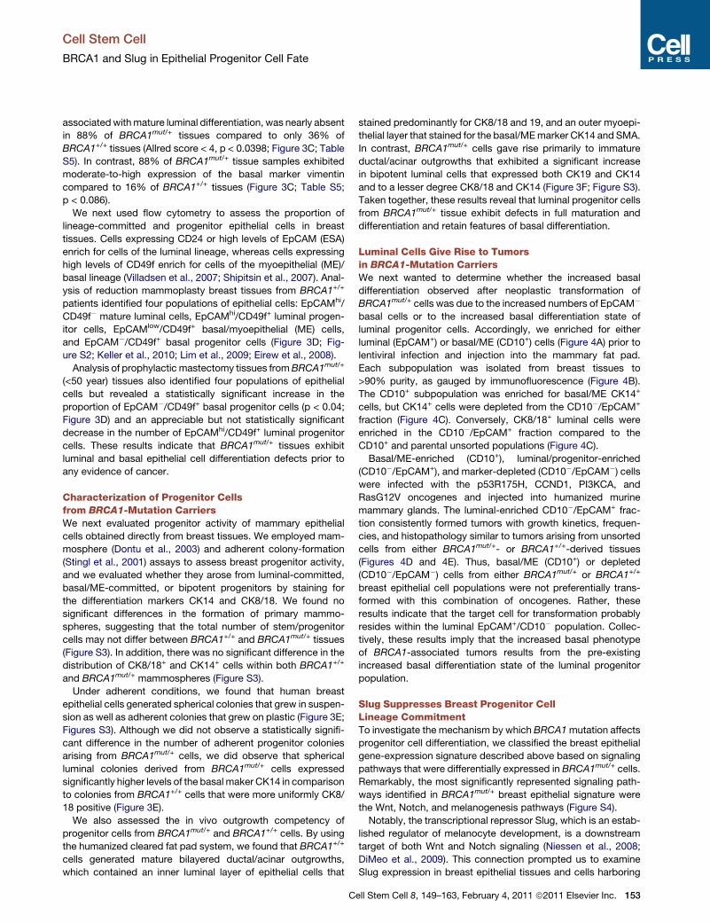
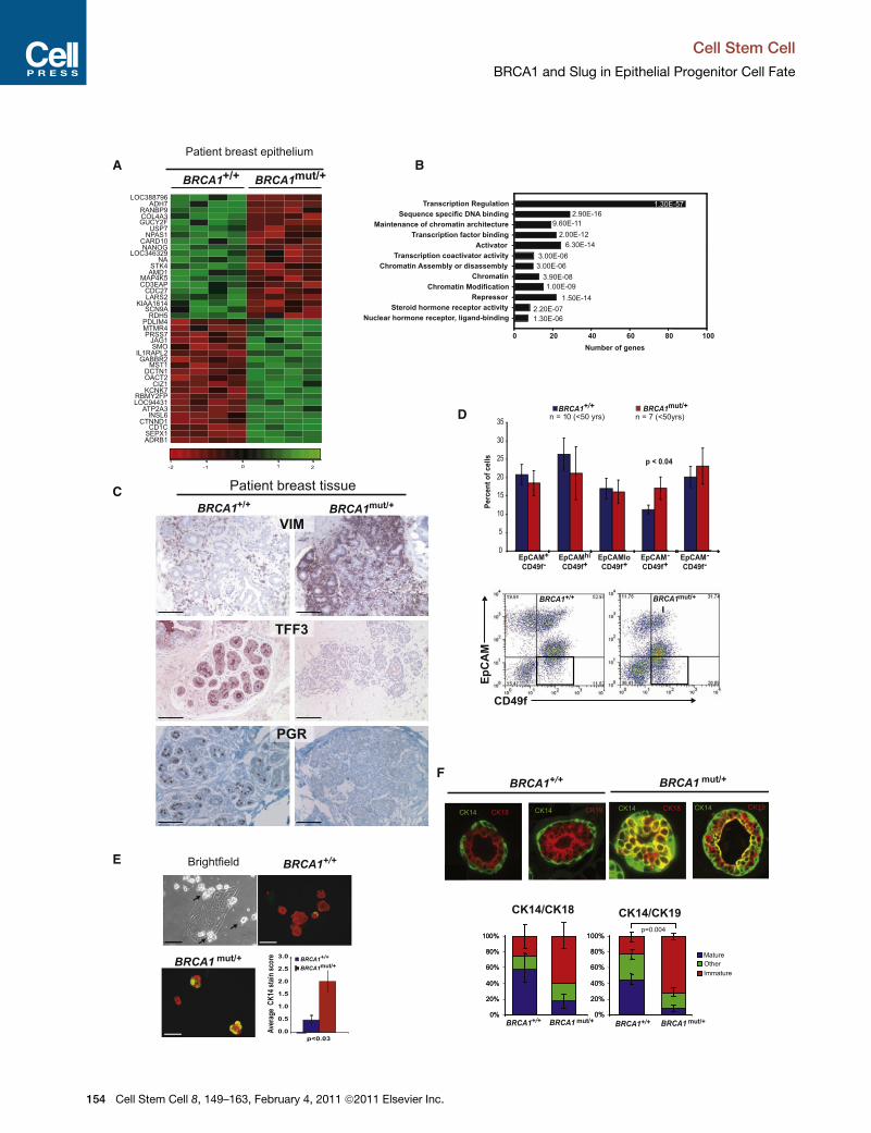
Figure 3. BRCA1mut/+ Breast Epithelial Cells Exhibit Defects in Lineage
(A) Heat map of hierarchical clustering of microarray data from epithelial cells is
samples (n = 4).
(B) Gene ontology biological process categories associated with BRCA1mut/+ bre
categories with an enrichment score >1.5, and the number of genes represented
shown.
(C) Immunoperoxidase staining of normal human breast tissue from BRCA1+/+ an
terone receptor (PGR) and basal-specific vimentin (VIM) antibodies. Scale bars rep
on age-matched BRCA1+/+ (n = 13) and BRCA1mut/+ (n = 10) disease-free breast
(D) Freshly dissociated, uncultured epithelial cells from age-matched (<50 years)B
CD49f expression by flow cytometry. Representative dot plots of a BRCA1+/+ or
(E) Human breast epithelial cells produce small (�30–50 mm) luminal suspension s
spheres were stained for CK8/18 (red) and CK14 (green). Scale bars represent 100
dures. At least 30 spheres were scored for each patient sample. The average sc
graph.
(F) Acinar structures frompatient-derivedBRCA1+/+ (n = 4) andBRCA1mut/+ patien
HIMmodel. Tissue outgrowths were double stained for CK14 and CK8/18 or CK19
basal/ME layer and CK8/18 and/or CK19+ luminal layer), immature (CK14+ basal/M
CK8/18/19 only, etc.). The average number of the three categories of structures
Error bars are ±SEM and p values were calculated by two-tailed t test. See also
C
basal progenitor cells and an increase in the numbers of
EpCAM+ luminal cells (Figure 5E). Given these findings, we
also examined whether inhibition of Slug might also affect
luminal differentiation in BRCA1+/+ cells. Indeed, inhibition of
Slug in BRCA1+/+ cells also led to a reduction in the proportion
of EpCAM�/CD49f+ basal progenitor cells and an increase in
luminal cells. Taken together, these findings indicate that Slug
is a regulator of human breast progenitor cell lineage commit-
ment and that its expression suppresses luminal differentiation.
BRCA1 Regulation of Slug Protein StabilityTo examine whether BRCA1 regulates Slug expression, we used
short interfering RNAs (siRNA) to inhibit BRCA1 expression in
human breast MCF10A cells, which express wild-type BRCA1
(Elstrodt et al., 2006). Quantitative RT-PCR and western blotting
confirmed knockdown of transcript and BRCA1 protein expres-
sion (Figure 6A). Knockdown of BRCA1 by siRNA led to amodest
but highly reproducible 2-fold increase in Slug protein expres-
sion, in the absence of increased mRNA expression (Figure 6A).
These results suggest that loss of BRCA1 may lead to increased
Slug protein expression by a posttranslational mechanism. We
therefore examined the stability of Slug protein in cells after
siRNA inhibition of BRCA1 as well as in cells with mutations in
BRCA1. We confirmed that Slug protein was highly unstable in
the BRCA1+/+ MCF10A cells (Figures 6B and 6C). BRCA1mut
cells (SUM149, SUM1315) and siBRCA1-MCF10A cells were
collected at regular time intervals subsequent to cyclohexamide
(CHX) treatment and subjected to western blot analysis.
Whereas Slug protein levels were turned over in siControl-
MCF10A cells, Slug protein was still detected up to 6 hr after
CHX treatment in siBRCA1-MCF10A cells and in cancer lines
harboring mutations in BRCA1 (Figure 6C). Importantly, the
difference in stability noted in Slug protein in BRCA1mut
SUM149 and SUM1315 cells was not due to a defect in protea-
some activity, as indicated by the fact that cyclin D1 protein was
still degraded. Taken together, these results indicate that BRCA1
regulates Slug protein stability.
To begin to understand the mechanism involved, we looked at
whether the ubiquitin ligase function of BRCA1 might be
Differentiation
olated from BRCA1+/+ breast patient samples (n = 4) and BRCA1mut/+ patient
ast epithelial cells. The DAVID Functional Annotation Tool was used to define
in the list and the p value of genes differentially expressed in the microarray are
d BRCA1mut/+ carriers with luminal-specific trefoil factor 3 (TFF3) and proges-
resent 100 mm. Immunohistochemistry for TFF3, PGR, and VIMwas performed
tissues. Differences in staining were observed primarily in lobules, not ducts.
RCA1+/+ (n = 10) andBRCA1mut/+ (n = 7) patients were analyzed for EpCAM and
BRCA1mut/+ patient are shown.
pheres when grown under adherent conditions (indicated by arrows). Cytospun
mm. CK14 content in spheres was scored as described in Experimental Proce-
ores from three BRCA1+/+ and BRCA1mut/+ patient samples are shown in the
t (n = 4) cells infectedwithGFP lentivirus to visualize outgrowth and grown in the
(representative photos, top). The staining was characterized asmature (CK14+
E layer and CK14 andCK8/19 and/or CK19+ luminal layer), or other (CK14 only,
are shown in the graph (n R 85 acini).
Figures S2 and S3 as well as Tables S4 and S5.
ell Stem Cell 8, 149–163, February 4, 2011 ª2011 Elsevier Inc. 155
BRCA1+/+
Unsorted CD10+ EpCAM+ Depleted
CD10+
Unsorted
CD10+
Unsorted
CD10+
Unsorted
CD10+ EpCAM +
Unsorted0
20406080
100
020406080
100
Unsorted CD10+ EpCAM+
Percen
t C
K14+
0
20
40
60
80
100
Fra
ctio
n p
urity
0
20
40
60
80
100
Unsorted Depleted
Pe
rc
en
t o
f to
ta
l
0
20
40
60
80
100
DoubleNeg
DoublePos
CK18 only
CK14 only
0204060
80100
0204060
80100
Unsorted CD10+ EpCAM+
Percen
t C
K18+
120
10080604020
0Tu
mo
r V
olu
me
-m
m3
120
10080604020
0
3
Tu
mo
r V
olu
me
-m
m3
35302520151050
3
35302520151050
BRCA1mut/+
Breast tissue organoids
Enzymatic and manual disaggregating
Filtration
Single celll suspension
CD10 beads
EpCAM beads
Depeted
Basal cells
Luminal cells
A B
C
D
E
Unsorted CD10+ EpCAM+ Depleted Unsorted CD10+ EpCAM+ Depleted
CD10+ EpCAM+CD10+
EpCAM+
EpCAM+
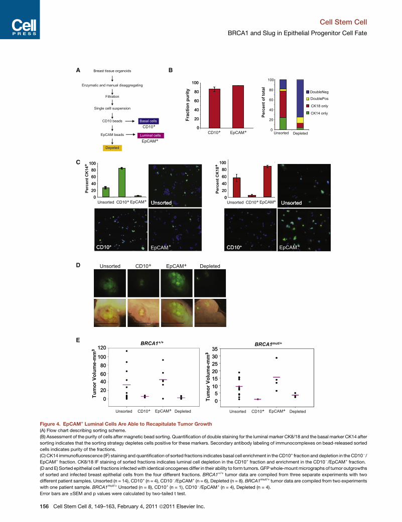
Figure 4. EpCAM+ Luminal Cells Are Able to Recapitulate Tumor Growth
(A) Flow chart describing sorting scheme.
(B) Assessment of the purity of cells after magnetic bead sorting. Quantification of double staining for the luminal marker CK8/18 and the basal marker CK14 after
sorting indicates that the sorting strategy depletes cells positive for these markers. Secondary antibody labeling of immunocomplexes on bead-released sorted
cells indicates purity of the fractions.
(C) CK14 immunofluorescence (IF) staining and quantification of sorted fractions indicates basal cell enrichment in the CD10+ fraction and depletion in the CD10�/EpCAM+ fraction. CK8/18 IF staining of sorted fractions indicates luminal cell depletion in the CD10+ fraction and enrichment in the CD10�/EpCAM+ fraction.
(D and E) Sorted epithelial cell fractions infectedwith identical oncogenes differ in their ability to form tumors. GFPwhole-mountmicrographs of tumor outgrowths
of sorted and infected breast epithelial cells from the four different fractions. BRCA1+/+ tumor data are compiled from three separate experiments with two
different patient samples. Unsorted (n = 14), CD10+ (n = 4), CD10�/EpCAM+ (n = 6), Depleted (n = 8). BRCA1mut/+ tumor data are compiled from two experiments
with one patient sample. BRCA1mut/+ Unsorted (n = 8), CD10+ (n = 1), CD10�/EpCAM+ (n = 4), Depleted (n = 4).
Error bars are ±SEM and p values were calculated by two-tailed t test.
Cell Stem Cell
BRCA1 and Slug in Epithelial Progenitor Cell Fate
156 Cell Stem Cell 8, 149–163, February 4, 2011 ª2011 Elsevier Inc.
A
0
1
2
3
4
5
6
7
Disease-free patient tissues
BRCA1mut/+
BRCA1+/+
20X
60X
1 2 3 4 5 6 7 8 9 10 111 2 3 4 5 6 7 8 9 10
p < 0.015
Allre
d sc
ore
BRCA1+/+
BRCA1mut/+
CD24
Patient pair 1 Patient pair 2
- + - + - + - + - +serum
Patient 1 Patient 2 Patient 1 Patient 2 Patient 3
mut/+BRCA1 Patient HMECsBRCA1 Patient HMECs+/+
B
Slug
β-actin
BRCA1 PatientTissuemut/+
012345678 shSlugshCntrl
EpCAM+CD49f -
EpCAM-CD49f +
Percen
t o
f C
ells
Patient 1
BRCA1 Patient HMECsmut/+
p<0.025
p<0.025
0
20
40
60
80
100
120
EpCAM+ EpCAM-CD49f + EpCAM-
CD49f +EpCAM+
p<0.003 p<0.003
0
5060708090
10203040
Percen
t o
f C
ells
Percen
t o
f C
ells
shSlugshCntrlshSlugshCntrl
-2.5-2.0-1.5-1.0-0.50.00.51.01.52.02.53.0
Patient 1Patient 2
No
rm
alized
Fo
ld C
han
ge
00.5
11.5
22.5
33.5
44.5
EpCAM+CD49f -
EpCAM-CD49f +
shSlugshCntrlPatient 3
Percen
t o
f C
ells
CD24
VIMSLUG
EpCAM+CD49f -
EpCAM-CD49f +
0
5
10
15
20
25
Percen
t o
f C
ells
shSlugshCntrl
Patient 2
D E
BRCA1+/+ Patient HMECs
EpCAM-CD49f +
EpCAM+ EpCAM-CD49f +EpCAM+
p<0.02
p<0.03 p<0.02
p<0.03
0
20
40
60
80
100
120
Percen
t o
f C
ells
0
20
40
60
80
100
120
Percen
t o
f C
ells
shSlugshCntrl
shSlugshCntrl
Patient 1 Patient 2
Patient 1 Patient 2
WTBRCA1
WTBRCA1
C
BRCA1+/+
BRCA1mut/+
serum
012345
67
Fold
Red
uctio
n
89
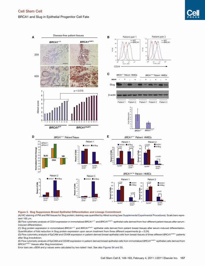
Figure 5. Slug Suppresses Breast Epithelial Differentiation and Lineage Commitment
(A) IHC staining of PM and RM tissues for Slug protein; staining was quantified by Allred scoring (see Supplemental Experimental Procedures). Scale bars repre-
sent 100 mm.
(B) Flow cytometry analysis of CD24 expression in immortalized BRCA1+/+ and BRCA1mut/+ epithelial cells derived from four different patient tissues after serum-
induced differentiation.
(C) Slug protein expression in immortalized BRCA1+/+ and BRCA1mut/+ epithelial cells derived from patient breast tissues after serum-induced differentiation.
Quantification of fold reduction in Slug protein expression upon serum treatment from three different experiments (p = 0.24).
(D) Flow cytometry analysis of EpCAM and CD49f expression in patient-derived breast epithelial cells from breast tissues of three different BRCA1mut/+ patients
after Slug knockdown.
(E) Flow cytometry analysis of EpCAM and CD49f expression in patient-derived breast epithelial cells from immortalized BRCA1mut/+ epithelial cells derived from
BRCA1mut/+ tissues after Slug knockdown.
Error bars are ±SEM and p values were calculated by two-tailed t test. See also Figures S4 and S5.
Cell Stem Cell
BRCA1 and Slug in Epithelial Progenitor Cell Fate
Cell Stem Cell 8, 149–163, February 4, 2011 ª2011 Elsevier Inc. 157
Slug
β-actin
BRCA1
siCntr
l
siBRCA1
Slug
Cyclin D1
DMSO6 h
r4 h
r3 h
r2 h
r1 h
r30
min
Time 0
Slug
β-actin
Slug
Cyclin D1
SUM149
SUM1315
MCF10A BRCA1
BRCA1
+/+
mut
BRCA1mut
DMSO6 h
r4 h
r3 h
r2 h
r1 h
r30
min
Time 0
DMSO6 h
r4 h
r3 h
r2 h
r1 h
r30
min
Time 0
C
β-actin
β-actin
Slug
BRCA1
6 hr
4 hr
2 hr
1 hr
Time 0
6 hr
4 hr
2 hr
1 hr
Time 0
siBRCA1siControl
β-actin
A
B
0.52 1.05 -0.4-0.2
00.20.40.60.81
1.21.41.6
BRCA1 SLUG
siCntrlsiBRCA1
Nor
mal
ized
fold
cha
nge
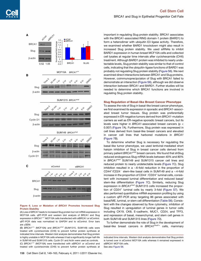
Figure 6. Loss or Mutation of BRCA1 Promotes Increased Slug
Protein Stability(A) Loss ofBRCA1 leads to increased Slug protein but not mRNA expression in
MCF10A cells. qRT-PCR and western blot analysis of BRCA1 and Slug
expression in BRCA1+/+ MCF10A cells transfected with siBRCA1 or siControl.
qRT-PCR data was normalized to GAPDH and to siControl. Error bars
are ±SEM.
(B) BRCA1+/+ (MCF10A) and BRCA1mut (SUM1315, SUM149) cells were
treated with cycloheximide (CHX) to prevent further protein synthesis at
indicated time intervals. Western blot analysis demonstrates that Slug protein
is highly unstable in MCF10A cells whereas it had a significantly longer half-life
in SUM149 and SUM1315 cells. Cyclin D1 and Actin were used as controls.
(C) BRCA1+/+ (MCF10A) were transfected with siBRCA1 or siControl and
treated with cycloheximide (CHX) to prevent further protein synthesis at
Cell Stem Cell
BRCA1 and Slug in Epithelial Progenitor Cell Fate
158 Cell Stem Cell 8, 149–163, February 4, 2011 ª2011 Elsevier Inc.
important in regulating Slug protein stability. BRCA1 associates
with the BRCA1-associated RING domain-1 protein (BARD1) to
form a heterodimer with ubiquitin E3 ligase activity. Therefore,
we examined whether BARD1 knockdown might also result in
increased Slug protein stability. We used siRNAs to inhibit
BARD1 expression in human breast MCF10A cells and collected
cell lysates at regular time intervals after cyclohexamide (CHX)
treatment. AlthoughBARD1 protein was inhibited to nearly unde-
tectable levels, Slug protein stability was similar to that of control
cells, indicating that the ubiquitin-ligase functions of BARD1 was
probably not regulating Slug protein stability (Figure S6). We next
examined direct interactions between BRCA1 and Slug proteins.
However, coimmunoprecipciaton of Slug with BRCA1 failed to
demonstrate an interaction (Figure S6), although we did observe
interaction between BRCA1 and BARD1. Further studies will be
needed to determine which BRCA1 functions are involved in
regulating Slug protein stability.
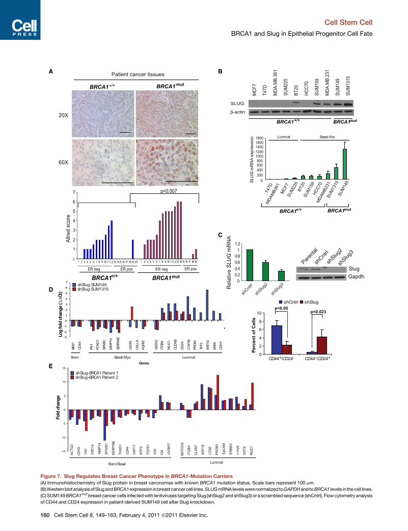
Slug Regulation of Basal-like Breast Cancer PhenotypeTo assess the role of Slug in basal-like breast cancer phenotype,
we first examined its expression in sporadic and BRCA1-associ-
ated breast tumor tissues. Slug protein was preferentially
expressed in ER-negative tumors derived from BRCA1-mutation
carriers as well as ER-negative sporadic breast cancers, but its
levels were higher in BRCA1-associated breast cancers (p <
0.007) (Figure 7A). Furthermore, Slug protein was expressed in
cell lines derived from basal-like breast cancers and elevated
in cancer cell lines that harbored mutations in BRCA1
(Figure 7B).
To determine whether Slug is necessary for regulating the
basal-like tumor phenotype, we used lentiviral-mediated short
hairpin inhibition of Slug in breast cancer cells derived from
primary patient BRCA1mut breast cancers. We found that shSlug
reduced endogenous Slug mRNA levels between 40% and 80%
in BRCA1mut SUM149 and SUM1315 cancer cell lines and
reduced protein to nearly undetectable levels (Figure 7C). Slug
inhibition resulted in a �6-fold reduction in the proportion of
CD44+/CD24� stem-like basal cells in SUM149 and a �4-fold
increase in the proportion of CD44�/CD24+ luminal cells, consis-
tent with increased luminal differentiation and reduced basal/
stem-like differentiation (Figure 7C). Similarly, reducing Slug
expression in BRCA1mut SUM1315 cells increased the propor-
tion of CD24+ luminal cells by nearly 3-fold (Figure S7). We
also performed quantitative mRNA expression profiling by using
a custom qRT-PCR array targeting 86 genes associated with
basal/ME, luminal, or stem cell differentiation (Table S6). Consis-
tent with the changes observed by flow cytometry, inhibition of
Slug resulted in upregulation of luminal genes in tumor cells
including CK19, CK8, E-cadherin, MUC1, CD24, and TFF3,
and repression of basal, mesenchymal, and stem cell genes in
both SUM149 and SUM1315 lines (Figure 7D).
To further demonstrate the role of Slug in the development of
basal-like breast cancers in BRCA1mut/+ cells, mammary
indicated time intervals. Western blot analysis demonstrates that Slug protein
is turned over in siControl MCF10A cells whereas it remained expressed in
siBRCA1 MCF10A cells.
See also Figure S6.
Cell Stem Cell
BRCA1 and Slug in Epithelial Progenitor Cell Fate
epithelial cells from disease-free prophylactic mastectomy
tissues from three different BRCA1mut/+ patients were trans-
duced with lentiviruses harboring oncogenes used in the crea-
tion of human breast cancers in vivo with or without targeting
Slug expression. Patient-derived BRCA1mut/+ cells expressing
shSlug showed increased expression of genes associated with
luminal tumors including CK19, CK8, MUC1, EpCAM, and
TFF3 with concomitant repression of many genes associated
with basal-like breast cancers including SPARC, SERPINE,
CD44, CK14, CK5, CK17, and vimentin compared to control
patient-derived BRCA1mut/+ cells (Figure 7E).
Finally, to demonstrate the connection between Slug expres-
sion and BRCA1 mutation before transformation, we examined
whether the genes that were induced upon Slug inhibition were
differentially expressed based on BRCA1 status in disease-free
tissues (Slug Gene Set, Table S3). We usedGSEA to evaluate the
expression of these Slug transcriptional targets in BRCA1mut/+
epithelium from four different patient samples. Six out of eight
Slug target genes were repressed in BRCA1mut/+ cells relative
to BRCA1+/+ cells (Figure S7), yielding significant enrichment
by GSEA (p < 0.0207). Collectively, these results show that upre-
gulation of Slug inhibits luminal lineage commitment and
increases the propensity for the formation of basal-like breast
tumors.
DISCUSSION
A fundamental difference between breast cancers arising in
BRCA1-mutation carriers compared to sporadic cancers is their
tendency toward a basal subtype. By using an in vivo model that
minimized cell culture, we were able to create human breast
cancers that recapitulated many features of clinically relevant
tumors to validate the previously untested idea that the predis-
position for basal-like tumors in BRCA1-mutation carriers arises
from perturbations in breast epithelial differentiation caused by
compromised BRCA1 function (Foulkes, 2003). Our results
show that breast epithelial cells isolated from BRCA1-mutation
carriers preferentially form tumors with increased basal differen-
tiation compared to cells isolated from noncarrier tissues. In
addition, we found that EpCAM+/CD10– luminal cells from both
BRCA1+/+ and BRCA1mut/+ tissues enriched for tumor-forming
ability in this model system, but that the latter exhibited
increased features of basal differentiation prior to transforma-
tion. However, since basal progenitor cells were also expanded
in disease-free breast tissues from BRCA1mut/+ tissues, it is
possible that these cells might also serve as targets of neoplastic
transformation in patients. Nevertheless, our findings are consis-
tent with the notion that tumor phenotype can be significantly
impacted by the pre-existing differentiation state of the normal
precursor (‘‘cell of origin’’) targeted for neoplastic transformation
(Gupta et al., 2005; Ince et al., 2007). However, since mutations
in a single allele ofBRCA1 can alter the differentiation potential of
the same cellular targets of transformation, leading to tumors
with different phenotypes, this indicates that the initiating
genetic mutation (‘‘mutation of origin’’) is a critical factor in
defining tumor subtype. Future studies will be needed to deter-
mine whether other combinations of cooperating oncogenes
give rise to BRCA1-associated basal-like tumors in basal/ME
cells and whether mutations in other tumor-suppressor genes
C
or oncogenes also affect the differentiation potential of progen-
itor cells that drive tumor phenotypes.
Although we have not excluded the possibility that LOH of the
wild-type BRCA1 allele is necessary for basal-like tumor forma-
tion, tumors in this model system were driven by ectopic onco-
genes, suggesting that LOH was not necessarily a rate-limiting
step. Furthermore, LOH is a stochastic event in BRCA1mut/+
patients, affecting the mutant or wild-type alleles with similar
frequencies (Clarke et al., 2006). Because the analysis of prophy-
lactic mastectomy tissues showed differentiation defects in
significant proportions of the breast tissue, this suggests that
LOHwas probably not responsible for the perturbations in breast
epithelial differentiation or basal tumor phenotype. Our findings,
combined with those of others (Burga et al., 2009; Lim et al.,
2009) indicate that haploinsufficiency of BRCA1 affects breast
epithelial differentiation and progenitor cells in patients.
The present study also provides several additional lines of
evidence that breast epithelial differentiation is altered in the
presence of BRCA1 mutations. First, genes involved in epige-
netic functions including DNA transcription and chromatin modi-
fication are overrepresented in the transcriptional signature of
BRCA1 mutant cells. Interestingly, many of the upregulated
genes are involved in the establishment and/or maintenance of
chromatin structure, including demethylases, methyltrans-
ferases, histones, acetyltransferases, and several components
of the ubiquitin pathway. These observations are consistent
with the idea that BRCA1mutations affect large-scale chromatin
unfolding (Ye et al., 2001), underscoring its role as an integral
component of multiprotein complexes that modulate gene
expression (Narod and Foulkes, 2004).
Second, the distinct transcriptional profile of BRCA1mut/+ cells
may reflect activation of signaling pathways associated with
progenitor/basal cells, increased basal differentiation, and
decreased luminal differentiation. Previous results suggest that
a reduction in BRCA1 leads to a failure of luminal lineage
commitment and increased expansion of an uncommitted
progenitor EpCAM� population (Liu et al., 2008). Consistent
with this observation, we found that BRCA1mut/+ breast tissue
exhibited an increase in the proportion of EpCAM�/CD49f+ basalprogenitor cells. However, in contrast to other findings (Lim et al.,
2009), we did not find an expansion of EpCAM+/CD49f+ luminal
progenitor cells in BRCA1mut/+ breast tissues, although we did
observe defects in luminal progenitor differentiation. This differ-
ence might reflect the genetic differences between the BRCA1
patient populations in the two studies. Nonetheless, overall
these studies reinforce the idea that BRCA1 is a critical regulator
of breast epithelial progenitor lineage commitment.
All of the BRCA1-mutation carrier samples used in this study
harbored frameshift mutations that compromise at a minimum
theC-terminal BRCT domain (Figure S2), which could destabilize
protein-protein interactions between BRCA1 and its C-terminal
binding partners. However, the overall levels of BRCA1 expres-
sion were surprisingly not affected. In addition, perturbations in
differentiation and increases in Slug expression could be de-
tected without changes in BRCA1 levels, implying that the
effects of BRCA1mutation may be at the level of protein-protein
interactions rather than overall BRCA1 expression. Consistent
with this notion, reduction of BRCA1 in MCF10A cells by RNA
interference impaired differentiation and could be rescued by
ell Stem Cell 8, 149–163, February 4, 2011 ª2011 Elsevier Inc. 159
BPatient cancer tissues
BRCA1mut
BRCA1+/+
20X
60X
SLUG
β-actin
MCF
7
T47D
MDA
.MB.
361
SUM
225
BT20
HCC7
0
SUM
159
MDA
.MB.
231
SUM
149
SUM
1315
T47D
MCF
7SU
M22
5
SUM
159
BT20
HCC7
0M
DAM
B231
SUM
149
MDA
MB3
61
BRCA1 BRCA1+/+ mut
BRCA1 BRCA1+/+ mut
600800
10001200140016001800
400200
0
SL
UG
mR
NA
expr
essi
on
A
SUM
1315
KRT1
9
KRT8
CDH
1
BMI1
CD44
FN1
FOXC
1
SPAR
C
MM
P14
SERP
INE
OXT
R
CALL
A
FGFR
1
GATA
3IT
GB4
PRO
M
MUC
1
TFF3
CLD
N8
CD24
C-M
YB
1
2
3
4
5
6
7
0
-3
-2
-1
shSlug-SUM149shSlug-SUM1315
LuminalBasal/MyoStemGenes
Log
fold
ch
an
ge (
ΔΔC
t)
0
2
4
6
8
10
shCntrl shSlugp<0.05
p<0.023
CD44+/CD24- CD44-/CD24+
Percen
t o
f C
ells
SlugGapdh
Parenta
l
shSlug
2
shSlug
3
00.20.40.60.81
1.2
Rel
ativ
eS
LU
Gm
RN
A
shCntr
l
shCntr
l
shSlug
2
shSlug
3
C
D
E
BRCA1 BRCA1+/+ mut
Allre
d sc
ore
0
1
2
3
4
5
6
7
ER pos
p<0.007
ER posER neg ER neg
10
-15
-10
-5
0
5
10
15
ACTG
2
CDH3
FN1
KRT1
4
MM
P14
SPAR
C
SERP
INE
THSD
1
CD44
KRT1
7
KRT5
TGFB
1
VIM
ID4
IGFB
P7
NOTC
H3
ITG
B4
CLDN
7
KRT1
9
LCN2
PRO
M1
EpCA
M
ERBB
3
c-m
yb
KRT8
MUC
1
LuminalStem/Basal
Fold
chang
e
shSlug-BRCA1 Patient 1shSlug-BRCA1 Patient 2
Luminal Basal-like
1 2 3 4 5 6 7 8 9 111213 1415 16 17 18 19 20 101 2 3 4 5 6 7 8 9 111213 1415 16 17 18 19
Figure 7. Slug Regulates Breast Cancer Phenotype in BRCA1-Mutation Carriers
(A) Immunohistochemistry of Slug protein in breast carcinomas with known BRCA1 mutation status. Scale bars represent 100 mm.
(B)Westernblot analysisofSlugandBRCA1expression inbreastcancercell lines.SLUGmRNA levelswerenormalized toGAPDHand toBRCA1 levels in thecell lines.
(C) SUM149BRCA1mutbreast cancer cells infectedwith lentiviruses targetingSlug (shSlug2and shSlug3) or a scrambled sequence (shCntrl). Flowcytometry analysis
of CD44 and CD24 expression in patient-derived SUM149 cell after Slug knockdown.
Cell Stem Cell
BRCA1 and Slug in Epithelial Progenitor Cell Fate
160 Cell Stem Cell 8, 149–163, February 4, 2011 ª2011 Elsevier Inc.
Cell Stem Cell
BRCA1 and Slug in Epithelial Progenitor Cell Fate
expression of a wild-type or a RAD51 mutant of BRCA1 but not
with a BRCA1 C-terminal BRCT domain mutant (Furuta et al.,
2005). Future studies will be necessary to fully dissect the
precise domains and mechanism by which BRCA1 regulates
breast epithelial differentiation. In addition, further experiments
will be needed to determine whether certain mutations in
BRCA1 affect differentiation and regulate progenitor cell fate
differently than others and whether different mutations alter the
propensity for the development of basal-like tumors.
The observation that BRCA1mut/+ epithelial cells express
genes involved in melanogenesis and stem cell biology promp-
ted us to examine Slug and its requirement for maintaining
progenitor cells and basal differentiation. We found that haploin-
sufficiency or knockdown of BRCA1 was associated with
increased expression and stability of the transcriptional
repressor Slug and that Slug regulated the basal epithelial and
tumor phenotype. This suggests that perturbations in luminal
differentiation due in part to Slug expression is probably respon-
sible for the increased propensity for the development of tumors
with basal-like features. Although our results do not address
whether Slug is sufficient to induce basal differentiation, Slug is
expressed in breast stem/progenitor cells, can promote
a basal-like phenotype in the luminal MCF-7 breast cancer cell
line, and can increase basal differentiation in the nontumorigenic
MCF10A breast line (Sarrio et al., 2008). Moreover, the fact that
Slug is expressed in basal-like breast cancers not associated
with BRCA1 mutations (Storci et al., 2008) also implies that
acquisition of its expression enables basal differentiation.
EXPERIMENTAL PROCEDURES
Detailed methods are described in Supplemental Experimental Procedures.
BRCA1-Mutation Carrier Tissues
All human breast tissue procurement for these experiments was obtained in
compliance with the laws and institutional guidelines, as approved by the insti-
tutional IRB committee from Beth Israel Deaconess Medical Center (BIDMC)
and Tufts University School of Medicine. Disease-free prophylactic mastec-
tomy (n = 31; 12 fresh, 19 formalin-fixed paraffin-embedded) and tumor tissues
(n = 19) derived from women carrying a known deleterious BRCA1 heterozy-
gousmutation were obtained with patient consent from the Surgical Pathology
files or immediately after prophylactic mastectomy surgery at BIDMC. Tissues
in which BRCA1 mutation was confirmed but not known were submitted for
sequence/genotyping at Myriad Genetic Laboratories. Non-BRCA1 tumor
tissues (n = 20) were obtained from discarded material at Tufts Medical Center
and noncancerous breast tissue was obtained from patients undergoing elec-
tive reduction mammoplasty at Tufts Medical Center or BIDMC (n = 38; 18
fresh, 24 formalin-fixed paraffin-embedded). BRCA1 mutation status and
clinical information are listed in Table S1. The range of patient ages for
fresh BRCA1+/+ tissue used in this study was 30–54 with a median age of
40; the range of patient ages for fresh BRCA1mut/+ tissue used in this study
was 35–53 with a median age of 44. All disease-free breast tissues were
verified by surgical pathologists prior to use in these studies.
(D)QuantitativeRT-PCRarrayagainstapanel of86genesexpressed inbreast lumina
and their respective scrambled controls. Genes differentially expressed in both cell
TFF3, KRT19, KRT8, and CDH1 genes are not expressed in SUM1315 cells.
(E) BRCA1mut/+ epithelial cells infected with oncogenes in the presence of Slug kn
patient-derived breast epithelial cells were infected with lentiviruses encoding mu
and a quantitative RT-PCR array was performed. Genes differentially expressed >2
Error bars are ±SEM and p values were calculated by two-tailed t test. See also Fig
C
Cell Lines and Tissue Culture
SUM cell lines were obtained from Dr. Stephen Ethier (Kramanos Institute, MI),
and the MCF10A cell lines were purchased from ATCC. MCF10A cells were
cultured in DMEM with 10% calf serum. SUM149PT cells were cultured
in Ham’s F12 with 5% calf serum, insulin (5 mg/mL), and hydrocortisone
(1 mg/mL) whereas SUM1315MO2 were in Ham’s F12 with 5% calf serum,
insulin (5 mg/mL), and EGF (10 ng/mL). All cell lines were grown at 37�C and
5% CO2. BRCA1mut/+ HMECs were immortalized with the catalytic subunit
of human telomerase as previously described (Elenbaas et al., 2001). BHME
cells were cultured in MEGM (Lonza) supplemented with bovine pituitary
extract (BPE), insulin (5 mg/mL), EGF (10 ng/mL), and hydrocortisone (1 mg/mL).
Lentiviral Constructs and Virus Production
Lentiviral constructs used for gene transduction into human mammary epithe-
lial cells were created with standard cloning techniques into the self-inactivat-
ing CS-CG (Miyoshi et al., 1998) viral vector generously provided by Inder
Verma (Salk Institute, La Jolla, CA). pLENTI-KRASG12V and pLenti-CMV-
PIK3CA-myr+CMV-CCND1 were obtained from Min Wu (Aveo Pharmaceuti-
cals, Cambridge, MA). A wild-type human p53 cDNA clone was generously
provided by Josh LaBaer (Harvard Institute of Proteomics, Harvard Medical
School, Cambridge, MA). Site-directed mutagenesis was employed to change
amino acid residue 175 fromR to H. The VSV-G-pseudotyped lentiviral vectors
were generated by transient cotransfection of the vector construct with the
VSV-G-expressing construct pCMV-VSV-G (Miyoshi et al., 1998) and the
packaging construct pCMV DR8.2Dvpr (Miyoshi et al., 1998), generously
provided by Inder Verma, into 293T cells with the FuGENE 6 transfection
reagent (Roche). High-titer stocks of the virus were prepared by ultracentrifu-
gation at 100,000 3 g. Lentiviral shRNA constructs targeting Slug (Addgene
plasmids 10904 and 10905) were prepared as previously described (Gupta
et al., 2005).
Breast tissues were minced and enzymatically digested overnight with
a mixture of collagenase and hyaluronidase as previously described (Kuper-
wasser et al., 2004; Proia and Kuperwasser, 2006) and dissociated to a single
cell suspension. Immediately after dissociation, cells were resuspended with
lentiviruses expressing the genes of interest and injected into cleared and
humanized mammary fat pads.
Animals and Surgery
All animal procedures were conducted in accordance with a protocol
approved by the Tufts University IACUC committee. A colony of immunocom-
promised NOD/SCID mice was maintained in house under aseptic sterile
conditions. Mice were administered autoclaved food and water ad libitum.
Surgeries were performed under sterile conditions, and animals received anti-
biotics in the drinking water up to 2 weeks after all surgical procedures.
Animals were humanized and injected as previously described (Kuperwasser
et al., 2004; Proia and Kuperwasser, 2006).
Immunohistochemistry
Immunohistochemistry was performed on formalin-fixed, paraffin-embedded
tissue sections with sodium citrate antigen retrieval, followed by visualization
with the ABC Elite peroxidase kit and NovaRed substrate (Vector Labs) for
detection of aSMA (1:500, clone asm-1), CK14 (1:500, clone LL002), CK8/18
(1:500, clone DC-10), Vimentin (1:500, clone V9), CK19 (1:500, clone b170)
(all, Vector Labs), TFF3 (Abnova, clone 3D9, 1:200), and Slug (1:200, Cell
Signaling). Staining for pan-cytokeratin (Ventana Medical Systems), p53 (Ven-
tana Medical Systems), cyclin D1 (NeoMarkers), pAKT (Cell Signaling, 1:100),
ER (Ventana Medical Systems), p63 (Ventana Medical Systems), and PR was
performed by the Histology Special Procedures Laboratory at Tufts Medical
l, basal,andstemcellswasperformedonSUM1315-shSlugandSUM149-shSlug
lines in the shSlug cells compared to the scrambled controls are plotted. PROM,
ockdown leads to cells with features of luminal-like breast cancers. BRCA1mut/+
tant p53, cyclin D1, and K-ras with shSlug or a scrambled sequence (shCntrl)
-fold in both patient samples compared to the scrambled controls are plotted.
ure S7 and Table S6.
ell Stem Cell 8, 149–163, February 4, 2011 ª2011 Elsevier Inc. 161
Cell Stem Cell
BRCA1 and Slug in Epithelial Progenitor Cell Fate
Center. IHC results were semiquantitatively analyzed (see Supplemental
Experimental Procedures for details).
Mammospheres and Adherent Colony Forming Assays
Viable cells dissociated from organoids derived from BRCA1+/+ (n = 4) and
BRCA1+/� (n = 4) patients were plated at 35,000 cells per well in 6-well plates
for adherent colony growth in MEGMmedia (Lonza) or at 20,000 cells per ml in
6-well ultralow attachment plates (Corning) inMEGMmediaminus the addition
of bovine pituitary extract for mammosphere growth. Colonies and mammo-
spheres were allowed to form for 8 days after which nonadherent suspension
colonies from adherent culture and mammospheres from nonadherent culture
were collected for analysis.
Mammospheres collected from nonadherent culture and suspension colo-
nies collected from adherent culture were cytospun onto glass slides and fixed
in methanol for immunofluorescence analysis. Quantification of mammo-
sphere and suspension colony numbers was accomplished with a Multisizer
3 COULTER COUNTER (Beckman-Coulter).
Immunomagnetic Bead Sorting
Epithelial organoids from BRCA1+/+ and BRCA1mut/+ patients were dissoci-
ated to a single cell suspension and sorted with CELLection pan-mouse IgG
magnetic beads according to the manufacturers’ instructions and as
described previously (Allinen et al., 2004) (Dynal, Invitrogen) conjugated to
a CD10 antibody (DAKO). CD10+ cells were released from the beads by DNase
treatment per the manufacturer’s instructions. Cells that did not bind to the
CD10-immunobeads were further sorted with magnetic beads conjugated to
an EpCAM antibody (Abcam).
Flow Cytometry and FACS
Uncultured cells from BRCA1+/+ (n = 10) or BRCA1mut/+ (n = 7) organoid prep-
arations were dissociated to a single-cell suspension as described above and
filtered through a 20 mm nylon mesh (Millipore). Endothelial, lymphocytic,
monocytic, and fibroblastic lineages were depleted with antibodies to CD31,
CD34, and CD45 (all Thermo/LabVision) and Fibroblast Specific Protein/IB10
(Sigma) and a cocktail of Pan-mouse IgG and IgM Dynabeads (Dynal, Invitro-
gen) according to the manufacturer’s instructions and as described previously
(Villadsen et al., 2007) and in Supplemental Experimental Procedures.
Nonconfluent cultures of SUM149, SUM1315, and immortalized HMECs
cells were trypsinized into single-cell suspension, counted, washed with
PBS, and stained with antibodies specific for human cell CD24 (PE) and
CD44 (APC) (BD Biosciences). Antibody-bound cells were washed and
resuspended at 1 3 106 cells/ml in FB and run on a FACSCalibur flow cytom-
eter (BD Biosciences) or sorted on a BD Influx FACS sorter (BD Biosciences).
Flow cytometry data was analyzed with the Flowjo software package
(TreeStar).
Microarray and RT-PCR Analysis
RNA was extracted from the tissues and cell lines with the RNAeasy Mini Kit
(QIAGEN). Standard RT-PCR to confirm expression of KRAS lentiviral
construct-specific transcript and quantitative real-time PCR was used for
detecting Slug, BRCA1, and Vimentin transcript in cell lines. See Supplemental
Experimental Procedures for all primer sequences and details. Custom
qRT-PCR arrays (Table S6) were obtained from SA Biosciences.
Total RNA for microarray expression studies was isolated from fibroblast-
depleted single cell suspensions of uncultured BRCA1+/+ or BRCA1mut/+ cells
or from tumors generated from infected BRCA1+/+ or BRCA1mut/+ cells via the
RNeasy Mini kit (QIAGEN). Synthesis of cDNA from total RNA and hybridiza-
tion/scanning of microarrays were performed with Affymetrix GeneChip
products (HGU133A) as described in the GeneChip manual. Raw data files
(.CEL) were converted into probe set values by RMA normalization. See
Supplemental Experimental Procedures for hierarchical clustering and GSEA
analysis.
ACCESSION NUMBERS
The accession number for the microarray data is GSE25835.
162 Cell Stem Cell 8, 149–163, February 4, 2011 ª2011 Elsevier Inc.
SUPPLEMENTAL INFORMATION
Supplemental Information includes Supplemental Experimental Procedures,
seven figures, and six tables and can be found with this article online at
doi:10.1016/j.stem.2010.12.007.
ACKNOWLEDGMENTS
We thank Gerbug Wulf, Steve Come, and Susan Troyan at Beth Israel
Deaconess Medical Center for assistance with BRCA1mut/+ tissue procure-
ment and Kai Tao and Lisa Arendt for technical assistance. We thank Myriad
Genetic Laboratories for gene-specific BRACAnalysis. We thank Annette
Shepard-Barry at Tufts Medical Center in the Histology-Special Procedures
Lab for histological and immunohistochemical staining. We thank Stephen
Kwok for assistance with cell sorting and Supriya Gupta for assistance with
gene expression experiments and data collection. We thank Josh LaBaer at
Harvard Medical School for generously providing us with human cyclin D1,
ras, p53, and PI3K cDNAs. This work was supported by grants from the
American Cancer Society-New England Division-Broadway on Beachside
Postdoctoral Fellowship (P.J.K.), the Raymond and Beverly Sackler Founda-
tion (P.J.K., C.K.), the Breast Cancer Research Foundation (C.K., T.P.), the
Department of Defense Breast Cancer Research Program (BC073866)
(P.J.K., T.P., C.K.), and the NIH/NCI (CA125554, CA125554) (C.K., I.K.). C.K.
is a Raymond and Beverly Sackler Foundation Scholar.
Received: February 17, 2010
Revised: August 19, 2010
Accepted: November 30, 2010
Published: February 3, 2011
REFERENCES
Allinen, M., Beroukhim, R., Cai, L., Brennan, C., Lahti-Domenici, J., Huang, H.,
Porter, D., Hu, M., Chin, L., Richardson, A., et al. (2004). Molecular character-
ization of the tumor microenvironment in breast cancer. Cancer Cell 6, 17–32.
Arnes, J.B., Brunet, J.S., Stefansson, I., Begin, L.R.,Wong, N., Chappuis, P.O.,
Akslen, L.A., and Foulkes, W.D. (2005). Placental cadherin and the basal
epithelial phenotype of BRCA1-related breast cancer. Clin. Cancer Res. 11,
4003–4011.
Burga, L.N., Tung, N.M., Troyan, S.L., Bostina, M., Konstantinopoulos, P.A.,
Fountzilas, H., Spentzos, D., Miron, A., Yassin, Y.A., Lee, B.T., and Wulf,
G.M. (2009). Altered proliferation and differentiation properties of primary
mammary epithelial cells from BRCA1 mutation carriers. Cancer Res. 69,
1273–1278.
Campbell, I.G., Russell, S.E., Choong, D.Y., Montgomery, K.G., Ciavarella,
M.L., Hooi, C.S., Cristiano, B.E., Pearson, R.B., and Phillips, W.A. (2004).
Mutation of the PIK3CA gene in ovarian and breast cancer. Cancer Res. 64,
7678–7681.
Clarke, C.L., Sandle, J., Jones, A.A., Sofronis, A., Patani, N.R., and Lakhani,
S.R. (2006). Mapping loss of heterozygosity in normal human breast cells
from BRCA1/2 carriers. Br. J. Cancer 95, 515–519.
DiMeo, T.A., Anderson, K., Phadke, P., Feng, C., Perou, C.M., Naber, S., and
Kuperwasser, C. (2009). A novel lung metastasis signature links Wnt signaling
with cancer cell self-renewal and epithelial-mesenchymal transition in basal-
like breast cancer. Cancer Res. 69, 5364–5373.
Dontu, G., Abdallah, W., Foley, J.F., Jackson, K., Clarke, M., Kawamura, M.,
and Wicha, M.S. (2003). In vitro propagation and transcriptional profiling of
human mammary stem/progenitor cells. Genes Dev. 17, 1253–1270.
Eirew, P., Stingl, J., Raouf, A., Turashvili, G., Aparicio, S., Emerman, J.T., and
Eaves, C.J. (2008). A method for quantifying normal human mammary epithe-
lial stem cells with in vivo regenerative ability. Nat. Med. 14, 1384–1389.
Elenbaas, B., Spirio, L., Koerner, F., Fleming, M.D., Zimonjic, D.B., Donaher,
J.L., Popescu, N.C., Hahn, W.C., and Weinberg, R.A. (2001). Human breast
cancer cells generated by oncogenic transformation of primary mammary
epithelial cells. Genes Dev. 15, 50–65.
Cell Stem Cell
BRCA1 and Slug in Epithelial Progenitor Cell Fate
Elstrodt, F., Hollestelle, A., Nagel, J.H., Gorin, M., Wasielewski, M., van den
Ouweland, A., Merajver, S.D., Ethier, S.P., and Schutte, M. (2006). BRCA1
mutation analysis of 41 human breast cancer cell lines reveals three new
deleterious mutants. Cancer Res. 66, 41–45.
Foulkes, W.D. (2003). BRCA1 functions as a breast stem cell regulator. J. Med.
Genet. 41, 1–5.
Foulkes, W.D., Brunet, J.S., Stefansson, I.M., Straume, O., Chappuis, P.O.,
Begin, L.R., Hamel, N., Goffin, J.R., Wong, N., Trudel, M., et al. (2004). The
prognostic implication of the basal-like (cyclin E high/p27 low/p53+/glomeru-
loid-microvascular-proliferation+) phenotype of BRCA1-related breast cancer.
Cancer Res. 64, 830–835.
Furuta, S., Jiang, X., Gu, B., Cheng, E., Chen, P.L., and Lee, W.H. (2005).
Depletion of BRCA1 impairs differentiation but enhances proliferation of
mammary epithelial cells. Proc. Natl. Acad. Sci. USA 102, 9176–9181.
Gauthier, M.L., Berman, H.K., Miller, C., Kozakeiwicz, K., Chew, K., Moore, D.,
Rabban, J., Chen, Y.Y., Kerlikowske, K., and Tlsty, T.D. (2007). Abrogated
response to cellular stress identifies DCIS associated with subsequent tumor
events and defines basal-like breast tumors. Cancer Cell 12, 479–491.
Gluz, O., Liedtke, C., Gottschalk, N., Pusztai, L., Nitz, U., and Harbeck, N.
(2009). Triple-negative breast cancer—current status and future directions.
Ann. Oncol. 20, 1913–1927.
Gupta, P.B., Kuperwasser, C., Brunet, J.P., Ramaswamy, S., Kuo, W.L., Gray,
J.W., Naber, S.P., and Weinberg, R.A. (2005). The melanocyte differentiation
program predisposes to metastasis after neoplastic transformation. Nat.
Genet. 37, 1047–1054.
Hu, Z., Fan, C., Oh, D.S., Marron, J.S., He, X., Qaqish, B.F., Livasy, C., Carey,
L.A., Reynolds, E., Dressler, L., et al. (2006). The molecular portraits of breast
tumors are conserved across microarray platforms. BMC Genomics 7, 96.
Ince, T.A., Richardson, A.L., Bell, G.W., Saitoh, M., Godar, S., Karnoub, A.E.,
Iglehart, J.D., and Weinberg, R.A. (2007). Transformation of different human
breast epithelial cell types leads to distinct tumor phenotypes. Cancer Cell
12, 160–170.
Keller, P.J., Lin, A.F., Arendt, L.M., Klebba, I., Jones, A.D., Rudnick, J.A.,
Dimeo, T.A., Gilmore, H., Jefferson, D.M., Graham, R.A., et al. (2010).
Mapping the cellular and molecular heterogeneity of normal and malignant
breast tissues and cultured cell lines. Breast Cancer Res. 12, R87.
Kubista, M., Rosner, M., Kubista, E., Bernaschek, G., and Hengstschlager, M.
(2002). Brca1 regulates in vitro differentiation of mammary epithelial cells.
Oncogene 21, 4747–4756.
Kuperwasser, C., Chavarria, T., Wu, M., Magrane, G., Gray, J.W., Richardson,
A., and Weinberg, R.A. (2004). Reconstruction of functionally normal and
malignant human breast tissues in mice. Proc. Natl. Acad. Sci. USA 101,
4966–4971.
Lim, E., Vaillant, F., Wu, D., Forrest, N.C., Pal, B., Hart, A.H., Asselin-Labat,
M.L., Gyorki, D.E., Ward, T., Partanen, A., et al. (2009). Aberrant luminal
progenitors as the candidate target population for basal tumor development
in BRCA1 mutation carriers. Nat. Med. 15, 907–913.
Liu, S., Ginestier, C., Charafe-Jauffret, E., Foco, H., Kleer, C.G., Merajver, S.D.,
Dontu, G., and Wicha, M.S. (2008). BRCA1 regulates human mammary stem/
progenitor cell fate. Proc. Natl. Acad. Sci. USA 105, 1680–1685.
Loi, S., Haibe-Kains, B., Lallemand, F., Pusztai, L., Bardelli, A., Gillett, C., Ellis,
P., Piccart-Gebhart, M.J., Phillips, W.A., McArthur, A., and Sotiriou, C. (2009).
Correlation of PIK3CA mutation-associated gene expression signature
(PIK3CA-GS) with deactivation of the PI3K pathway and with prognosis within
the luminal-B ER+ breast cancers. J. Clin. Oncol. 27, 533.
C
Miyakis, S., Sourvinos, G., and Spandidos, D.A. (1998). Differential expression
and mutation of the ras family genes in human breast cancer. Biochem.
Biophys. Res. Commun. 251, 609–612.
Miyoshi, H., Blomer, U., Takahashi, M., Gage, F.H., and Verma, I.M. (1998).
Development of a self-inactivating lentivirus vector. J. Virol. 72, 8150–8157.
Narod, S.A., and Foulkes, W.D. (2004). BRCA1 and BRCA2: 1994 and beyond.
Nat. Rev. Cancer 4, 665–676.
Niessen, K., Fu, Y., Chang, L., Hoodless, P.A., McFadden, D., and Karsan, A.
(2008). Slug is a direct Notch target required for initiation of cardiac cushion
cellularization. J. Cell Biol. 182, 315–325.
Peppercorn, J., Perou, C.M., and Carey, L.A. (2008). Molecular subtypes in
breast cancer evaluation and management: divide and conquer. Cancer
Invest. 26, 1–10.
Proia, D.A., and Kuperwasser, C. (2006). Reconstruction of human mammary
tissues in a mouse model. Nat. Protoc. 1, 206–211.
Rakha, E.A., Reis-Filho, J.S., and Ellis, I.O. (2008). Basal-like breast cancer: A
critical review. J. Clin. Oncol. 26, 2568–2581.
Saal, L.H., Holm, K., Maurer, M., Memeo, L., Su, T., Wang, X., Yu, J.S.,
Malmstrom, P.O., Mansukhani, M., Enoksson, J., et al. (2005). PIK3CA muta-
tions correlate with hormone receptors, node metastasis, and ERBB2, and are
mutually exclusive with PTEN loss in human breast carcinoma. Cancer Res.
65, 2554–2559.
Sarrio, D., Rodriguez-Pinilla, S.M., Hardisson, D., Cano, A., Moreno-Bueno,
G., and Palacios, J. (2008). Epithelial-mesenchymal transition in breast cancer
relates to the basal-like phenotype. Cancer Res. 68, 989–997.
Shipitsin, M., Campbell, L.L., Argani, P., Weremowicz, S., Bloushtain-Qimron,
N., Yao, J., Nikolskaya, T., Serebryiskaya, T., Beroukhim, R., Hu, M., et al.
(2007). Molecular definition of breast tumor heterogeneity. Cancer Cell 11,
259–273.
Stingl, J., Eaves, C.J., Zandieh, I., and Emerman, J.T. (2001). Characterization
of bipotent mammary epithelial progenitor cells in normal adult human breast
tissue. Breast Cancer Res. Treat. 67, 93–109.
Storci, G., Sansone, P., Trere, D., Tavolari, S., Taffurelli, M., Ceccarelli, C.,
Guarnieri, T., Paterini, P., Pariali, M., Montanaro, L., et al. (2008). The basal-
like breast carcinoma phenotype is regulated by SLUG gene expression.
J. Pathol. 214, 25–37.
Villadsen, R., Fridriksdottir, A.J., Ronnov-Jessen, L., Gudjonsson, T., Rank, F.,
Labarge, M.A., Bissell, M.J., and Petersen, O.W. (2007). Evidence for a stem
cell hierarchy in the adult human breast. J. Cell Biol. 177, 87–101.
Welcsh, P.L., and King, M.C. (2001). BRCA1 and BRCA2 and the genetics of
breast and ovarian cancer. Hum. Mol. Genet. 10, 705–713.
Wu,M., Jung, L., Cooper, A.B., Fleet, C., Chen, L., Breault, L., Clark, K., Cai, Z.,
Vincent, S., Bottega, S., et al. (2009). Dissecting genetic requirements of
human breast tumorigenesis in a tissue transgenic model of human breast
cancer in mice. Proc. Natl. Acad. Sci. USA 106, 7022–7027.
Ye, Q., Hu, Y.F., Zhong, H., Nye, A.C., Belmont, A.S., and Li, R. (2001). BRCA1-
induced large-scale chromatin unfolding and allele-specific effects of cancer-
predisposing mutations. J. Cell Biol. 155, 911–921.
Zhao, X., Malhotra, G.K., Lele, S.M., Lele, M.S., West, W.W., Eudy, J.D., Band,
H., and Band, V. (2010). Telomerase-immortalized human mammary stem/
progenitor cells with ability to self-renew and differentiate. Proc. Natl. Acad.
Sci. USA 107, 14146–14151.
ell Stem Cell 8, 149–163, February 4, 2011 ª2011 Elsevier Inc. 163