Embed Size (px)
Citation preview

1
GRAFT SELECTION IN ANTERIOR CRUCIATE LIGAMENT
RECONSTRUCTION
1Nathan A. Mall, MD 1,3Geoffrey S. Van Thiel, MD, MBA
2Asheesh Bedi, MD 1Brian J. Cole, MD, MBA
1Rush University Medical Center, Department of Orthopaedics, Division of Sports Medicine
2 University of Michigan, Department of Orthopaedics, MedSport 3 Rockford Orthopedic Associates, Rockford, IL
Introduction Anterior cruciate ligament (ACL) reconstruction is one of the most studied surgeries performed within any surgical specialty. The focus of much of this research has been related to graft selection and its impact on clinical outocmes. Favorable characteristics of graft options for ACL reconstruction include those that have similar structural and biomechanical characteristics of the native ligament, allow for secure fixation, permit rapid biologic incorporation, and have limited donor site morbidity.36, 61 Ideally, the graft selected should allow for early, active rehabilitation and maintain its biomechanical properties and strength until full graft incorporation and biologic maturation. Ultimately, any graft construct should approximate or exceed the physical and mechanical properties of the native ACL. (Table 1) There are many graft sources available to the surgeon, and these can be broadly categorized into autografts and allografts. Autologous grafts can be harvested from the ipsilateral or contralateral patellar tendon, the semitendonosis and gracilis tendons, or from the quadriceps tendon with or without a bone plug. Allografts typically used for ACL reconstruction include cadaveric forms of these same types of autologous grafts as well as tibialis anterior and Achilles tendon grafts. Many surgeons prefer a certain graft based on their training and observed clinical outcomes in their practice. Regardless of personal preference, however, each graft has relative advantages and disadvantages and should be appropriately and individually considered based on patient age, size, activity level, pain tolerance, and return to sport goals. The goal of this chapter is to review the peer-reviewed literature on ACL graft sources and allow the surgeon to make an educated decision with his or her patients as to the appropriate graft for each individual patient. Autograft
2
Bone patellar tendon bone (BTB) autograft and hamstring autograft are two of the most popular graft sources for ACL reconstruction. Figure 1. These grafts have reported donor site morbidity; however, most surgeons would agree that the risk of catastrophic failure with allograft may outweigh the risks of donor site morbidity in the young, active patient. The quadriceps tendon autograft is not as widely used, but is favored by some authors, particularly in the revision setting, due to its large cross-sectional area and the presence of a bone plug. Table 1, Figure 1. Use of autografts have been shown to decrease retear rates substantially in the younger population, although in all patients had a failure rate of 3.5% compared with 8.9% in patients in which allografts were used.19 Bone-Tendon-Bone: The central third of the patellar tendon along with bone plugs from the patella and the tibial tubercle is considered the “gold standard” by many surgeons. Bone-patellar tendon-bone (BTB or BPTB) grafts allow for bone-to-bone healing within the tunnels, have favorable time-zero biomechanical strength, and have a longstanding track record of good-to-excellent clinical outcomes. BTB grafts have the advantage of bone-to-bone healing, which has been shown to be faster and more reliable than tendon to bone healing.10, 41, 55 Histologic studies have revealed that osteonecrosis occurs at the graft-tunnel interface, followed by creeping substitution and rapid incorporation into surrounding host bone. Dense fibrous tissue surrounds the bone block at 3 weeks with complete incorporation of the grafted bone to the host bone by 6 weeks.41 The intact ACL has an ultimate tensile load, stiffness, and cross-sectional area of 2160 Newtons (N), 242 N/mm, and 44 mm2, respectively. A 10mm wide BTB graft was found to have an ultimate tensile load, stiffness, and cross-sectional area of 2977 N, 620 N/mm, and 35mm2, respectively. Thus the BTB autograft has the intrinsic properties to allow immediate active rehabilitation, and these favorable biomechanical properties combined with bone to bone healing and rigid, aperture fixation with interference screws account for the graft’s excellent initial and continued long term performance. Regardless of the bone tunnel healing, a ligamentization process must occur, which entails an initial phase of acellular and avascular necrosis with the collagen scaffold remaining intact. The second phase is repopulation by host synovial cells, followed by revascularization and maturation. By 1 month post-operative the graft is completely acellular but has intact collagen, and at 3 months cellular repopulation with some vascular proliferation is seen. At 6 months the number of cells is closer to a normal ligament, and at 9 months the intraarticular portion of the graft is histologically similar to native ACL.40 Therefore, the bone to bone healing provided by the BTB graft helps prevent initial graft slippage and healing within the bone tunnel allowing safe resumption of activities of daily living, but the process of ligamentization proceeds independent of the graft-tunnel healing characteristics.. Length and width of BTB autografts can be adjusted to match patient size and footprint anatomy and may be an advantage compared to hamstring autograft. Depending on the size of the patient’s patellar tendon, in some cases a 12mm graft can be obtained while still safely retaining sufficient

3
tendon on either side of the graft. Also, while the surgeon is relatively limited on the size of the bone plug that can be taken from the patella, the tibial bone plug can be widened or lengthened to fill larger tunnels in revision setting or to help prevent graft tunnel mismatch. The length of the soft tissue component cannot be adjusted and thus, if the tendon is extremely long or extremely short, the surgeon must be prepared with different fixation options for graft-tunnel mismatch. BTB autografts have been used for years with excellent results at long term follow-up.38 A Cochrane review comparing patellar tendon to hamstring autografts, with the search criteria of randomized or quasi-randomized controlled trials, demonstrated that static stability testing in the form of Lachman, Pivot Shift, and instrumented laxity favored BTB grafts over hamstring grafts.37 In the same critical review, no differences were found between the graft sources when evaluating single leg hop, return to activity, Tegner score, Lysholm score, IKDC, or re-tear rates.37 Another recent systematic review of level I studies found a higher risk of failure in hamstrings autografts compared with BTB autografts, and only one of five studies found a higher likelihood of anterior knee complaints in patients in which BTB autografts were used.44 Patellar tendon autograft does have several disadvantages that have been described in the literature. First, the bone-patellar tendon-bone graft is a fixed length graft and, as such, is vulnerable to graft-tunnel mismatch when tunnel length is not accordingly adjusted. There are subtle intraoperative maneuvers, however, (recessing the femoral bone plug, drilling a longer tibial tunnel, graft rotation) that can reduce the impact of graft-tunnel mismatch.7, 54, 58.7, 47, 59 Graft-tunnel mismatch may be more problematic with anatomic ACL reconstruction where the femoral tunnel is placed low on the lateral wall of the femoral notch, reducing the intra-articular graft length relative to previous, vertical graft configurations. The surgeon should be particularly aware of this problem with back-up fixation options available in the setting of patella alta or baja. Graft-tunnel mismatch is not an issue with soft tissue grafts such as hamstrings or quad tendons as they can be readily shortened to the desired length. Another well documented disadvantage of BTB autografts is a higher incidence of anterior knee pain. Dissatisfaction and potential inability to return to work have been reported in patients that perform kneeling as part of their occupation or religion. Other conditions that may preclude use of a BTB autograft include Osgood-Schlatter or patellar tendinosis or tendonitis; however, there is no good data to recommend against BTB use in this population. A Cochrane review noted more anterior knee discomfort, especially with kneeling after BTB autografts. BTB grafts had a clinically insignificant loss of extension, whereas hamstring reconstructions had a trend towards a loss of knee flexion. BTB grafts demonstrated a trend towards reduced extension strength, while hamstrings grafts had a statistically significant reduction in knee flexion strength.37 Patella fracture is another concern following ACL reconstruction using BTB autograft. Stein et al reported an incidence of patella fracture following graft harvest of 1.3%; however, these all occurred at least 3 weeks post-operatively.52 Another study reported only 2 of 1725 ACL reconstruction (one

4
intra-operative and one post-operative).25 While the incidence is low, there is a risk that should be discussed when counseling the patient regarding graft choice. The surgeon can minimize risk of this by assuring a bone plug no more than half the length of the patella is taken, making cuts angled and no more than 10mm deep, avoiding cross-hatching at the corners, and creating a trapezoidal cut rather than triangular or square. Typically, fractures are vertically oriented and thus are less likely to completely disrupt the extensor mechanism. These fractures can be treated non-operatively if minimally displaced and with a preserved extensor mechanism. Transverse fractures can also occur as well and may be more likely to require fixation. These may often result from postoperative trauma or aggressive rehabilitation in the setting of a significant stress riser. In the pediatric population, many surgeons believe that the use of BTB autografts in patients with open physes can result in angular deformity or apophyseal closure. However, several recent studies have demonstrated that this may not be the case. In a recent study, using BTB autografts that traversed the open physis in patients Tanner Stage 3 or greater found no growth disturbance; however fixation was placed extraphyseal.50 This is likely less of an issue with the tibia as the graft is placed more central within the physis. The more vertical the tunnel, the more circular the aperture, reducing the cross-sectional area of physeal injury. Furthermore, a centrally positioned physeal injury or bar resulting from graft placement may restrict growth but will minimize the risk of angular deformity. However, when the femoral tunnel is placed anatomic, the tunnel is oblique and affects more total volume of the physis.20 A single bundle anatomic reconstruction is not likely to affect more than 7% of the physis,48 which has been shown to be the critical amount for increased risk of partial physeal closure in an animal model.34 The risk of angular deformity obviously lessens the closer the patient is to skeletal maturity. Hamstrings: Hamstring grafts are the other most common autograft used for a variety of reasons. Need some statement about the width, stiffness, UTS, etc. similar to what you have for the BTB auto. The gracilis and semitendonosis are typically harvested separately and then combined and doubled over themselves to create a 4-strand graft. Some authors describe creating 5 or 6-strand grafts to improve strength. Hamstring grafts are typically stretched in order to remove some of the creep in the tendons prior to inserting and tensioning the graft. There are numerous fixation techniques and products available for soft tissue grafts, which include in broad categories: cortical suspensory fixation, aperture interference fixation, and a combination of these methods. One advantage of hamstrings autograft fixation is the cosmesis. As the surgeon becomes more familiar with this technique a smaller incision can be used, which can be almost as small as an allograft incision. The location of the semitendonosis and gracilis tendons’ insertion conveniently is in the same location as the starting point for the tibial tunnel, and thus the same incision can be used for both graft harvest and tunnel drilling. Another advantage of hamstrings grafts is that it avoids the disadvantages reported for bone patellar bone autograft. In a long term follow-up study, hamstrings autografts were found to have better IKDC, kneeling, knee walking, and single leg hop compared to BTB autografts; however,

5
hamstrings grafts did have inferior knee flexion strength. This study found no difference in range of motion, KT-1000 anthropometric data, or pivot shift examination.62 At an average of 86 months follow-up, another study found no difference in osteoarthritis, Lachman and anthropometric testing, kneeling, IKDC, Tegner, and single leg hop testing between patellar tendon and hamstrings grafts. This study did find that hamstring tendon graft patients were better in knee walking and Lysholm scores.31 Another long-term follow-up study29 found superior results in the hamstring tendon group in terms of osteoarthritis, motion loss, single leg hop, and kneeling pain, but no difference in IKDC scores. The authors also found that 17% of their hamstrings reconstructions failed while only 8% of their BTB grafts failed.29 Thus, hamstring grafts may be a better option in patients at risk of anterior knee pain or with professional demands that require extensive kneeling. Soft tissue grafts are also favored by several authors for a transphyseal approach in skeletally immature patients to minimize the risk of bar formation and a secondary growth deformity. Bone-tendon-bone grafts can risk injury the tibial tubercle apophysis, resulting in a severe recurvatum deformity. Bone plug placement across the physis can also increase the risk of bar formation and an angular growth deformity. Kocher et al retrospectively reviewed 59 Tanner stage 3 patients in which hamstring trans-physeal ACL reconstruction was used. The reported only 2 late failures secondary to reinjury, and no growth disturbance (both angular and leg length inequality).22 There are likely more factors than simply graft selection that play a role in whether or not premature physeal closure occurs, including placement of the tunnels. A recent study demonstrated that the more anatomic or oblique the tunnel is made, the greater volumetric destruction of the physis. In this regard, a relatively central and vertical tunnel trajectory into the anatomic ligament footprint is favorable to minimize the volume of physeal injury.49 Less post-operative pain is another commonly cited advantage of hamstring tendon use by proponents of this technique. A study of the early post-operative morbidity associated with ACL reconstruction demonstrated significantly better pain scores in hamstrings compared to patellar tendon group up to 8 weeks post-operatively; however, the differences were relatively small and the clinical significance was questioned by the authors.5 While the incision is typically smaller, and there is less bone destruction involved with hamstring graft harvest, there is a paucity of literature supporting this difference in early post-operative pain reduction. Hamstrings tendon grafts, however, are not without their disadvantages. Hamstring tendon grafts also may increase in laxity over time. When reviewing the literature related to hamstring tendon autograft, however, one must pay particular attention to the number of graft strands. In the 6 manuscripts included in a recent systematic review, both of the two studies evaluating 2-strand hamstring grafts demonstrated increased laxity compared to patellar tendon grafts; whereas, only one of the 4 comparing quadrupled hamstring grafts to patellar tendons was able to find a statistical difference in laxity at follow-up.44

6
Another disadvantage to using hamstrings tendon grafts is that soft-tissue to bone healing takes longer than bone-to-bone healing and is less reliable. The intact ACL inserts into bone via direct insertion, which has 4 zones: tendon, unmineralized fibrocartilage, mineralized fibrocartilage, and bone.10 The fibrocartilage has a mixture of collagen II, IX, X, and XI, with type X being the most significant. ACL tendon-bone healing occurs with a layer of fibrovascular scar between tendon and bone at graft-tunnel interface. This eventually organizes into perpendicular fibers that resemble Sharpey’s fibers. Presence and number of these fibers are directly correlated with pull-out strength.8-
9, 46 This process takes approximately 12 weeks, until which time hamstrings autografts demonstrated reduced ultimate failure load when compared to BTB autografts.55 Several other studies have demonstrated similar results with soft tissue autografts taking longer to fully incorporate as compared to the bone-patellar tendon-bone autograft.41-42 Soft tissue grafts may also have a propensity to cause tunnel widening.23, 60 However, this is unknown to be a function of the soft tissue graft or the suspensory fixation1 often used with soft tissue grafts.13 Likely, tunnel expansion is caused by a variety of factors, including graft micromotion, stress shielding, drill-related necrosis, accelerated rehabilitation, bone resorption and remodeling, graft swelling, synovial fluid propagation, and increased synovial cytokine production.3, 18, 32, 45, 64 However, there is little evidence that tunnel widening affects clinical or arthrometric outcomes following ACL reconstruction.4 Likely, the only clinical significance of tunnel widening is at the time of revision when vastly expanded tunnels may compromise graft fixation necessitating a two-stage revision approach. Many studies evaluating the morbidity of hamstring tendon harvest have demonstrated reduced knee flexion strength compared with the contralateral extremity, but similar to the loss of extension strength seen in some studies after BTB autografts. It is unknown if this difference in clinically significant.21 Weakness in the knee flexors has been associated with inferior knee function in ACL-deficient knees30, 56 likely due to the hamstrings role as an ACL agonist and dynamic muscle protector. However, in ACL reconstructed knees, a flexion strength deficit occurs but has no effect on IKDC scores.24 Quadriceps tendon The quadriceps tendon graft is a less common but viable graft option for primary and, perhaps more commonly, revision ACL reconstructive surgery.6 This graft does have several theoretical advantages. The large cross-sectional area affords a favorable time-zero biomechanical strength and is particularly useful to fill expanded tunnels at the time of a single-stage revision ACL reconstruction. While both quadriceps tendon and patellar tendon grafts are stronger than the native ACL, several studies have demonstrated weaker mechanical characteristics for the quadriceps tendon graft when compared to a 10mm patellar tendon autograft.12, 51 Another advantage of the quadriceps tendon graft is that it can be harvested with a bone plug from the superior pole of the patella and thus bone-to-bone healing can occur at one of the tunnels. Also, the length of the graft can be adjusted more easily than can a BTB autograft, avoiding

7
concerns and risks of graft-tunnel mismatch. The quadriceps graft can also be used in adolescents with open growth plates as it would not violate the tibial tubercle apophysis. The major disadvantage to the patellar tendon graft is the possible complication of anterior knee pain. This is a “catch-all” term that describes many different diagnoses and clinical problems, some of which are scar sensitivity, fat pad herniation, and neuroma of the infrapatellar branch of the saphenous nerve. By moving the incision for graft harvest superior to the patella as in the quadriceps tendon graft harvest, the patient may no longer have to kneel directly on the scar. Also, an incision at the superior pole of the patella should not affect the saphenous nerve and will not affect the fat pad. Also, a smaller proportion of the extensor mechanism is disrupted, which could potentially prevent some of the extension weakness or extension loss noted in some studies evaluating patellar tendon. However, the quadriceps graft has not been studied as extensively as the patellar tendon graft, and it is therefore difficult to directly compare it to the more commonly used patellar tendon and hamstring grafts. The major disadvantage of the quadriceps graft is the lack of level I clinical studies with long-term follow-up that can demonstrate no functional loss of extensor mechanism strength, similar graft incorporation and longevity as the patellar tendon graft, and minimal donor site morbidity or complications related to the graft harvest. Future studies are necessary to define these outcomes and the role of quadriceps graft in primary ACL reconstruction. It is clear, however, that this graft does have several advantages and is the favored autograft option for revision ACL surgery in the setting of well-placed but expanded sockets for which tunnel fill is critical. Allograft: The reduced pain and morbidity along with the reliable clinical results of allograft in the older population may favor its consideration as a graft choice in older or lower demand patients. With no harvest site morbidity, the surgeon can use a small incision and the muscle weakness, kneeling pain, and risk of patellar fracture are minimized. Several different allograft sources are available, including: BTB (hemi or whole), Achilles, hamstrings, tibialis anterior, tibialis posterior and quadriceps tendon. Figure 1. Allografts can be irradiated or non-irradiated; however, there is concern of compromised graft integrity and decreased time-zero biomechanical strength with irradiated grafts.53 Low dose irradiation < 20 kGy has been shown to affectively eliminate bacteria, but only irradiation greater than 30 kGy has been shown to eliminate both bacterial and viral pathogens.43 Irradiation greater than 20 kGy in a single dose causes substantial changes in the structural properties of the graft,14 which likely causes its inferior clinical outcomes. While the risk of contamination is greater with non-irradiated tissue, this may be negated with modern day nucleic acid testing and use of sterile processing.57 The current reported risk of transferring tissue from an HIV-infected donor is reported as between 1 and 4 in 1 million, and while the number of infected donors is unknown for Hepatitis B and C, there are more Americans infected with these viruses than HIV. Most grafts

8
currently undergo low dose irradiation that allows for the destruction of bacteria and rely on donor screening and nucleic acid testing to reduce the risk of viral contamination as irradiation at the level needed for viral elimination would significantly compromise graft biomechanical properties. Allografts are used frequently in revision ACL surgery, especially when the surgeon’s “go to” graft has been used in the primary surgery. Large allografts, such as Achilles or quadriceps tendon, affords the additional advantage of a large cross-sectional area to fill large tunnels, have favorable time-zero biomechanical strength, and have a bone plug for bone-to-bone healing and fixation in at least a single tunnel. A study of the epidemiology of the Multicenter ACL revision study (MARS) cohort demonstrated that 54% of the surgeons used an allograft at the time of revision compared with 27% of the patients having had an allograft at the time of their primary reconstruction.63 BTB allografts were the most frequent allograft used in the MARS cohort at 50%, followed by tibialis anterior (23%), Achilles tendon (12%), and tibialis posterior (11%).63 However, a significant number of grafts were autografts at the time of revision (45%), of which 49% were BTB autografts and 40% quadrupled hamstrings autografts, indicating that many surgeons will still favor autografts when available in the revision setting, especially in the young, high demand athlete. Another common use for allograft is when more collagen is needed than can be obtained from autograft sources, such as the multi-ligament injured knee. Despite undergoing operative repair within 3 weeks from surgery, many surgeons are now recommending augmentation with additional collagen for lateral collateral or posterolateral injuries. Levy et al found a significantly increased incidence of failures when comparing repairs of the lateral collateral and posterolateral corner injuries compared to reconstructions using allografts.26-27 A combination of allografts can be used depending on the number of ligaments injured and reconstruction technique. There are several disadvantages to allograft use, however, and the benefits of convenience and reduced donor site morbidity may come at the cost of less favorable healing and a greater risk of failure in the young athlete. First, there have been several studies that have shown that allograft tissue takes longer to incorporate.10 While no direct correlation between graft incorporation and clinical performance has been clearly shown, allograft tissue has without question demonstrated inferior performance in the younger patient population.19 Kaeding et al demonstrated a 2.3 times greater risk of graft rupture with allograft reconstructions for each 10-year decrease in age, with the highest risk being in those aged 10-19 years.19 Secondly, while the risk is small, there is still a potential risk of disease transmission. There currently is a lack of standardized protocols for testing and cleansing of allograft tissue. The risk can be minimized by using a reputable tissue bank and the surgeon must be fully aware of the protocol used by the tissue bank to assure it meets the standards outlined by the AAOS. Allografts must undergo the same process of avascular necrosis, creeping substitution, and ligamentization as autografts and are repopulated by host synovial cells. Jackson et al found that donor DNA was replaced completely by host DNA by 4 weeks.17 While allografts heal by the same
9
process as their autograft counterparts (bone vs. tendon to bone healing), they do so at a much slower rate. Compared with autografts, allografts lose more of their time zero strength.15 At 6 months autografts had better restraint to AP translation, more cross sectional area, twice the load-to-failure strength, and more small-diameter collagen fibrils (more like normal).16 At the same time, typically allograft patients have less pain and are more likely to place additional stress on the reconstruction. Patients in which allograft was used for their ACL reconstruction should be counseled that the graft may take longer to fully incorporate and that longer protection may be warranted. This is another reason why surgeons may want to avoid using allograft in high-level athletes. A recent study produced by the MOON group demonstrated an overall retear rate of 8.9% in allograft primary reconstructions compared with 3.5% when autografts were used. This difference was more profound in the younger patient population with over a 10% difference in failure rates in patients less than 18 years of age.19 The surgeon has many choices when it comes to graft selection for ACL reconstruction. There are certain situations in which one graft may be favored over another, such as in the young, athletic population where autograft tissue should be used. However, there is good literature that excellent results can be achieved with each type of graft, and thus the surgeon must inform his or her patients of the advantages and disadvantages of each graft and help them make an informed decision. In summary, BTB autograft is generally accepted as the “gold standard” due to its biomechanical profile and reliable, fast bone-to-bone healing; however, hamstring tendons offer certain theoretical advantages in those that do a lot of kneeling, pre-existing patellofemoral pain, patella alta, or in those with open physes. Quadriceps tendon grafts have several advantages, including a large cross-sectional area and associated bone plug, but more long term, prospective studies must be done to determine its incorporation properties and ensure its long-term survival compared to patellar tendon and hamstring autografts. Allograft tissue is an excellent choice in many revision situations, in the older recreational athlete, and in those with low demands but need to return to work faster with less pain and dysfunction immediately post-operatively.
10
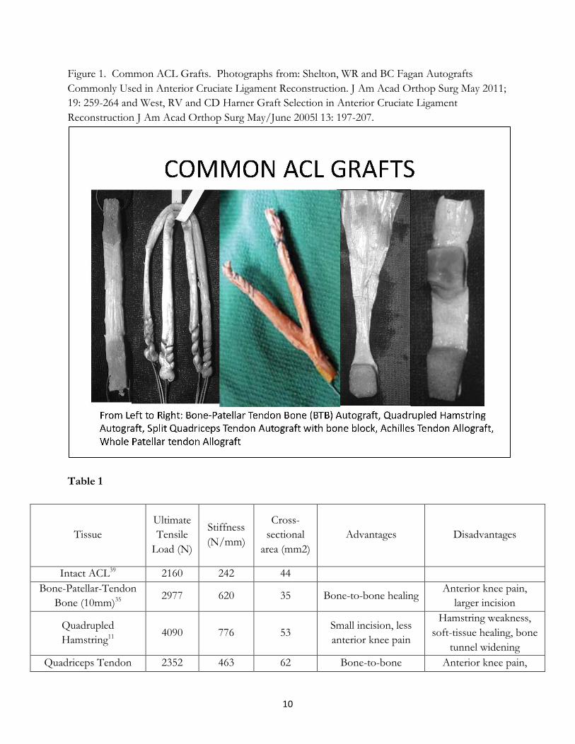
Figure 1. Common ACL Grafts. Photographs from: Shelton, WR and BC Fagan Autografts Commonly Used in Anterior Cruciate Ligament Reconstruction. J Am Acad Orthop Surg May 2011; 19: 259-264 and West, RV and CD Harner Graft Selection in Anterior Cruciate Ligament Reconstruction J Am Acad Orthop Surg May/June 2005l 13: 197-207.
Table 1
Tissue Ultimate Tensile
Load (N)
Stiffness (N/mm)
Cross-sectional
area (mm2) Advantages Disadvantages
Intact ACL39 2160 242 44 Bone-Patellar-Tendon
Bone (10mm)35 2977 620 35 Bone-to-bone healing
Anterior knee pain, larger incision
Quadrupled Hamstring11
4090 776 53 Small incision, less anterior knee pain
Hamstring weakness, soft-tissue healing, bone
tunnel widening Quadriceps Tendon 2352 463 62 Bone-to-bone Anterior knee pain,

11
(10mm)12, 51 healing, thick, can be made into two
bundles
larger incision, patella fracture if take bone
plug, soft-tissue healing Patellar Tendon
Allograft2 1403 224 Bone-to-bone healing Longer incorporation
Achilles Allograft28, 33 1189 7413 105 Longer incorporation,
soft-tissue healing Tibialis Anterior
Allograft2 3012 343
Longer incorporation, soft-tissue healing
12
References 1. Baumfeld JA, Diduch DR, Rubino LJ, et al. Tunnel widening following anterior cruciate ligament reconstruction using hamstring autograft: a comparison between double cross-pin and suspensory graft fixation. Knee Surg Sports Traumatol Arthrosc 2008;16:1108-1113. 2. Chan DB, Temple HT, Latta LL, Mahure S, Dennis J, Kaplan LD. A biomechanical comparison of fan-folded, single-looped fascia lata with other graft tissues as a suitable substitute for anterior cruciate ligament reconstruction. Arthroscopy 2010;26:1641-1647. 3. Fahey M, Indelicato PA. Bone tunnel enlargement after anterior cruciate ligament replacement. Am J Sports Med 1994;22:410-414. 4. Fauno P, Kaalund S. Tunnel widening after hamstring anterior cruciate ligament reconstruction is influenced by the type of graft fixation used: a prospective randomized study. Arthroscopy 2005;21:1337-1341. 5. Feller JA, Webster KE, Gavin B. Early post-operative morbidity following anterior cruciate ligament reconstruction: patellar tendon versus hamstring graft. Knee Surg Sports Traumatol Arthrosc 2001;9:260-266. 6. Fulkerson JP, Langeland R. An alternative cruciate reconstruction graft: the central quadriceps tendon. Arthroscopy 1995;11:252-254. 7. Goldstein JL, Verma N, McNickle AG, Zelazny A, Ghodadra N, Bach BR, Jr. Avoiding mismatch in allograft anterior cruciate ligament reconstruction: correlation between patient height and patellar tendon length. Arthroscopy 2010;26:643-650. 8. Goradia VK, Rochat MC, Grana WA, Rohrer MD, Prasad HS. Tendon-to-bone healing of a semitendinosus tendon autograft used for ACL reconstruction in a sheep model. Am J Knee Surg 2000;13:143-151. 9. Grana WA, Egle DM, Mahnken R, Goodhart CW. An analysis of autograft fixation after anterior cruciate ligament reconstruction in a rabbit model. Am J Sports Med 1994;22:344-351. 10. Gulotta LV, Rodeo SA. Biology of autograft and allograft healing in anterior cruciate ligament reconstruction. Clin Sports Med 2007;26:509-524. 11. Hamner DL, Brown CH, Jr., Steiner ME, Hecker AT, Hayes WC. Hamstring tendon grafts for reconstruction of the anterior cruciate ligament: biomechanical evaluation of the use of multiple strands and tensioning techniques. J Bone Joint Surg Am 1999;81:549-557. 12. Harris NL, Smith DA, Lamoreaux L, Purnell M. Central quadriceps tendon for anterior cruciate ligament reconstruction. Part I: Morphometric and biomechanical evaluation. Am J Sports Med 1997;25:23-28. 13. Hersekli MA, Akpinar S, Ozalay M, et al. Tunnel enlargement after arthroscopic anterior cruciate ligament reconstruction: comparison of bone-patellar tendon-bone and hamstring autografts. Adv Ther 2004;21:123-131. 14. Hoburg A, Keshlaf S, Schmidt T, et al. Fractionation of high-dose electron beam irradiation of BPTB grafts provides significantly improved viscoelastic and structural properties compared to standard gamma irradiation. Knee Surg Sports Traumatol Arthrosc 2011;19:1955-1961. 15. Jackson DW, Corsetti J, Simon TM. Biologic incorporation of allograft anterior cruciate ligament replacements. Clin Orthop Relat Res 1996:126-133.

13
16. Jackson DW, Grood ES, Goldstein JD, et al. A comparison of patellar tendon autograft and allograft used for anterior cruciate ligament reconstruction in the goat model. Am J Sports Med 1993;21:176-185. 17. Jackson DW, Simon TM, Kurzweil PR, Rosen MA. Survival of cells after intra-articular transplantation of fresh allografts of the patellar and anterior cruciate ligaments. DNA-probe analysis in a goat model. J Bone Joint Surg Am 1992;74:112-118. 18. Jackson DW, Windler GE, Simon TM. Intraarticular reaction associated with the use of freeze-dried, ethylene oxide-sterilized bone-patella tendon-bone allografts in the reconstruction of the anterior cruciate ligament. Am J Sports Med 1990;18:1-10; discussion 10-11. 19. Kaeding CC, Aros BC, Pedroza A, et al. Allograft Versus Autograft Anterior Cruciate Ligament Reconstruction: Predictors of Failure from a MOON Prospective Longitudinal Cohort. Sports Health 2011;3:73-81. 20. Kercher J, Xerogeanes J, Tannenbaum A, Al-Hakim R, Black JC, Zhao J. Anterior cruciate ligament reconstruction in the skeletally immature: an anatomical study utilizing 3-dimensional magnetic resonance imaging reconstructions. J Pediatr Orthop 2009;29:124-129. 21. Kim JG, Yang SJ, Lee YS, Shim JC, Ra HJ, Choi JY. The effects of hamstring harvesting on outcomes in anterior cruciate ligament-reconstructed patients: a comparative study between hamstring-harvested and -unharvested patients. Arthroscopy 2011;27:1226-1234. 22. Kocher MS, Smith JT, Zoric BJ, Lee B, Micheli LJ. Transphyseal anterior cruciate ligament reconstruction in skeletally immature pubescent adolescents. J Bone Joint Surg Am 2007;89:2632-2639. 23. L'Insalata JC, Klatt B, Fu FH, Harner CD. Tunnel expansion following anterior cruciate ligament reconstruction: a comparison of hamstring and patellar tendon autografts. Knee Surg Sports Traumatol Arthrosc 1997;5:234-238. 24. Landes S, Nyland J, Elmlinger B, Tillett E, Caborn D. Knee flexor strength after ACL reconstruction: comparison between hamstring autograft, tibialis anterior allograft, and non-injured controls. Knee Surg Sports Traumatol Arthrosc 2010;18:317-324. 25. Lee GH, McCulloch P, Cole BJ, Bush-Joseph CA, Bach BR, Jr. The incidence of acute patellar tendon harvest complications for anterior cruciate ligament reconstruction. Arthroscopy 2008;24:162-166. 26. Levy BA, Dajani KA, Morgan JA, Shah JP, Dahm DL, Stuart MJ. Repair versus reconstruction of the fibular collateral ligament and posterolateral corner in the multiligament-injured knee. Am J Sports Med 2010;38:804-809. 27. Levy BA, Dajani KA, Whelan DB, et al. Decision making in the multiligament-injured knee: an evidence-based systematic review. Arthroscopy 2009;25:430-438. 28. Lewis G, Shaw KM. Modeling the tensile behavior of human Achilles tendon. Biomed Mater Eng 1997;7:231-244. 29. Leys T, Salmon L, Waller A, Linklater J, Pinczewski L. Clinical Results and Risk Factors for Reinjury 15 Years After Anterior Cruciate Ligament Reconstruction: A Prospective Study of Hamstring and Patellar Tendon Grafts. Am J Sports Med 2011.

14
30. Li RC, Maffulli N, Hsu YC, Chan KM. Isokinetic strength of the quadriceps and hamstrings and functional ability of anterior cruciate deficient knees in recreational athletes. Br J Sports Med 1996;30:161-164. 31. Liden M, Sernert N, Rostgard-Christensen L, Kartus C, Ejerhed L. Osteoarthritic changes after anterior cruciate ligament reconstruction using bone-patellar tendon-bone or hamstring tendon autografts: a retrospective, 7-year radiographic and clinical follow-up study. Arthroscopy 2008;24:899-908. 32. Linn RM, Fischer DA, Smith JP, Burstein DB, Quick DC. Achilles tendon allograft reconstruction of the anterior cruciate ligament-deficient knee. Am J Sports Med 1993;21:825-831. 33. Louis-Ugbo J, Leeson B, Hutton WC. Tensile properties of fresh human calcaneal (Achilles) tendons. Clin Anat 2004;17:30-35. 34. Makela EA, Vainionpaa S, Vihtonen K, Mero M, Rokkanen P. The effect of trauma to the lower femoral epiphyseal plate. An experimental study in rabbits. J Bone Joint Surg Br 1988;70:187-191. 35. Markolf KL, Burchfield DM, Shapiro MM, Cha CW, Finerman GA, Slauterbeck JL. Biomechanical consequences of replacement of the anterior cruciate ligament with a patellar ligament allograft. Part II: forces in the graft compared with forces in the intact ligament. J Bone Joint Surg Am 1996;78:1728-1734. 36. Miller SL, Gladstone JN. Graft selection in anterior cruciate ligament reconstruction. Orthop Clin North Am 2002;33:675-683. 37. Mohtadi NG, Chan DS, Dainty KN, Whelan DB. Patellar tendon versus hamstring tendon autograft for anterior cruciate ligament rupture in adults. Cochrane Database Syst Rev 2011;9:CD005960. 38. Moller E, Weidenhielm L, Werner S. Outcome and knee-related quality of life after anterior cruciate ligament reconstruction: a long-term follow-up. Knee Surg Sports Traumatol Arthrosc 2009;17:786-794. 39. Noyes FR, Butler DL, Grood ES, Zernicke RF, Hefzy MS. Biomechanical analysis of human ligament grafts used in knee-ligament repairs and reconstructions. J Bone Joint Surg Am 1984;66:344-352. 40. Panni AS, Milano G, Lucania L, Fabbriciani C. Graft healing after anterior cruciate ligament reconstruction in rabbits. Clin Orthop Relat Res 1997:203-212. 41. Papageorgiou CD, Ma CB, Abramowitch SD, Clineff TD, Woo SL. A multidisciplinary study of the healing of an intraarticular anterior cruciate ligament graft in a goat model. Am J Sports Med 2001;29:620-626. 42. Park MJ, Lee MC, Seong SC. A comparative study of the healing of tendon autograft and tendon-bone autograft using patellar tendon in rabbits. Int Orthop 2001;25:35-39. 43. Pruss A, Kao M, Gohs U, Koscielny J, von Versen R, Pauli G. Effect of gamma irradiation on human cortical bone transplants contaminated with enveloped and non-enveloped viruses. Biologicals 2002;30:125-133.
15
44. Reinhardt KR, Hetsroni I, Marx RG. Graft selection for anterior cruciate ligament reconstruction: a level I systematic review comparing failure rates and functional outcomes. Orthop Clin North Am 2010;41:249-262. 45. Roberts TS, Drez D, Jr., McCarthy W, Paine R. Anterior cruciate ligament reconstruction using freeze-dried, ethylene oxide-sterilized, bone-patellar tendon-bone allografts. Two year results in thirty-six patients. Am J Sports Med 1991;19:35-41. 46. Rodeo SA, Arnoczky SP, Torzilli PA, Hidaka C, Warren RF. Tendon-healing in a bone tunnel. A biomechanical and histological study in the dog. J Bone Joint Surg Am 1993;75:1795-1803. 47. Sekiya JK, Ong BC, Bradley JP. Complications in anterior cruciate ligament surgery. Orthop Clin North Am 2003;34:99-105. 48. Shea KG, Belzer J, Apel PJ, Nilsson K, Grimm NL, Pfeiffer RP. Volumetric injury of the physis during single-bundle anterior cruciate ligament reconstruction in children: a 3-dimensional study using magnetic resonance imaging. Arthroscopy 2009;25:1415-1422. 49. Shea KG, Grimm NL, Belzer JS. Volumetric injury of the distal femoral physis during double-bundle ACL reconstruction in children: a three-dimensional study with use of magnetic resonance imaging. J Bone Joint Surg Am 2011;93:1033-1038. 50. Shelbourne KD, Gray T, Wiley BV. Results of transphyseal anterior cruciate ligament reconstruction using patellar tendon autograft in tanner stage 3 or 4 adolescents with clearly open growth plates. Am J Sports Med 2004;32:1218-1222. 51. Staubli HU, Schatzmann L, Brunner P, Rincon L, Nolte LP. Mechanical tensile properties of the quadriceps tendon and patellar ligament in young adults. Am J Sports Med 1999;27:27-34. 52. Stein DA, Hunt SA, Rosen JE, Sherman OH. The incidence and outcome of patella fractures after anterior cruciate ligament reconstruction. Arthroscopy 2002;18:578-583. 53. Sun K, Tian S, Zhang J, Xia C, Zhang C, Yu T. Anterior cruciate ligament reconstruction with BPTB autograft, irradiated versus non-irradiated allograft: a prospective randomized clinical study. Knee Surg Sports Traumatol Arthrosc 2009;17:464-474. 54. Taylor DE, Dervin GF, Keene GC. Femoral bone plug recession in endoscopic anterior cruciate ligament reconstruction. Arthroscopy 1996;12:513-515. 55. Tomita F, Yasuda K, Mikami S, Sakai T, Yamazaki S, Tohyama H. Comparisons of intraosseous graft healing between the doubled flexor tendon graft and the bone-patellar tendon-bone graft in anterior cruciate ligament reconstruction. Arthroscopy 2001;17:461-476. 56. Tsepis E, Vagenas G, Giakas G, Georgoulis A. Hamstring weakness as an indicator of poor knee function in ACL-deficient patients. Knee Surg Sports Traumatol Arthrosc 2004;12:22-29. 57. Vangsness CJ. How safe are soft-tissue allografts? . AAOS Now 2007. 58. Verma N, Noerdlinger MA, Hallab N, Bush-Joseph CA, Bach BR, Jr. Effects of graft rotation on initial biomechanical failure characteristics of bone-patellar tendon-bone constructs. Am J Sports Med 2003;31:708-713. 59. Verma NN, Dennis MG, Carreira DS, Bojchuk J, Hayden JK, Bach BR, Jr. Preliminary clinical results of two techniques for addressing graft tunnel mismatch in endoscopic anterior cruciate ligament reconstruction. J Knee Surg 2005;18:183-191.

16
60. Webster KE, Feller JA, Hameister KA. Bone tunnel enlargement following anterior cruciate ligament reconstruction: a randomised comparison of hamstring and patellar tendon grafts with 2-year follow-up. Knee Surg Sports Traumatol Arthrosc 2001;9:86-91. 61. West RV, Harner CD. Graft selection in anterior cruciate ligament reconstruction. J Am Acad Orthop Surg 2005;13:197-207. 62. Wipfler B, Donner S, Zechmann CM, Springer J, Siebold R, Paessler HH. Anterior cruciate ligament reconstruction using patellar tendon versus hamstring tendon: a prospective comparative study with 9-year follow-up. Arthroscopy 2011;27:653-665. 63. Wright RW, Huston LJ, Spindler KP, et al. Descriptive epidemiology of the Multicenter ACL Revision Study (MARS) cohort. Am J Sports Med 2010;38:1979-1986. 64. Zysk SP, Fraunberger P, Veihelmann A, et al. Tunnel enlargement and changes in synovial fluid cytokine profile following anterior cruciate ligament reconstruction with patellar tendon and hamstring tendon autografts. Knee Surg Sports Traumatol Arthrosc 2004;12:98-103.


![The Evolution of Anatomic Anterior Cruciate Ligament ... · The Evolution of Anatomic Anterior Cruciate Ligament Reconstruction ... tunnel placement in the axial plane [23]. These](https://img.dokumen.tips/doc/110x75/5f03ed437e708231d40b74ae/the-evolution-of-anatomic-anterior-cruciate-ligament-the-evolution-of-anatomic.jpg)