Embed Size (px)
Citation preview
Acta Biomaterialia xxx (2010) xxx–xxx
ARTICLE IN PRESS
Contents lists available at ScienceDirect
Acta Biomaterialia
journal homepage: www.elsevier .com/locate /actabiomat
Functional biomimetic analogs help remineralize apatite-depleteddemineralized resin-infiltrated dentin via a bottom–up approach
Jongryul Kim a, Dwayne D. Arola b, Lisha Gu c, Young Kyung Kim d, Sui Mai c, Yan Liu e,David H. Pashley f, Franklin R. Tay f,*
a Department of Conservative Dentistry, School of Dentistry, Kyung Hee University, 1 Hoegi-dong, Dongdaemun-gu, Seou, 130-701, South Koreab Department of Mechanical Engineering, University of Maryland, 1000 Hilltop Circle, Baltimore, MD 21250, USAc Department of Operative Dentistry and Endodontics, Guanghua School of Stomatology, Sun Yat-sen University, 56 Lingyuan Road West, Guangzhou 510055, Chinad Department of Conservative Dentistry, School of Dentistry, Kyungpook National University, Samduk-dong 2-ga, Jung-gu, Daegu 700-412, South Koreae Department of Stomatology, Tongji Hospital, Huazhong University of Science and Technology, Wuhan, Chinaf Department of Oral Biology, School of Dentistry, Medical College of Georgia, 1120,15th Street, CL 2120, Augusta, GA 30912-1129, USA
a r t i c l e i n f o
Article history:Received 25 September 2009Received in revised form 18 November 2009Accepted 30 December 2009Available online xxxx
Keywords:CollagenDentinIntrafibrillarRemineralizationSelf-etching adhesive
1742-7061/$ - see front matter � 2009 Acta Materialdoi:10.1016/j.actbio.2009.12.052
* Corresponding author. Tel.: +1 706 721 2033; faxE-mail address: [email protected] (F.R. Tay).
Please cite this article in press as: Kim J et al. Funbottom–up approach. Acta Biomater (2010), do
a b s t r a c t
Natural biominerals are formed through metastable amorphous precursor phases via a bottom–up,nanoparticle-mediated mineralization mechanism. Using an acid-etched human dentin model to createa layer of completely demineralized collagen matrix, a bio-inspired mineralization scheme has beendeveloped based on the use of dual biomimetic analogs. These analogs help to sequester fluidic amor-phous calcium phosphate nanoprecursors and function as templates for guiding homogeneous apatitenucleation within the collagen fibrils. By adopting this scheme for remineralizing adhesive resin-bonded,completely demineralized dentin, we have been able to redeposit intrafibrillar and extrafibrillar apatitesin completely demineralized collagen matrices that are imperfectly infiltrated by resins. This study uti-lizes a spectrum of completely and partially demineralized dentin collagen matrices to further validatethe necessity for using a biomimetic analog-containing medium for remineralizing resin-infiltrated par-tially demineralized collagen matrices in which remnant seed crystallites are present. In control speci-mens in which biomimetic analogs are absent from the remineralization medium, remineralizationcould only be seen in partially demineralized collagen matrices, probably by epitaxial growth via atop–down crystallization approach. Conversely, in the presence of biomimetic analogs in the remineral-ization medium, intrafibrillar remineralization of completely demineralized collagen matrices via a bot-tom–up crystallization mechanism can additionally be identified. The latter is characterized by thetransition of intrafibrillar minerals from an inchoate state of continuously braided microfibrillar elec-tron-dense amorphous strands to discrete nanocrystals, and ultimately into larger crystalline plateletswithin the collagen fibrils. Biomimetic remineralization via dual biomimetic analogs has the potentialto be translated into a functional delivery system for salvaging failing resin–dentin bonds.
� 2009 Acta Materialia Inc. Published by Elsevier Ltd. All rights reserved.
1. Introduction
Dental caries is a transmittable infectious disease which repre-sents pathologic destruction of hard and soft dental tissues by oralmicroorganisms. It affects individuals of all ages, cultures andsocioeconomic backgrounds, and is a major problem in dentistrydespite significant advances in the development of preventivestrategies over the past few decades [1]. Acids produced by cario-genic microorganisms diffuse through calcified dental tissues andresult in dissolution of the apatite crystallites. Dental caries maybe initiated from either the crown enamel or the root cementum,and eventually progresses to the underlying dentin. Bacteria pro-
ia Inc. Published by Elsevier Ltd. A
: +1 706 721 6252.
ctional biomimetic analogs heli:10.1016/j.actbio.2009.12.052
duce lactic acid until the local pH reaches about 4.5–5.0, at whichtime they cease to form any more lactic acid. The lactic acid tendsto preferentially dissolve some, but not all, interfibrillar crystal-lites, creating a zone of partially demineralized dentin. The processalso activates endogenous collagenolytic enzymes known asmatrix metalloproteinases (MMPs) that break down the deminer-alized collagen matrix. Dentin contains approximately 70 wt.%mineral phase, 20 wt.% organic phase and 10 wt.% water [2]. TypeI collagen constitutes 90 wt.% of the dentin organic phase and theremaining 10% is composed of noncollagenous proteins (NCPs) [3].If demineralization by bacterial acids is not halted or reversed byre-deposition of minerals derived from salivary secretions, a cavitywill form that will require a dental filling [1]. Contemporarymanagement of caries has progressed historically from extractionof the caries-involved tooth to the use of minimally invasive
ll rights reserved.
p remineralize apatite-depleted demineralized resin-infiltrated dentin via a
2 J. Kim et al. / Acta Biomaterialia xxx (2010) xxx–xxx
ARTICLE IN PRESS
techniques that maximize the conservation of tooth structures.Filling of carious teeth with tooth-colored resin composites thatare coupled to the dentin substrate via the use of dentin adhesivesis the current treatment of choice.
Dentin adhesives rely on micromechanical entanglement ofresin polymers within partially or completely demineralizedcollagen matrices for retention of the resin composite fillings. Infil-tration of resins into the demineralized dentin creates a so-calledinterdiffusion zone. Demineralization of dentin substrates for thesake of micromechanical retention may be accomplished by a sep-arate phosphoric acid-etching step in the so-called etch-and-rinseadhesives or by utilizing acidic resin monomers with carboxylicacid or phosphoric acid functional groups in the so-called self-etching adhesives. When 37% phosphoric acid is used, the pH ofthe etched surface can fall below pH 1.0. At such low pHs, apatitecrystallites are dissolved from both interfibrillar and intrafibrillarcompartments, leaving the type I collagen completely demineral-ized. Conversely, when milder formers of self-etching adhesivesare used, zones of partially demineralized dentin that contain seedapatite crystallites are produced. Resin–dentin bonds created bycontemporary dentin adhesives are susceptible to degradationin vivo after aging [4–6] by endogenous MMPs that are bound tothe demineralized collagen fibrils [7]. A potential strategy to im-prove the durability of these bonds is to replace the residual watertrapped within the non-resin encapsulated demineralized collagenfibrils by new apatitic minerals. The newly formed apatitic miner-als probably fossilize the MMPs and prevent the collagen fibrilsfrom enzymatic degradation. This protection concept is based onthe rationale that natural mineralized dentin that is protected byintrafibrillar and extrafibrillar apatite crystallites do not undergodegradation over time [8]. Since collagen matrix does not initiatemineralization on its own [9], noncollagenous extracellular matrixproteins are required for regulating bone and dentin mineraliza-tion and for controlling the dimension, order and hierarchy ofapatite deposition within mineralized hard tissues [10]. As thetherapeutic use of native or recombinant ECM proteins for in situbiomineralization is not yet economically viable, research scien-tists have resorted to the use of polyelectrolyte and poly(amino)acid macromolecules to mimic the functional domains of thesenaturally occurring proteins [11].
Most of the techniques involved in the synthesis of nanomate-rials and nanodevices may be classified as bottom–up and top–down approaches [12]. Bottom–up approaches start with one ormore defined molecular species, which undergo certain processesthat result in a higher-ordered, highly organized structure. Exam-ples of bottom–up approaches include self-assembly and molecu-lar patterning. Top–down approaches begin with a bulk materialthat incorporates nanoscale details, such as nanolithography andetching techniques. In the top–down approach, biomaterials aregenerated by stripping down a complex entity into its componentparts, such as paring of a virus particle down to its capsid to form aviral cage. In terms of crystallization, creating smaller crystals fromlarger crystals in partially demineralized dentin by the aforemen-tioned bacterial acids and self-etching primers represents exam-ples of the top–down approach.
A nanotechnology-inspired, biomimetic mineralization tech-nique that generates calcium and phosphate ions with a high pHhas recently been developed [13]. This biomimetic mineralizationscheme uses two polyanionic analogs to mimic the dual functionsof dentin matrix proteins [14] in sequestering and stabilizingamorphous nanoprecursors that are generated from prenucleationclusters [15–17], and acting as template molecules [18,19] forguiding the bottom–up assembly [20] of intrafibrillar apatite crys-tallites within the collagen fibrils. The aforementioned biomimeticmineralization scheme has been adopted for remineralization ofimperfect, water permeable resin–dentin interfaces created by
Please cite this article in press as: Kim J et al. Functional biomimetic analogs helbottom–up approach. Acta Biomater (2010), doi:10.1016/j.actbio.2009.12.052
etch-and-rinse dentin adhesives that utilize an aggressive phos-phoric acid etchant to create a zone of completely demineralizeddentin on top of a mineralized dentin base [21,22]. The resultsderived from this completely demineralized, resin-infiltrated den-tin model led to the conclusion that intrafibrillar remineralizationof water-rich, resin-sparse collagen matrices cannot occur in thepresence of a remineralization medium that is devoid of biomi-metic analogs. The idea of dentin remineralization is certainlynot new; the dental literature abounds with studies on a plethoraof filling materials that are capable of remineralizing partiallydemineralized dentin. An obvious difference between completelyand partially demineralized dentin is the presence of remnantapatite seed crystallites in the latter that can act as centers for het-erogeneous nucleation. Thus, remineralization of a partiallydemineralized collagen matrix is thermodynamically more favor-able than homogeneous nucleation within a completely deminer-alized collagen matrix. Part of the success reported with the useof contemporary calcium- and phosphate-releasing remineralizingmaterials may be related to the remineralization of the collagenmatrix via epitaxial growth of calcium phosphate salts on remnantseed crystallites [23]. Epitaxial growth may be regarded as a top–down approach according to the classical crystallization theory,via ion-by-ion addition to pre-existing seed crystallites [14]. Con-versely, the bottom–up mineralization approach is based on thenon-classical theory of crystallization, which involves the use ofbiomimetic analogs for generating metastable amorphous mineralprecursors and mesocrystals. As a partially demineralized collagenmatrix invariably exhibits a gradient of demineralization from thesurface to the base of the matrix, it is not known whether theaforementioned biomimetic analogs are required for remineraliz-ing partially demineralized collagen matrices. Intrafibrillar remin-eralization, in particular, has been surmised to be responsible forrestoring the mechanical properties of dentin [24].
Unlike etch-and-rinse adhesives, self-etching adhesives containpolymerizable, methacrylate-based acidic resin monomers thatsimultaneously etch and infiltrate the dentin substrate. They varywidely in their aggressiveness and may create completely or par-tially demineralized collagen matrices of variable thickness [25].The use of hydrophilic self-etching adhesives of variable aggres-siveness provides the opportunity for designing a phenomenologi-cal model consisting of a spectrum of completely and partiallydemineralized collagen matrices for validating the necessity ofusing a biomimetic analog-containing medium for remineraliza-tion of partially demineralized dentin. Thus, the objective of thiswork was to examine the ultrastructural characteristics of com-pletely demineralized vs. partially demineralized resin-infiltrateddentin collagen matrices (i.e. interdiffusion zones) that had beensubjected to biomimetic remineralization. Understanding theultrastructural differences between top–down and bottom–upapproaches in their ability to remineralize partially demineralizedresin-infiltrated dentin sets the stage for the development of thecurrent biomimetic remineralization strategy as a functionaldelivery system for remineralizing partially demineralized, resin-bonded caries-affected dentin.
2. Materials and methods
2.1. Dentin bonding
Twenty-four extracted non-carious human third molars wereused for the study. The teeth were collected after the parents’informed consents were obtained under a protocol reviewed andapproved by the Human Assurance Committee of the MedicalCollege of Georgia. A flat dentin surface was prepared perpendicu-lar to the longitudinal axis of each tooth using a low-speed Isomet
p remineralize apatite-depleted demineralized resin-infiltrated dentin via a
J. Kim et al. / Acta Biomaterialia xxx (2010) xxx–xxx 3
ARTICLE IN PRESS
diamond saw (Buehler Ltd., Lake Bluff, IL, USA) under water cool-ing. The occlusal dentin surface was polished with a 320-grit sili-con carbide paper under running water to create a bondingsurface that was devoid of enamel. The specimens were randomlydivided into three groups (eight teeth each): (I) Adper Prompt (3MESPE, St. Paul, MN, USA), the most aggressive self-etching adhesive;(II) Adper Scotchbond SE (3M ESPE), a moderately aggressiveself-etching adhesive; and (III) Adper Easy Bond (3M ESPE), themildest of the three self-etching adhesives. After the applicationof these adhesives to dentin according to the manufacturers’instructions, they were polymerized using a quartz-tungsten-halo-gen light-curing unit with an output intensity of 600 mW cm�2.This was followed by incremental placement of two 2-mm thicklayers of a resin composite that was light-cured separately for40 s each. The bonded teeth were stored at 100% relative humidityfor 24 h. Each tooth was sectioned occluso-gingivally into 0.3-mmthick slabs, each containing the resin–dentin interface.
2.2. Remineralization medium
White Portland cement (Lehigh Cement Company, Allentown,Pennsylvania, USA) was mixed with deionized water in a water-to-powder ratio of 0.35:1, placed in flexible silicone molds andallowed to set and aged at 100% relative humidity for 1 weekbefore use. A simulated body fluid (SBF) was prepared by dissolv-ing 136.8 mM NaCl, 4.2 mM NaHCO3, 3.0 mM KCl, 1.0 mMK2HPO4�3H2O, 1.5 mM MgCl2�6H2O, 2.5 mM CaCl2 and 0.5 mMNa2SO4 in deionized water and adding 3.08 mM sodium azide toprevent bacterial growth. This SBF also served as the controlremineralization medium which contained no biomimetic analog.For the biomimetic remineralization medium, 500 lg ml�1 of poly-acrylic acid (Mw = 1800; Sigma–Aldrich, St. Louis, Illinois, USA) and200 lg ml�1 of polyvinylphosphonic acid (Mw = 24,000; Sigma–Aldrich) were added to the SBF as dual biomimetic analogs. Allsolutions were buffered to pH 7.4 with 0.1 M Tris base or 0.1 MHCl.
2.3. Biomimetic remineralization
Each control and experimental specimen slab was placed over aset Portland cement block (ca. 1 g) inside a glass scintillation vial.The latter was filled with 15 ml of SBF containing the two biomi-metic analogs. Each glass vial was capped to prevent evaporationof the solution and stored in an incubator at 37 �C. The remineral-ization medium was changed every month, with its pH (after inclu-sion of Portland cement blocks) monitored weekly so that it wasabove 9.25. This ensured that apatite was formed instead of octa-calcium phosphate [26]. Experimental and control specimens wereretrieved after 1–4 months (four slabs per month) for ultrastruc-tural examination of the extent of remineralization. Baseline spec-imens were examined before immersion for examination of theinterdiffusion zone and for the presence of water-rich, resin-sparseregions using a silver nitrate tracer protocol [27].
2.4. Transmission electron microscopy
Following retrieval, the control and experimental specimenslabs were fixed in Karnovsky’s fixative, rinsed in cacodylate bufferand post-fixed in 1% osmium tetroxide. After fixation, each slabwas rinsed three times with sodium cacodylate buffer. The slabwas dehydrated in an ascending ethanol series (50–100%),immersed in propylene oxide as a transitional medium andembedded in epoxy resin [28]. For each monthly examinationperiod, non-demineralized, 90 nm thick sections were preparedand examined without further staining using a JEM-1230 transmis-sion electron microscope (JEOL, Tokyo, Japan) at 110 kV.
Please cite this article in press as: Kim J et al. Functional biomimetic analogs helbottom–up approach. Acta Biomater (2010), doi:10.1016/j.actbio.2009.12.052
3. Results
3.1. Baseline specimens
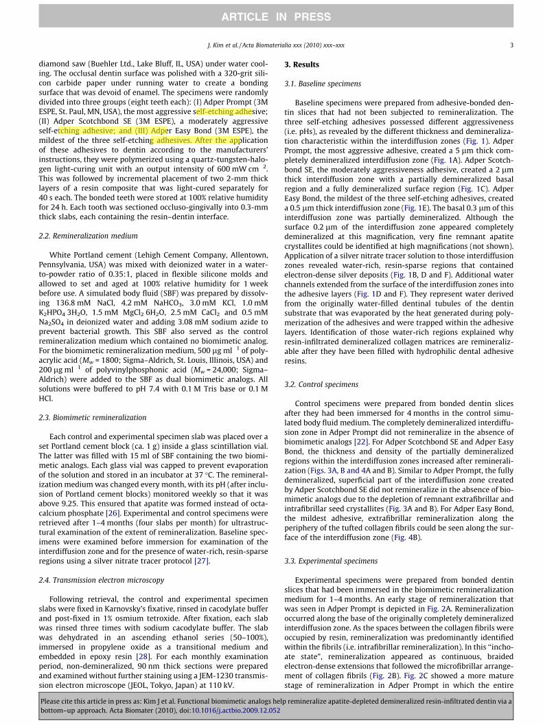
Baseline specimens were prepared from adhesive-bonded den-tin slices that had not been subjected to remineralization. Thethree self-etching adhesives possessed different aggressiveness(i.e. pHs), as revealed by the different thickness and demineraliza-tion characteristic within the interdiffusion zones (Fig. 1). AdperPrompt, the most aggressive adhesive, created a 5 lm thick com-pletely demineralized interdiffusion zone (Fig. 1A). Adper Scotch-bond SE, the moderately aggressiveness adhesive, created a 2 lmthick interdiffusion zone with a partially demineralized basalregion and a fully demineralized surface region (Fig. 1C). AdperEasy Bond, the mildest of the three self-etching adhesives, createda 0.5 lm thick interdiffusion zone (Fig. 1E). The basal 0.3 lm of thisinterdiffusion zone was partially demineralized. Although thesurface 0.2 lm of the interdiffusion zone appeared completelydemineralized at this magnification, very fine remnant apatitecrystallites could be identified at high magnifications (not shown).Application of a silver nitrate tracer solution to those interdiffusionzones revealed water-rich, resin-sparse regions that containedelectron-dense silver deposits (Fig. 1B, D and F). Additional waterchannels extended from the surface of the interdiffusion zones intothe adhesive layers (Fig. 1D and F). They represent water derivedfrom the originally water-filled dentinal tubules of the dentinsubstrate that was evaporated by the heat generated during poly-merization of the adhesives and were trapped within the adhesivelayers. Identification of those water-rich regions explained whyresin-infiltrated demineralized collagen matrices are remineraliz-able after they have been filled with hydrophilic dental adhesiveresins.
3.2. Control specimens
Control specimens were prepared from bonded dentin slicesafter they had been immersed for 4 months in the control simu-lated body fluid medium. The completely demineralized interdiffu-sion zone in Adper Prompt did not remineralize in the absence ofbiomimetic analogs [22]. For Adper Scotchbond SE and Adper EasyBond, the thickness and density of the partially demineralizedregions within the interdiffusion zones increased after reminerali-zation (Figs. 3A, B and 4A and B). Similar to Adper Prompt, the fullydemineralized, superficial part of the interdiffusion zone createdby Adper Scotchbond SE did not remineralize in the absence of bio-mimetic analogs due to the depletion of remnant extrafibrillar andintrafibrillar seed crystallites (Fig. 3A and B). For Adper Easy Bond,the mildest adhesive, extrafibrillar remineralization along theperiphery of the tufted collagen fibrils could be seen along the sur-face of the interdiffusion zone (Fig. 4B).
3.3. Experimental specimens
Experimental specimens were prepared from bonded dentinslices that had been immersed in the biomimetic remineralizationmedium for 1–4 months. An early stage of remineralization thatwas seen in Adper Prompt is depicted in Fig. 2A. Remineralizationoccurred along the base of the originally completely demineralizedinterdiffusion zone. As the spaces between the collagen fibrils wereoccupied by resin, remineralization was predominantly identifiedwithin the fibrils (i.e. intrafibrillar remineralization). In this ‘‘incho-ate state”, remineralization appeared as continuous, braidedelectron-dense extensions that followed the microfibrillar arrange-ment of collagen fibrils (Fig. 2B). Fig. 2C showed a more maturestage of remineralization in Adper Prompt in which the entire
p remineralize apatite-depleted demineralized resin-infiltrated dentin via a
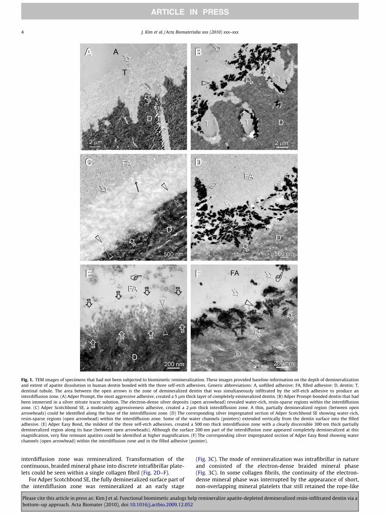
Fig. 1. TEM images of specimens that had not been subjected to biomimetic remineralization. These images provided baseline information on the depth of demineralizationand extent of apatite dissolution in human dentin bonded with the three self-etch adhesives. Generic abbreviations: A, unfilled adhesive; FA, filled adhesive; D, dentin; T,dentinal tubule. The area between the open arrows is the zone of demineralized dentin that was simultaneously infiltrated by the self-etch adhesive to produce aninterdiffusion zone. (A) Adper Prompt, the most aggressive adhesive, created a 5 lm thick layer of completely emineralized dentin. (B) Adper Prompt-bonded dentin that hadbeen immersed in a silver nitrate tracer solution. The electron-dense silver deposits (open arrowhead) revealed water-rich, resin-sparse regions within the interdiffusionzone. (C) Adper Scotchbond SE, a moderately aggressiveness adhesive, created a 2 lm thick interdiffusion zone. A thin, partially demineralized region (between openarrowheads) could be identified along the base of the interdiffusion zone. (D) The corresponding silver impregnated section of Adper Scotchbond SE showing water-rich,resin-sparse regions (open arrowhead) within the interdiffusion zone. Some of the water channels (pointers) extended vertically from the dentin surface into the filledadhesive. (E) Adper Easy Bond, the mildest of the three self-etch adhesives, created a 500 nm thick interdiffusion zone with a clearly discernible 300 nm thick partiallydemineralized region along its base (between open arrowheads). Although the surface 200 nm part of the interdiffusion zone appeared completely demineralized at thismagnification, very fine remnant apatites could be identified at higher magnification. (F) The corresponding silver impregnated section of Adper Easy Bond showing waterchannels (open arrowhead) within the interdiffusion zone and in the filled adhesive (pointer).
4 J. Kim et al. / Acta Biomaterialia xxx (2010) xxx–xxx
ARTICLE IN PRESS
interdiffusion zone was remineralized. Transformation of thecontinuous, braided mineral phase into discrete intrafibrillar plate-lets could be seen within a single collagen fibril (Fig. 2D–F).
For Adper Scotchbond SE, the fully demineralized surface part ofthe interdiffusion zone was remineralized at an early stage
Please cite this article in press as: Kim J et al. Functional biomimetic analogs helbottom–up approach. Acta Biomater (2010), doi:10.1016/j.actbio.2009.12.052
(Fig. 3C). The mode of remineralization was intrafibrillar in natureand consisted of the electron-dense braided mineral phase(Fig. 3C). In some collagen fibrils, the continuity of the electron-dense mineral phase was interrupted by the appearance of short,non-overlapping mineral platelets that still retained the rope-like
p remineralize apatite-depleted demineralized resin-infiltrated dentin via a
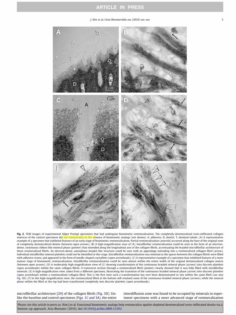
Fig. 2. TEM images of experimental Adper Prompt specimens that had undergone biomimetic remineralization. The completely demineralized resin-infiltrated collagenmatrices of the control specimens did not remineralize in the absence of biomimetic analogs (not shown). A, adhesive; D, dentin; T, dentinal tubule. (A) A representativeexample of a specimen that exhibited features of an early stage of biomimetic remineralization. Partial remineralization (asterisk) occurred along the base of the original zoneof completely demineralized dentin (between open arrows). (B) A high-magnification view of (A). Intrafibrillar remineralization could be seen in the form of an electron-dense, continuous ribbon-like mineral phase (pointer) that extended along the longitudinal axis of the collagen fibrils, accentuating the braided microfibrillar architecture ofthose remineralized fibrils. An electron-dense, amorphous droplet-like structure could be seen with an appendage extending into a remineralized collagen fibril (arrow).Individual intrafibrillar mineral platelets could not be identified at this stage. Extrafibrillar remineralization was minimal as the spaces between the collagen fibrils were filledwith adhesive resins, and appeared in the form of needle-shaped crystallites (open arrowheads). (C) A representative example of a specimen that exhibited features of a moremature stage of biomimetic remineralization. Intrafibrillar remineralization could be seen almost within the entire width of the original demineralized collagen matrix(between open arrows). (D) A moderately high-magnification view of (C) showing transformation of the continuous braided mineral phase (arrows) into discrete platelets(open arrowheads) within the same collagen fibrils. A transverse section through a remineralized fibril (pointer) clearly showed that it was fully filled with intrafibrillarminerals. (E) A high-magnification view, taken from a different specimen, illustrating the transition of the continuous braided mineral phase (arrow) into discrete platelets(open arrowhead) within a remineralized collagen fibril. This is the first time such a transformation has ever been demonstrated in situ within the same fibril (see alsoFig. 3D). (F) In this high-magnification view, the remineralized fibril at the bottom still retained some of the continuous braided mineral phase (arrows), while the mineralphase within the fibril at the top had been transformed completely into discrete platelets (open arrowheads).
J. Kim et al. / Acta Biomaterialia xxx (2010) xxx–xxx 5
ARTICLE IN PRESS
microfibrillar architecture [29] of the collagen fibrils (Fig. 3D). Un-like the baseline and control specimens (Figs. 1C and 3A), the entire
Please cite this article in press as: Kim J et al. Functional biomimetic analogs helbottom–up approach. Acta Biomater (2010), doi:10.1016/j.actbio.2009.12.052
interdiffusion zone was found to be occupied by minerals in exper-iment specimens with a more advanced stage of remineralization
p remineralize apatite-depleted demineralized resin-infiltrated dentin via a
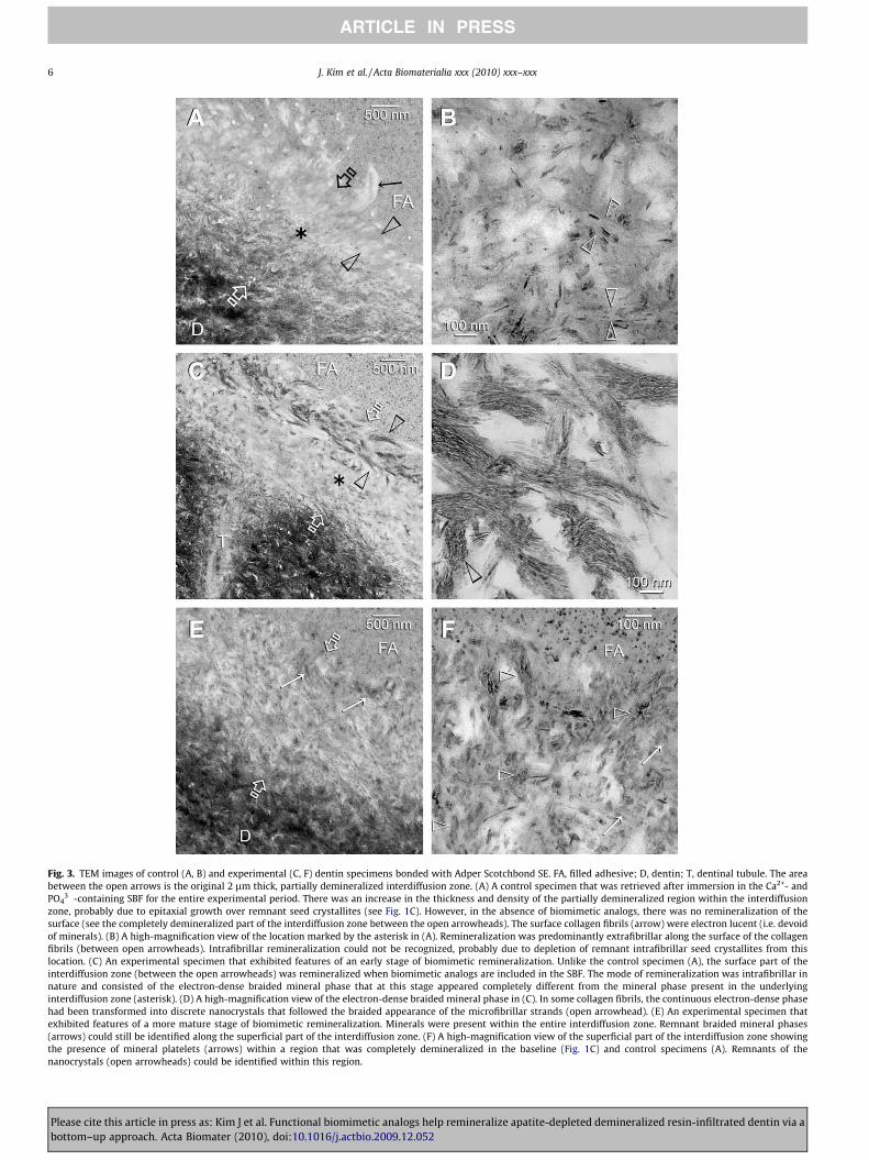
Fig. 3. TEM images of control (A, B) and experimental (C, F) dentin specimens bonded with Adper Scotchbond SE. FA, filled adhesive; D, dentin; T, dentinal tubule. The areabetween the open arrows is the original 2 lm thick, partially demineralized interdiffusion zone. (A) A control specimen that was retrieved after immersion in the Ca2+- andPO4
3�-containing SBF for the entire experimental period. There was an increase in the thickness and density of the partially demineralized region within the interdiffusionzone, probably due to epitaxial growth over remnant seed crystallites (see Fig. 1C). However, in the absence of biomimetic analogs, there was no remineralization of thesurface (see the completely demineralized part of the interdiffusion zone between the open arrowheads). The surface collagen fibrils (arrow) were electron lucent (i.e. devoidof minerals). (B) A high-magnification view of the location marked by the asterisk in (A). Remineralization was predominantly extrafibrillar along the surface of the collagenfibrils (between open arrowheads). Intrafibrillar remineralization could not be recognized, probably due to depletion of remnant intrafibrillar seed crystallites from thislocation. (C) An experimental specimen that exhibited features of an early stage of biomimetic remineralization. Unlike the control specimen (A), the surface part of theinterdiffusion zone (between the open arrowheads) was remineralized when biomimetic analogs are included in the SBF. The mode of remineralization was intrafibrillar innature and consisted of the electron-dense braided mineral phase that at this stage appeared completely different from the mineral phase present in the underlyinginterdiffusion zone (asterisk). (D) A high-magnification view of the electron-dense braided mineral phase in (C). In some collagen fibrils, the continuous electron-dense phasehad been transformed into discrete nanocrystals that followed the braided appearance of the microfibrillar strands (open arrowhead). (E) An experimental specimen thatexhibited features of a more mature stage of biomimetic remineralization. Minerals were present within the entire interdiffusion zone. Remnant braided mineral phases(arrows) could still be identified along the superficial part of the interdiffusion zone. (F) A high-magnification view of the superficial part of the interdiffusion zone showingthe presence of mineral platelets (arrows) within a region that was completely demineralized in the baseline (Fig. 1C) and control specimens (A). Remnants of thenanocrystals (open arrowheads) could be identified within this region.
6 J. Kim et al. / Acta Biomaterialia xxx (2010) xxx–xxx
ARTICLE IN PRESS
Please cite this article in press as: Kim J et al. Functional biomimetic analogs help remineralize apatite-depleted demineralized resin-infiltrated dentin via abottom–up approach. Acta Biomater (2010), doi:10.1016/j.actbio.2009.12.052
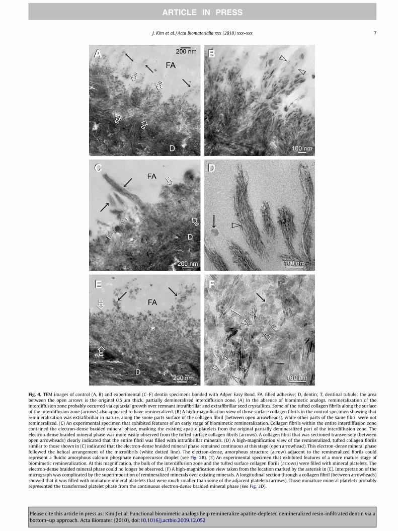
Fig. 4. TEM images of control (A, B) and experimental (C–F) dentin specimens bonded with Adper Easy Bond. FA, filled adhesive; D, dentin; T, dentinal tubule; the areabetween the open arrows is the original 0.5 lm thick, partially demineralized interdiffusion zone. (A) In the absence of biomimetic analogs, remineralization of theinterdiffusion zone probably occurred via epitaxial growth over remnant intrafibrillar and extrafibrillar seed crystallites. Some of the tufted collagen fibrils along the surfaceof the interdiffusion zone (arrows) also appeared to have remineralized. (B) A high-magnification view of those surface collagen fibrils in the control specimen showing thatremineralization was extrafibrillar in nature, along the some parts surface of the collagen fibril (between open arrowheads), while other parts of the same fibril were notremineralized. (C) An experimental specimen that exhibited features of an early stage of biomimetic remineralization. Collagen fibrils within the entire interdiffusion zonecontained the electron-dense braided mineral phase, masking the existing apatite platelets from the original partially demineralized part of the interdiffusion zone. Theelectron-dense braided mineral phase was more easily observed from the tufted surface collagen fibrils (arrows). A collagen fibril that was sectioned transversely (betweenopen arrowheads) clearly indicated that the entire fibril was filled with intrafibrillar minerals. (D) A high-magnification view of the remineralized, tufted collagen fibrilssimilar to those shown in (C) indicated that the electron-dense braided mineral phase remained continuous at this stage (open arrowhead). This electron-dense mineral phasefollowed the helical arrangement of the microfibrils (white dotted line). The electron-dense, amorphous structure (arrow) adjacent to the remineralized fibrils couldrepresent a fluidic amorphous calcium phosphate nanoprecursor droplet (see Fig. 2B). (E) An experimental specimen that exhibited features of a more mature stage ofbiomimetic remineralization. At this magnification, the bulk of the interdiffusion zone and the tufted surface collagen fibrils (arrows) were filled with mineral platelets. Theelectron-dense braided mineral phase could no longer be observed. (F) A high-magnification view taken from the location marked by the asterisk in (E). Interpretation of themicrograph was complicated by the superimposition of remineralized minerals over existing minerals. A longitudinal section through a collagen fibril (between arrowheads)showed that it was filled with miniature mineral platelets that were much smaller than some of the adjacent platelets (arrows). Those miniature mineral platelets probablyrepresented the transformed platelet phase from the continuous electron-dense braided mineral phase (see Fig. 3D).
J. Kim et al. / Acta Biomaterialia xxx (2010) xxx–xxx 7
ARTICLE IN PRESS
Please cite this article in press as: Kim J et al. Functional biomimetic analogs help remineralize apatite-depleted demineralized resin-infiltrated dentin via abottom–up approach. Acta Biomater (2010), doi:10.1016/j.actbio.2009.12.052
8 J. Kim et al. / Acta Biomaterialia xxx (2010) xxx–xxx
ARTICLE IN PRESS
(Fig. 3E). Although remnant fibrils with the electron-dense braidedmineral phase could still be seen, the surface part of the interdiffu-sion zone was predominantly filled with platelet-shaped mineralsat this stage of remineralization (Fig. 3F).
For Adper Easy Bond, difference between the control and exper-imental specimens was most notable during the early stage of bio-mimetic remineralization. The extrafibrillar remineralization thatwas observed on the tufted fibrils along the surface of the interdif-fusion zone in the control specimens (Fig. 4B) was replaced byintrafibrillar remineralization of similar tufted fibrils in the exper-imental specimens (Fig. 4C). In those experimental specimens, thecharacteristic electron-dense braided mineral phase could be seenwithin the fibrils, with occasional appearance of amorphous, elec-tron-dense globular bodies adjacent to the braided fibrils (Fig. 4D).With a more mature stage of biomimetic remineralization, thebraided fibrils were transformed into a denser conglomerate ofmineral platelets that could be more easily identified along the sur-face tufted collagen fibrils (Fig. 4E). A high-magnification view ofthe subsurface of the remineralized interdiffusion zone revealedcollagen fibrils packed with mineral platelets that were muchsmaller than the adjacent platelets derived from the original par-tially demineralized dentin (Fig. 4F). These miniature mineralplatelets probably represent an intermediate stage of transforma-tion of the ‘‘inchoate” braided mineral phase to the final ‘‘mature”platelet phase.
4. Discussion
The results obtained from the control specimens confirmed thatremineralization of completely demineralized dentin does notoccur when biomimetic analogs are absent from a remineralizationmedium. Conversely, the increase in thickness and density of thepartially demineralized regions in control specimens preparedwith Adper Scotchbond SE and Adper Easy Bond (Figs. 3A, B and4A, B) indirectly indicated that remineralization of partially demin-eralized dentin can occur in the absence of biomimetic analogs.This is probably due to the epitaxial growth over remnant seedcrystallites which act as templates for mineral deposition [30],with the orientation of newly formed mineral lattice determinedby the lattice of the underlying crystal. This process is quite com-mon in the formation of calculus [31] and remineralization ofenamel [32,33]. Such a top–down mechanism of crystal growth isprobably thermodynamically more favorable based on the classicalcrystallization theory. Crystallization proceeds via the one-steproute of simply overcoming the free energy required for crystalgrowth (DGgrowth), without the need to overcome simultaneouslythe activation-energy barrier for nucleation (DGnucleation). Thus,remineralization occurs without the need for the alternative kinet-ically driven protein/polymer-modulated pathway for lowering theGibbs free energy via sequential steps of phase transformations, asdepicted by the non-classical crystallization theory [15,34].Although noncollagenous proteins may be associated with theremaining minerals in partially demineralized dentin [35], wedid not observe additional intrafibrillar remineralization of thecompletely demineralized regions of the interdiffusion zonescreated by these two adhesives, contrary to the results suggestedby Saito et al. [36]. However, it is possible that some of these non-collagenous proteins may participate in the top–down reminerali-zation process by inhibiting the dimensions of the apatitecrystallites during the process of epitaxial growth [37].
In this study, we could only speculate the occurrence of epitax-ial crystal growth based on the increase in thickness and density ofthe partially demineralized regions of the interdiffusion zones. Thisis due to our inability to distinguish newly precipitated mineralphase from the existing mineral core with the use of conventional
Please cite this article in press as: Kim J et al. Functional biomimetic analogs helbottom–up approach. Acta Biomater (2010), doi:10.1016/j.actbio.2009.12.052
transmission electron microscopy (TEM). Direct evidence ofepitaxial crystal growth requires the use of high-resolution TEM(HR-TEM), which permits the examination of the orientation oflattice fringes in those crystals and the identification of possibleinterfacial dislocations and lattice mismatches between the precip-itating phase and the existing crystal core [38,39]. Another methodwhich we have recently adopted in our laboratory is to induceheteroepitaxial growth on apatite seed crystallites. This may beachieved by substituting the calcium ions in the apatite hexagonalP63/m lattice with lead or strontium [40,41], or the phosphate ionswith vanadate [42], by incorporating inorganic salts containingthese replacement ions in the remineralization media. This shouldenable us to detect evidence of epitaxial growth derived from atop–down remineralization approach using energy-dispersiveX-ray microanalysis in conjunction with a conventional TEM.
The aforementioned top–down remineralization mechanism isapplicable, on a nanoscopical scale, only to those parts of a collagenfibril that contain seed crystallites. This is exemplified by the high-magnification TEM image in Fig. 4B. Extrafibrillar crystallites wereattached only to the some parts of the collagen fibril surface, whileother locations along the same fibril were completely devoid ofextrafibrillar mineral platelets. This mode of remineralization dif-fered drastically from results achieved with the bottom–up ap-proach when the dual biomimetic analogs were included in theremineralization medium. Intrafibrillar remineralization in theform of the electron-dense braided mineral phase was the first signof bottom–up remineralization to appear in the apatite-depletedregions of all three adhesives. This ‘‘inchoate” state of mineraliza-tion, depicted at high magnification in Fig. 4D, has the unique char-acteristic of recapitulating the microfibrillar subunits of a collagenfibril [43,44] and is schematically represented by Fig. A of Scheme1. Similar features were also observed in the recent study by Desh-pande and Beniash [45] after non-resin-infiltrated, reconstituted,single collagen fibrils were remineralized in the presence ofpoly(L-aspartic acid). Although the natures of the two studies weredifferent, important conclusions may be drawn when Deshpandeand Beniash’s findings are compared with our present results.
In the Deshpande and Beniash study, the authors used selectedarea electron diffraction to show that the minerals that were formedwithin 2 h were initially amorphous. The amorphous nature of thismineral phase gradually decreased and became exclusively crystal-line after 16 h. In the present study, the initially continuous nature ofthe braided mineral phase became discrete nanocrystals that stillcaptured the microfibrillar arrangement of the collagen fibrils. Weinterpreted these findings as complementary manifestations of thesame phenomenon. The only difference is that, in the Deshpandeand Beniash study, the authors examined whole remineralized col-lagen fibrils without sectioning; in the present study, sectionsthrough the remineralized fibrils were examined, thereby enablinga clearer depiction of the internal structure of the transformed crys-talline phase. Although we had performed selected area electron dif-fraction on these remineralized phases (not shown), our distinctionbetween the amorphous and crystalline nature of these mineralphases was less clear cut due to the three-dimensional, closelyapproximated nature of a natural collagen matrix. In the Deshpandeand Beniash study, the collagen fibrils were in direct contact withthe poly(aspartic acid)-containing solution. Thus, amorphous cal-cium phosphate (ACP) nanoprecursor phases required minimal dif-fusion distances to reach the collagen fibrils. In the present study,the collagen matrices were encapsulated by hydrophilic adhesiveresins. Although the latter permits the ingress of water via diffusionchannels (Fig. 1B, D and F), they were considerably more tortuous.The water-rich, resin-sparse channels between the collagen fibrilswere also occupied by water-sorbed, space-filling glycosaminogly-cans [46] that could have prolonged the time required for the fluidicACP nanoprecursors to reach the collagen fibrils. Collectively, these
p remineralize apatite-depleted demineralized resin-infiltrated dentin via a
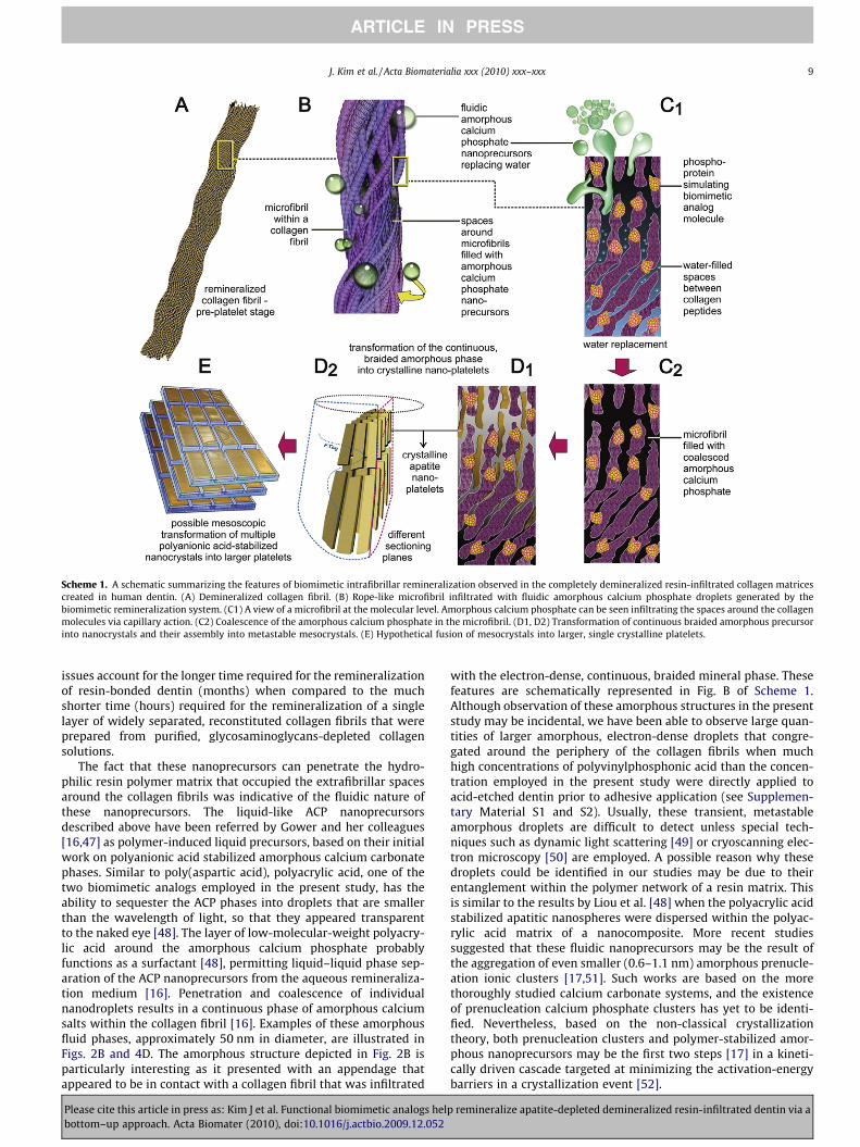
Scheme 1. A schematic summarizing the features of biomimetic intrafibrillar remineralization observed in the completely demineralized resin-infiltrated collagen matricescreated in human dentin. (A) Demineralized collagen fibril. (B) Rope-like microfibril infiltrated with fluidic amorphous calcium phosphate droplets generated by thebiomimetic remineralization system. (C1) A view of a microfibril at the molecular level. Amorphous calcium phosphate can be seen infiltrating the spaces around the collagenmolecules via capillary action. (C2) Coalescence of the amorphous calcium phosphate in the microfibril. (D1, D2) Transformation of continuous braided amorphous precursorinto nanocrystals and their assembly into metastable mesocrystals. (E) Hypothetical fusion of mesocrystals into larger, single crystalline platelets.
J. Kim et al. / Acta Biomaterialia xxx (2010) xxx–xxx 9
ARTICLE IN PRESS
issues account for the longer time required for the remineralizationof resin-bonded dentin (months) when compared to the muchshorter time (hours) required for the remineralization of a singlelayer of widely separated, reconstituted collagen fibrils that wereprepared from purified, glycosaminoglycans-depleted collagensolutions.
The fact that these nanoprecursors can penetrate the hydro-philic resin polymer matrix that occupied the extrafibrillar spacesaround the collagen fibrils was indicative of the fluidic nature ofthese nanoprecursors. The liquid-like ACP nanoprecursorsdescribed above have been referred by Gower and her colleagues[16,47] as polymer-induced liquid precursors, based on their initialwork on polyanionic acid stabilized amorphous calcium carbonatephases. Similar to poly(aspartic acid), polyacrylic acid, one of thetwo biomimetic analogs employed in the present study, has theability to sequester the ACP phases into droplets that are smallerthan the wavelength of light, so that they appeared transparentto the naked eye [48]. The layer of low-molecular-weight polyacry-lic acid around the amorphous calcium phosphate probablyfunctions as a surfactant [48], permitting liquid–liquid phase sep-aration of the ACP nanoprecursors from the aqueous remineraliza-tion medium [16]. Penetration and coalescence of individualnanodroplets results in a continuous phase of amorphous calciumsalts within the collagen fibril [16]. Examples of these amorphousfluid phases, approximately 50 nm in diameter, are illustrated inFigs. 2B and 4D. The amorphous structure depicted in Fig. 2B isparticularly interesting as it presented with an appendage thatappeared to be in contact with a collagen fibril that was infiltrated
Please cite this article in press as: Kim J et al. Functional biomimetic analogs helbottom–up approach. Acta Biomater (2010), doi:10.1016/j.actbio.2009.12.052
with the electron-dense, continuous, braided mineral phase. Thesefeatures are schematically represented in Fig. B of Scheme 1.Although observation of these amorphous structures in the presentstudy may be incidental, we have been able to observe large quan-tities of larger amorphous, electron-dense droplets that congre-gated around the periphery of the collagen fibrils when muchhigh concentrations of polyvinylphosphonic acid than the concen-tration employed in the present study were directly applied toacid-etched dentin prior to adhesive application (see Supplemen-tary Material S1 and S2). Usually, these transient, metastableamorphous droplets are difficult to detect unless special tech-niques such as dynamic light scattering [49] or cryoscanning elec-tron microscopy [50] are employed. A possible reason why thesedroplets could be identified in our studies may be due to theirentanglement within the polymer network of a resin matrix. Thisis similar to the results by Liou et al. [48] when the polyacrylic acidstabilized apatitic nanospheres were dispersed within the polyac-rylic acid matrix of a nanocomposite. More recent studiessuggested that these fluidic nanoprecursors may be the result ofthe aggregation of even smaller (0.6–1.1 nm) amorphous prenucle-ation ionic clusters [17,51]. Such works are based on the morethoroughly studied calcium carbonate systems, and the existenceof prenucleation calcium phosphate clusters has yet to be identi-fied. Nevertheless, based on the non-classical crystallizationtheory, both prenucleation clusters and polymer-stabilized amor-phous nanoprecursors may be the first two steps [17] in a kineti-cally driven cascade targeted at minimizing the activation-energybarriers in a crystallization event [52].
p remineralize apatite-depleted demineralized resin-infiltrated dentin via a
10 J. Kim et al. / Acta Biomaterialia xxx (2010) xxx–xxx
ARTICLE IN PRESS
The observation that intrafibrillar remineralization occurred inour present systems indicated that the internal environment ofthose remineralized fibrils were occupied by water instead of resin.Remineralization of the collagen fibrils would require that the freewater within the collagen fibrils and loosely bound water withinthe collagen molecules be replaced by liquid-like ACP nanoprecur-sors (Fig. C1 of Scheme 1). A recent magnetic response microscopystudy showed that there was a reduction in the mobility of watermolecules, as indicated by a reduction in the water proton trans-verse (T2) relaxation times, as water in the collagen fibrils wasreplaced by amorphous calcium phosphate precursors [53]. Thusmineralization of collagen fibrils may be perceived as a dehydra-tion process.
Template-directed transformation of the coalesced amorphousprecursor phase into polycrystalline phases [17,18] could beidentified along different parts of the same collagen fibrils inthe present study (Fig. 2D and E). This is schematically repre-sented by Figs. C2, D1 and D2 in Scheme 1. Polyvinylphosphonicacid (PVPA), the other analog present in the biomimetic reminer-alization medium, is a polyanion that mimics phosphoproteinssuch as DMP-1, phosphophoryn or bone sialoprotein [37]. Unlikephosphoprotein molecules that bind to specific sites along thecollagen molecules, PVPA is conjectured to bind nonspecificallyto the collagen molecules (Gu et al., unpublished results). Similarto natural phosphoproteins, they may act as templates that trig-ger transformation of the coalesced amorphous precursors intonanocrystalline domains inside the amorphous structures [54].Stabilized by the templating molecules, these nanocrystalline do-mains undergo subsequent growth into oriented single crystals[17]. This protein/polymer-dependent bottom–up assembly ofnanocrystalline domains into single crystals has been referred toas mesoscopic transformation [15,52]. In the present study, thissequence of events is exemplified by the conversion of the contin-uous braided mineral phase into discrete, non-overlapping nano-crystals (Fig. 3D), and ultimately into larger overlapping plateletsor needles (Fig. 2E and F), depending on the plane of sectioning(Fig. D2 of Scheme 1).
It is not possible to provide direct evidence of mesoscopic trans-formation based on electron microscopy observations alone.However, indirect evidence may be obtained by comparing theconfiguration of the continuous braided mineral phase vs. thepost-transformation overlapping platelet phase identified fromthe TEM images. The microfibril model proposed by Smith andKing et al. [55,56] considered packing of a bundle of five intertwin-ing triple helical collagen molecules into microfibrillar subunitswithin a collagen fibril. According to Holmes et al. [57], a cross-sec-tion of 100 nm diameter collagen fibril would contain approxi-mately 3900 collagen molecules. This is equivalent to 780microfibrils in a 100 nm diameter collagen fibril. Indeed, a longitu-dinal section through a collagen fibril that was infiltrated thecontinuous braided mineral phase (Fig. 4D) appeared to supportthe presence of a large number of microfibrils within the collagenfibril. However, irrespective of the plane of sectioning (Figs. 3D and4F), the number of transformed nanocrystalline phases that weresequentially produced within a 100 nm diameter fibril was lessthan 10 in number. These nanocrystalline phases, in turn, weremuch smaller in dimensions when compared to the more matureplatelets observed in Figs. 2F and 4F. The only way to account forthis reduction in the number of electron-dense strands to the num-ber of platelets seen in the mature remineralized fibrils is the ini-tial condensation of the amorphous mineral phase into metastablenanocrystalline phases, followed by their mesoscopic transforma-tion into larger crystallites. These observations appear to supportthe cascade of events proposed by the mechanism of bottom–up,template-directed, particle-based assembly of metastable nano-components into single crystalline structures [17,18]. A hypothet-
Please cite this article in press as: Kim J et al. Functional biomimetic analogs helbottom–up approach. Acta Biomater (2010), doi:10.1016/j.actbio.2009.12.052
ical depiction of this transition from nanocrystalline phase intolarger crystalline platelets is depicted in Fig. E of Scheme 1.
5. Conclusion
Biomimetic remineralization research has advanced consider-ably over the last decade. From the earlier studies that demon-strated predominantly extrafibrillar remineralization [58,59] tothe more recent studies that verified that reconstituted collagenscaffolds or individual fibrils are capable of intrafibrillar remineral-ization [45,47], the progress has been nothing short of phenome-nal. The discovery of a cascade of metastable phases such asliquid-like amorphous precursors and mesocrystals has furtheradvanced our knowledge on how biomineralization may be biomi-metically recapitulated based on the non-classical theory ofcrystallization.
Calcium- and phosphate-releasing materials have been avail-able for almost three decades for remineralizing the apatite miner-als lost due to the carious process. The majority of studiesreporting this were based on the deposition of newly formedcalcium phosphate phases over remnant seed crystallites, in com-pliance with the principles of the classical theory of crystallization.The question exists whether existing systems can remineralizethose parts of a collagen matrix that are devoid of seed crystallites.In this study, it was found that, in the absence biomimetic analogsin a remineralization medium, remineralization only occurred inpartially demineralized collagen matrices, probably by epitaxialgrowth. Conversely, in the presence of biomimetic analogs in theremineralization medium, intrafibrillar remineralization of com-pletely demineralized collagen matrices can additionally be identi-fied. The success of the current proof-of-concept, laterally diffusingremineralization protocol warrants the development of a clinicallyapplicable delivery system by incorporating these biomimetic ana-logs into the steps involved in the application of these adhesivesand filling materials.
Acknowledgements
This study was supported by Grant R21 DE019213-01 from theNational Institute of Dental and Craniofacial Research (to F.R.T.).We thank Thomas Bryan for epoxy resin embedding, Robert Smithfor TEM support and Michelle Barnes for secretarial support.
Appendix A. Figures with essential color discrimination
Certain figures in this article, particularly Scheme 1 is difficultto interpret in black and white. The full color images can be foundin the on-line version, at doi: 10.1016/j.actbio.2009.12.052.
Appendix B. Supplementary data
Supplementary data associated with this article can be found, inthe online version, at doi:10.1016/j.actbio.2009.12.052.
References
[1] Featherstone JD. The science and practice of caries prevention. J Am Dent Assoc2000;131:887–99.
[2] Linde A. Dentin matrix proteins: composition and possible functions incalcification. Anat Rec 1989;224:154–66.
[3] Goldberg M, Takagi M. Dentine proteoglycans: composition, ultrastructure andfunctions. Histochem J 1993;25:781–806.
[4] Hebling J, Pashley DH, Tjäderhane L, Tay FR. Chlorhexidine arrests subclinicaldegradation of dentin hybrid layers in vivo. J Dent Res 2005;84:741–6.
[5] Carrilho MR, Geraldeli S, Tay FR, de Goes MF, Carvalho RM, Tjäderhane L, et al.In vivo preservation of the hybrid layer by chlorhexidine. J Dent Res2007;86:529–33.
p remineralize apatite-depleted demineralized resin-infiltrated dentin via a
J. Kim et al. / Acta Biomaterialia xxx (2010) xxx–xxx 11
ARTICLE IN PRESS
[6] Brackett WW, Tay FR, Brackett MG, Dib AR, Sword J, Pashley DH. The effect ofchlorhexidine on dentin hybrid layers in vivo. Oper Dent 2007;32:107–11.
[7] Pashley DH, Tay FR, Yiu C, Hashimoto M, Breschi L, Carvalho RM, et al. Collagendegradation by host-derived enzymes during aging. J Dent Res2004;83:216–21.
[8] Trebacz H, Wójtowicz K. Thermal stabilization of collagen molecules in bonetissue. Int J Biol Macromol 2005;37:257–62.
[9] Honda Y, Kamakura S, Sasaki K, Suzuki O. Formation of bone-like apatiteenhanced by hydrolysis of octacalcium phosphate crystals deposited incollagen matrix. J Biomed Mater Res B Appl Biomater 2007;80:281–9.
[10] Boskey AL. Biomineralization: an overview. Connect Tissue Res 2003;44(Suppl.1):5–9.
[11] Goldberg HA, Warner KJ, Li MC, Hunter GK. Binding of bone sialoprotein,osteopontin and synthetic polypeptides to hydroxyapatite. Connect Tissue Res2001;42:25–37.
[12] Zhang S. Fabrication of novel biomaterials through molecular self-assembly.Nat Biotechnol 2003;21:1171–8.
[13] Tay FR, Pashley DH. Guided tissue remineralisation of partially demineralisedhuman dentine. Biomaterials 2008;29:1127–37.
[14] Gajjeraman S, Narayanan K, Hao J, Qin C, George A. Matrix macromolecules inhard tissues control the nucleation and hierarchical assembly ofhydroxyapatite. J Biol Chem 2007;282:1193–204.
[15] Xu A, Ma Y, Cölfen H. Biomimetic mineralization. J Mater Chem2007;17:415–49.
[16] Gower LB. Biomimetic model systems for investigating the amorphousprecursor pathway and its role in biomineralization. Chem Rev2008;108:4551–627.
[17] Pouget EM, Bomans PH, Goos JA, Frederik PM, de With G, Sommerdijk NA. Theinitial stages of template-controlled CaCO3 formation revealed by cryo-TEM.Science 2009;323:1455–8.
[18] Tsuji T, Onuma K, Yamamoto A, Iijima M, Shiba K. Direct transformation fromamorphous to crystalline calcium phosphate facilitated by motif-programmedartificial proteins. Proc Natl Acad Sci USA 2008;105:16866–70.
[19] Yu S. Bio-inspired crystal growth by synthetic templates. Top Curr Chem2007;271:79–118.
[20] Wong TS, Brough B, Ho CM. Creation of functional micro/nano systemsthrough top–down and bottom–up approaches. Mol Cell Biomech2009;6:1–55.
[21] Tay FR, Pashley DH. Biomimetic remineralization of resin-bonded acid-etcheddentin. J Dent Res 2009;88:719–24.
[22] Mai S, Kim YK, Toledano M, Breschi L, Ling JQ, Pashley DH, et al. Phosphoricacid esters cannot replace polyvinylphosphonic acid as phosphoproteinanalogs in biomimetic remineralization of resin-bonded dentin. Dent Mater2009;25:1230–9.
[23] Doi Y, Eanes ED. Transmission electron microscopic study of calciumphosphate formation in supersaturated solutions seeded with apatite. CalcifTissue Int 1984;36:39–47.
[24] Bertassoni LE, Habelitz S, Kinney JH, Marshall SJ, Marshall Jr GW.Biomechanical perspective on the remineralization of dentin. Caries Res2009;43:70–7.
[25] Tay FR, Pashley DH. Aggressiveness of contemporary self-etching systems. I.Depth of penetration beyond dentin smear layers. Dent Mater2001;17:296–308.
[26] Meyer JL, Eanes ED. A thermodynamic analysis of the secondary transition inthe spontaneous precipitation of calcium phosphate. Calcif Tissue Res1978;25:209–16.
[27] Tay FR, Pashley DH, Yoshiyama M. Two modes of nanoleakage expression insingle-step adhesives. J Dent Res 2002;81:472–6.
[28] Tay FR, Moulding KM, Pashley DH. Distribution of nanofillers from asimplified-step adhesive in acid-conditioned dentin. J Adhes Dent1999;1:103–17.
[29] Bozec L, van der Heijden G, Horton M. Collagen fibrils: nanoscale ropes.Biophys J 2007;92:70–5.
[30] Donnet M, Bowen P, Jongen N, Lemaître J, Hofmann H. Use of seeds to controlprecipitation of calcium carbonate and determination of seed nature.Langmuir 2005;21:100–8.
[31] Rohanizadeh R, Legeros RZ. Ultrastructural study of calculus–enamel andcalculus–root interfaces. Arch Oral Biol 2005;50:89–96.
Please cite this article in press as: Kim J et al. Functional biomimetic analogs helbottom–up approach. Acta Biomater (2010), doi:10.1016/j.actbio.2009.12.052
[32] Iijima M, Tohda H, Suzuki H, Yangisawa T, Moriwaki Y. Effects of F- on apatite–octacalcium phosphate intergrowth and crystal morphology in a modelsystem of tooth enamel formation. Cal Tissue Int 1992;50:357–61.
[33] Fan Y, Sun Z, Moradian-Oldak J. Controlled remineralization of enamel in thepresence of amelogenin and fluoride. Biomaterials 2009;30:478–83.
[34] Wang L, Nancollas GH. Pathways to biomineralization andbiodemineralization of calcium phosphates: the thermodynamic and kineticcont rols. Dalton Trans 2009;15:2665–72.
[35] Clarkson BH, Feagin FF, McCurdy SP, Sheetz JH, Speirs R. Effects ofphosphoprotein moieties on the remineralization of human root caries.Caries Res 1991;25:166–73.
[36] Saito T, Yamauchi M, Crenshaw MA. Apatite induction by insoluble dentincollagen. J Bone Miner Res 1998;13:265–70.
[37] George A, Veis A. Phosphorylated proteins and control over apatite nucleation,crystal growth, and inhibition. Chem Rev 2008;108:4670–93.
[38] Aoba T, Yoshioka C, Yagi T, Moreno EC. High-resolution electron microscopy ofhydroxyapatite grown in dilute solutions. J Dent Res 1984;63:1348–54.
[39] Nelson DG, Barry JC. High resolution electron microscopy of nonstoichiometricapatite crystals. Anat Rec 1989;224:265–76.
[40] Ellis DE, Terra J, Warschkow O, Jiang M, González GB, Okasinski JS, et al. Atheoretical and experimental study of lead substitution in calciumhydroxyapatite. Phys Chem Chem Phys 2006;8:967–76.
[41] O’Donnell MD, Fredholm Y, de Rouffignac A, Hill RG. Structural analysis of aseries of strontium-substituted apatites. Acta Biomater 2008;4:1455–64.
[42] Pizzala H, Caldarelli S, Eon JG, Rossi AM, Laurencin D, Smith ME. A solid-stateNMR study of lead and vanadium substitution into hydroxyapatite. J Am ChemSoc 2009;131:5145–52.
[43] Trus BL, Piez KA. Compressed microfibril models of the native collagen fibril.Nature 1980;286:300–1.
[44] Castellani PP, Morocutti M, Franchi M, Ruggeri A, Bigi A, Roveri N.Arrangement of microfibrils in collagen fibrils of tendons in the rat tail.Ultrastructural and X-ray diffraction investigation. Cell Tissue Res1983;234:735–43.
[45] Deshpande AS, Beniash E. Bioinspired synthesis of mineralized collagen fibrils.Crystal Growth Design 2008;8:3084–90.
[46] Mazzoni A, Pashley DH, Ruggeri Jr A, Vita F, Falconi M, Di Lenarda R, et al.Adhesion to chondroitinase ABC treated dentin. J Biomed Mater Res B ApplBiomater 2008;86:228–36.
[47] Olszta MJ et al. Bone structure and formation: a new perspective. Mat Sc Eng R2007;58:77–116.
[48] Liou SC, Chen SY, Liu DM. Manipulation of nanoneedle and nanosphere apatite/poly(acrylic acid) nanocomposites. J Biomed Mater Res B Appl Biomater2005;73:117–22.
[49] DiMasi E, Liu T, Olszta MJ, Gower LB. Laser light scattering observations ofliquid–liquid phase separation in a polymer-induced liquid-precursor (PILP)mineralization process. Mater Res Soc Symp Proc 2005;873E:K10.6.
[50] Wolf SE, Leiterer J, Kappl M, Emmerling F, Tremel W. Early homogenousamorphous precursor stages of calcium carbonate and subsequent crystalgrowth in levitated droplets. J Am Chem Soc 2008;130:12342–7.
[51] Gebauer D, Völkel A, Cölfen H. Stable prenucleation calcium carbonateclusters. Science 2008;322:1819–22.
[52] Cölfen H, Mann S. Higher-order organization by mesoscale self-assembly andtransformation of hybrid nanostructures. Angew Chem Int Ed2003;42:2350–65.
[53] Chesnick IE, Mason JT, Giuseppetti AA, Eidelman N, Potter K. Magneticresonance microscopy of collagen mineralization. Biophys J 2008;95:2017–26.
[54] Zhang TH, Liu XY. How does a transient amorphous precursor templatecrystallization? J Am Chem Soc 2007;129:13520–6.
[55] Smith JW. Molecular pattern in native collagen. Nature 1968;219:157–8.[56] King G, Brown EM, Chen JM. Computer model of a bovine type I collagen
microfibril. Protein Eng 1996;9:43–9.[57] Holmes DF, Graham HK, Trotter JA, Kadler KE. STEM/TEM studies of collagen
fibril assembly. Micron 2001;32:273–85.[58] Bradt J-H, Mertig M, Teresiak A, Pompe W. Biomimetic mineralization of
collagen by combined fibril assembly and calcium phosphate formation. ChemMater 1999;11:2694–701.
[59] Zhang W, Liao SS, Cui FZ. Hierarchical self-assembly of nano-fibrils inmineralized collagen. Chem Mater 2003;15:3221–6.
p remineralize apatite-depleted demineralized resin-infiltrated dentin via a