Embed Size (px)
Citation preview
2014/2015
Pedro Ribeiro Queirós
Full coding sequence mutation screening
of KRAS gene in gastric cancer
março, 2015
Mestrado Integrado em Medicina
Área: Patologia e Oncologia
Tipologia: Dissertação
Trabalho efetuado sob a Orientação de:
Doutora Carla Isabel Gonçalves de Oliveira
E sob a Coorientação de:
Doutora Gabriela Martinho Almeida
Trabalho organizado de acordo com as normas da revista:
Virchows Archiv
Pedro Ribeiro Queirós
Full coding sequence mutation screening
of KRAS gene in gastric cancer
março, 2015
1
KRAS MUTATIONS IN MICROSATELLITE INSTABLE GASTRIC TUMOURS: IMPACT OF TARGETED
TREATMENT AND INTRATUMOURAL HETEROGENEITY
Pedro Queirós1,2,3, Hugo Pinheiro1,2, Joana Carvalho1,2, Patrícia Oliveira1,2, Irene Gullo4, Fátima Carneiro1,2,3,4,
Gabriela M Almeida1,2, Carla Oliveira1,2,3,*
1Instituto de Investigação e Inovação em Saúde, Universidade do Porto, Porto, Portugal;
2IPATIMUP, Institute of Molecular Pathology and Immunology of the University of Porto, Porto, Portugal;
3Faculty of Medicine, University of Porto, Porto, Portugal.
4Department of Pathology, Hospital de São João, Porto, Portugal;
*Corresponding author:
Carla Oliveira, PhD; E-mail: [email protected]; Phone: +351225570700; Fax: +351225570799

2
Abstract: In gastric cancer (GC), epidermal growth factor receptor (EGFR) overexpression associates with poor
prognosis. Addition of a chimeric monoclonal antibody against EGFR (cetuximab) to first-line treatment of
metastatic colorectal tumours improved outcomes of patients (stratified for KRAS wild-type cancers), whereas
GC patients did not benefit from this approach. In GC, however, stratification based on KRAS mutations was
not performed, and the 30% KRAS mutation frequency in Microsatellite instable cancers (MSI), which represents
~4% of total GC was disregarded. Further, intratumoural heterogeneity regarding KRAS mutant subpopulations
might also contribute to anti-EGFR therapy failure. We assessed the mutational status of the KRAS entire coding
sequence in 19 MSI-GC cases by multiplex PCR/Sequencing, and used peak height ratio determined from
electropherograms from KRAS heterozygous mutants and histopathological evaluation to infer tumour
heterogeneity in GC. Using 2 multiplex reactions per sample we found that 26% (5/19) of MSI-GC cases
harboured KRAS mutations (2 G12D, 2 G13D, 1 G12V). No mutations were found outside the codon 12 and 13
hotspots. Our analysis supported the co-existence of KRAS-positive and -negative tumour populations in 4/5
MSI-GC cases. In conclusion, the method developed stands as a cost-effective and practical way for mutation
screening of the entire KRAS coding sequence. KRAS mutations are frequent in our series of MSI cases, and
are often found in a subpopulation of the tumour and not in the whole tumour. Further studies are needed to
access the implications of this heterogeneity in KRAS mutant and wild-type tumour clones in anti-EGFR therapy
response.
Keywords (4-6): KRAS, microsatellite instability, MSI, gastric cancer, gastric cancer treatment
3
Introduction
In 2012, gastric cancer’s (GC) incidence was reported to be of 952,000 cases (representing 6.8% of
total), with a mortality of 723,000 patients (8.8% of total) worldwide. Even though over 70% of cases occur in
developing countries, it ranks sixth in developed countries’ numbers of both incidence and mortality [1]. Both
incidence and mortality have been declining appreciably over the last decades, but they remain high mainly due
to advanced stage of disease at presentation, lack of understanding of the complex biology, and morphologic
and molecular heterogeneity.
The progress in GC therapy has been limited. Currently, surgery is the only method capable of curing
patients and remains the first choice for early-stage disease, although endoscopic resection is being
increasingly used in selected early-stage tumours [2]. Adjuvant chemo and radiotherapy may result in improved
survival for some of these early-stage patients, but neither the methods nor the outcomes of this approach have
been consensual in previous reports [2]. As for advanced-stage GC, chemotherapy should be considered for
all patients, given its proven benefits in survival and quality of life, with patients with HER2+ tumours also
meriting targeted therapy such as trastuzumab [3]. However, the prognosis remains very poor for patients with
GC, with 5-year survival rates of only 30% in Western countries [3]. Thus, the need to better comprehend the
molecular behaviour of the disease is urgent, and the current paradigm begets the need of a more appropriate
and individualized therapy [2,4].
It has been accepted for many years that there are at least two mutually exclusive molecular types of
GC: the chromosomal instability (CIN) type, characterized by structural alterations, namely large deletions,
amplifications and translocations; and the microsatellite instability (MSI) type, with instability at the nucleotide
level. The MSI type is characterized by alterations in the length of nucleotide repeat sequences, and marks an
underlying mismatch repair (MMR) deficiency, meaning that cells fail to recognize errors introduced during DNA
replication, which are prone to occur at those nucleotide repeat sequences (microsatellites) [5].
MSI is observed in approximately 15-20% of sporadic GC and also in sporadic colorectal cancer (CRC)
with a similar frequency, appearing to be associated with a more favourable prognosis in both cancer types
[6,7]. MSI tumours from both colon and stomach are associated with specific profiles of target gene mutations
that include insertions/deletions at microsatellite stretches in coding and noncoding sequences, but also with
hotspot missense mutations at the KRAS (stomach and colon) and BRAF genes (colon) [8,6,9]. KRAS, a
molecule of the MAP kinase (MAPK) pathway and a target gene of the MSI molecular phenotype, was reported
to have no difference in mutation frequency between MSI and non-MSI (i.e. microsatellite stable (MSS)) CRC
cases [10], or to have lower frequency in MSI as compared with MSS tumours [11,9,12]. Interestingly, in GC,
KRAS mutations are generally restricted to the MSI group of cancers, and have been described in nearly 30%
of these cases using specific analysis of KRAS exon 2, affecting the hotspot codons 12 and 13 of the gene
[9,13]. Aside from these exon 2 hotspots, KRAS mutation frequency seems to be lacklustre. Although in GC,
the information on KRAS mutations outside these hotspots is scarce, 6.5% of a CRC series of 276 cases
demonstrated KRAS mutations in other codons of the gene [14].
In CRC, the addition of cetuximab (a chimeric monoclonal antibody against the epidermal growth factor
receptor (EGFR)) to irinotecan, fluorouracil, and leucovorin, as first-line treatment for metastatic tumours, was
shown to reduce the risk of disease progression, and to increase the chance of response in patients with KRAS
4
wild-type (WT) disease [15]. EGFR positivity, determined by immunohistochemistry, is however regarded as a
non-relevant predictive marker of response to this targeted therapy [16]. Indeed, anti-EGFR therapy is reserved
for RAS WT tumours, since CRC bearing mutations of either KRAS or NRAS are irresponsive to this treatment
[16]. These are gain-of-function mutations that activate the EGFR signalling pathway at a downstream location,
rendering the antibodies against this molecule useless [17]. Since this targeted therapy is currently used in CRC
patients, predictive testing for RAS mutational status is needed, generally using a tumour biopsy sample [17].
However, these specimens are often single, and thus represent only a small region of the tumour, limiting the
possibility of detecting positive RAS-clones at different sites of the tumour [17]; nonetheless, RAS mutational
screening is definitive to guide anti-EGFR treatment in CRC patients [16].
In GC, EGFR overexpression occurs and is associated with poor prognosis [18]; this finding, together
with the observed improved outcomes regarding anti-EGFR therapy in KRAS WT metastatic CRC [15], recurrent
or metastatic squamous-cell cancer of the head and neck and advanced non-small-cell lung carcinoma,
supported the rationale to perform the EXPAND trial in GC [19]. Cetuximab was added to first-line chemotherapy
treatment in advanced non-resectable or metastatic GC, but failed to produce improved outcomes. Also, a
subpopulation analysis in this trial revealed no association between EGFR positivity (determined by
immunohistochemistry) and overall survival in patients treated either with or without cetuximab, suggesting a
non-predictive role for EGFR positivity in GC regarding anti-EGFR therapy, similarly to what occurs in CRC.
However, the EXPAND trial failed to account for the presence of KRAS mutations in at least 4-10% of GC overall
[9,13,20]. Taking in consideration the KRAS mutation-mediated anti-EGFR resistance in CRC, this fact could
explain, to a certain extent, the failure of the EXPAND trial. Therefore, in case anti-EGFR therapy trials are
reconsidered in GC, it would be important to use MSI-positivity (which represents roughly 15-20% of all cases),
and its accompanying frequency of KRAS mutation in hotspot codons 12 and 13, as part of the GC treatment
stratification plan. Also, if a more thorough stratification is to be performed, rigorously checking for mutations,
both inside and outside the KRAS hotspot regions of the gene, could also prove worthy.
Another important issue in therapy response is intratumoural molecular heterogeneity, a feature of
malignant neoplasias that experienced competition between different tumour cells (or clones) for survival,
leading to the evolutionary selection of different cell kindred and their progeny [21-23]. A study on metastatic
CRC, using a highly sensitive picodroplet digital PCR (dPCR), found that some KRAS WT cases actually
harboured KRAS mutations. Moreover, the fraction of KRAS-mutated allele, quantified by dPCR, was inversely
correlated with anti-EGFR therapy response rate [24]. These authors hypothesised that while the majority of
KRAS WT tumour cells in these cases would respond to anti-EGFR therapy, tumour subclones bearing KRAS
mutations would not. These observations could explain why nearly 50% of CRC cases considered KRAS WT,
and regarded as responders to anti-EGFR therapy, do not benefit from this approach [24]. This same fact might
also contribute to explain the failure of the addition of anti-EGFR therapy to first-line chemotherapy in producing
improved outcomes in GC.
With this work, we aim to assess the mutational status of the entire coding sequence of KRAS in MSI
GC cases, and use peak height ratio determined from electropherograms from KRAS heterozygous mutants
and histopathological evaluation to infer the tumour heterogeneity in GC cases. To achieve these aims, we
developed a multiplex PCR reaction for practical and cost-effective mutation screening of the whole KRAS
coding sequence; we determined the frequency and type of KRAS coding sequence mutations in a series of 19
5
MSI-positive GC cases; and we inferred tumour heterogeneity in mutated cases by comparing KRAS WT and
mutated allelic fractions, as determined by electropherogram analysis, with the percentage of tumour cells in
matched histological slides.
Materials and Methods
DNA Samples and Extraction
DNA from AGS, IPA220 and MKN28 gastric cancer cell lines was used for optimization of the multiplex
PCR. The DNA was extracted from in vitro cultured cells using Spin Tissue Mini Kit (Invisiorb®) according to
manufacturer’s instructions. DNA quantification and purity were determined using a spectrophotometer
(Nanodrop®), and aliquots were created by diluting the DNA of each cell line as to create a working solution of
50ng/µl.
Primary gastric cancer DNA samples were obtained from 19 cases of the Tumour Bank from Centro
Hospitalar São João and IPATIMUP. Informed consent was obtained from all patients, and the study was
approved by the Hospital's Ethics Committee. These tumour samples had been previously characterized for the
presence of MSI phenotype using Pentaplex Kit (Roche®). The DNA content of each sample was determined
using the method described above, and aliquots were created in the same manner.
Polymerase Chain Reaction & Primers
Primers were designed in intronic regions flanking KRAS exons, and were produced by SIGMA® (Table
1). Working solutions for each primer at a concentration of 100 µM were created. QIAGEN® Multiplex PCR Kit
was used according to the manufacturer’s instructions. Briefly, the Polymerase Chain Reaction (PCR) was
conducted using the following ingredients for each sample: 12.5 µl of QIAGEN® Multiplex Master Mix, 1 µl of
primer mix (composed of reverse and forward primers for the corresponding exons in equal proportions), 5 µl
of QIAGEN® Q-solution, 1 µl of DNA at working solution and 5.5 µl of DNAse and RNAse-free water. The
following cycling conditions were used for PCR: 15 min of 95 ºC for enzyme activation, followed by 30 sec at 94
ºC, 90 sec at 62 ºC and 90 sec at 72 ºC for 3 cycles, 30 sec at 95 ºC, 90 sec at 60 ºC and 90 sec at 72 ºC for 3
cycles, 30 sec at 95 ºC, 90 sec at 58 ºC and 90 sec at 72 ºC for 24 cycles and an extension phase of 72 ºC for
the final 10 min.
Sequencing Reaction
After the multiplex PCR, product clean-up was achieved by mixing 10 µl of each multiplex product with
1 µl of Exonuclease I (ExoI) and 2 µl of Thermosensitive Alkaline Phosphatase (FastAP™), and exposing the
solution to 37 ºC for 15 min and 85 ºC for 15 min. This was performed separately for each sample. Each exon
was sequenced independently by using the corresponding primer (shown in bold in Table 1). For each
sequencing reaction 2 µl of cleaned-up DNA were mixed with 0.5 µl of sequencing buffer, 0.4 µl of BigDye®
terminator (both of Applied Biosystems®), 0.4 µl of primer (at working solution) and 1.7 µl of DNAse and RNAse-
free water. The cycling conditions used for the reaction were the following: 96 ºC for 2 min, followed by 30 cycles
of 30 sec at 96 ºC, 15 sec at 54 ºC and 150 sec at 60 ºC; the final extension was performed at 60 ºC for 10 min.
6
Post-sequencing purification was performed by cross-link dextran gel filtration. A tube for each exon
from all the samples was centrifuged at 2000 x g for 4 min with a Sephadex® solution at a concentration of 0.66
mg/ml. The sequencing product was then purified using 5 µl of it under the same centrifugation conditions. The
purified product was analysed using an automated sequencer.
Histological & Electropherogram Analysis
Frozen tumour fragments, deposited at the Tumour Bank from Centro Hospitalar São João and
IPATIMUP, used for DNA sampling were matched with fragments that were paraffin embedded for
histopathological analysis. Haematoxylin and eosin (H&E) stained slides from KRAS mutant GC cases were
examined by a pathologist from the Department of Pathology of Centro Hospitalar São João (Fátima Carneiro),
who determined the approximate percentage of tumour cells present in each slide. The minimal percentage of
tumour cells in each case was registered. Then, the (lowest) expected mutant-to-WT allelic ratio was
determined, that is, the ratio when considering that the whole tumour cell population is mutated for KRAS, and
that all tumour cells are heterozygous for the mutation (meaning that each cell bears one WT and one mutated
allele). The following formula was used (in percentage):
𝑃2⁄
𝑃2⁄ + (100 − 𝑃)
× 100
Where 𝑃 represents the approximate percentage of tumour cells in the slide. With the assumptions
made above, 𝑃 2⁄ should represent both the mutated and WT allelic fraction of the tumour cell population, while
(100 − 𝑃) should represent the WT allelic fraction of non-tumoural cells in the slide.
For each sample submitted to the automated sequencer, an electropherogram was obtained. Peaks on
the electropherogram were manually analysed for alterations in the WT sequence of each exon, and altered
cases were classified as mutated. For the mutated cases, the peak height for each nucleotide variant was
determined using Photoshop® (CC version, Adobe), and the observed mutant-to-WT allelic ratio (in percentage)
was determined by the following formula:
(𝐻1𝐻2
) × 100
Where H1 represents the height of the peak for the mutated nucleotide, and H2 represents the height of
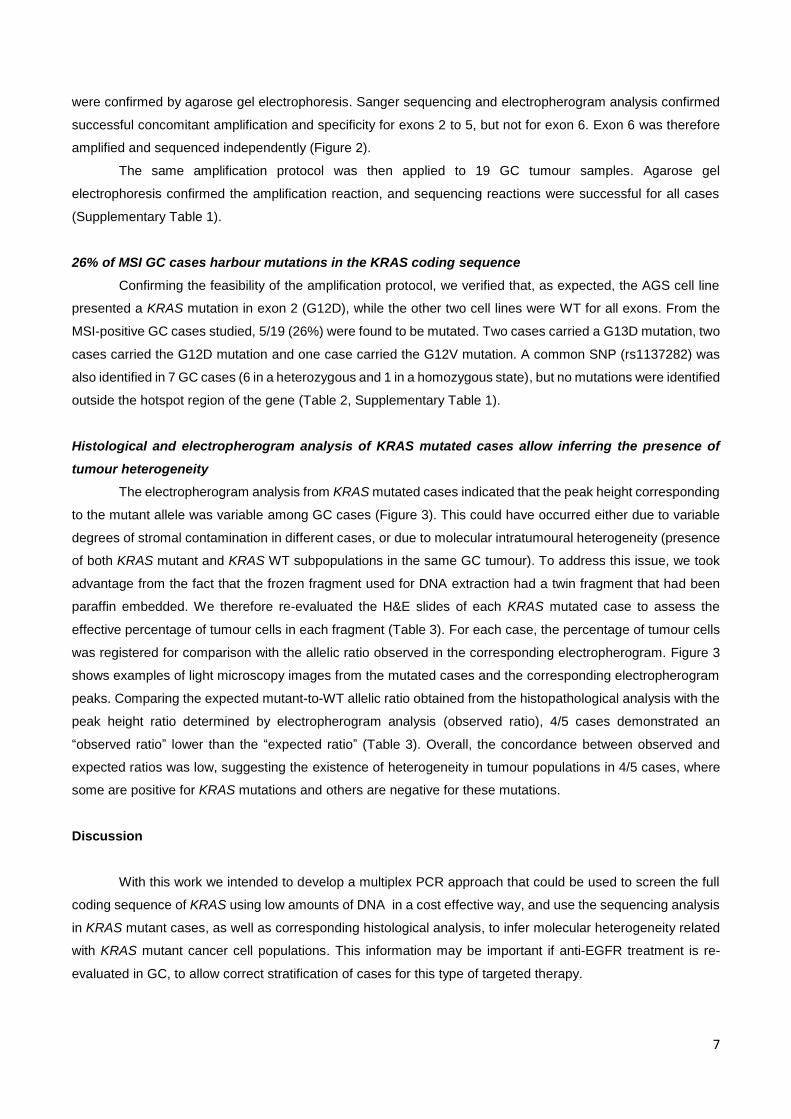
the peak for the WT nucleotide. This followed the strategy described by Jiang et al [25] (Figure 1a). The
electropherogram from a common KRAS single nucleotide polymorphism (SNP) rs1137282 found in 6/19 GC
cases studied was used as a control to determine the expected peak height ratio of the nucleotides (representing
each allele) from a constitutional SNP (Figure 1b). The allelic ratios obtained for this SNP in three different GC
cases were 89%, 120% and 100%. These were therefore confirmed to represent constitutional heterozygous
states.
Results
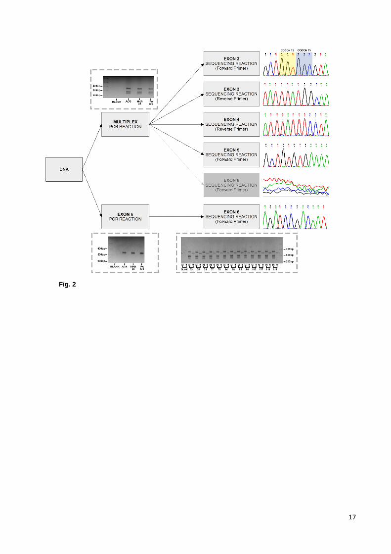
Full coding sequence mutation screening of KRAS with two PCR reactions
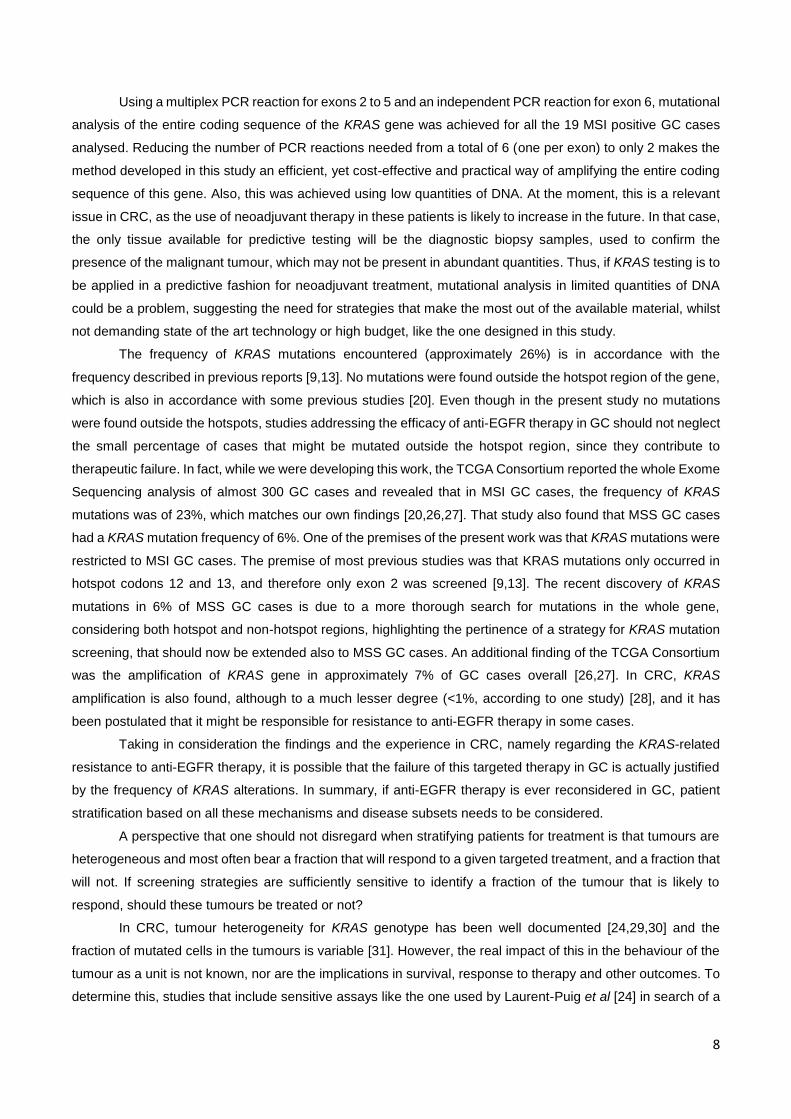
We started by the optimization of a multiplex PCR approach to amplify the full coding sequence of
KRAS, from exon 2 to exon 6, using three GC cell lines. Following a PCR reaction with 5 primer-pairs, products
7
were confirmed by agarose gel electrophoresis. Sanger sequencing and electropherogram analysis confirmed
successful concomitant amplification and specificity for exons 2 to 5, but not for exon 6. Exon 6 was therefore
amplified and sequenced independently (Figure 2).
The same amplification protocol was then applied to 19 GC tumour samples. Agarose gel
electrophoresis confirmed the amplification reaction, and sequencing reactions were successful for all cases
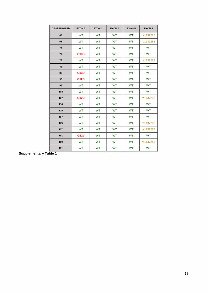
(Supplementary Table 1).
26% of MSI GC cases harbour mutations in the KRAS coding sequence
Confirming the feasibility of the amplification protocol, we verified that, as expected, the AGS cell line
presented a KRAS mutation in exon 2 (G12D), while the other two cell lines were WT for all exons. From the
MSI-positive GC cases studied, 5/19 (26%) were found to be mutated. Two cases carried a G13D mutation, two
cases carried the G12D mutation and one case carried the G12V mutation. A common SNP (rs1137282) was
also identified in 7 GC cases (6 in a heterozygous and 1 in a homozygous state), but no mutations were identified
outside the hotspot region of the gene (Table 2, Supplementary Table 1).
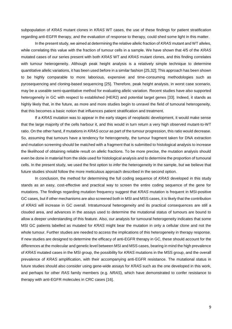
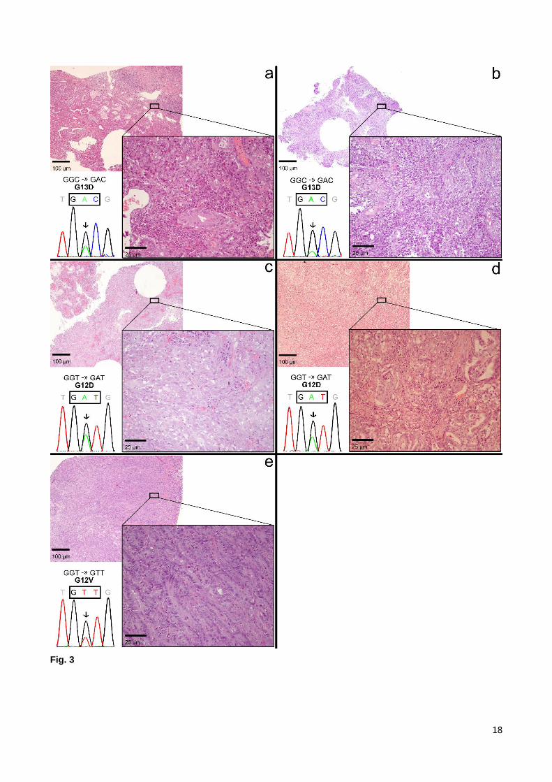
Histological and electropherogram analysis of KRAS mutated cases allow inferring the presence of
tumour heterogeneity
The electropherogram analysis from KRAS mutated cases indicated that the peak height corresponding
to the mutant allele was variable among GC cases (Figure 3). This could have occurred either due to variable
degrees of stromal contamination in different cases, or due to molecular intratumoural heterogeneity (presence
of both KRAS mutant and KRAS WT subpopulations in the same GC tumour). To address this issue, we took
advantage from the fact that the frozen fragment used for DNA extraction had a twin fragment that had been
paraffin embedded. We therefore re-evaluated the H&E slides of each KRAS mutated case to assess the
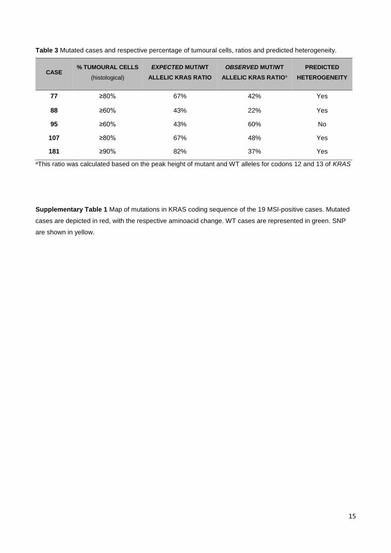
effective percentage of tumour cells in each fragment (Table 3). For each case, the percentage of tumour cells
was registered for comparison with the allelic ratio observed in the corresponding electropherogram. Figure 3
shows examples of light microscopy images from the mutated cases and the corresponding electropherogram
peaks. Comparing the expected mutant-to-WT allelic ratio obtained from the histopathological analysis with the
peak height ratio determined by electropherogram analysis (observed ratio), 4/5 cases demonstrated an
“observed ratio” lower than the “expected ratio” (Table 3). Overall, the concordance between observed and
expected ratios was low, suggesting the existence of heterogeneity in tumour populations in 4/5 cases, where
some are positive for KRAS mutations and others are negative for these mutations.
Discussion
With this work we intended to develop a multiplex PCR approach that could be used to screen the full
coding sequence of KRAS using low amounts of DNA in a cost effective way, and use the sequencing analysis
in KRAS mutant cases, as well as corresponding histological analysis, to infer molecular heterogeneity related
with KRAS mutant cancer cell populations. This information may be important if anti-EGFR treatment is re-
evaluated in GC, to allow correct stratification of cases for this type of targeted therapy.
8
Using a multiplex PCR reaction for exons 2 to 5 and an independent PCR reaction for exon 6, mutational
analysis of the entire coding sequence of the KRAS gene was achieved for all the 19 MSI positive GC cases
analysed. Reducing the number of PCR reactions needed from a total of 6 (one per exon) to only 2 makes the
method developed in this study an efficient, yet cost-effective and practical way of amplifying the entire coding
sequence of this gene. Also, this was achieved using low quantities of DNA. At the moment, this is a relevant
issue in CRC, as the use of neoadjuvant therapy in these patients is likely to increase in the future. In that case,
the only tissue available for predictive testing will be the diagnostic biopsy samples, used to confirm the
presence of the malignant tumour, which may not be present in abundant quantities. Thus, if KRAS testing is to
be applied in a predictive fashion for neoadjuvant treatment, mutational analysis in limited quantities of DNA
could be a problem, suggesting the need for strategies that make the most out of the available material, whilst
not demanding state of the art technology or high budget, like the one designed in this study.
The frequency of KRAS mutations encountered (approximately 26%) is in accordance with the
frequency described in previous reports [9,13]. No mutations were found outside the hotspot region of the gene,
which is also in accordance with some previous studies [20]. Even though in the present study no mutations
were found outside the hotspots, studies addressing the efficacy of anti-EGFR therapy in GC should not neglect
the small percentage of cases that might be mutated outside the hotspot region, since they contribute to
therapeutic failure. In fact, while we were developing this work, the TCGA Consortium reported the whole Exome
Sequencing analysis of almost 300 GC cases and revealed that in MSI GC cases, the frequency of KRAS
mutations was of 23%, which matches our own findings [20,26,27]. That study also found that MSS GC cases
had a KRAS mutation frequency of 6%. One of the premises of the present work was that KRAS mutations were
restricted to MSI GC cases. The premise of most previous studies was that KRAS mutations only occurred in
hotspot codons 12 and 13, and therefore only exon 2 was screened [9,13]. The recent discovery of KRAS
mutations in 6% of MSS GC cases is due to a more thorough search for mutations in the whole gene,
considering both hotspot and non-hotspot regions, highlighting the pertinence of a strategy for KRAS mutation
screening, that should now be extended also to MSS GC cases. An additional finding of the TCGA Consortium
was the amplification of KRAS gene in approximately 7% of GC cases overall [26,27]. In CRC, KRAS
amplification is also found, although to a much lesser degree (<1%, according to one study) [28], and it has
been postulated that it might be responsible for resistance to anti-EGFR therapy in some cases.
Taking in consideration the findings and the experience in CRC, namely regarding the KRAS-related
resistance to anti-EGFR therapy, it is possible that the failure of this targeted therapy in GC is actually justified
by the frequency of KRAS alterations. In summary, if anti-EGFR therapy is ever reconsidered in GC, patient
stratification based on all these mechanisms and disease subsets needs to be considered.
A perspective that one should not disregard when stratifying patients for treatment is that tumours are
heterogeneous and most often bear a fraction that will respond to a given targeted treatment, and a fraction that
will not. If screening strategies are sufficiently sensitive to identify a fraction of the tumour that is likely to
respond, should these tumours be treated or not?
In CRC, tumour heterogeneity for KRAS genotype has been well documented [24,29,30] and the
fraction of mutated cells in the tumours is variable [31]. However, the real impact of this in the behaviour of the
tumour as a unit is not known, nor are the implications in survival, response to therapy and other outcomes. To
determine this, studies that include sensitive assays like the one used by Laurent-Puig et al [24] in search of a
9
subpopulation of KRAS mutant clones in KRAS WT cases, the use of these findings for patient stratification
regarding anti-EGFR therapy, and the evaluation of response to therapy, could shed some light in this matter.
In the present study, we aimed at determining the relative allelic fraction of KRAS mutant and WT alleles,
while correlating this value with the fraction of tumour cells in a sample. We have shown that 4/5 of the KRAS
mutated cases of our series present with both KRAS WT and KRAS mutant clones, and this finding correlates
with tumour heterogeneity. Although peak height analysis is a relatively simple technique to determine
quantitative allelic variations, it has been used before in a similar fashion [25,32]; This approach has been shown
to be highly comparable to more laborious, expensive and time-consuming methodologies such as
pyrosequencing and cloning-based sequencing [25]. Therefore, peak height analysis, in worst case scenario,
may be a useable semi-quantitative method for evaluating allelic variation. Recent studies have also supported
heterogeneity in GC with respect to established (HER2) and potential target genes [33]. Indeed, it stands as
highly likely that, in the future, as more and more studies begin to unravel the field of tumoural heterogeneity,
that this becomes a basic notion that influences patient stratification and treatment.
If a KRAS mutation was to appear in the early stages of neoplastic development, it would make sense
that the large majority of the cells harbour it, and this would in turn return a very high observed mutant-to-WT
ratio. On the other hand, if mutations in KRAS occur as part of the tumour progression, this ratio would decrease.
So, assuming that tumours have a tendency for heterogeneity, the tumour fragment taken for DNA extraction
and mutation screening should be matched with a fragment that is submitted to histological analysis to increase
the likelihood of obtaining reliable result on allelic fractions. To be more precise, the mutation analysis should
even be done in material from the slide used for histological analysis and to determine the proportion of tumoural
cells. In the present study, we used the first option to infer the heterogeneity in the sample, but we believe that
future studies should follow the more meticulous approach described in the second option.
In conclusion, the method for determining the full coding sequence of KRAS developed in this study
stands as an easy, cost-effective and practical way to screen the entire coding sequence of the gene for
mutations. The findings regarding mutation frequency suggest that KRAS mutation is frequent in MSI-positive
GC cases, but if other mechanisms are also screened both in MSI and MSS cases, it is likely that the contribution
of KRAS will increase in GC overall. Intratumoural heterogeneity and its practical consequences are still a
clouded area, and advances in the assays used to determine the mutational status of tumours are bound to
allow a deeper understanding of this feature. Also, our analysis for tumoural heterogeneity indicates that some
MSI GC patients labelled as mutated for KRAS might bear the mutation in only a cellular clone and not the
whole tumour. Further studies are needed to access the implications of this heterogeneity in therapy response.
If new studies are designed to determine the efficacy of anti-EGFR therapy in GC, these should account for the
differences at the molecular and genetic level between MSI and MSS cases, bearing in mind the high prevalence
of KRAS mutated cases in the MSI group, the possibility for KRAS mutations in the MSS group, and the overall
prevalence of KRAS amplification, with their accompanying anti-EGFR resistance. The mutational status in
future studies should also consider using gene-wide assays for KRAS such as the one developed in this work,
and perhaps for other RAS family members (e.g. NRAS), which have demonstrated to confer resistance to
therapy with anti-EGFR molecules in CRC cases [16].
10
Acknowledgments
IPATIMUP integrates the i3S Research Unit, which is partially supported by FCT, the Portuguese
Foundation for Science and Technology. This work is funded by: 1) FEDER funds through the Operational
Programme for Competitiveness Factors - COMPETE and National Funds through the FCT - Foundation for
Science and Technology, under the projects "PEst-C/SAU/LA0003/2013”; 2) NORTE-07-0162-FEDER-00018 -
Contributos para o reforço da capacidade do IPATIMUP enquanto actor do sistema regional de inovação" and
NORTE-07-0162-FEDER-000067 - Reforço e consolidação da capacidade infraestrutural do IPATIMUP para o
sistema regional de inovação", both supported by Programa Operacional Regional do Norte (ON.2 – O Novo
Norte), through FEDER funds under the Quadro de Referência Estratégico Nacional (QREN); 3) Coimbra
Genomics and BGI in the frame of the project “NGSGC - Using NGS to uncover structural and regulatory
variation in gastric cancer”; 4) FCT Fellowships (SFRH/BPD/89764/2012 to PO; SFRH/BPD/86543/2012 to JC;
SFRH/BPD/79499/2011 to HP) and Salary support to GMA from iFCT Program 2013, POPH - QREN Type 4.2,
European Social Fund and Portuguese Ministry of Science and Technology (MCTES).
Conflict of Interest
The authors declare that they have no conflict of interest.
References
1. Torre LA, Bray F, Siegel RL, Ferlay J, Lortet-Tieulent J, Jemal A (2015) Global cancer statistics, 2012. CA: a cancer journal for clinicians. doi:10.3322/caac.21262 2. Lordick F, Allum W, Carneiro F, Mitry E, Tabernero J, Tan P, Van Cutsem E, van de Velde C, Cervantes A (2014) Unmet needs and challenges in gastric cancer: the way forward. Cancer treatment reviews 40 (6):692-700. doi:10.1016/j.ctrv.2014.03.002 3. Rivera F, Gravalos C, Garcia-Carbonero R, Seom (2012) SEOM clinical guidelines for the diagnosis and treatment of gastric adenocarcinoma. Clinical & translational oncology : official publication of the Federation of Spanish Oncology Societies and of the National Cancer Institute of Mexico 14 (7):528-535. doi:10.1007/s12094-012-0836-9 4. Ferro A, Peleteiro B, Malvezzi M, Bosetti C, Bertuccio P, Levi F, Negri E, La Vecchia C, Lunet N (2014) Worldwide trends in gastric cancer mortality (1980-2011), with predictions to 2015, and incidence by subtype. European journal of cancer 50 (7):1330-1344. doi:10.1016/j.ejca.2014.01.029 5. Suraweera N, Duval A, Reperant M, Vaury C, Furlan D, Leroy K, Seruca R, Iacopetta B, Hamelin R (2002) Evaluation of tumor microsatellite instability using five quasimonomorphic mononucleotide repeats and pentaplex PCR. Gastroenterology 123 (6):1804-1811. doi:10.1053/gast.2002.37070 6. Boland CR, Thibodeau SN, Hamilton SR, Sidransky D, Eshleman JR, Burt RW, Meltzer SJ, Rodriguez-Bigas MA, Fodde R, Ranzani GN, Srivastava S (1998) A National Cancer Institute Workshop on Microsatellite Instability for cancer detection and familial predisposition: development of international criteria for the determination of microsatellite instability in colorectal cancer. Cancer research 58 (22):5248-5257 7. Oliveira C, Seruca R, Seixas M, Sobrinho-Simoes M (1998) The clinicopathological features of gastric carcinomas with microsatellite instability may be mediated by mutations of different "target genes": a study of the TGFbeta RII, IGFII R, and BAX genes. The American journal of pathology 153 (4):1211-1219
11
8. Duval A, Reperant M, Compoint A, Seruca R, Ranzani GN, Iacopetta B, Hamelin R (2002) Target gene mutation profile differs between gastrointestinal and endometrial tumors with mismatch repair deficiency. Cancer research 62 (6):1609-1612 9. Oliveira C, Pinto M, Duval A, Brennetot C, Domingo E, Espin E, Armengol M, Yamamoto H, Hamelin R, Seruca R, Schwartz S, Jr. (2003) BRAF mutations characterize colon but not gastric cancer with mismatch repair deficiency. Oncogene 22 (57):9192-9196. doi:10.1038/sj.onc.1207061 10. Olschwang S, Hamelin R, Laurent-Puig P, Thuille B, De Rycke Y, Li YJ, Muzeau F, Girodet J, Salmon RJ, Thomas G (1997) Alternative genetic pathways in colorectal carcinogenesis. Proceedings of the National Academy of Sciences of the United States of America 94 (22):12122-12127 11. Samowitz WS, Holden JA, Curtin K, Edwards SL, Walker AR, Lin HA, Robertson MA, Nichols MF, Gruenthal KM, Lynch BJ, Leppert MF, Slattery ML (2001) Inverse relationship between microsatellite instability and K-ras and p53 gene alterations in colon cancer. The American journal of pathology 158 (4):1517-1524. doi:10.1016/S0002-9440(10)64102-8 12. Velho S, Moutinho C, Cirnes L, Albuquerque C, Hamelin R, Schmitt F, Carneiro F, Oliveira C, Seruca R (2008) BRAF, KRAS and PIK3CA mutations in colorectal serrated polyps and cancer: primary or secondary genetic events in colorectal carcinogenesis? BMC cancer 8:255. doi:10.1186/1471-2407-8-255 13. Brennetot C, Duval A, Hamelin R, Pinto M, Oliveira C, Seruca R, Schwartz S, Jr. (2003) Frequent Ki-ras mutations in gastric tumors of the MSI phenotype. Gastroenterology 125 (4):1282 14. Cancer Genome Atlas N (2012) Comprehensive molecular characterization of human colon and rectal cancer. Nature 487 (7407):330-337. doi:10.1038/nature11252 15. Van Cutsem E, Kohne CH, Lang I, Folprecht G, Nowacki MP, Cascinu S, Shchepotin I, Maurel J, Cunningham D, Tejpar S, Schlichting M, Zubel A, Celik I, Rougier P, Ciardiello F (2011) Cetuximab plus irinotecan, fluorouracil, and leucovorin as first-line treatment for metastatic colorectal cancer: updated analysis of overall survival according to tumor KRAS and BRAF mutation status. Journal of clinical oncology : official journal of the American Society of Clinical Oncology 29 (15):2011-2019. doi:10.1200/JCO.2010.33.5091 16. Van Cutsem E, Cervantes A, Nordlinger B, Arnold D, Group EGW (2014) Metastatic colorectal cancer: ESMO Clinical Practice Guidelines for diagnosis, treatment and follow-up. Annals of oncology : official journal of the European Society for Medical Oncology / ESMO 25 Suppl 3:iii1-9. doi:10.1093/annonc/mdu260 17. Fadhil W, Ibrahem S, Seth R, AbuAli G, Ragunath K, Kaye P, Ilyas M (2012) The utility of diagnostic biopsy specimens for predictive molecular testing in colorectal cancer. Histopathology 61 (6):1117-1124. doi:10.1111/j.1365-2559.2012.04321.x 18. Nicholson RI, Gee JM, Harper ME (2001) EGFR and cancer prognosis. European journal of cancer 37 Suppl 4:S9-15 19. Lordick F, Kang YK, Chung HC, Salman P, Oh SC, Bodoky G, Kurteva G, Volovat C, Moiseyenko VM, Gorbunova V, Park JO, Sawaki A, Celik I, Gotte H, Melezinkova H, Moehler M, Arbeitsgemeinschaft Internistische O, Investigators E (2013) Capecitabine and cisplatin with or without cetuximab for patients with previously untreated advanced gastric cancer (EXPAND): a randomised, open-label phase 3 trial. The Lancet Oncology 14 (6):490-499. doi:10.1016/S1470-2045(13)70102-5 20. Cancer Genome Atlas Research N (2014) Comprehensive molecular characterization of gastric adenocarcinoma. Nature 513 (7517):202-209. doi:10.1038/nature13480 21. Merlo LM, Pepper JW, Reid BJ, Maley CC (2006) Cancer as an evolutionary and ecological process. Nature reviews Cancer 6 (12):924-935. doi:10.1038/nrc2013 22. Anaka M, Hudson C, Lo PH, Do H, Caballero OL, Davis ID, Dobrovic A, Cebon J, Behren A (2013) Intratumoral genetic heterogeneity in metastatic melanoma is accompanied by variation in malignant behaviors. BMC medical genomics 6:40. doi:10.1186/1755-8794-6-40 23. Maley CC, Galipeau PC, Finley JC, Wongsurawat VJ, Li X, Sanchez CA, Paulson TG, Blount PL, Risques RA, Rabinovitch PS, Reid BJ (2006) Genetic clonal diversity predicts progression to esophageal adenocarcinoma. Nature genetics 38 (4):468-473. doi:10.1038/ng1768 24. Laurent-Puig P, Pekin D, Normand C, Kotsopoulos SK, Nizard P, Perez-Toralla K, Rowell R, Olson J, Srinivasan P, Le Corre D, Hor T, El Harrak Z, Li X, Link DR, Bouche O, Emile JF, Landi B, Boige V, Hutchison JB,
12
Taly V (2014) Clinical Relevance of KRAS-Mutated Subclones Detected with Picodroplet Digital PCR in Advanced Colorectal Cancer Treated with Anti-EGFR Therapy. Clinical cancer research : an official journal of the American Association for Cancer Research. doi:10.1158/1078-0432.CCR-14-0983 25. Jiang M, Zhang Y, Fei J, Chang X, Fan W, Qian X, Zhang T, Lu D (2010) Rapid quantification of DNA methylation by measuring relative peak heights in direct bisulfite-PCR sequencing traces. Laboratory investigation; a journal of technical methods and pathology 90 (2):282-290. doi:10.1038/labinvest.2009.132 26. Gao J, Aksoy BA, Dogrusoz U, Dresdner G, Gross B, Sumer SO, Sun Y, Jacobsen A, Sinha R, Larsson E, Cerami E, Sander C, Schultz N (2013) Integrative analysis of complex cancer genomics and clinical profiles using the cBioPortal. Science signaling 6 (269):pl1. doi:10.1126/scisignal.2004088 27. Cerami E, Gao J, Dogrusoz U, Gross BE, Sumer SO, Aksoy BA, Jacobsen A, Byrne CJ, Heuer ML, Larsson E, Antipin Y, Reva B, Goldberg AP, Sander C, Schultz N (2012) The cBio cancer genomics portal: an open platform for exploring multidimensional cancer genomics data. Cancer discovery 2 (5):401-404. doi:10.1158/2159-8290.CD-12-0095 28. Valtorta E, Misale S, Sartore-Bianchi A, Nagtegaal ID, Paraf F, Lauricella C, Dimartino V, Hobor S, Jacobs B, Ercolani C, Lamba S, Scala E, Veronese S, Laurent-Puig P, Siena S, Tejpar S, Mottolese M, Punt CJ, Gambacorta M, Bardelli A, Di Nicolantonio F (2013) KRAS gene amplification in colorectal cancer and impact on response to EGFR-targeted therapy. International journal of cancer Journal international du cancer 133 (5):1259-1265. doi:10.1002/ijc.28106 29. Baldus SE, Schaefer KL, Engers R, Hartleb D, Stoecklein NH, Gabbert HE (2010) Prevalence and heterogeneity of KRAS, BRAF, and PIK3CA mutations in primary colorectal adenocarcinomas and their corresponding metastases. Clinical cancer research : an official journal of the American Association for Cancer Research 16 (3):790-799. doi:10.1158/1078-0432.CCR-09-2446 30. Richman SD, Chambers P, Seymour MT, Daly C, Grant S, Hemmings G, Quirke P (2011) Intra-tumoral heterogeneity of KRAS and BRAF mutation status in patients with advanced colorectal cancer (aCRC) and cost-effectiveness of multiple sample testing. Analytical cellular pathology 34 (1-2):61-66. doi:10.3233/ACP-2011-0005 31. Goranova TE, Ohue M, Shimoharu Y, Kato K (2011) Dynamics of cancer cell subpopulations in primary and metastatic colorectal tumors. Clinical & experimental metastasis 28 (5):427-435. doi:10.1007/s10585-011-9381-0 32. Carr IM, Robinson JI, Dimitriou R, Markham AF, Morgan AW, Bonthron DT (2009) Inferring relative proportions of DNA variants from sequencing electropherograms. Bioinformatics 25 (24):3244-3250. doi:10.1093/bioinformatics/btp583 33. Stahl P, Seeschaaf C, Lebok P, Kutup A, Bockhorn M, Izbicki JR, Bokemeyer C, Simon R, Sauter G, Marx AH (2015) Heterogeneity of amplification of HER2, EGFR, CCND1 and MYC in gastric cancer. BMC gastroenterology 15 (1):7. doi:10.1186/s12876-015-0231-4
13
Figure Legends
Fig. 1 Determination of peak heights. a. A sample electropherogram is shown to illustrate how peak height
measurements were made. H1 represents the height of the peak for the mutated nucleotide and H2 represents
the height of the peak for the WT nucleotide. Below the electropherogram the formula used to calculate the
allelic ratio is shown b. Three electropherograms used as controls and displaying an example of a common
KRAS SNP found in the same cases. For this SNP, the height of each peak remains the same in each GC case,
representing equivalent allelic fractions from both alleles
Fig. 2 KRAS amplification strategy. A representative picture of agarose gel electrophoresis for each reaction is
shown. On the right, an example of the obtained electropherograms is shown
Fig. 3 Electropherogram and histological analysis of the mutated cases. In each part of the image, the upper
left corner shows a 4x magnification light microscopy image of a slide of the tumour sample, reflecting the global
aspect of the piece; the lower right corner shows a 20x magnification of a region in the same slide, demonstrating
particular features for each case; the lower left corner shows the respective electropherogram and the
corresponding mutation. a. case number 77. Upper left corner: image shows areas of tubular aspect (middle of
the image), progressing to solid aspect and a region of fibrosis (upper right corner); Lower right corner: a few
tubular structures are visible, along with the solid aspect (upper left corner) and a mild lymphoplasmocytic
infiltrate. Tumoural cells make up at least 80% of the content. b. case number 88. Upper left corner: high cellular
content is predictable, together with areas of fibrosis; Lower right corner: tubular structures are sparse and with
low differentiation, and an abundant lymphoplasmocytic infiltrate is present, which accounts for the estimated
percentage of tumoural cells of at least 60%. c. case number 95. Upper left corner: solid appearance with soft
basophilic staining and areas of fibrosis; Lower right corner: solid tumour with adenosquamous differentiation.
Tumoural cell percentage estimated to be at least 60%. d. case number 107. Upper left corner: some tubular-
like structures are visible together with fibrosis; Lower right corner: a mix of tubular and isolated cell patterns is
observed, with a desmoplastic reaction. Tumoural cells should account for at least 80% of the cellular content.
e. case number 181. Upper left corner: image filled with tubular appearing structures; Lower right corner:
abundant tubular structures occupy the whole photograph. Tumoural cell percentage estimated to be of at least
90%
14
Tables
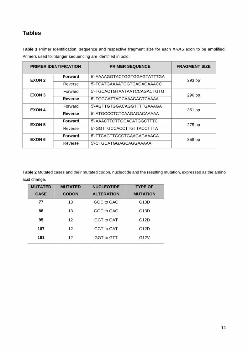
Table 1 Primer Identification, sequence and respective fragment size for each KRAS exon to be amplified.
Primers used for Sanger sequencing are identified in bold.
PRIMER IDENTIFICATION PRIMER SEQUENCE FRAGMENT SIZE
EXON 2 Forward 5’-AAAAGGTACTGGTGGAGTATTTGA
293 bp Reverse 5’-TCATGAAAATGGTCAGAGAAACC
EXON 3 Forward 5’-TGCACTGTAATAATCCAGACTGTG
296 bp Reverse 5’-TGGCATTAGCAAAGACTCAAAA
EXON 4 Forward 5’-AGTTGTGGACAGGTTTTGAAAGA
351 bp Reverse 5’-ATGCCCTCTCAAGAGACAAAAA
EXON 5 Forward 5’-AAACTTCTTGCACATGGCTTTC
275 bp Reverse 5’-GGTTGCCACCTTGTTACCTTTA
EXON 6 Forward 5’-TTCAGTTGCCTGAAGAGAAACA
358 bp Reverse 5’-CTGCATGGAGCAGGAAAAA
Table 2 Mutated cases and their mutated codon, nucleotide and the resulting mutation, expressed as the amino
acid change.
MUTATED
CASE
MUTATED
CODON
NUCLEOTIDE
ALTERATION
TYPE OF
MUTATION
77 13 GGC to GAC G13D
88 13 GGC to GAC G13D
95 12 GGT to GAT G12D
107 12 GGT to GAT G12D
181 12 GGT to GTT G12V
15
Table 3 Mutated cases and respective percentage of tumoural cells, ratios and predicted heterogeneity.
CASE % TUMOURAL CELLS
(histological)
EXPECTED MUT/WT
ALLELIC KRAS RATIO
OBSERVED MUT/WT
ALLELIC KRAS RATIOa
PREDICTED
HETEROGENEITY
77 ≥80% 67% 42% Yes
88 ≥60% 43% 22% Yes
95 ≥60% 43% 60% No
107 ≥80% 67% 48% Yes
181 ≥90% 82% 37% Yes
aThis ratio was calculated based on the peak height of mutant and WT alleles for codons 12 and 13 of KRAS
Supplementary Table 1 Map of mutations in KRAS coding sequence of the 19 MSI-positive cases. Mutated
cases are depicted in red, with the respective aminoacid change. WT cases are represented in green. SNP
are shown in yellow.
16
Fig. 1
17
Fig. 2
18
Fig. 3
19
CASE NUMBER EXON 2 EXON 3 EXON 4 EXON 5 EXON 6 63 WT WT WT WT rs1137282 65 WT WT WT WT rs1137282 74 WT WT WT WT WT 77 G13D WT WT WT WT 78 WT WT WT WT rs1137282 86 WT WT WT WT WT 88 G13D WT WT WT WT 95 G12D WT WT WT WT 96 WT WT WT WT WT 103 WT WT WT WT WT 107 G12D WT WT WT rs1137282 114 WT WT WT WT WT 119 WT WT WT WT WT 157 WT WT WT WT WT 175 WT WT WT WT rs1137282 177 WT WT WT WT rs1137282 181 G12V WT WT WT WT 189 WT WT WT WT rs1137282 191 WT WT WT WT WT
Supplementary Table 1

Agradecimentos:
À Doutora Carla Oliveira, à Doutora Gabriela Almeida e à Doutora Fátima Carneiro por
toda a atenção e tempo dispensado.
A toda a equipa do Expression Regulation In Cancer Group, por me terem ajudado das
mais diversas formas.
À Irene Gullo, por me ter ajudado com as preciosas fotografias.

ANEXOS
Instructions for Authors
Manuscript Structure
The following types of articles will be considered for publication:
All manuscripts are subject to copy editing.
Original articles
Original articles should not exceed 10 printed pages. This equals about 30 typewritten pages (A4 paper) with
33 lines per page and 65 strokes per line.
Case reports
Case reports specially written-up single cases which present original observations, new insights into
pathogenetic mechanisms and/or information based on novel or developing technology may be published after
peer review. Such reports must not exceed three printed pages with 3-4 illustrations and 1 table. The text is to
be composed in Abstract, Introduction, Clinical history, Materials and Methods, Results, and Discussion. Case
reports that do not conform to these requirements will be returned without review.
Letters to the Editors
Letters to the Editors will be published at the discretion of the editor and are subject to copy editing. They
should be limited to 500 words and should include no more than 5 pertinent references. If they concern a case
study they must not exceed 2 printed pages (1,600 words) with 2–3 illustrations and no more than 5 references.
Only invited Editorials, Review articles and Reports on meetings and clinico-pathologic trials will be published.
Review articles for Virchows Archiv should comprise no more than 5 printed pages including literature (50- 60
citations). The review should begin with an introduction and end with a conclusion or a perspective. It should be
accompanied by a short informative abstract including key words. Four to 5 illustrations are welcome. Please let me
know if you need more space for illustrations.
Reports on meetings, symposia and conferences that deal with surgical, experimental or molecular pathology should
comprise no more than 5 printed pages; they should include both a statement of the purpose of the meeting or the trial
and a summary of the findings. In addition, these reports should include a critical commentary on whether or not a
consensus was reached, as well as any recommendations for future research.
EDITORIAL PROCEDURE
Further correspondence should be addressed to:
Editorial Office Virchows Archiv
Institute of Pathology , University of Kiel
Michaelisstrasse 11, 24105 Kiel , Germany
e-mail: [email protected]
Tel.: +49 431 597 3423
Fax: +49 431 597 3428
MANUSCRIPT SUBMISSION
Manuscript Submission
Submission of a manuscript implies: that the work described has not been published before; that it is not under
consideration for publication anywhere else; that its publication has been approved by all co-authors, if any, as well as
by the responsible authorities – tacitly or explicitly – at the institute where the work has been carried out. The publisher
will not be held legally responsible should there be any claims for compensation.
Permissions
Authors wishing to include figures, tables, or text passages that have already been published elsewhere are required to
obtain permission from the copyright owner(s) for both the print and online format and to include evidence that such
permission has been granted when submitting their papers. Any material received without such evidence will be
assumed to originate from the authors.
Online Submission
Authors should submit their manuscripts online. Electronic submission substantially reduces the editorial processing
and reviewing times and shortens overall publication times. Please follow the hyperlink “Submit online” on the right
and upload all of your manuscript files following the instructions given on the screen.
TITLE PAGE
Title Page
The title page should include:
The name(s) of the author(s)
A concise and informative title
The affiliation(s) and address(es) of the author(s)
The e-mail address, telephone and fax numbers of the corresponding author
Abstract
Please provide an abstract of 150 to 250 words. The abstract should not contain any undefined abbreviations or
unspecified references.
Keywords
Please provide 4 to 6 keywords which can be used for indexing purposes.
TEXT
Text Formatting
Manuscripts should be submitted in Word.
Use a normal, plain font (e.g., 10-point Times Roman) for text.
Use italics for emphasis.
Use the automatic page numbering function to number the pages.
Do not use field functions.
Use tab stops or other commands for indents, not the space bar.
Use the table function, not spreadsheets, to make tables.
Use the equation editor or MathType for equations.
Save your file in docx format (Word 2007 or higher) or doc format (older Word versions).
Manuscripts with mathematical content can also be submitted in LaTeX.
LaTeX macro package (zip, 182 kB)
Headings
Please use no more than three levels of displayed headings.
Abbreviations
Abbreviations should be defined at first mention and used consistently thereafter.
Footnotes
Footnotes can be used to give additional information, which may include the citation of a reference included in the
reference list. They should not consist solely of a reference citation, and they should never include the bibliographic
details of a reference. They should also not contain any figures or tables.
Footnotes to the text are numbered consecutively; those to tables should be indicated by superscript lower-case letters
(or asterisks for significance values and other statistical data). Footnotes to the title or the authors of the article are not
given reference symbols.
Always use footnotes instead of endnotes.
Acknowledgments
Acknowledgments of people, grants, funds, etc. should be placed in a separate section before the reference list. The
names of funding organizations should be written in full.
REFERENCES
Citation
Reference citations in the text should be identified by numbers in square brackets. Some examples:
1. Negotiation research spans many disciplines [3].
2. This result was later contradicted by Becker and Seligman [5].
3. This effect has been widely studied [1-3, 7].
Reference list
The list of references should only include works that are cited in the text and that have been published or accepted for
publication. Personal communications and unpublished works should only be mentioned in the text. Do not use
footnotes or endnotes as a substitute for a reference list.
The entries in the list should be numbered consecutively.
Journal article
Gamelin FX, Baquet G, Berthoin S, Thevenet D, Nourry C, Nottin S, Bosquet L (2009) Effect of
high intensity intermittent training on heart rate variability in prepubescent children. Eur J Appl
Physiol 105:731-738. doi: 10.1007/s00421-008-0955-8
Ideally, the names of all authors should be provided, but the usage of “et al” in long author lists
will also be accepted:
Smith J, Jones M Jr, Houghton L et al (1999) Future of health insurance. N Engl J Med 965:325–
329
Article by DOI
Slifka MK, Whitton JL (2000) Clinical implications of dysregulated cytokine production. J Mol
Med. doi:10.1007/s001090000086
Book
South J, Blass B (2001) The future of modern genomics. Blackwell, London
Book chapter
Brown B, Aaron M (2001) The politics of nature. In: Smith J (ed) The rise of modern genomics,
3rd edn. Wiley, New York, pp 230-257
Online document
Cartwright J (2007) Big stars have weather too. IOP Publishing PhysicsWeb.
http://physicsweb.org/articles/news/11/6/16/1. Accessed 26 June 2007
Dissertation
Trent JW (1975) Experimental acute renal failure. Dissertation, University of California
Always use the standard abbreviation of a journal’s name according to the ISSN List of Title Word Abbreviations, see
ISSN.org LTWA
If you are unsure, please use the full journal title.
For authors using EndNote, Springer provides an output style that supports the formatting of in-text citations and
reference list.
EndNote style (zip, 2 kB)
Authors preparing their manuscript in LaTeX can use the bibtex file spbasic.bst which is included in Springer’s LaTeX
macro package.
TABLES
All tables are to be numbered using Arabic numerals.
Tables should always be cited in text in consecutive numerical order.
For each table, please supply a table caption (title) explaining the components of the table.
Identify any previously published material by giving the original source in the form of a reference
at the end of the table caption.
Footnotes to tables should be indicated by superscript lower-case letters (or asterisks for
significance values and other statistical data) and included beneath the table body.
ARTWORK AND ILLUSTRATIONS GUIDELINES
Electronic Figure Submission
Supply all figures electronically.
Indicate what graphics program was used to create the artwork.
For vector graphics, the preferred format is EPS; for halftones, please use TIFF format. MSOffice
files are also acceptable.
Vector graphics containing fonts must have the fonts embedded in the files.
Name your figure files with "Fig" and the figure number, e.g., Fig1.eps.
Line Art
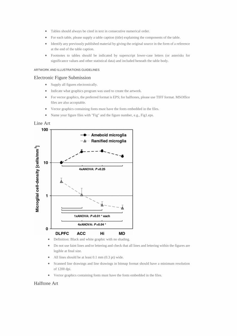
Definition: Black and white graphic with no shading.
Do not use faint lines and/or lettering and check that all lines and lettering within the figures are
legible at final size.
All lines should be at least 0.1 mm (0.3 pt) wide.
Scanned line drawings and line drawings in bitmap format should have a minimum resolution
of 1200 dpi.
Vector graphics containing fonts must have the fonts embedded in the files.
Halftone Art
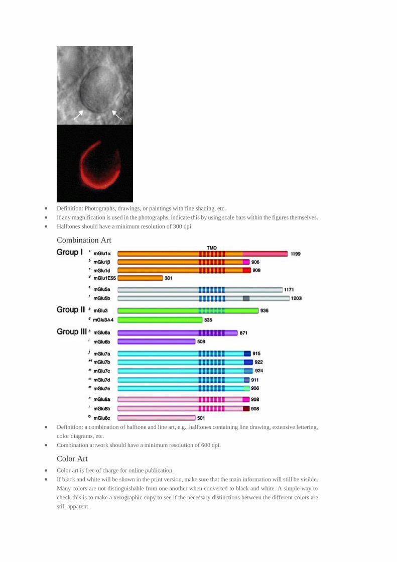
Definition: Photographs, drawings, or paintings with fine shading, etc.
If any magnification is used in the photographs, indicate this by using scale bars within the figures themselves.
Halftones should have a minimum resolution of 300 dpi.
Combination Art
Definition: a combination of halftone and line art, e.g., halftones containing line drawing, extensive lettering,
color diagrams, etc.
Combination artwork should have a minimum resolution of 600 dpi.
Color Art
Color art is free of charge for online publication.
If black and white will be shown in the print version, make sure that the main information will still be visible.
Many colors are not distinguishable from one another when converted to black and white. A simple way to
check this is to make a xerographic copy to see if the necessary distinctions between the different colors are
still apparent.
If the figures will be printed in black and white, do not refer to color in the captions.
Color illustrations should be submitted as RGB (8 bits per channel).
Figure Lettering
To add lettering, it is best to use Helvetica or Arial (sans serif fonts).
Keep lettering consistently sized throughout your final-sized artwork, usually about 2–3 mm (8–12 pt).
Variance of type size within an illustration should be minimal, e.g., do not use 8-pt type on an axis and 20-
pt type for the axis label.
Avoid effects such as shading, outline letters, etc.
Do not include titles or captions within your illustrations.
Figure Numbering
All figures are to be numbered using Arabic numerals.
Figures should always be cited in text in consecutive numerical order.
Figure parts should be denoted by lowercase letters (a, b, c, etc.).
If an appendix appears in your article and it contains one or more figures, continue the consecutive numbering
of the main text. Do not number the appendix figures,
"A1, A2, A3, etc." Figures in online appendices (Electronic Supplementary Material) should, however, be
numbered separately.
Figure Captions
Each figure should have a concise caption describing accurately what the figure depicts. Include the
captions in the text file of the manuscript, not in the figure file.
Figure captions begin with the term Fig. in bold type, followed by the figure number, also in bold type.
No punctuation is to be included after the number, nor is any punctuation to be placed at the end of the
caption.
Identify all elements found in the figure in the figure caption; and use boxes, circles, etc., as coordinate
points in graphs.
Identify previously published material by giving the original source in the form of a reference citation at
the end of the figure caption.
Figure Placement and Size
When preparing your figures, size figures to fit in the column width.
For most journals the figures should be 39 mm, 84 mm, 129 mm, or 174 mm wide and not higher than 234
mm.
For books and book-sized journals, the figures should be 80 mm or 122 mm wide and not higher than 198
mm.
Permissions
If you include figures that have already been published elsewhere, you must obtain permission from the copyright
owner(s) for both the print and online format. Please be aware that some publishers do not grant electronic rights for
free and that Springer will not be able to refund any costs that may have occurred to receive these permissions. In such
cases, material from other sources should be used.
Accessibility
In order to give people of all abilities and disabilities access to the content of your figures, please make sure that
All figures have descriptive captions (blind users could then use a text-to-speech software or a text-to-Braille
hardware)
Patterns are used instead of or in addition to colors for conveying information (colorblind users would then
be able to distinguish the visual elements)
Any figure lettering has a contrast ratio of at least 4.5:1

ELECTRONIC SUPPLEMENTARY MATERIAL
Springer accepts electronic multimedia files (animations, movies, audio, etc.) and other supplementary files to be
published online along with an article or a book chapter. This feature can add dimension to the author's article, as
certain information cannot be printed or is more convenient in electronic form.
Submission
Supply all supplementary material in standard file formats.
Please include in each file the following information: article title, journal name, author names; affiliation and
e-mail address of the corresponding author.
To accommodate user downloads, please keep in mind that larger-sized files may require very long download
times and that some users may experience other problems during downloading.
Audio, Video, and Animations
Always use MPEG-1 (.mpg) format.
Text and Presentations
Submit your material in PDF format; .doc or .ppt files are not suitable for long-term viability.
A collection of figures may also be combined in a PDF file.
Spreadsheets
Spreadsheets should be converted to PDF if no interaction with the data is intended.
If the readers should be encouraged to make their own calculations, spreadsheets should be submitted as .xls
files (MS Excel).
Specialized Formats
Specialized format such as .pdb (chemical), .wrl (VRML), .nb (Mathematica notebook), and .tex can also be
supplied.
Collecting Multiple Files
It is possible to collect multiple files in a .zip or .gz file.
Numbering
If supplying any supplementary material, the text must make specific mention of the material as a citation,
similar to that of figures and tables.
Refer to the supplementary files as “Online Resource”, e.g., "... as shown in the animation (Online Resource
3)", “... additional data are given in Online Resource 4”.
Name the files consecutively, e.g. “ESM_3.mpg”, “ESM_4.pdf”.
Captions
For each supplementary material, please supply a concise caption describing the content of the file.
Processing of supplementary files
Electronic supplementary material will be published as received from the author without any conversion,
editing, or reformatting.
Accessibility
In order to give people of all abilities and disabilities access to the content of your supplementary files, please make
sure that
The manuscript contains a descriptive caption for each supplementary material
Video files do not contain anything that flashes more than three times per second (so that users prone to
seizures caused by such effects are not put at risk)
ETHICAL RESPONSIBILITIES OF AUTHORS
This journal is committed to upholding the integrity of the scientific record. As a member of the Committee on
Publication Ethics (COPE) the journal will follow the COPE guidelines on how to deal with potential acts of
misconduct.
Authors should refrain from misrepresenting research results which could damage the trust in the journal, the
professionalism of scientific authorship, and ultimately the entire scientific endeavour. Maintaining integrity of the
research and its presentation can be achieved by following the rules of good scientific practice, which include:
The manuscript has not been submitted to more than one journal for simultaneous consideration.
The manuscript has not been published previously (partly or in full), unless the new work concerns an
expansion of previous work (please provide transparency on the re-use of material to avoid the hint of text-
recycling (“self-plagiarism”)).
A single study is not split up into several parts to increase the quantity of submissions and submitted to
various journals or to one journal over time (e.g. “salami-publishing”).
No data have been fabricated or manipulated (including images) to support your conclusions
No data, text, or theories by others are presented as if they were the author’s own (“plagiarism”). Proper
acknowledgements to other works must be given (this includes material that is closely copied (near
verbatim), summarized and/or paraphrased), quotation marks are used for verbatim copying of material,
and permissions are secured for material that is copyrighted.
Important note: the journal may use software to screen for plagiarism.
Consent to submit has been received explicitly from all co-authors, as well as from the responsible
authorities - tacitly or explicitly - at the institute/organization where the work has been carried
out, before the work is submitted.
Authors whose names appear on the submission have contributed sufficiently to the scientific work and
therefore share collective responsibility and accountability for the results.
In addition:
Changes of authorship or in the order of authors are not accepted after acceptance of a manuscript.
Requesting to add or delete authors at revision stage, proof stage, or after publication is a serious matter and
may be considered when justifiably warranted. Justification for changes in authorship must be compelling and
may be considered only after receipt of written approval from all authors and a convincing, detailed
explanation about the role/deletion of the new/deleted author. In case of changes at revision stage, a letter
must accompany the revised manuscript. In case of changes after acceptance or publication, the request and
documentation must be sent via the Publisher to the Editor-in-Chief. In all cases, further documentation may
be required to support your request. The decision on accepting the change rests with the Editor-in-Chief of
the journal and may be turned down. Therefore authors are strongly advised to ensure the correct author group,
corresponding author, and order of authors at submission.
Upon request authors should be prepared to send relevant documentation or data in order to verify the validity
of the results. This could be in the form of raw data, samples, records, etc.
If there is a suspicion of misconduct, the journal will carry out an investigation following the COPE guidelines. If, after
investigation, the allegation seems to raise valid concerns, the accused author will be contacted and given an opportunity
to address the issue. If misconduct has been established beyond reasonable doubt, this may result in the Editor-in-
Chief’s implementation of the following measures, including, but not limited to:
If the article is still under consideration, it may be rejected and returned to the author.
If the article has already been published online, depending on the nature and severity of the infraction, either
an erratum will be placed with the article or in severe cases complete retraction of the article will occur. The
reason must be given in the published erratum or retraction note.
The author’s institution may be informed.
COMPLIANCE WITH ETHICAL STANDARDS
To ensure objectivity and transparency in research and to ensure that accepted principles of ethical and professional
conduct have been followed, authors should include information regarding sources of funding, potential conflicts of
interest (financial or non-financial), informed consent if the research involved human participants, and a statement on
welfare of animals if the research involved animals.
Authors should include the following statements (if applicable) in a separate section entitled “Compliance with Ethical
Standards” before the References when submitting a paper:
Disclosure of potential conflicts of interest
Research involving Human Participants and/or Animals
Informed consent
Please note that standards could vary slightly per journal dependent on their peer review policies (i.e. double blind peer
review) as well as per journal subject discipline. Before submitting your article check the Instructions for Authors
carefully.
The corresponding author should be prepared to collect documentation of compliance with ethical standards and send
if requested during peer review or after publication.
The Editors reserve the right to reject manuscripts that do not comply with the above-mentioned guidelines. The author
will be held responsible for false statements or failure to fulfill the above-mentioned guidelines.
DISCLOSURE OF POTENTIAL CONFLICTS OF INTEREST
Authors must disclose all relationships or interests that could have direct or potential influence or impart bias on the
work. Although an author may not feel there is any conflict, disclosure of relationships and interests provides a more
complete and transparent process, leading to an accurate and objective assessment of the work. Awareness of a real or
perceived conflicts of interest is a perspective to which the readers are entitled. This is not meant to imply that a
financial relationship with an organization that sponsored the research or compensation received for consultancy work
is inappropriate. Examples of potential conflicts of interests that are directly or indirectly related to the
research may include but are not limited to the following:
Research grants from funding agencies (please give the research funder and the grant number)
Honoraria for speaking at symposia
Financial support for attending symposia
Financial support for educational programs
Employment or consultation
Support from a project sponsor
Position on advisory board or board of directors or other type of management relationships
Multiple affiliations
Financial relationships, for example equity ownership or investment interest
Intellectual property rights (e.g. patents, copyrights and royalties from such rights)
Holdings of spouse and/or children that may have financial interest in the work
In addition, interests that go beyond financial interests and compensation (non-financial interests) that may be important
to readers should be disclosed. These may include but are not limited to personal relationships or competing interests
directly or indirectly tied to this research, or professional interests or personal beliefs that may influence your research.
The corresponding author collects the conflict of interest disclosure forms from all authors. In author collaborations
where formal agreements for representation allow it, it is sufficient for the corresponding author to sign the disclosure
form on behalf of all authors. Examples of forms can be found
here:
The corresponding author will include a summary statement in the text of the manuscript in a separate section before
the reference list, that reflects what is recorded in the potential conflict of interest disclosure form(s).
See below examples of disclosures:
Funding: This study was funded by X (grant number X).
Conflict of Interest: Author A has received research grants from Company A. Author B has received a speaker
honorarium from Company X and owns stock in Company Y. Author C is a member of committee Z.
If no conflict exists, the authors should state:
Conflict of Interest: The authors declare that they have no conflict of interest.
DOES SPRINGER PROVIDE ENGLISH LANGUAGE SUPPORT?
Manuscripts that are accepted for publication will be checked by our copyeditors for spelling and formal style. This
may not be sufficient if English is not your native language and substantial editing would be required. In that case, you
may want to have your manuscript edited by a native speaker prior to submission. A clear and concise language will
help editors and reviewers concentrate on the scientific content of your paper and thus smooth the peer review process.
The following editing service provides language editing for scientific articles in all areas Springer
publishes in:
Edanz English editing for scientists
Use of an editing service is neither a requirement nor a guarantee of acceptance for publication.
Please contact the editing service directly to make arrangements for editing and payment.
Edanz English editing for scientists
For Authors from China
文章在投稿前进行专业的语言润色将对作者的投稿进程有所帮助。作者可自愿选择使用Springer推荐的编辑
服务,使用与否并不作为判断文章是否被录用的依据。提高文章的语言质量将有助于审稿人理解文章的内容
,通过对学术内容的判断来决定文章的取舍,而不会因为语言问题导致直接退稿。作者需自行联系Springer
推荐的编辑服务公司,协商编辑事宜。
理文编辑
For Authors from Japan
ジャーナルに論文を投稿する前に、ネイティブ・スピーカーによる英文校閲を希望されている方には、
Edanz社をご紹介しています。サービス内容、料金および申込方法など、日本語による詳しい説明はエダ
ンズグループジャパン株式会社の下記サイトをご覧ください。
エダンズグループジャパン
For Authors from Korea
영어 논문 투고에 앞서 원어민에게 영문 교정을 받고자 하시는 분들께 Edanz 회사를 소개해 드립니다. 서비스
내용, 가격 및
신청 방법 등에 대한 자세한 사항은 저희 Edanz Editing Global 웹사이트를 참조해 주시면 감사하겠습니다.
Edanz Editing Global
AFTER ACCEPTANCE
Upon acceptance of your article you will receive a link to the special Author Query Application at Springer’s web page
where you can sign the Copyright Transfer Statement online and indicate whether you wish to order OpenChoice and
offprints.
Once the Author Query Application has been completed, your article will be processed and you will receive the proofs.
Open Choice
In addition to the normal publication process (whereby an article is submitted to the journal and access to that article is
granted to customers who have purchased a subscription), Springer now provides an alternative publishing option:
Springer Open Choice. A Springer Open Choice article receives all the benefits of a regular subscription-based article,
but in addition is made available publicly through Springer’s online platform SpringerLink.
Springer Open Choice
Copyright transfer
Authors will be asked to transfer copyright of the article to the Publisher (or grant the Publisher exclusive publication
and dissemination rights). This will ensure the widest possible protection and dissemination of information under
copyright laws.
Open Choice articles do not require transfer of copyright as the copyright remains with the author. In opting for open
access, the author(s) agree to publish the article under the Creative Commons Attribution License..
Offprints
Offprints can be ordered by the corresponding author.
Color illustrations
Publication of color illustrations is free of charge.
Proof reading
The purpose of the proof is to check for typesetting or conversion errors and the completeness and accuracy of the text,
tables and figures. Substantial changes in content, e.g., new results, corrected values, title and authorship, are not
allowed without the approval of the Editor.
After online publication, further changes can only be made in the form of an Erratum, which will be hyperlinked to the
article.
Online First
The article will be published online after receipt of the corrected proofs. This is the official first publication citable with
the DOI. After release of the printed version, the paper can also be cited by issue and page numbers.