Embed Size (px)
Citation preview
Frontotemporal LobarDegeneration: NewUnderstanding BringsNew Approaches
Maria Carmela Tartaglia, MDKey Learning Points
1. Frontotemporal lobar degeneration (FTLD) is the pathologic term associated with several clinicalsyndromes.
2. Frontotemporal dementia (FTD) is a clinical term referring to behavioral variant FTD (bvFTD), nonflu-ent variant primary progressive aphasia (nfvPPA), and semantic variant primary progressive aphasia(svPPA)
3. Mutations in multiple genes cause FTLD.
4. Three different pathologic substrates can cause FTLD: tau, transactive DNA-binding protein 43(TDP-43), and fused in sarcoma (FUS).
KEYWORDS
� Frontotemporal dementia� Semantic variant primary progressive aphasia� Nonfluent variant primary progressive aphasia� Frontotemporal lobar degeneration
clinics.com
FTLD: A FAMILY OF SYNDROMES
FTLD is associated with several clinical syndromesinvolving behavior, language, and motor function.Until recently, the main syndromes encompassedby the clinical term FTD were bvFTD, nfvPPA,and svPPA. Recent discoveries revealed overlapat a clinical, genetic, and pathologic level betweenthese syndromes and 3 other syndromes: FTDwith motor neuron disease (FTD-MND), progres-sive supranuclear palsy (PSP), and corticobasalsyndrome (CBS). The clinical expression of thesesyndromes is markedly different, reflecting selec-tive injury of specific areas of the brain, whichleads to the diverse signs and symptoms.1
Recent clinical-pathologic studies all emphasizethat proper clinical assessments are paramountto clinicopathologic prediction.2–4 Accurate diag-nosis is the first step in the determination of the
Tanz Centre for Research in Neurodegenerative Diseases,Cres West, Room 231, Toronto, ON, Canada M5S 3H2E-mail address: [email protected]
Neuroimag Clin N Am 22 (2012) 83–97doi:10.1016/j.nic.2011.11.0091052-5149/12/$ – see front matter Crown Copyright � 20
pathologic substrate because different syndromesare associated with different pathologies. In aneffort to improve diagnosis, new criteria havebeen developed in the last year for bvFTD and thePPAs (semantic variant and nonfluent variant).5,6
bvFTD is characterized by dramatic personalityand behavioral changes with prominent loss ofsocial cognition.7–11 New international researchcriteria have been established for bvFTD and focuson the behavioral/executive deficits (Box 1).5
As its name implies, bvFTD begins with promi-nent changes in social cognition, emotion, andbehavior. Typical early symptoms include apathy,disinhibition, repetitive and compulsive behaviors,and progressive inability to represent the self andothers, manifesting as shallow insight and lack ofempathy. Disinhibition can lead to sociopathicbehaviors such as being overly familiar with
University of Toronto, Tanz Building, 6 Queen’s Park
12 Published by Elsevier Inc. All rights reserved. neuroimaging.the
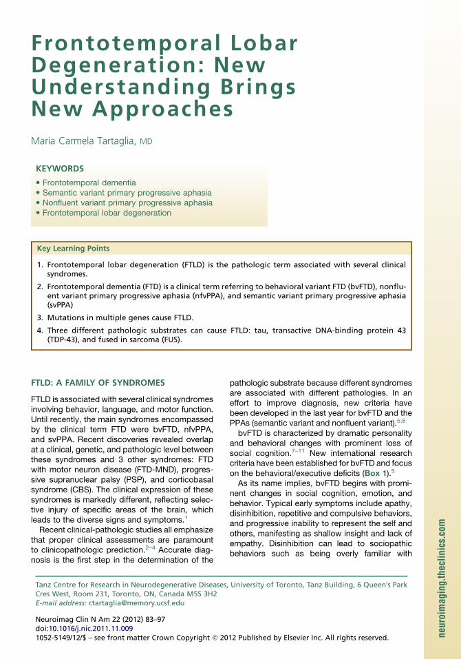
Box 1Inclusion and exclusion criteria for diagnosis ofbvFTD
I. Neurodegenerative disease
The following symptom must be present fordiagnosis:
A. Progressive deterioration of behavior orcognition
II. Possible bvFTD
Three of the following symptoms must bepresent early (A-F):
A. Behavioral disinhibition
B. Apathy or inertia
C. Loss of sympathy or empathy
D. Perseverative, stereotyped, or compulsive/ritualistic behavior
E. Hyperorality or dietary changes
F. Executive/generation deficits with relativesparing of visuospatial functions
III. Probable bvFTD
Must meet criteria A to C:
A. Meets possible bvFTD
B. Significant functional decline
C. Imaging results consistent with bvFTD
IV. bvFTD with definite FTLD disease
Must meet A and either B or C:
A. Meets possible bvFTD
B. Histopathologic evidence of FTLD
C. Presence of known pathogenic mutation
V. Exclusion criteria for bvFTD
A and B must both be negative; C can be posi-tive for possible bvFTD but must be negativefor probable bvFTD:
A. Better accounted for by nondegenerativedisorders
B. Better accounted for by psychiatric diagnosis
C. Biomarkers strongly indicates Alzheimer’sdisease or other neurodegenerative process
From Rascovsky K, Hodges JR, Knopman D, et al.Sensitivity of revised diagnostic criteria for the behav-ioral variant of frontotemporal dementia. Brain 2011;134(Pt 9):2456–77.
Tartaglia84
strangers, unsolicited sexual approaches, publicurination, traffic violations, and shoplifting.12–14
There can be dramatic personality changes, suchas change in religious beliefs, political conviction,
dress, and social style.15 In bvFTD, overeating,weight gain, overstuffing the mouth, and idiosyn-cratic food fads occur.16 Patients often displayutilization behavior, manifested by graspingat items in view or repeatedly switching lightson and off.12 Cognitive complaints, unlike inAlzheimer’s disease (AD), are typically less dra-matic than the behavioral changes, and the maindeficits are in executive function.9,11 bvFTD is themost common of the 3 main clinical subtypes ofFTD, accounting for 56% of cases. It showsa male predominance (2:1), has the earliest ageof onset (58 years at diagnosis), and progressesthe most rapidly (3.4 years from diagnosis todeath). bvFTD has the highest genetic suscepti-bility and is strongly associated with MND.17,18
nfvPPA (previously known as progressivenonfluent aphasia) is a disorder of expressivelanguage and speech production.11,19 New inter-national criteria for PPA were recently published6
and highlight the expressive language deficitswith relative sparing of single sentence or wordcomprehension (Boxes 2–4). nfvPPA accountsfor 25% of FTD and has an intermediate rate ofprogression (4.3 years from diagnosis) and geneticcause.17 Nonfluent speech is often accompaniedby agrammatism, phonemic paraphasias, anomia,and speech apraxia.20 Speech is slow, effortful,and telegraphic, and in contrast to bvFTD, per-sonal and interpersonal conduct, behavior, andinsight are preserved early on.svPPA, previously known as semantic dementia
or temporal variant FTD, is a disorder with loss ofsemantic knowledge for words. It presents differ-ently depending on which hemisphere is the siteof pathology. Left-sided atrophy produces pro-gressive loss of meaning for words, objects, andemotions.11,19 In contrast, right-sided pathologyis associated with behavioral changes. svPPA ac-counts for less than 20% of all FTD cases andshares an earlier age of onset with bvFTD butshows the slowest rate of progression (5.2 yearsfrom diagnosis to death).17,18 svPPA has fewercases of autosomal-dominant inheritance.Although the term FTD was established to
include only bvFTD, nfvPPA, and svPPA, it isnow known that there are strong links and consid-erable overlap between FTD clinical syndromesand other conditions including PSP and CBS.21
Many patients begin with an nfvPPA syndromeand later evolve into a clinical disorder suggestiveof CBS or PSP.22,23
There are no consensus clinical research criteriafor CBS but previous studies have describeda progressive, asymmetric, akinetic-rigid syn-drome that does not respond to levodopa treat-ment. Individuals have a combination of deficits

Box 2Inclusion and exclusion criteria for thediagnosis of PPA
Inclusion: criteria 1 to 3 must be answeredpositively
1. Most prominent clinical feature is difficultywith language (word-finding deficits, para-phasias, effortful speech, grammatical orcomprehension deficits)
2. These deficits are the principal cause ofimpaired daily living activities (eg, problemswithcommunicationactivity relatedto speechand language, such as using the telephone)
3. Aphasia shouldbe themostprominentdeficitfor approximately 2 years since symptomonset.
Exclusion: criteria 1 to 4 must be answerednegatively for a PPA diagnosis
1. Pattern of deficits is better accounted for byother nondegenerative nervous system ormedical disorders (eg, neoplasm, cerebrovas-cular disease, hypothyroidism)
2. Cognitive disturbance is better accountedfor by a psychiatric diagnosis (eg, depression,bipolar disorder, schizophrenia, preexistingpersonality disorder)
3. Prominent initial episodic memory, visualmemory, and visuoperceptual impairments(eg, inability to copy simple line drawings)
4. Prominent initial behavioral disturbance(eg, marked disinhibition, emotional detach-ment, hyperorality, or repetitive/compulsivebehaviors)
From Gorno-Tempini M, Hillis AE, Weintraub S, et al.Classification of primary progressive aphasia and its vari-ants. Neurology 2011;76(11):1006–14.
Box 3Diagnostic features for nfvPPA (also known asprogressive nonfluent aphasia or PNFA, and asagrammatic PPA or PPA-G)
I. Clinical diagnosis of nfvPPA
At least one of the following core features mustbe present:
1. Grammatical errors and simplification inlanguage production
2. Effortful, halting speech with inconsistentdistortions, deletions, substitutions, inser-tions, or transpositions of speech sounds,particularly in polysyllabic words (oftenconsidered to reflect apraxia of speech)
At least 2 of the following 3 features must bepresent:
1. Impaired comprehension of syntacticallycomplex sentences, with relatively sparedcomprehension of syntactically simplersentences
2. Spared content, single-word comprehension
3. Spared object knowledge
II. Imaging-supported nfvPPA diagnosis
Both of the following criteria must be present:
1. Clinical diagnosis of nfvPPA
2. Imaging must show one or more of thefollowing results:
a. Predominant left posterior frontoinsularatrophy on magnetic resonance (MR)imaging
b. Predominant left posterior frontoinsularhypoperfusion or hypometabolism onsingle-photon emission computed tomography (SPECT) or positron emissiontomography (PET)
III. nfvPPA with definite pathology
Clinical diagnosis (criterion 1) and either crite-rion 2 or 3 must be present:
1. Clinical diagnosis of nfvPPA
2. Histopathologic evidence of a specificpathology (eg, FTLD-tau, FTLD-TDP)onbiopsyor post mortem
3. Presence of a known pathogenic mutation
Frontotemporal Lobar Degeneration 85
attributable to cortical dysfunction, such asapraxia, language difficulties, and cortical sensoryloss or neglect as well as symptoms attributable tobasal ganglia dysfunction such as rigidity or dysto-nia.24 Cognitively, planning and other aspects ofexecutive function are impaired in CBS.25 InOctober 2009, a group of corticobasal degenera-tion (CBD)/CBS researchers met to determineconsensus clinical research criteria for CBS. Thenew criteria (Litvan and colleagues, manuscript inpreparation) will be similar to the previous criteriabut will also include dementia syndromes thathave been associated with CBD.
Consensus research criteria have been devel-oped for PSP and have been reported to haveexcellent predictive power for underlying PSP
pathology.26,27 PSP is named for its characteristiceye movement abnormalities, and a diagnosis ofprobable PSP requires a slowly progressivedisorder with onset after age 40 years anda vertical supranuclear gaze palsy of eye move-ments and falls within the first year of diagnosis.
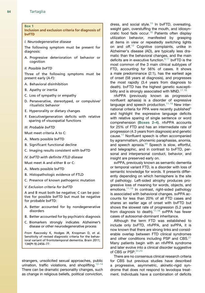
Box 4Diagnostic criteria for svPPA (also known assemantic dementia or PPA-S)
I. Clinical diagnosis of svPPA
Both of the following core features must bepresent:
1. Poor confrontation naming (of pictures orobjects), particularly for low familiarity orlow frequency items
2. Impaired single-word comprehension
At least 3 of the following other diagnosticfeatures must be present:
1. Poor object or person knowledge, particu-larly for low frequency or low familiarityitems
2. Surface dyslexia or dysgraphia
3. Spared repetition
4. Spared motor speech
II. Imaging-supported svPPA diagnosis
Both of the following criteria must be present:
1. Clinical diagnosis of svPPA
2. Imaging must show one or more of thefollowing results:
a. Predominant anterior temporal lobeatrophy
b. Predominant anterior temporal hypoper-fusion or hypometabolism on SPECT orPET
III. svPPA with definite pathology
Clinical diagnosis (criterion 1) and either crite-rion 2 or 3 must be present:
1. Clinical diagnosis of svPPA
2. Histopathologic evidence of a specificpathology (eg, FTLD-tau, FTLD-TDP) onbiopsyor post mortem
3. Presence of a known pathogenic mutation
Tartaglia86
Other supportive criteria include prominent axialrigidity, early dysphagia and dysarthria, apathy,and cognitive impairments consisting mainly ofexecutive dysfunction. Significant behavioralchanges including impulsivity, perseveration, anddiminished judgment also feature in PSP.MND may co-occur with bvFTD-like symptoms,
or, less commonly, with svPPA or nfvPPA,28,29
and the term FTD-MND has been applied to thesecases. FTD-MND most commonly affects lowermotor neurons to the bulbar and upper limb mus-culature. The close relationship between dementia
and MND (also known as amyotrophic lateralsclerosis) surfaced with the discovery of over-lapping genetics,30 including a recent discoverythat the expanded hexanucleotide repeat in a non-coding region of chromosome 9 is associatedwith MND and FTD.31,32 In addition, commonneuropathologic substrates exist between the 2syndromes,33 suggesting a strong connectionbetween them. In prospective studies, up to 50%of patients with FTD had possible or probableMND,28 and 50%of patients with MNDwho under-went behavioral and neuropsychological evalua-tion had measurable (mainly frontal/executiveand behavioral) cognitive deficits, and many metcriteria for an FTD syndrome.34
FTLD: UNDERSTANDING THESYNDROME-PATHOLOGY RELATIONSHIP
The underlying pathology of FTLD has provedmore complex than anticipated. It is now knownthat at least 3 different molecular pathologies existconsisting of abnormal protein aggregation: tau,TDP-43, and FUS.Abnormal tau deposition or aggregation was the
first pathologic substrate described and, untilrecently, it was believed to be the main cause ofFTLD. Tau is a protein that binds to and stabilizesmicrotubules that are necessary for maintainingneuronal shape and for transport of cellularcargo.35 The abnormal tau can be seen in neurons,glia, or both. The syndromes that associate withabnormal tau deposition are known as tauopa-thies. PSP is associated exclusively with taupathology, whereas in the case of CBS, althoughmost cases are caused by abnormal tau aggrega-tion, there is recognition that other diseases cancause the syndrome such as TDP-43 or AD. Non-fluent variant PPA is also primarily a tauopathy butcan also be associated with TDP-43 or AD. Avariety of mutations have been identified in themicrotubule-associated protein tau (MAPT) genethat lead to bvFTD, nfvPPA, PSP, and CBS.36
Tau-negative cases of FTLD stain for ubiquitin,an integral part of the degradative system.37
Most ubiquitin-positive inclusions observed inFTLD result from the accumulation of inclusionsthat stain for TDP-43, a widely expressed nuclearprotein with presumed functions in transcriptionregulation and exon skipping.38 These neuronalor cytoplasmic ubiquitinated inclusions are usuallyseen in affected cortex, dentate granule cells,39
and primary motor cortex, spinal cord, or brainstem motor neurons in FTD-MND.40
Four distinct patterns of pathologic featuresand regional variability were observed in cases ofTDP-43 proteinopathy,41–43 and these patterns
Frontotemporal Lobar Degeneration 87
have clinical relevance, because they are specificto some, although not all, of the syndromes.44
FTLD-TDP A, which features many neuronal cyto-plasmic inclusions and short dystrophic neurites inlayer 2 of the cortex, is seen in bvFTD, nfvPPA, andnearly all patients with granulin (GRN) mutations.45
Patients with FTLD-TDP and a GRN polymorphismof the T-allele of rs5848 often show an FTLD-TDPA pathologic pattern.46 FTLD-TDP B is transcorti-cal and features some neuronal cytoplasmic inclu-sions and few dystrophic neurites and is seen inbvFTD and FTD-MND. FTLD-TDP C is alsopredominantly in layer 2, features many, long,dystrophic neurites and few neuronal cytoplasmicinclusions, and is seen in svPPA and bvFTD. Afourth subtype, FTLD-TDP D, is seen only invalosin-containing protein (VCP) mutations andconsists of many short dystrophic neurites, manylentiform neuronal intranuclear inclusions, andfew neuronal cytoplasmic inclusions. The pathol-ogy are seen in all layers. The clinical phenotypeassociated with the VCP mutations is inclusionbody myopathy with Paget disease of bone andFTD.
The inclusions inubiquitin-positive, TDP-43–nega-tive cases of FTLD were recently discovered tostain for the FUS protein. FUS is a ubiquitouslyexpressed DNA/RNA-binding protein involved inmultiple aspects of gene expression, transcriptionregulation,RNAsplicing, transport, and translation.47
FUS pathology is reportedly associated witha distinct clinical phenotype that includes youngonset,prominentobsessionality, repetitivebehaviorsand rituals, social withdrawal and lack of engage-ment, hyperorality with pica, and marked stimulus-bound behavior including use behavior.48,49 Inaddition, FUS cases show severe caudate atrophy.This pathologic subtypemaybe the easiest to disen-tangle from the other FTLD pathologic substrates.
Clinicopathologic correlations have shown thatcertain FTLD syndromes are reliably associatedwith specific proteinopathies: FTD-MND is associ-ated almost uniquely with FTLD-TDP, with onlya few cases ascribed to FUS. svPPA also is almostalways associated with FTLD-TDP, with only rarecases of FTLD-tau described. PSP is exclusivelya tauopathy.2–4 However, the rest of the syndromesaredifficult to predict becausenfvPPA ismost oftenassociated with a tauopathy,50 but FTLD-TDP andAD pathology can cause this clinical syndrome insome cases.2–4 bvFTD is difficult to predict,because it can be associated with FTLD-tau,FTLD-TDP, FTLD-FUS, or AD. CBS is similarly diffi-cult to predict, because although most cases areassociated with FTLD-tau, there are reports ofFTLD-TDP and AD as the pathologic substrate ofCBS.2–4,51,52
GENETICS OF FTLD
Although most FTLD cases are sporadic, unlikethe other dementing illnesses, FTLD has a strongfamilial component because up to 40% to 50%of cases are diagnosed as familial and 10%show an autosomal-dominant pattern of inheri-tance.53 Multiple genes have been implicated inFTLD, including MAPT (chromosome 17), GRN(chromosome 17), VCP (chromosome 9),chromatin-modifying protein 2B (also known ascharged multivesicular body protein 2B gene)CHMP2B (chromosome 3), and FUS gene (chro-mosome 16).54–56 Recently, a novel mutation hasbeen discovered as the most common cause offamilial FTD and MND; it consists of an expandedhexanucleotide repeat in a noncoding region ofchromosome 9 open reading frame 72.31,32 Thegene encodes an uncharacterized protein with noknown domains or function, but which is highlyconserved across species.
These genetic abnormalities are associated withspecific proteinopathies so that MAPT mutationleads to a tauopathy, VCP and GRN mutationsand expanded hexanucleotide repeat to TDP-43proteinopathy, and FUS mutation to a FUSop-athy.38,47 The abnormal protein in CMP2B muta-tions remains elusive to date; no specific proteinhas been discovered to identify the inclusions.
FTLD was first linked to chromosome 17 (MAPTgene) by Wilhelmsen and colleagues.54 MAPTmutations are associated with tau pathology, andmore than 40 different mutations in the MAPTgene have been reported in association withfamilial FTD syndromes.57 Humans carry an equalratio of 3R and 4R tau, but mutations in the tauintron lead to increases in the ratio of 4R to 3Rtau. A wide spectrum of disorders has been re-ported with tau inclusions, including bvFTD,nfvPPA, CBD, and PSP.58
Although MAPT mutations were the first discov-ered, epidemiologic studies have identified GRNmutations in 5% to 11% of all FTD and 11% to26% of patients with a family history,59 making itthe most common cause of genetic FTLD. Thismutation is also on chromosome 17 but at 17q21in the progranulin (GRN) gene.60 More than 50 pro-granulin mutations have been found since it wasdiscovered in 2006,57 and these mutations leadto deficient protein levels of progranulin.60 Therole of progranulin in neurodegeneration is stillunknown, although progranulin is a trophic factor,and is implicated in wound healing, tumor growth,inflammation, and brain development in mice; itpromotes neuronal survival and stimulates neuriticoutgrowth in cultures of rat motor and corticalneurons.61 Progranulin mutations are associated
Tartaglia88
with phenotypic heterogeneity, even within familymembers with the same mutation. bvFTD is themost common presentation of GRN mutation fol-lowed by nfvPPA and svPPA although parkinso-nian and AD syndromes are also seen.57 Theubiquitinated inclusions associated with progranu-lin mutations stain for TDP-43.38
The likelihood of a genetic mutation associatedwith the FTLD syndrome varies across the differentsubtypes. bvFTD and FTD-MND are the moststrongly familial of the FTLD syndromes. Semanticvariant is the least familial, whereas nfvPPA lies inbetween bvFTD and svPPA.62 As a risk for FTD,the role of apolipoprotein E4 seems to be small,63
although E4may expand the pathologic damage infrontal regions in FTD.64
Polymorphisms, in the absence ofmutation, havegained attention as possible contributors topathology. Rademakers and colleagues46 showeda significant reduction in progranulin protein levelin homozygous GRN T-allele carriers in vivo, andthe neuropathology of homozygous GRN rs5848T-allelecarriers frequently resembled thepathologicFTLD-ubiquitin subtype of GRN mutation carriers.Structural differences in brain areas important forsocial cognition (ie, left medial orbital white matter[WM], right fusiform WM, and right supramarginaltotal volume) were observed between normalcontrols with homozygous T-alleles of a commongenetic variant rs5848 in the GRN gene comparedwith the CT and CC polymorphisms.65 The MAPTH1H1 haplotype is overly represented in patientswith CBS and PSP; healthy white controls showbetween 60% and 70% homozygosity for H1,approximately 90% of patients with CBS and PSPare H1H1.66 How polymorphisms interact with themolecular pathology to determine the specificpathology as well as its distribution is unknown.The ubiquitinated inclusions associated with
progranulin mutations stain not for progranulin butinstead for TDP-43.38,60 In contrast to sporadiccases of FTD with ubiquitin-TDP-43 pathology, inwhich the inclusions occur in the cytoplasm, withprogranulin mutations, TDP-43 is found in thenucleus. This naturally occurring protein is foundin the nucleus and is implicated in exon skippingand transcription regulation.47 Mutations in theTARDBP-43 gene localized on chromosome 1have recently been identified in a few families withautosomal-dominant MND,38,67 which supportsthe role of TDP-43 in the pathophysiology of MND.
IMAGING FTLD AND THE PATHOLOGICSUBSTRATES
The FTLD syndromes share similar pathologicsubstrates but their clinical expression is markedly
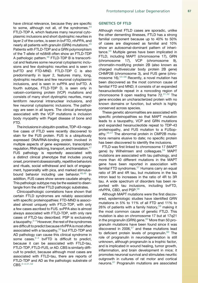
different and reflects selective injury of certainareas of the brain, which leads to the diverse signsand symptoms.1 Specific atrophy patterns evidenton structural images have been the basis of muchof the current research in FTD,68–71 and recentresearch shows that imaging has prognostic impli-cations.72 bvFTD is associated with loss of graymatter (GM) in the frontal and temporal lobes andin particular the ventromedial frontal cortex, theposterior orbital frontal regions, the insula bilater-ally, and the anterior cingulate cortex (ACC)(Fig. 1).68 These regions are components of theemotional processing systems of the brain,73 sothat their involvement in FTD explains the uniquebehavioral symptoms seen in that disorder.74–77
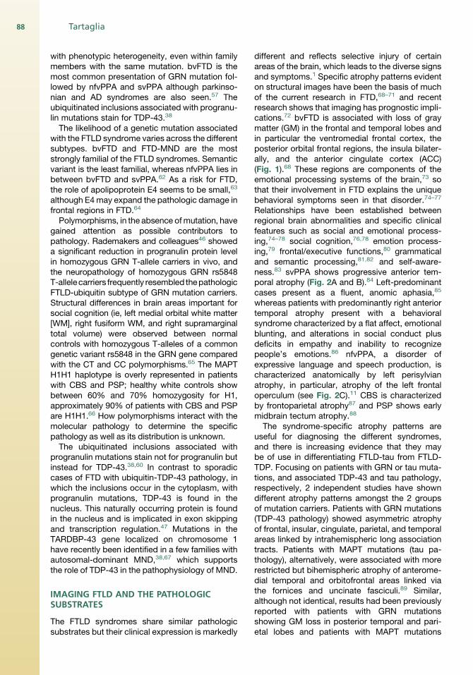
Relationships have been established betweenregional brain abnormalities and specific clinicalfeatures such as social and emotional process-ing,74–78 social cognition,76,78 emotion process-ing,79 frontal/executive functions,80 grammaticaland semantic processing,81,82 and self-aware-ness.83 svPPA shows progressive anterior tem-poral atrophy (Fig. 2A and B).84 Left-predominantcases present as a fluent, anomic aphasia,85
whereas patients with predominantly right anteriortemporal atrophy present with a behavioralsyndrome characterized by a flat affect, emotionalblunting, and alterations in social conduct plusdeficits in empathy and inability to recognizepeople’s emotions.86 nfvPPA, a disorder ofexpressive language and speech production, ischaracterized anatomically by left perisylvianatrophy, in particular, atrophy of the left frontaloperculum (see Fig. 2C).11 CBS is characterizedby frontoparietal atrophy87 and PSP shows earlymidbrain tectum atrophy.88
The syndrome-specific atrophy patterns areuseful for diagnosing the different syndromes,and there is increasing evidence that they maybe of use in differentiating FTLD-tau from FTLD-TDP. Focusing on patients with GRN or tau muta-tions, and associated TDP-43 and tau pathology,respectively, 2 independent studies have showndifferent atrophy patterns amongst the 2 groupsof mutation carriers. Patients with GRN mutations(TDP-43 pathology) showed asymmetric atrophyof frontal, insular, cingulate, parietal, and temporalareas linked by intrahemispheric long associationtracts. Patients with MAPT mutations (tau pa-thology), alternatively, were associated with morerestricted but bihemispheric atrophy of anterome-dial temporal and orbitofrontal areas linked viathe fornices and uncinate fasciculi.89 Similar,although not identical, results had been previouslyreported with patients with GRN mutationsshowing GM loss in posterior temporal and pari-etal lobes and patients with MAPT mutations
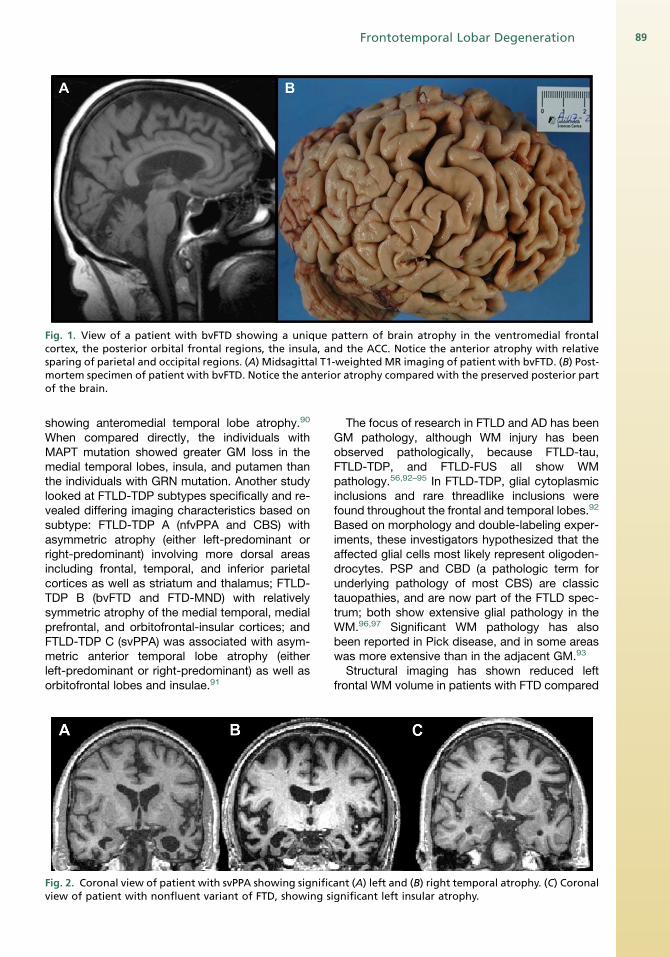
Fig. 1. View of a patient with bvFTD showing a unique pattern of brain atrophy in the ventromedial frontalcortex, the posterior orbital frontal regions, the insula, and the ACC. Notice the anterior atrophy with relativesparing of parietal and occipital regions. (A) Midsagittal T1-weighted MR imaging of patient with bvFTD. (B) Post-mortem specimen of patient with bvFTD. Notice the anterior atrophy compared with the preserved posterior partof the brain.
Frontotemporal Lobar Degeneration 89
showing anteromedial temporal lobe atrophy.90
When compared directly, the individuals withMAPT mutation showed greater GM loss in themedial temporal lobes, insula, and putamen thanthe individuals with GRN mutation. Another studylooked at FTLD-TDP subtypes specifically and re-vealed differing imaging characteristics based onsubtype: FTLD-TDP A (nfvPPA and CBS) withasymmetric atrophy (either left-predominant orright-predominant) involving more dorsal areasincluding frontal, temporal, and inferior parietalcortices as well as striatum and thalamus; FTLD-TDP B (bvFTD and FTD-MND) with relativelysymmetric atrophy of the medial temporal, medialprefrontal, and orbitofrontal-insular cortices; andFTLD-TDP C (svPPA) was associated with asym-metric anterior temporal lobe atrophy (eitherleft-predominant or right-predominant) as well asorbitofrontal lobes and insulae.91
Fig. 2. Coronal view of patient with svPPA showing significview of patient with nonfluent variant of FTD, showing s
The focus of research in FTLD and AD has beenGM pathology, although WM injury has beenobserved pathologically, because FTLD-tau,FTLD-TDP, and FTLD-FUS all show WMpathology.56,92–95 In FTLD-TDP, glial cytoplasmicinclusions and rare threadlike inclusions werefound throughout the frontal and temporal lobes.92
Based on morphology and double-labeling exper-iments, these investigators hypothesized that theaffected glial cells most likely represent oligoden-drocytes. PSP and CBD (a pathologic term forunderlying pathology of most CBS) are classictauopathies, and are now part of the FTLD spec-trum; both show extensive glial pathology in theWM.96,97 Significant WM pathology has alsobeen reported in Pick disease, and in some areaswas more extensive than in the adjacent GM.93
Structural imaging has shown reduced leftfrontal WM volume in patients with FTD compared
ant (A) left and (B) right temporal atrophy. (C) Coronalignificant left insular atrophy.
Tartaglia90
with controls.98 Diffusion tensor imaging (DTI)informs on the integrity of WM by providingdifferent metrics related to the diffusivity of water:axial diffusivity is parallel to the axonal directionand alterations are believed to reflect primarilyaxonal injury; radial diffusivity is perpendicular tothe axon and believed to relate primarily to myelindamage.99,100 The axial and radial parameters canbe averaged to provide amean diffusivity (MD) andcan be used to compute the fractional anisotropy(FA).101 One of the advantages of DTI is that it ispossible to investigate specific WM tracts suchas the uncinate fasciculus (Unc) (Fig. 3) and evensegregate out subcomponents of complex tractslike the superior longitudinal fasciculus (SLF)(Fig. 4). Comparing bvFTD and AD with normalcontrols and each other, reduced FA wasobserved in the anterior corpus callosum, bilateralanterior and descending cingulum (Cg) tracts, andUnc in patients with bvFTD compared with normalage-matched controls and in the anterior corpuscallosum and bilateral Unc fasciculus comparedwith patients with AD.102 Patients with AD alsoshowed areas of decreased FA compared withcontrols but in a more restricted fashion and theyshowed no areas of worse damage whencompared with bvFTD.A DTI study evaluated WM alterations in specific
tracts in patients with PPA and found strikingdifferences according to the clinical syndrome. InnfvPPA, focal injury was observed in the SLF andwhen looking closely at all the subcomponents ofthe SLF, all subcomponents were affected.103
Patients with semantic variant showed severeinvolvement of the Unc bilaterally and the anteriorportion of the inferior longitudinal fasciculus (ILF).
Fig. 3. A three-dimensional reconstruction of DTI-derived uncinate in normal controls. (Courtesy ofS. Galantucci.)
The logopenic patients showed the most consis-tent DTI changes in the left temporoparietalcomponent of the SLF. These findings are impor-tant for clinical expression of these PPA variantsand so the dorsal network injury in nfvPPA maybe partially responsible for the motor speech diffi-culties and agrammatism typical of this variant,whereas a dysfunction of the ventral languagesystem accounts for the typical combination ofimpaired semantics and spared phonology,grammar, and fluency language domains insvPPA. In logopenic patients, tracts important forsentence repetition and phonological short-termmemory are most injured.104 Furthermore, nonflu-ent and semantic variants showed differentpatterns of alterations in their DTI metrics. ThenfvPPA was sparing of their axial diffusivitycompared with controls but the semantic variantshad significantly different FA, MD, axial, and radialdiffusivity compared with controls, suggesting thatthe svPPA may have both axonal and myelindamage; in the nonfluent variant, the pathologymay be restricted to the myelin.104
In an effort to try to understand the contributionof WM tract injury to some of the cognitive andbehavioral deficits observed in FTLD, severalstudies have included both GM and WM metrics.A look at executive dysfunction in bvFTD revealedthe left anterior Cg tract to be a significantpredictor of executive impairments, even after cor-recting for GM volumes adjacent to the tract.105 Arecent study examining syntax in patients withPPA showed that the arcuate fasciculus/SLFcorrelated significantly with syntax productionand comprehension and not with semantic func-tion, whereas the extreme capsule fiber system,a more recently describe WM tract, whichconnects the frontal operculum to midposteriortemporal cortex, was predictive of lexical process-ing but not syntax (Fig. 5).106
We have used DTI and GM volumetrics toassess the relative contribution of WM and GMinjury to social cognitive dysfunction in PPA.107
We found a relationship between emotion detec-tion and WM tract integrity in bilateral Unc andbilateral anterior ILF in addition to the bilateral or-bitofrontal GM, and bilateral anterior temporallobe GM. Certain personality traits like coldnesscorrelated with WM tract injury in right anteriorILF, right Unc, and right arcuate in addition toGM in the anterior temporal, posterior temporal,and orbitofrontal cortex. Overall, these resultssuggest that WM connections in right anteriortemporal and ventromedial frontal regions mayalso be important for emotional sensitivityand expressiveness, in addition to the knownGM-personality relationships in these regions.
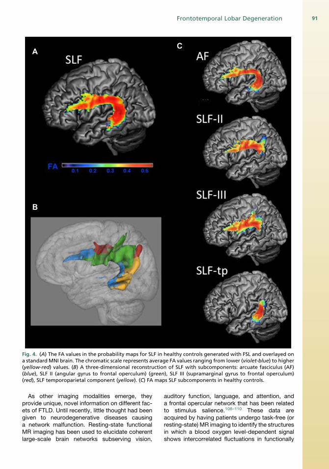
Fig. 4. (A) The FA values in the probability maps for SLF in healthy controls generated with FSL and overlayed ona standardMNI brain. The chromatic scale represents average FA values ranging from lower (violet-blue) to higher(yellow-red) values. (B) A three-dimensional reconstruction of SLF with subcomponents: arcuate fasciculus (AF)(blue), SLF II (angular gyrus to frontal operculum) (green), SLF III (supramarginal gyrus to frontal operculum)(red), SLF temporoparietal component (yellow). (C) FA maps SLF subcomponents in healthy controls.
Frontotemporal Lobar Degeneration 91
As other imaging modalities emerge, theyprovide unique, novel information on different fac-ets of FTLD. Until recently, little thought had beengiven to neurodegenerative diseases causinga network malfunction. Resting-state functionalMR imaging has been used to elucidate coherentlarge-scale brain networks subserving vision,
auditory function, language, and attention, anda frontal opercular network that has been relatedto stimulus salience.108–110 These data areacquired by having patients undergo task-free (orresting-state) MR imaging to identify the structuresin which a blood oxygen level-dependent signalshows intercorrelated fluctuations in functionally
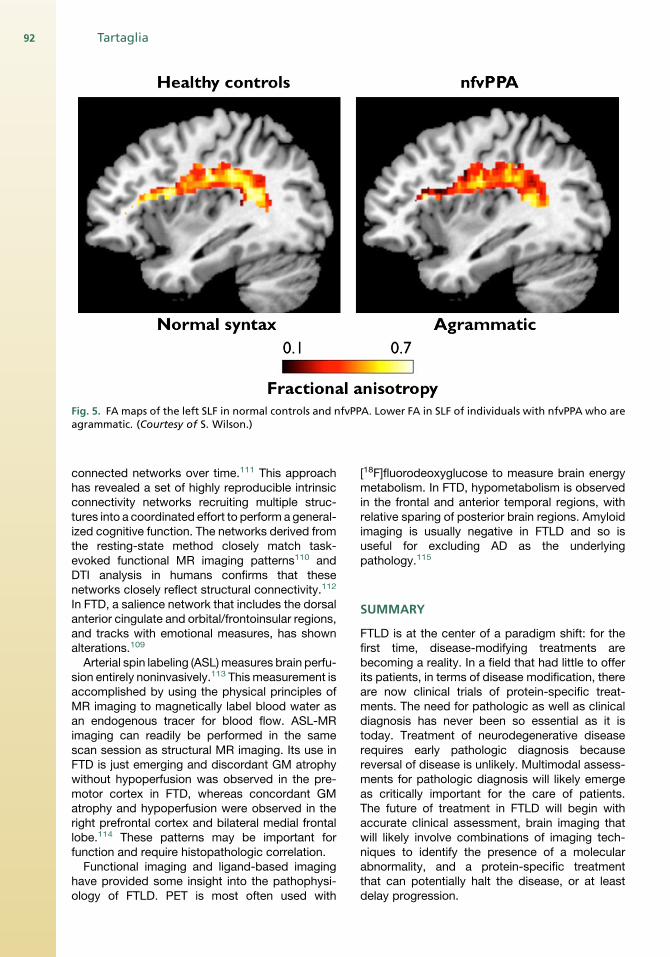
Fig. 5. FA maps of the left SLF in normal controls and nfvPPA. Lower FA in SLF of individuals with nfvPPA who areagrammatic. (Courtesy of S. Wilson.)
Tartaglia92
connected networks over time.111 This approachhas revealed a set of highly reproducible intrinsicconnectivity networks recruiting multiple struc-tures into a coordinated effort to perform a general-ized cognitive function. The networks derived fromthe resting-state method closely match task-evoked functional MR imaging patterns110 andDTI analysis in humans confirms that thesenetworks closely reflect structural connectivity.112
In FTD, a salience network that includes the dorsalanterior cingulate and orbital/frontoinsular regions,and tracks with emotional measures, has shownalterations.109
Arterial spin labeling (ASL)measures brain perfu-sion entirely noninvasively.113 This measurement isaccomplished by using the physical principles ofMR imaging to magnetically label blood water asan endogenous tracer for blood flow. ASL-MRimaging can readily be performed in the samescan session as structural MR imaging. Its use inFTD is just emerging and discordant GM atrophywithout hypoperfusion was observed in the pre-motor cortex in FTD, whereas concordant GMatrophy and hypoperfusion were observed in theright prefrontal cortex and bilateral medial frontallobe.114 These patterns may be important forfunction and require histopathologic correlation.Functional imaging and ligand-based imaging
have provided some insight into the pathophysi-ology of FTLD. PET is most often used with
[18F]fluorodeoxyglucose to measure brain energymetabolism. In FTD, hypometabolism is observedin the frontal and anterior temporal regions, withrelative sparing of posterior brain regions. Amyloidimaging is usually negative in FTLD and so isuseful for excluding AD as the underlyingpathology.115
SUMMARY
FTLD is at the center of a paradigm shift: for thefirst time, disease-modifying treatments arebecoming a reality. In a field that had little to offerits patients, in terms of disease modification, thereare now clinical trials of protein-specific treat-ments. The need for pathologic as well as clinicaldiagnosis has never been so essential as it istoday. Treatment of neurodegenerative diseaserequires early pathologic diagnosis becausereversal of disease is unlikely. Multimodal assess-ments for pathologic diagnosis will likely emergeas critically important for the care of patients.The future of treatment in FTLD will begin withaccurate clinical assessment, brain imaging thatwill likely involve combinations of imaging tech-niques to identify the presence of a molecularabnormality, and a protein-specific treatmentthat can potentially halt the disease, or at leastdelay progression.
Frontotemporal Lobar Degeneration 93
SUMMARY CONCEPTS
1. FTD is a clinical term that encompassesa heterogeneous group of patients who sharefocal degeneration within the anterior frontal,temporal, and insular regions. This termincludes bvFTD, nfvPPA, and svPPA.
2. FTLD is the pathologic term that encompassesseveral syndromes that involve focal atrophy.Recently, the discovery of overlapping geneticsand disease has led to the incorporation of 3other clinical phenotypes under this rubric sothat now, in addition to bvFTD, nfvPPA, andsvPPA, PSP, CBD, and FTD with MND havebeen added.
3. bvFTD is associated with an early change inpersonality and behavior. bvFTD is associatedwith GM atrophy in the frontal and temporallobes, in particular the ventromedial frontalcortex, the posterior orbital frontal regions, theinsula bilaterally, and the ACC.
4. nfvPPA presents with nonfluent speech andlanguage. nfvPPA is characterized anatomicallyby left perisylvian atrophy, in particular, atrophyof the left frontal operculum (Broca areas 44,45, and 47).
5. svPPA is a disorder of semantic knowledge forwords. svPPA shows progressive anteriortemporal atrophy with the clinical syndromedetermined by the side of the brain withthe greatest atrophy. Left-predominant casespresent as a fluent, anomic aphasia, whereasright anterior temporal atrophy presents witha behavioral syndrome characterized by a flataffect, emotional blunting, and alterations insocial conduct plus deficits in empathy andinability to recognize people’s emotions.
6. FTD is the second most common dementia inthose less than 65 years of age; it accountsfor 5% to 6% of all dementias and is respon-sible for up to 17% of early-onset (<70 years)dementias in autopsy series.
7. FTLD can be classified into 2 main pathologicsubtypes based on the pattern of neuronaland glial inclusion: (1) tau-positive lesions withtau-positive inclusions (Pick disease, CBD,PSP) and (2) tau-negative, ubiquitin-positiveinclusions, including TDP-43 and FUS.
8. GM and WM pathology contribute to some ofthe deficits seen in FTLD.
9. GM andWMpathology can be evaluated in vivousing MR imaging.
REFERENCES
1. Brun A. Frontal lobe degeneration of non-Alzheimer
type revisited. Dementia 1993;4:126–31.
2. Snowden JS, Thompson JC, Stopford CL, et al. The
clinical diagnosis of early-onset dementias: diag-
nostic accuracy and clinicopathological relation-
ships. Brain 2011;134(Pt 9):2478–92.
3. Josephs KA, Hodges JR, Snowden JS, et al.
Neuropathological background of phenotypical
variability in frontotemporal dementia. Acta Neuro-
pathol 2011;122:137–53.
4. Rohrer J, Lashley T, Schott J, et al. Clinical and
neuroanatomical signatures of tissue pathology in
frontotemporal lobar degeneration. Brain 2011;
134(Pt 9):2565–81.
5. Rascovsky K, Hodges JR, Knopman D, et al. Sensi-
tivity of revised diagnostic criteria for the behaviou-
ral variant of frontotemporal dementia. Brain 2011;
134(Pt 9):2456–77.
6. Gorno-Tempini M, Hillis AE, Weintraub S, et al.
Classification of primary progressive aphasia
and its variants. Neurology 2011;76(11):1006–14.
7. Gustafson L. Frontal lobe degeneration of non-
Alzheimer type. II. Clinical picture and differential
diagnosis. Arch Gerontol Geriatr 1987;6:209–23.
8. Neary D, Snowden JS, Northen B, et al. Dementia
of frontal lobe type. J Neurol Neurosurg Psychiatry
1988;51:353–61.
9. Miller BL, Cummings JL, Villanueva-Meyer J, et al.
Frontal lobe degeneration: clinical, neuropsycho-
logical, and SPECT characteristics. Neurology
1991;41:1374–82.
10. The Lund and Manchester Groups. Clinical
and neuropathological criteria for frontotemporal
dementia. J Neurol Neurosurg Psychiatry 1994;
57:416–8.
11. NearyD, SnowdenJS,GustafsonL, et al. Frontotem-
poral lobar degeneration: a consensus on clinical
diagnostic criteria. Neurology 1998;51:1546–54.
12. Gustafson L. Clinical picture of frontal lobe degener-
ationofnon-Alzheimer type.Dementia1993;4:143–8.
13. Miller BL, Darby A, Benson DF, et al. Aggressive,
socially disruptive and antisocial behaviour associ-
ated with fronto-temporal dementia. Br J Psychiatry
1997;170:150–4.
14. Mendez MF, Chen AK, Shapira JS, et al. Acquired
sociopathy and frontotemporal dementia. Dement
Geriatr Cogn Disord 2005;20:99–104.
15. Miller BL, Seeley WW, Mychack P, et al. Neuro-
anatomyof the self: evidence frompatientswith fron-
totemporal dementia. Neurology 2001;57:817–21.
16. Miller BL, Darby AL, Swartz JR, et al. Dietary
changes, compulsions and sexual behavior in fron-
totemporal degeneration. Dementia 1995;6:195–9.
17. Johnson JK, Diehl J, Mendez MF, et al. Frontotem-
poral lobar degeneration: demographic character-
istics of 353 patients. Arch Neurol 2005;62:925–30.
18. Roberson ED, Hesse JH, Rose KD, et al. Frontotem-
poral dementia progresses to death faster than
Alzheimer disease. Neurology 2005;65:719–25.
Tartaglia94
19. Hodges JR, Miller B. The classification, genetics
and neuropathology of frontotemporal dementia.
Introduction to the special topic papers: part I.
Neurocase 2001;7:31–5.
20. Gorno-Tempini ML, Ogar JM, Brambati SM, et al.
Anatomical correlates of early mutism in pro-
gressive nonfluent aphasia. Neurology 2006;67:
1849–51.
21. Boeve BF, Lang AE, Litvan I. Corticobasal degener-
ation and its relationship to progressive supranu-
clear palsy and frontotemporal dementia. Ann
Neurol 2003;54(Suppl 5):S15–9.
22. Gorno-Tempini ML, Dronkers NF, Rankin KP, et al.
Cognition and anatomy in three variants of primary
progressive aphasia. Ann Neurol 2004;55:335–46.
23. Josephs KA, Petersen RC, Knopman DS, et al.
Clinicopathologic analysis of frontotemporal and
corticobasal degenerations and PSP. Neurology
2006;66:41–8.
24. Litvan I, Bhatia KP, Burn DJ, et al. Movement Disor-
ders Society Scientific Issues Committee report:
SIC Task Force appraisal of clinical diagnostic
criteria for Parkinsonian disorders. Mov Disord
2003;18:467–86.
25. Murray R, Neumann M, Forman MS, et al. Cognitive
and motor assessment in autopsy-proven cortico-
basal degeneration. Neurology 2007;68:1274–83.
26. Litvan I, Agid Y, Calne D, et al. Clinical research
criteria for thediagnosisof progressive supranuclear
palsy (Steele-Richardson-Olszewski syndrome):
report of the NINDS-SPSP international workshop.
Neurology 1996;47:1–9.
27. Litvan I, Agid Y, Jankovic J, et al. Accuracy of clinical
criteria for thediagnosisof progressive supranuclear
palsy (Steele-Richardson-Olszewski syndrome).
Neurology 1996;46:922–30.
28. Lomen-Hoerth C, Anderson T, Miller B. The overlap
of amyotrophic lateral sclerosis and frontotemporal
dementia. Neurology 2002;59:1077–9.
29. Lomen-Hoerth C, Murphy J, Langmore S, et al. Are
amyotrophic lateral sclerosis patients cognitively
normal? Neurology 2003;60:1094–7.
30. Foster NL, Wilhelmsen K, Sima AA, et al. Fronto-
temporal dementia and parkinsonism linked to
chromosome 17: a consensus conference. Confer-
ence Participants. Ann Neurol 1997;41:706–15.
31. Renton AE, Waite A, Simon-Sanchez J, et al.
A hexanucleotide repeat expansion in C9ORF72
is the cause of chromosome 9p21-linked ALS-
FTD. Neuron 2011;72(2):257–68.
32. DeJesus-Hernandez M, Boeve BF, Boxer AL, et al.
Expanded GGGGCC hexanucleotide repeat in
noncoding region of C9ORF72 causes chromo-
some 9p-Linked FTD and ALS. Neuron 2011;
72(2):245–56.
33. Cooper PN, Jackson M, Lennox G, et al. Tau, ubiq-
uitin, and alpha B-crystallin immunohistochemistry
define the principal causes of degenerative fronto-
temporal dementia. Arch Neurol 1995;52:1011–5.
34. Strong MJ, Lomen-Hoerth C, Caselli RJ, et al.
Cognitive impairment, frontotemporal dementia,
and the motor neuron diseases. Ann Neurol 2003;
54(Suppl 5):S20–3.
35. Brunden KR, Trojanowski JQ, Lee VM. Advances in
tau-focused drug discovery for Alzheimer’s
disease and related tauopathies. Nat Rev Drug
Discov 2009;8:783–93.
36. Kertesz A. Pick’s complex and FTDP-17. Mov Dis-
ord 2003;18(Suppl 6):S57–62.
37. Tai HC, Schuman EM. Ubiquitin, the proteasome
and protein degradation in neuronal function and
dysfunction. Nat Rev Neurosci 2008;9:826–38.
38. Neumann M, Sampathu DM, Kwong LK, et al.
Ubiquitinated TDP-43 in frontotemporal lobar
degeneration and amyotrophic lateral sclerosis.
Science 2006;314:130–3.
39. Josephs KA, Holton JL, Rossor MN, et al. Fronto-
temporal lobar degeneration and ubiquitin immu-
nohistochemistry. Neuropathol Appl Neurobiol
2004;30:369–73.
40. Bigio EH, Lipton AM, White CL 3rd, et al. Frontotem-
poral and motor neurone degeneration with neurofi-
lament inclusion bodies: additional evidence for
overlap between FTD and ALS. Neuropathol Appl
Neurobiol 2003;29:239–53.
41. Mackenzie IR, Baborie A, Pickering-Brown S, et al.
Heterogeneity of ubiquitin pathology in frontotem-
poral lobar degeneration: classification and relation
to clinical phenotype. Acta Neuropathol 2006;112:
539–49.
42. Sampathu DM, Neumann M, Kwong LK, et al. Path-
ological heterogeneity of frontotemporal lobar
degeneration with ubiquitin-positive inclusions
delineated by ubiquitin immunohistochemistry
and novel monoclonal antibodies. Am J Pathol
2006;169:1343–52.
43. Mackenzie IR, Neumann M, Baborie A, et al.
A harmonized classification system for FTLD-TDP
pathology. Acta Neuropathol 2011;122:111–3.
44. Cairns NJ, Neumann M, Bigio EH, et al. TDP-43 in
familial and sporadic frontotemporal lobar degen-
eration with ubiquitin inclusions. Am J Pathol
2007;171:227–40.
45. Mackenzie IR, Baker M, Pickering-Brown S, et al.
The neuropathology of frontotemporal lobar degen-
eration caused by mutations in the progranulin
gene. Brain 2006;129:3081–90.
46. Rademakers R, Eriksen JL, Baker M, et al. Common
variation in the miR-659 binding-site of GRN is
a major risk factor for TDP43-positive frontotem-
poral dementia. Hum Mol Genet 2008;17:3631–42.
47. Buratti E, Baralle FE. Multiple roles of TDP-43 in
gene expression, splicing regulation, and human
disease. Front Biosci 2008;13:867–78.
Frontotemporal Lobar Degeneration 95
48. Rohrer JD, Lashley T, Holton J, et al. The clinical
and neuroanatomical phenotype of FUS associ-
ated frontotemporal lobar degeneration. J Neurol
Neurosurg Psychiatry 2011;82(12):1405–7.
49. Snowden JS, Hu Q, Rollinson S, et al. The
most common type of FTLD-FUS (aFTLD-U) is
associated with a distinct clinical form of frontotem-
poral dementia but is not related to mutations in the
FUS gene. Acta Neuropathol 2011;122:99–110.
50. Grossman M. Primary progressive aphasia: clinico-
pathological correlations. Nat Rev Neurol 2010;6:
88–97.
51. Neumann M, Tolnay M, Mackenzie IR. The molec-
ular basis of frontotemporal dementia. Expert Rev
Mol Med 2009;11:e23.
52. Tartaglia MC, Sidhu M, Laluz V, et al. Sporadic cor-
ticobasal syndrome due to FTLD-TDP. Acta Neuro-
pathol 2009;119:365–74.
53. Seelaar H, Kamphorst W, Rosso SM, et al. Distinct
genetic forms of frontotemporal dementia. Neurology
2008;71:1220–6.
54. Wilhelmsen KC, Lynch T, Pavlou E, et al. Localization
of disinhibition-dementia-parkinsonism-amyotrophy
complex to 17q21-22. Am J Hum Genet 1994;55:
1159–65.
55. Bigio EH. Update on recent molecular and genetic
advances in frontotemporal lobar degeneration.
J Neuropathol Exp Neurol 2008;67:635–48.
56. Neumann M, Rademakers R, Roeber S, et al.
A new subtype of frontotemporal lobar degenera-
tion with FUS pathology. Brain 2009;132:2922–31.
57. Rademakers R, Hutton M. The genetics of fronto-
temporal lobar degeneration. Curr Neurol Neurosci
Rep 2007;7:434–42.
58. Bugiani O. The many ways to frontotemporal
degeneration and beyond. Neurol Sci 2007;28:
241–4.
59. Mackenzie IR. The neuropathology and clinical
phenotype of FTD with progranulin mutations.
Acta Neuropathol 2007;114:49–54.
60. Baker M, Mackenzie IR, Pickering-Brown SM, et al.
Mutations in progranulin cause tau-negative fronto-
temporal dementia linked to chromosome 17.
Nature 2006;442:916–9.
61. Ahmed Z, Mackenzie IR, Hutton ML, et al. Progra-
nulin in frontotemporal lobar degeneration and
neuroinflammation. J Neuroinflammation 2007;4:7.
62. Goldman JS, Farmer JM, Van Deerlin VM, et al.
Frontotemporal dementia: genetics and genetic
counseling dilemmas. Neurologist 2004;10:227–34.
63. Geschwind D, Karrim J, Nelson SF, et al. The apoli-
poprotein E epsilon4 allele is not a significant risk
factor for frontotemporal dementia. Ann Neurol
1998;44:134–8.
64. Agosta F, Vossel KA, Miller BL, et al. Apolipoprotein
E epsilon4 is associated with disease-specific
effects on brain atrophy in Alzheimer’s disease
and frontotemporal dementia. Proc Natl Acad Sci
U S A 2009;106:2018–22.
65. Tartaglia MC, Laluz V, Rademakers R, et al. Poly-
morphism in Progranulin gene associated with
structural differences in brains of normal controls.
Neurology 2010;74.
66. Houlden H, Baker M, Morris HR, et al. Corticobasal
degeneration and progressive supranuclear palsy
share a common tau haplotype. Neurology 2001;
56:1702–6.
67. Arai T, Hasegawa M, Akiyama H, et al. TDP-43 is
a component of ubiquitin-positive tau-negative
inclusions in frontotemporal lobar degeneration
and amyotrophic lateral sclerosis. Biochem Bio-
phys Res Commun 2006;351:602–11.
68. Rosen HJ, Gorno-Tempini ML, Goldman WP, et al.
Patterns of brain atrophy in frontotemporal
dementia and semantic dementia. Neurology
2002;58:198–208.
69. Whitwell JL, Josephs KA, Rossor MN, et al.
Magnetic resonance imaging signatures of tissue
pathology in frontotemporal dementia. Arch Neurol
2005;62:1402–8.
70. Rabinovici GD, Seeley WW, Kim EJ, et al. Distinct
MRI atrophy patterns in autopsy-proven Alzheimer’s
disease and frontotemporal lobar degeneration. Am
J Alzheimers Dis Other Demen 2007;22:474–88.
71. Seeley WW, Crawford RK, Zhou J, et al. Neurode-
generative diseases target large-scale human
brain networks. Neuron 2009;62:42–52.
72. Kipps CM, Mioshi E, Hodges JR. Emotion, social
functioning and activities of daily living in fronto-
temporal dementia. Neurocase 2009;15:182–9.
73. Phillips ML, Drevets WC, Rauch SL, et al. Neurobi-
ology of emotion perception I: the neural basis of
normal emotion perception. Biol Psychiatry 2003;
54:504–14.
74. Rosen HJ, Allison SC, Ogar JM, et al. Behavioral
features in semantic dementia vs other forms of
progressive aphasias. Neurology 2006;67:1752–6.
75. Rosen HJ, Allison SC, Schauer GF, et al. Neuroan-
atomical correlates of behavioural disorders in
dementia. Brain 2005;128:2612–25.
76. Rankin KP, Gorno-Tempini ML, Allison SC, et al.
Structural anatomy of empathy in neurodegenera-
tive disease. Brain 2006;129:2945–56.
77. Rankin KP, Salazar A, Gorno-Tempini ML, et al. De-
tecting sarcasm from paralinguistic cues: anatomic
and cognitive correlates in neurodegenerative
disease. Neuroimage 2009;47:2005–15.
78. Sollberger M, Stanley CM, Wilson SM, et al. Neural
basis of interpersonal traits in neurodegenerative
diseases. Neuropsychologia 2009;47:2812–27.
79. Rosen HJ, Wilson MR, Schauer GF, et al. Neuroan-
atomical correlates of impaired recognition of
emotion in dementia. Neuropsychologia 2006;44:
365–73.
Tartaglia96
80. Carey CL, Woods SP, Damon J, et al. Discriminant
validity andneuroanatomical correlates of rulemoni-
toring in frontotemporal dementia and Alzheimer’s
disease. Neuropsychologia 2008;46:1081–7.
81. Amici S, Brambati SM, Wilkins DP, et al. Anatomical
correlates of sentence comprehension and verbal
working memory in neurodegenerative disease.
J Neurosci 2007;27:6282–90.
82. Wilson SM, Brambati SM, Henry RG, et al. The
neural basis of surface dyslexia in semantic
dementia. Brain 2009;132:71–86.
83. RosenHJ,AlcantarO,Rothlind J, et al. Neuroanatom-
ical correlates of cognitive self-appraisal in neurode-
generative disease. Neuroimage 2009;49:3358–64.
84. Thompson PM, Hayashi KM, de Zubicaray G, et al.
Dynamics of gray matter loss in Alzheimer’s
disease. J Neurosci 2003;23:994–1005.
85. Seeley WW, Bauer AM, Miller BL, et al. The natural
history of temporal variant frontotemporal dementia.
Neurology 2005;64:1384–90.
86. Gorno-Tempini ML, Rankin KP, Woolley JD, et al.
Cognitive and behavioral profile in a case of right
anterior temporal lobe neurodegeneration. Cortex
2004;40:631–44.
87. Lee SE, Rabinovici GD, Mayo MC, et al. Clinico-
pathological correlations in corticobasal degenera-
tion. Ann Neurol 2011;70:327–40.
88. Boxer AL, Geschwind MD, Belfor N, et al. Patterns
of brain atrophy that differentiate corticobasal
degeneration syndrome from progressive supranu-
clear palsy. Arch Neurol 2006;63:81–6.
89. Rohrer JD, Ridgway GR, Modat M, et al. Distinct
profiles of brain atrophy in frontotemporal lobar
degeneration caused by progranulin and tau muta-
tions. Neuroimage 2010;53:1070–6.
90. Whitwell JL, Jack CR Jr, Boeve BF, et al. Voxel-
based morphometry patterns of atrophy in FTLD
with mutations in MAPT or PGRN. Neurology
2009;72:813–20.
91. Rohrer JD, Geser F, Zhou J, et al. TDP-43 subtypes
are associated with distinct atrophy patterns
in frontotemporal dementia. Neurology 2010;75:
2204–11.
92. Neumann M, Kwong LK, Truax AC, et al. TDP-43-
positive white matter pathology in frontotemporal
lobar degeneration with ubiquitin-positive inclu-
sions. J Neuropathol Exp Neurol 2007;66:177–83.
93. Zhukareva V, Mann D, Pickering-Brown S, et al.
Sporadic Pick’s disease: a tauopathy characterized
by a spectrum of pathological tau isoforms in gray
and white matter. Ann Neurol 2002;51:730–9.
94. Englund E. Neuropathology of white matter
changes in Alzheimer’s disease and vascular
dementia. Dement Geriatr Cogn Disord 1998;
9(Suppl 1):6–12.
95. Braak H, Braak E. Cortical and subcortical argyro-
philic grains characterize a disease associated
with adult onset dementia. Neuropathol Appl Neu-
robiol 1989;15:13–26.
96. Zhukareva V, Joyce S, Schuck T, et al. Unexpected
abundance of pathological tau in progressive
supranuclear palsy white matter. Ann Neurol
2006;60:335–45.
97. Dickson DW, Bergeron C, Chin SS, et al. Office of
Rare Diseases neuropathologic criteria for cortico-
basal degeneration. J Neuropathol Exp Neurol
2002;61:935–46.
98. Chao LL, Schuff N, Clevenger EM, et al. Patterns
of white matter atrophy in frontotemporal lobar
degeneration. Arch Neurol 2007;64:1619–24.
99. Beaulieu C. The basis of anisotropic water diffusion
in the nervous system–a technical review. NMR
Biomed 2002;15:435–55.
100. Song SK, Sun SW, Ramsbottom MJ, et al. Dysmye-
lination revealed through MRI as increased radial
(but unchanged axial) diffusion of water. Neuro-
image 2002;17:1429–36.
101. Basser PJ, Mattiello J, LeBihan D. MR diffusion
tensor spectroscopy and imaging. Biophys J
1994;66:259–67.
102. Zhang Y, Schuff N, Du AT, et al. White matter
damage in frontotemporal dementia and Alz-
heimer’s disease measured by diffusion MRI. Brain
2009;132:2579–92.
103. Galantucci S, Tartaglia MC, Wilson SM, et al. White
matter damage in primary progressive aphasias:
a diffusion tensor tractography study. Brain 2011;
134(Pt 10):3011–29.
104. Gorno-Tempini ML, Brambati SM, Ginex V, et al.
The logopenic/phonological variant of primary
progressive aphasia. Neurology 2008;71:1227–34.
105. Tartaglia MC, Zhang Y, Racine C, et al. Executive
dysfunction in frontotemporal dementia is related
to abnormalities in frontal white matter tracts.
Neurology 2009;72:A437.
106. Wilson SM, Galantucci S, Tartaglia MC, et al.
Syntactic processing depends on dorsal language
tracts. 2011;72(2):397–403.
107. Tartaglia MC, Galantucci S, Wilson SM, et al. White
matter tract integrity corresponds with individual
differences in emotional expression and sensitivity.
Dement Geriatr Cognit Disord 2010;30(Suppl 1):84.
108. Fox MD, Corbetta M, Snyder AZ, et al. Sponta-
neous neuronal activity distinguishes human dorsal
and ventral attention systems. Proc Natl Acad Sci
U S A 2006;103:10046–51.
109. Seeley WW, Menon V, Schatzberg AF, et al. Disso-
ciable intrinsic connectivity networks for salience
processing and executive control. J Neurosci
2007;27:2349–56.
110. Smith SM, Fox PT, Miller KL, et al. Correspondence
of the brain’s functional architecture during activa-
tion and rest. Proc Natl Acad Sci U S A 2009;106:
13040–5.

Frontotemporal Lobar Degeneration 97
111. Fox MD, Snyder AZ, Vincent JL, et al. The human
brain is intrinsically organized into dynamic, anti-
correlated functional networks. Proc Natl Acad
Sci U S A 2005;102:9673–8.
112. Greicius MD, Supekar K, Menon V, et al. Resting-
state functional connectivity reflects structural
connectivity in the default mode network. Cereb
Cortex 2009;19:72–8.
113. Roberts DA, Detre JA, Bolinger L, et al. Quantitative
magnetic resonance imaging of human brain
perfusion at 1.5 T using steady-state inversion of
arterial water. Proc Natl Acad Sci U S A 1994;91:
33–7.
114. Shimizu S, Zhang Y, Laxamana J, et al. Concor-
dance and discordance between brain perfusion
and atrophy in frontotemporal dementia. Brain
Imaging Behav 2010;4:46–54.
115. RabinoviciGD,FurstAJ,O’Neil JP, et al. 11C-PIBPET
imaging in Alzheimer disease and frontotemporal
lobar degeneration. Neurology 2007;68:1205–12.