Embed Size (px)
Citation preview

Copyright © 2008 Pearson Education, Inc., publishing as Pearson Benjamin Cummings
PowerPoint® Lecture Presentations for
BiologyEighth Edition
Neil Campbell and Jane Reece
Lectures by Chris Romero, updated by Erin Barley with contributions from Joan Sharp
Chapter 17
From Gene to Protein
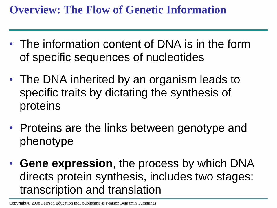
Overview: The Flow of Genetic Information
• The information content of DNA is in the form of specific sequences of nucleotides
• The DNA inherited by an organism leads to specific traits by dictating the synthesis of proteins
• Proteins are the links between genotype and phenotype
• Gene expression, the process by which DNA directs protein synthesis, includes two stages: transcription and translation
Copyright © 2008 Pearson Education Inc., publishing as Pearson Benjamin Cummings
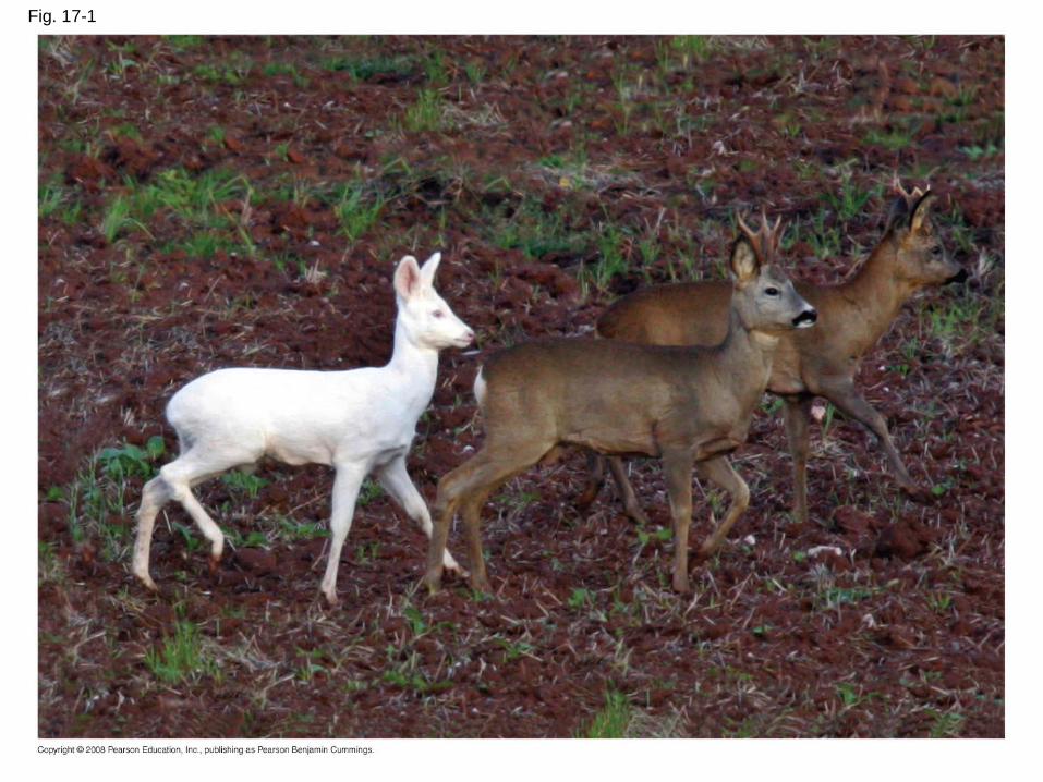
Fig. 17-1

Concept 17.1: Genes specify proteins via transcription and translation
• How was the fundamental relationship between
genes and proteins discovered?
Copyright © 2008 Pearson Education Inc., publishing as Pearson Benjamin Cummings
Evidence from the Study of Metabolic Defects
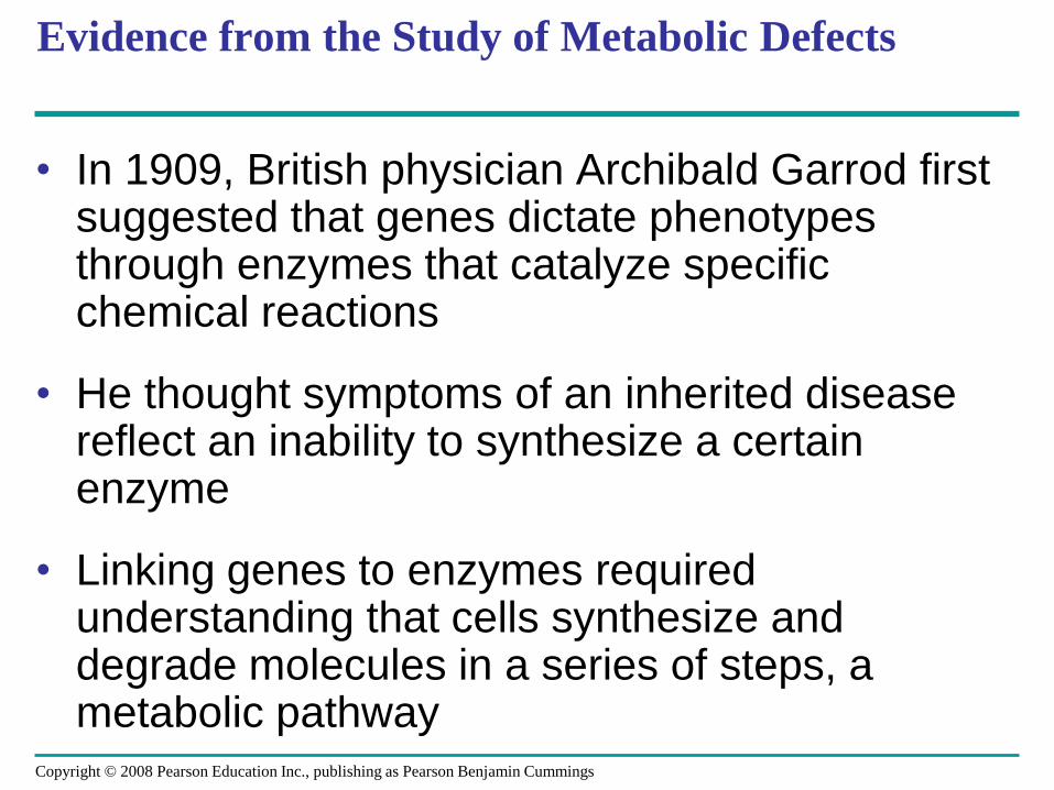
• In 1909, British physician Archibald Garrod first suggested that genes dictate phenotypes through enzymes that catalyze specific chemical reactions
• He thought symptoms of an inherited disease reflect an inability to synthesize a certain enzyme
• Linking genes to enzymes required understanding that cells synthesize and degrade molecules in a series of steps, a metabolic pathway
Copyright © 2008 Pearson Education Inc., publishing as Pearson Benjamin Cummings
Nutritional Mutants in Neurospora: Scientific Inquiry
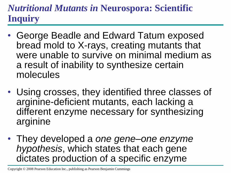
• George Beadle and Edward Tatum exposed bread mold to X-rays, creating mutants that were unable to survive on minimal medium as a result of inability to synthesize certain molecules
• Using crosses, they identified three classes of arginine-deficient mutants, each lacking a different enzyme necessary for synthesizing arginine
• They developed a one gene–one enzyme hypothesis, which states that each gene dictates production of a specific enzyme
Copyright © 2008 Pearson Education Inc., publishing as Pearson Benjamin Cummings
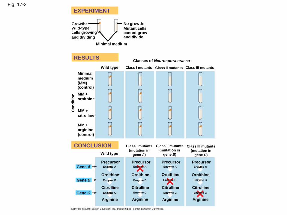
Fig. 17-2
RESULTS
EXPERIMENT
CONCLUSION
Growth:Wild-typecells growing
and dividing
No growth:
Mutant cellscannot grow and divide
Minimal medium
Classes of Neurospora crassa
Wild type Class I mutants Class II mutants Class III mutants
Minimal
medium
(MM)(control)
MM +
ornithine
MM +
citrulline
Co
nd
itio
n
MM +
arginine
(control)
Class I mutants
(mutation in
gene A)Wild type
Class II mutants
(mutation in
gene B)
Class III mutants
(mutation in
gene C)
Gene A
Gene B
Gene C
Precursor Precursor Precursor PrecursorEnzyme A Enzyme AEnzyme A Enzyme A
Enzyme B
Ornithine Ornithine Ornithine Ornithine
Enzyme B Enzyme B Enzyme B
Citrulline Citrulline Citrulline Citrulline
Enzyme C Enzyme C Enzyme C Enzyme C
Arginine Arginine Arginine Arginine
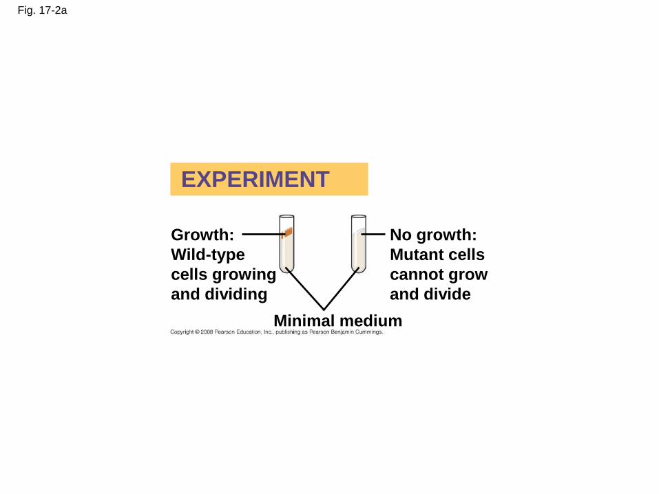
Fig. 17-2a
EXPERIMENT
Growth:
Wild-type
cells growing
and dividing
No growth:
Mutant cells
cannot grow
and divide
Minimal medium
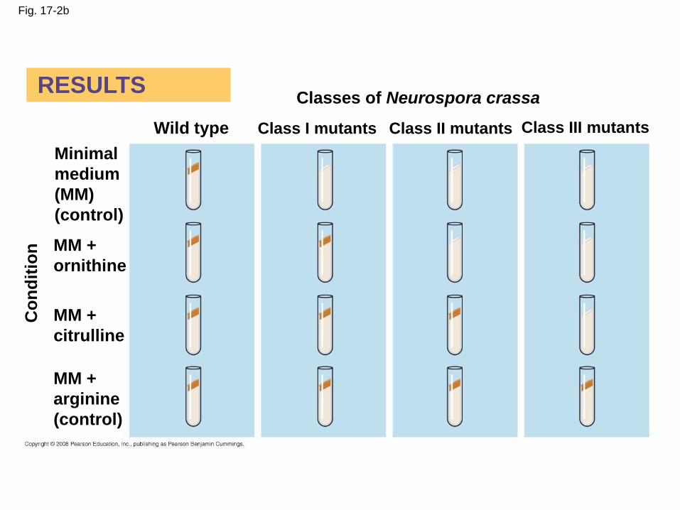
Fig. 17-2b
RESULTSClasses of Neurospora crassa
Wild type Class I mutants Class II mutants Class III mutants
Minimal
medium
(MM)
(control)
MM +
ornithine
MM +
citrulline
MM +
arginine
(control)
Co
nd
itio
n

Fig. 17-2c
CONCLUSION Class I mutants
(mutation in
gene A)
Class II mutants
(mutation in
gene B)
Class III mutants
(mutation in
gene C)Wild type
Precursor Precursor Precursor PrecursorEnzyme AEnzyme AEnzyme AEnzyme A
Ornithine Ornithine Ornithine Ornithine
Enzyme BEnzyme B Enzyme BEnzyme B
Citrulline Citrulline Citrulline Citrulline
Enzyme CEnzyme CEnzyme CEnzyme C
Arginine Arginine Arginine Arginine
Gene A
Gene B
Gene C
The Products of Gene Expression: A Developing Story
• Some proteins aren’t enzymes, so researchers later revised the hypothesis: one gene–one protein
• Many proteins are composed of several polypeptides, each of which has its own gene
• Therefore, Beadle and Tatum’s hypothesis is now restated as the one gene–one polypeptide hypothesis
• Note that it is common to refer to gene products as proteins rather than polypeptides
Copyright © 2008 Pearson Education Inc., publishing as Pearson Benjamin Cummings
Basic Principles of Transcription and Translation
• RNA is the intermediate between genes and
the proteins for which they code
• Transcription is the synthesis of RNA under
the direction of DNA
• Transcription produces messenger RNA
(mRNA)
• Translation is the synthesis of a polypeptide,
which occurs under the direction of mRNA
• Ribosomes are the sites of translationCopyright © 2008 Pearson Education Inc., publishing as Pearson Benjamin Cummings
• In prokaryotes, mRNA produced by
transcription is immediately translated without
more processing
• In a eukaryotic cell, the nuclear envelope
separates transcription from translation
• Eukaryotic RNA transcripts are modified
through RNA processing to yield finished
mRNA
Copyright © 2008 Pearson Education Inc., publishing as Pearson Benjamin Cummings
• A primary transcript is the initial RNA
transcript from any gene
• The central dogma is the concept that cells are
governed by a cellular chain of command: DNA
RNA protein
Copyright © 2008 Pearson Education Inc., publishing as Pearson Benjamin Cummings
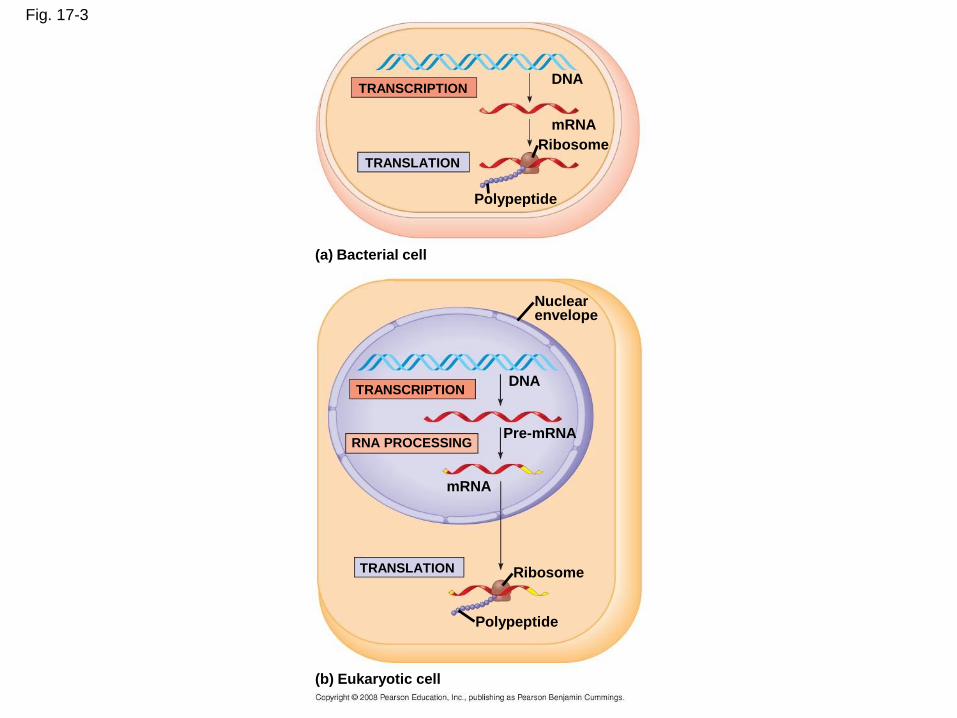
Fig. 17-3
TRANSCRIPTION
TRANSLATION
DNA
mRNA
Ribosome
Polypeptide
(a) Bacterial cell
Nuclearenvelope
TRANSCRIPTION
RNA PROCESSINGPre-mRNA
DNA
mRNA
TRANSLATION Ribosome
Polypeptide
(b) Eukaryotic cell
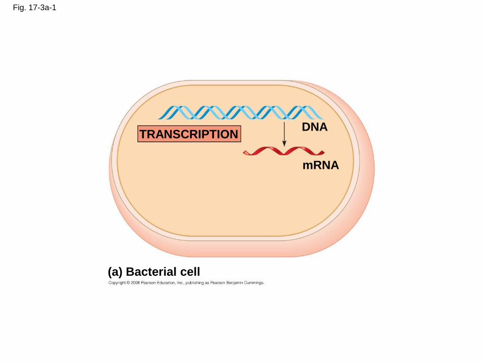
Fig. 17-3a-1
TRANSCRIPTIONDNA
mRNA
(a) Bacterial cell
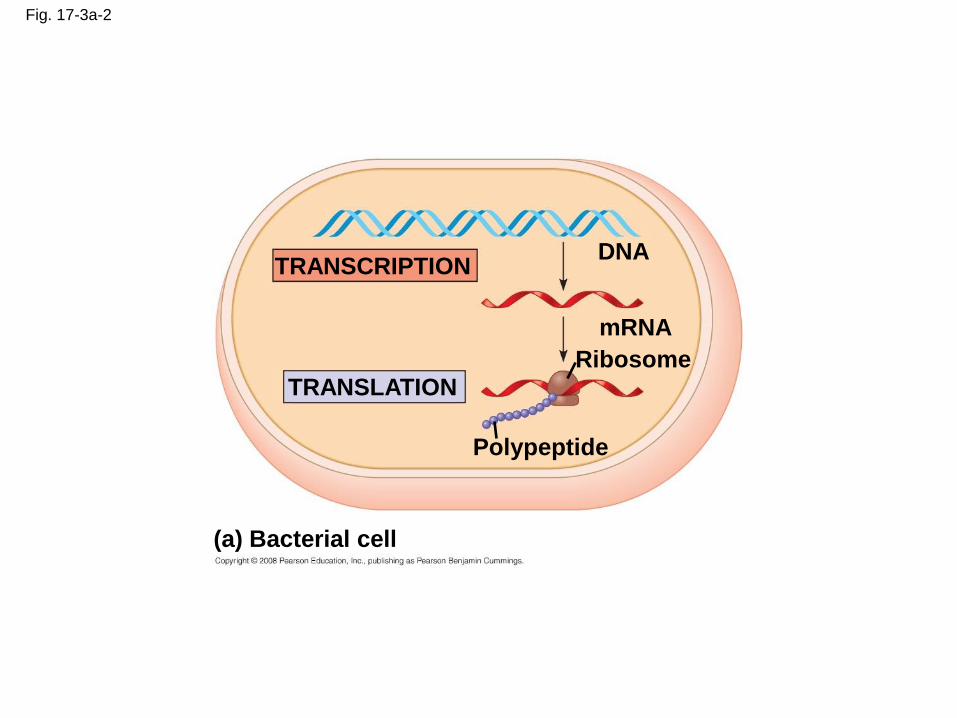
Fig. 17-3a-2
(a) Bacterial cell
TRANSCRIPTIONDNA
mRNA
TRANSLATIONRibosome
Polypeptide

Fig. 17-3b-1
(b) Eukaryotic cell
TRANSCRIPTION
Nuclearenvelope
DNA
Pre-mRNA
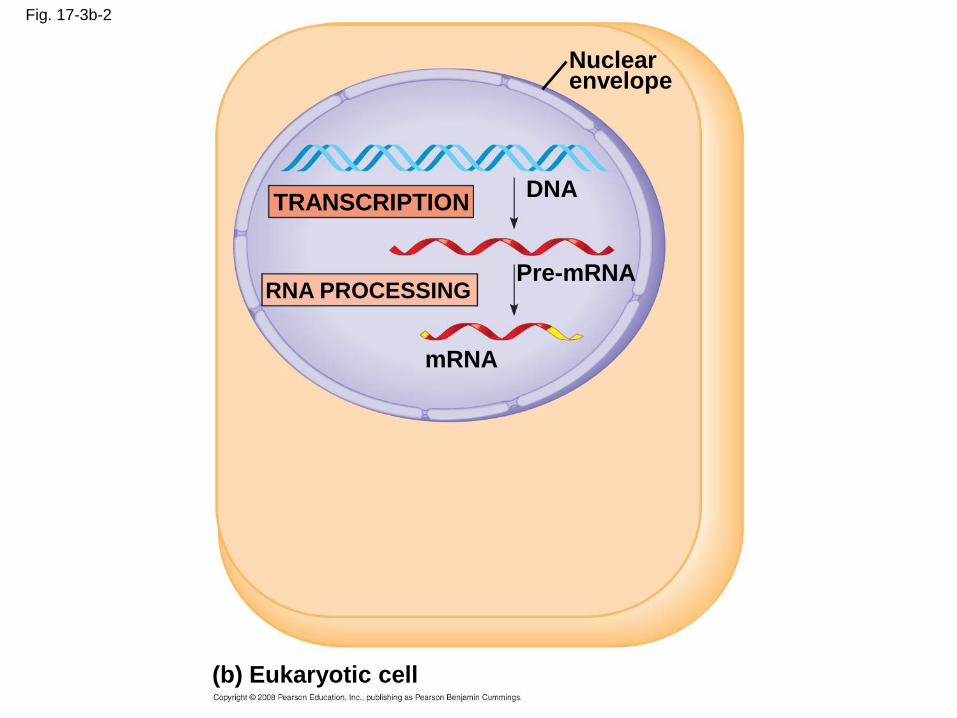
Fig. 17-3b-2
(b) Eukaryotic cell
TRANSCRIPTION
Nuclearenvelope
DNA
Pre-mRNARNA PROCESSING
mRNA
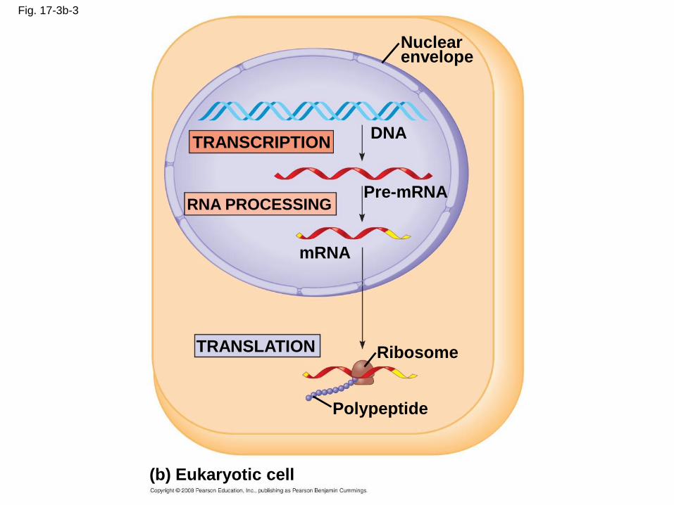
Fig. 17-3b-3
(b) Eukaryotic cell
TRANSCRIPTION
Nuclearenvelope
DNA
Pre-mRNARNA PROCESSING
mRNA
TRANSLATION Ribosome
Polypeptide
The Genetic Code
• How are the instructions for assembling amino
acids into proteins encoded into DNA?
• There are 20 amino acids, but there are only
four nucleotide bases in DNA
• How many bases correspond to an amino
acid?
Copyright © 2008 Pearson Education Inc., publishing as Pearson Benjamin Cummings

Codons: Triplets of Bases
• The flow of information from gene to protein is
based on a triplet code: a series of
nonoverlapping, three-nucleotide words
• These triplets are the smallest units of uniform
length that can code for all the amino acids
• Example: AGT at a particular position on a
DNA strand results in the placement of the
amino acid serine at the corresponding position
of the polypeptide to be produced
Copyright © 2008 Pearson Education Inc., publishing as Pearson Benjamin Cummings
• During transcription, one of the two DNA
strands called the template strand provides a
template for ordering the sequence of
nucleotides in an RNA transcript
• During translation, the mRNA base triplets,
called codons, are read in the 5 to 3 direction
• Each codon specifies the amino acid to be
placed at the corresponding position along a
polypeptide
Copyright © 2008 Pearson Education Inc., publishing as Pearson Benjamin Cummings
• Codons along an mRNA molecule are read by
translation machinery in the 5 to 3 direction
• Each codon specifies the addition of one of 20
amino acids
Copyright © 2008 Pearson Education Inc., publishing as Pearson Benjamin Cummings

Fig. 17-4
DNAmolecule
Gene 1
Gene 2
Gene 3
DNAtemplatestrand
TRANSCRIPTION
TRANSLATION
mRNA
Protein
Codon
Amino acid
Cracking the Code
• All 64 codons were deciphered by the mid-1960s
• Of the 64 triplets, 61 code for amino acids; 3 triplets are “stop” signals to end translation
• The genetic code is redundant but not ambiguous; no codon specifies more than one amino acid
• Codons must be read in the correct reading frame (correct groupings) in order for the specified polypeptide to be produced
Copyright © 2008 Pearson Education Inc., publishing as Pearson Benjamin Cummings
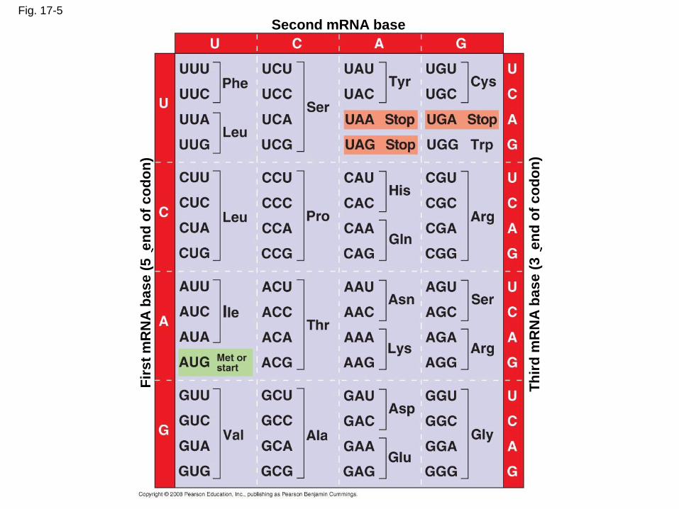
Fig. 17-5
Second mRNA base
Fir
st
mR
NA
ba
se
(5
en
d o
f c
od
on
)
Th
ird
mR
NA
ba
se
(3
en
d o
f c
od
on
)

Evolution of the Genetic Code
• The genetic code is nearly universal, shared by
the simplest bacteria to the most complex
animals
• Genes can be transcribed and translated after
being transplanted from one species to another
Copyright © 2008 Pearson Education Inc., publishing as Pearson Benjamin Cummings
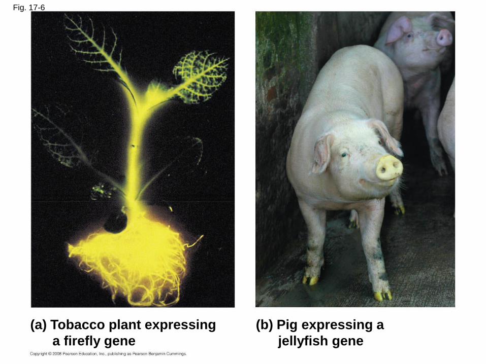
Fig. 17-6
(a) Tobacco plant expressing
a firefly gene
(b) Pig expressing a
jellyfish gene
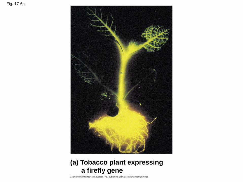
Fig. 17-6a
(a) Tobacco plant expressing
a firefly gene
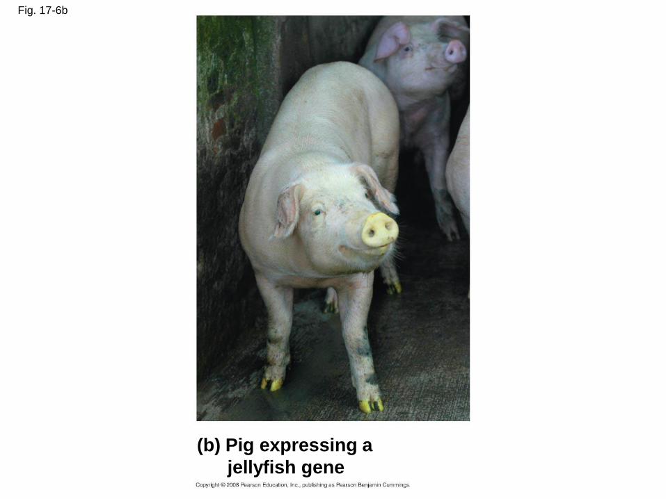
Fig. 17-6b
(b) Pig expressing a
jellyfish gene
Concept 17.2: Transcription is the DNA-directed synthesis of RNA: a closer look
• Transcription, the first stage of gene
expression, can be examined in more detail
Copyright © 2008 Pearson Education Inc., publishing as Pearson Benjamin Cummings
Molecular Components of Transcription
• RNA synthesis is catalyzed by RNA
polymerase, which pries the DNA strands
apart and hooks together the RNA nucleotides
• RNA synthesis follows the same base-pairing
rules as DNA, except uracil substitutes for
thymine
Copyright © 2008 Pearson Education Inc., publishing as Pearson Benjamin Cummings
• The DNA sequence where RNA polymerase
attaches is called the promoter; in bacteria,
the sequence signaling the end of transcription
is called the terminator
• The stretch of DNA that is transcribed is called
a transcription unit
Copyright © 2008 Pearson Education Inc., publishing as Pearson Benjamin Cummings
Transcription
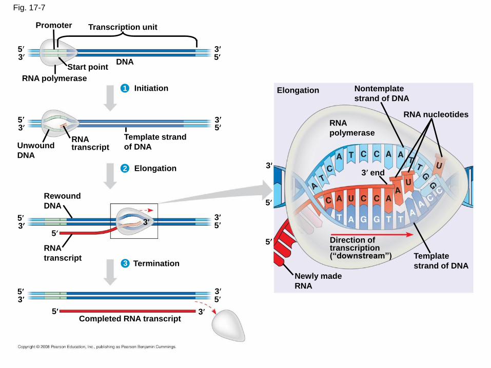
Fig. 17-7
Promoter Transcription unit
Start pointDNA
RNA polymerase
5533
Initiation1
2
3
5533
Unwound
DNA
RNAtranscript
Template strand
of DNA
Elongation
Rewound
DNA
5
55
5
5
33
3
3
RNA
transcriptTermination
5533
35Completed RNA transcript
Newly made
RNA
Template
strand of DNA
Direction oftranscription(“downstream”)
3 end
RNA
polymerase
RNA nucleotides
Nontemplate
strand of DNAElongation
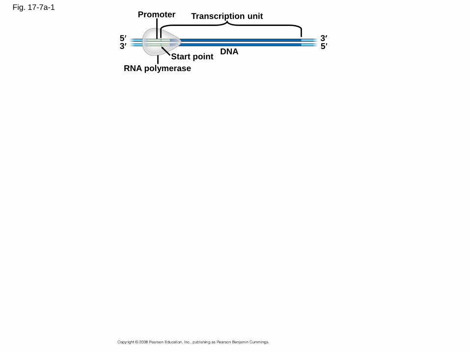
Fig. 17-7a-1Promoter Transcription unit
DNAStart point
RNA polymerase
553
3
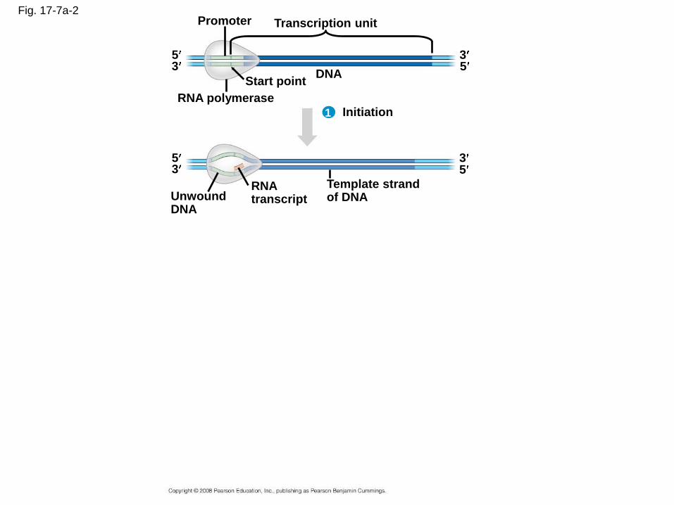
Fig. 17-7a-2Promoter Transcription unit
DNAStart point
RNA polymerase
553
3
Initiation
33
1
RNAtranscript
55
UnwoundDNA
Template strandof DNA
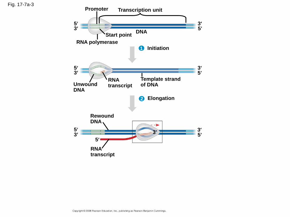
Fig. 17-7a-3Promoter Transcription unit
DNAStart point
RNA polymerase
553
3
Initiation
33
1
RNAtranscript
55
UnwoundDNA
Template strandof DNA
2 Elongation
RewoundDNA
5
553 3 3
RNAtranscript
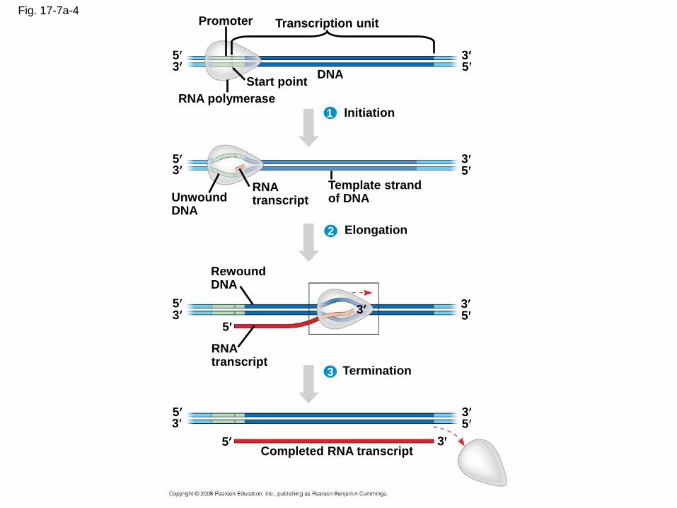
Fig. 17-7a-4Promoter Transcription unit
DNAStart point
RNA polymerase
553
3
Initiation
33
1
RNAtranscript
55
UnwoundDNA
Template strandof DNA
2 Elongation
RewoundDNA
5
553 3 3
RNAtranscript
3 Termination
5
5
533
3Completed RNA transcript
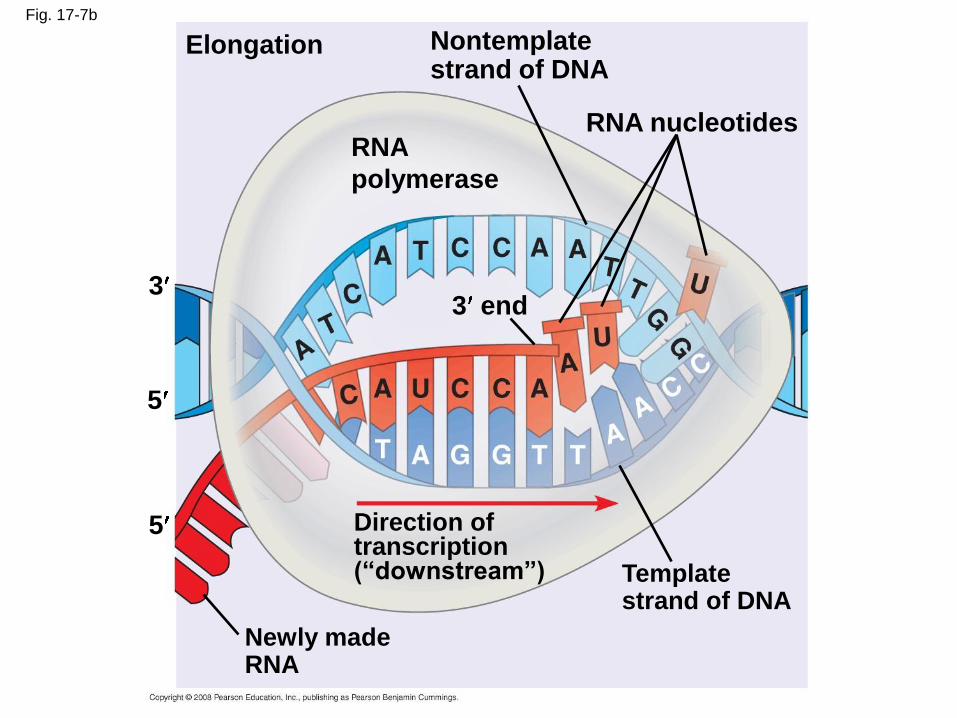
Fig. 17-7b
Elongation
RNA
polymerase
Nontemplatestrand of DNA
RNA nucleotides
3 end
Direction oftranscription(“downstream”) Template
strand of DNA
Newly madeRNA
3
5
5
Synthesis of an RNA Transcript
• The three stages of transcription:
– Initiation
– Elongation
– Termination
Copyright © 2008 Pearson Education Inc., publishing as Pearson Benjamin Cummings
RNA Polymerase Binding and Initiation of Transcription
• Promoters signal the initiation of RNA synthesis
• Transcription factors mediate the binding of RNA polymerase and the initiation of transcription
• The completed assembly of transcription factors and RNA polymerase II bound to a promoter is called a transcription initiation complex
• A promoter called a TATA box is crucial in forming the initiation complex in eukaryotes
Copyright © 2008 Pearson Education Inc., publishing as Pearson Benjamin Cummings
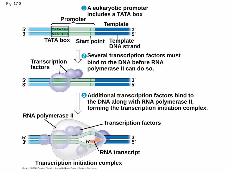
Fig. 17-8
A eukaryotic promoterincludes a TATA box
3
1
2
3
Promoter
TATA box Start point
Template
TemplateDNA strand
535
Transcriptionfactors
Several transcription factors must
bind to the DNA before RNApolymerase II can do so.
5533
Additional transcription factors bind tothe DNA along with RNA polymerase II,forming the transcription initiation complex.
RNA polymerase II
Transcription factors
55 53
3
RNA transcript
Transcription initiation complex
Elongation of the RNA Strand
• As RNA polymerase moves along the DNA, it
untwists the double helix, 10 to 20 bases at a
time
• Transcription progresses at a rate of 40
nucleotides per second in eukaryotes
• A gene can be transcribed simultaneously by
several RNA polymerases
Copyright © 2008 Pearson Education Inc., publishing as Pearson Benjamin Cummings
Termination of Transcription
• The mechanisms of termination are different in
bacteria and eukaryotes
• In bacteria, the polymerase stops transcription
at the end of the terminator
• In eukaryotes, the polymerase continues
transcription after the pre-mRNA is cleaved
from the growing RNA chain; the polymerase
eventually falls off the DNA
Copyright © 2008 Pearson Education Inc., publishing as Pearson Benjamin Cummings
Concept 17.3: Eukaryotic cells modify RNA after transcription
• Enzymes in the eukaryotic nucleus modify pre-
mRNA before the genetic messages are
dispatched to the cytoplasm
• During RNA processing, both ends of the
primary transcript are usually altered
• Also, usually some interior parts of the
molecule are cut out, and the other parts
spliced together
Copyright © 2008 Pearson Education Inc., publishing as Pearson Benjamin Cummings
Alteration of mRNA Ends
• Each end of a pre-mRNA molecule is modified in a particular way:
– The 5 end receives a modified nucleotide 5cap
– The 3 end gets a poly-A tail
• These modifications share several functions:
– They seem to facilitate the export of mRNA
– They protect mRNA from hydrolytic enzymes
– They help ribosomes attach to the 5 endCopyright © 2008 Pearson Education Inc., publishing as Pearson Benjamin Cummings

Fig. 17-9
Protein-coding segment Polyadenylation signal
3
3 UTR5 UTR
5
5 Cap Start codon Stop codon Poly-A tail
G P PP AAUAAA AAA AAA…
Split Genes and RNA Splicing
• Most eukaryotic genes and their RNA transcripts have long noncoding stretches of nucleotides that lie between coding regions
• These noncoding regions are called intervening sequences, or introns
• The other regions are called exons because they are eventually expressed, usually translated into amino acid sequences
• RNA splicing removes introns and joins exons, creating an mRNA molecule with a continuous coding sequence
Copyright © 2008 Pearson Education Inc., publishing as Pearson Benjamin Cummings

Fig. 17-10
Pre-mRNA
mRNA
Codingsegment
Introns cut out andexons spliced together
5 Cap
Exon Intron5
1 30 31 104
Exon Intron
105
Exon
146
3
Poly-A tail
Poly-A tail5 Cap
5 UTR 3 UTR1 146
• In some cases, RNA splicing is carried out by
spliceosomes
• Spliceosomes consist of a variety of proteins
and several small nuclear ribonucleoproteins
(snRNPs) that recognize the splice sites
Copyright © 2008 Pearson Education Inc., publishing as Pearson Benjamin Cummings

Fig. 17-11-1
RNA transcript (pre-mRNA)
Exon 1 Exon 2Intron
Protein
snRNA
snRNPs
Otherproteins
5
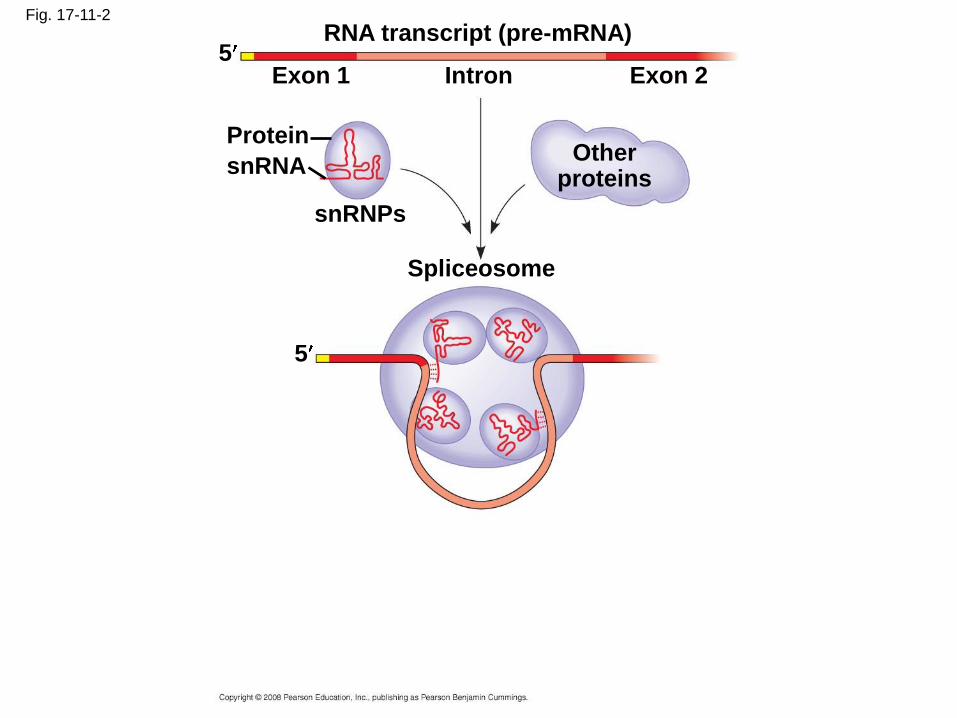
Fig. 17-11-2
RNA transcript (pre-mRNA)
Exon 1 Exon 2Intron
Protein
snRNA
snRNPs
Otherproteins
5
5
Spliceosome
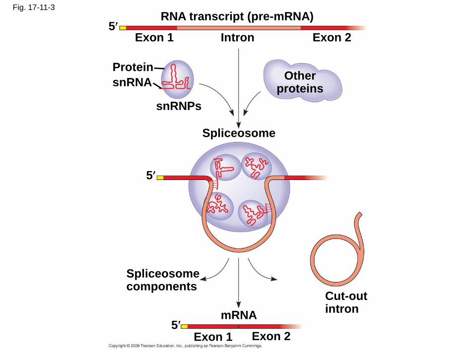
Fig. 17-11-3
RNA transcript (pre-mRNA)
Exon 1 Exon 2Intron
Protein
snRNA
snRNPs
Otherproteins
5
5
Spliceosome
Spliceosomecomponents
Cut-outintron
mRNA
Exon 1 Exon 25
Ribozymes
• Ribozymes are catalytic RNA molecules that
function as enzymes and can splice RNA
• The discovery of ribozymes rendered obsolete
the belief that all biological catalysts were
proteins
Copyright © 2008 Pearson Education Inc., publishing as Pearson Benjamin Cummings
• Three properties of RNA enable it to function
as an enzyme
– It can form a three-dimensional structure
because of its ability to base pair with itself
– Some bases in RNA contain functional groups
– RNA may hydrogen-bond with other nucleic
acid molecules
Copyright © 2008 Pearson Education Inc., publishing as Pearson Benjamin Cummings
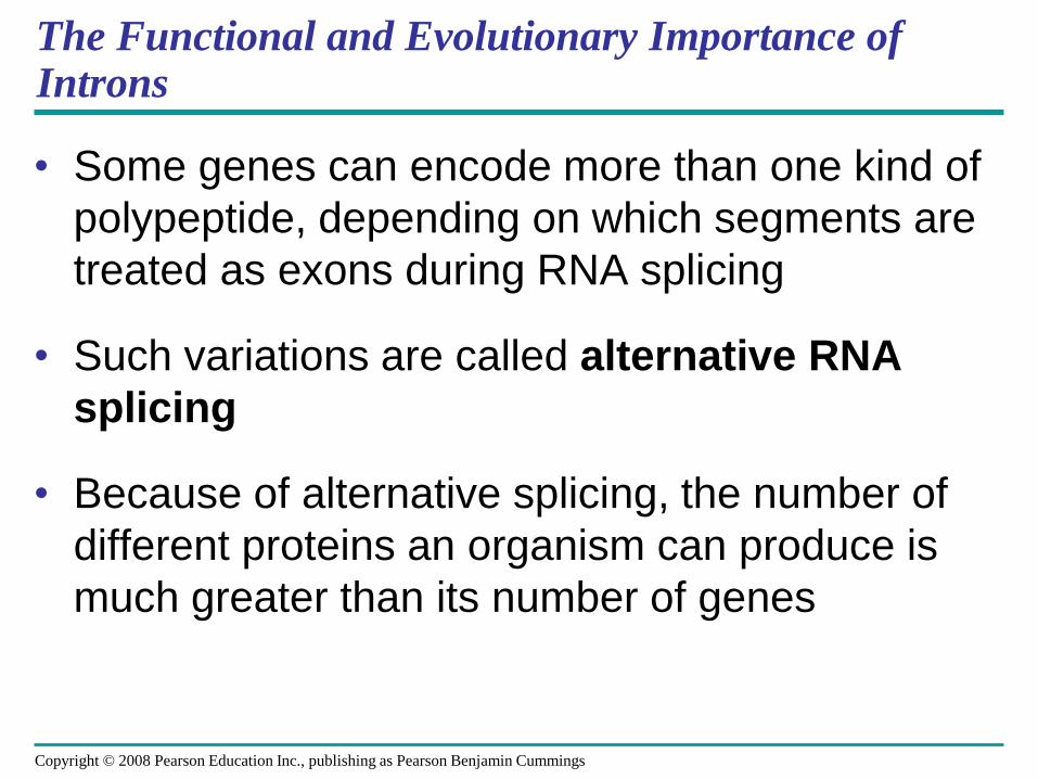
The Functional and Evolutionary Importance of Introns
• Some genes can encode more than one kind of
polypeptide, depending on which segments are
treated as exons during RNA splicing
• Such variations are called alternative RNA
splicing
• Because of alternative splicing, the number of
different proteins an organism can produce is
much greater than its number of genes
Copyright © 2008 Pearson Education Inc., publishing as Pearson Benjamin Cummings
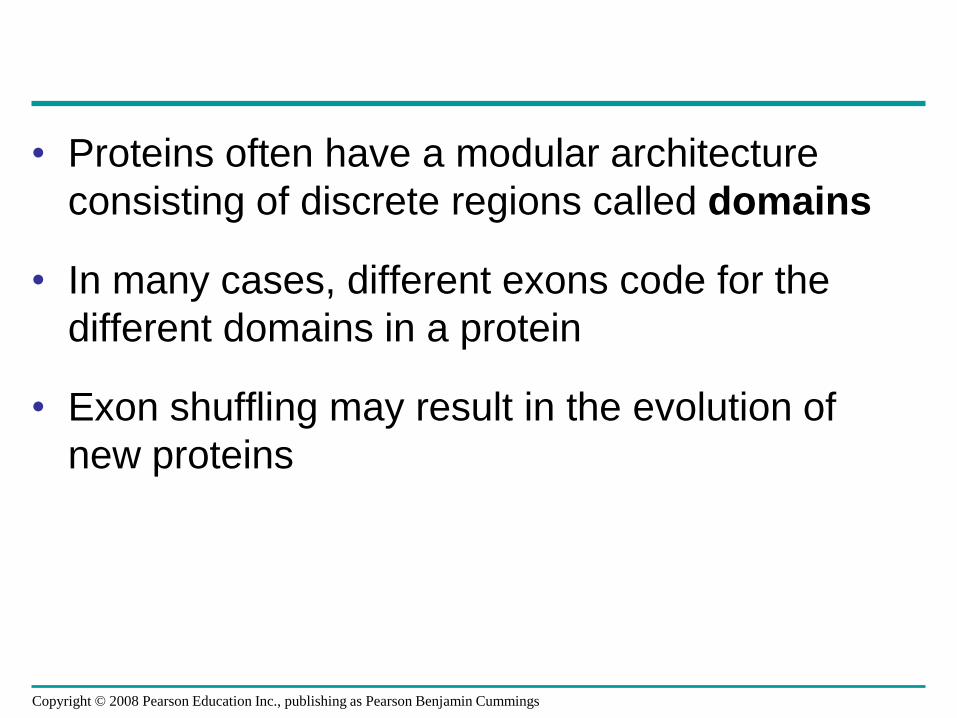
• Proteins often have a modular architecture
consisting of discrete regions called domains
• In many cases, different exons code for the
different domains in a protein
• Exon shuffling may result in the evolution of
new proteins
Copyright © 2008 Pearson Education Inc., publishing as Pearson Benjamin Cummings

Fig. 17-12
Gene
DNA
Exon 1 Exon 2 Exon 3Intron Intron
Transcription
RNA processing
Translation
Domain 2
Domain 3
Domain 1
Polypeptide

Concept 17.4: Translation is the RNA-directed synthesis of a polypeptide: a closer look
• The translation of mRNA to protein can be
examined in more detail
Copyright © 2008 Pearson Education Inc., publishing as Pearson Benjamin Cummings

Molecular Components of Translation
• A cell translates an mRNA message into
protein with the help of transfer RNA (tRNA)
• Molecules of tRNA are not identical:
– Each carries a specific amino acid on one end
– Each has an anticodon on the other end; the
anticodon base-pairs with a complementary
codon on mRNA
Copyright © 2008 Pearson Education Inc., publishing as Pearson Benjamin Cummings
BioFlix: Protein Synthesis

Fig. 17-13
Polypeptide
Ribosome
Aminoacids
tRNA withamino acidattached
tRNA
Anticodon
Codons 35
mRNA

The Structure and Function of Transfer RNA
ACC
• A tRNA molecule consists of a single RNA
strand that is only about 80 nucleotides long
• Flattened into one plane to reveal its base
pairing, a tRNA molecule looks like a
cloverleaf
Copyright © 2008 Pearson Education Inc., publishing as Pearson Benjamin Cummings
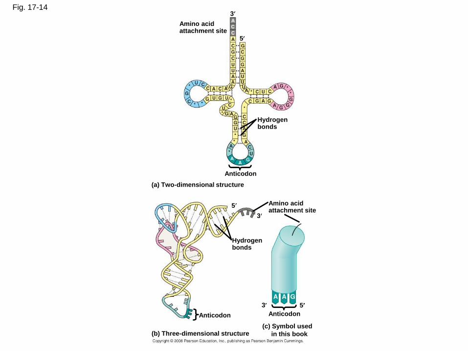
Fig. 17-14
Amino acidattachment site
3
5
Hydrogenbonds
Anticodon
(a) Two-dimensional structure
Amino acidattachment site
5
3
Hydrogenbonds
3 5
AnticodonAnticodon
(c) Symbol used
in this book(b) Three-dimensional structure
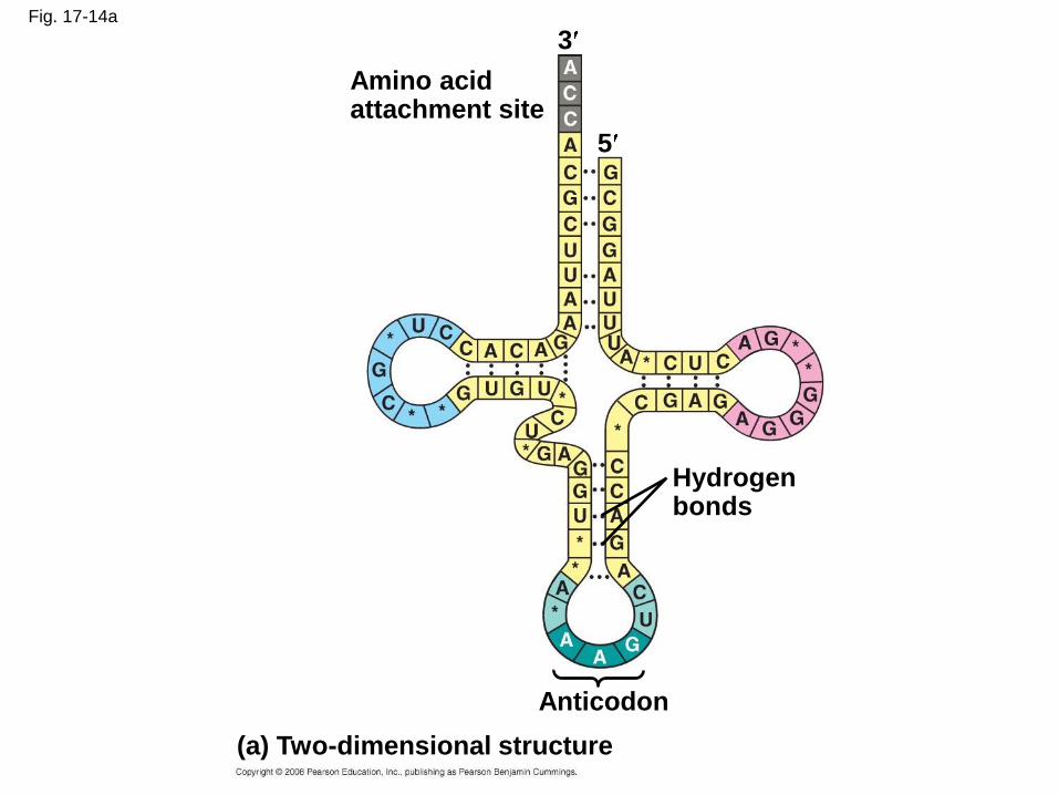
Fig. 17-14a
Amino acidattachment site
(a) Two-dimensional structure
Hydrogenbonds
Anticodon
3
5
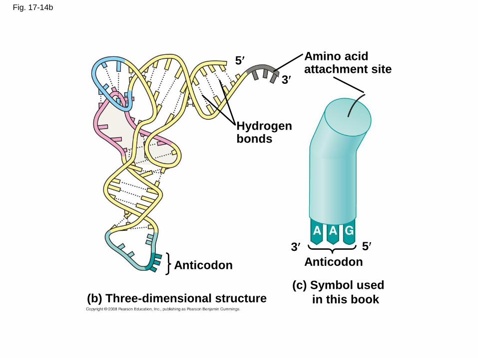
Fig. 17-14b
Amino acidattachment site
3
3
5
5
Hydrogenbonds
Anticodon Anticodon
(b) Three-dimensional structure(c) Symbol used
in this book
• Because of hydrogen bonds, tRNA actually
twists and folds into a three-dimensional
molecule
• tRNA is roughly L-shaped
Copyright © 2008 Pearson Education Inc., publishing as Pearson Benjamin Cummings
• Accurate translation requires two steps:
– First: a correct match between a tRNA and an
amino acid, done by the enzyme aminoacyl-
tRNA synthetase
– Second: a correct match between the tRNA
anticodon and an mRNA codon
• Flexible pairing at the third base of a codon is
called wobble and allows some tRNAs to bind
to more than one codon
Copyright © 2008 Pearson Education Inc., publishing as Pearson Benjamin Cummings

Fig. 17-15-1
Amino acid Aminoacyl-tRNAsynthetase (enzyme)
ATP
AdenosineP P P
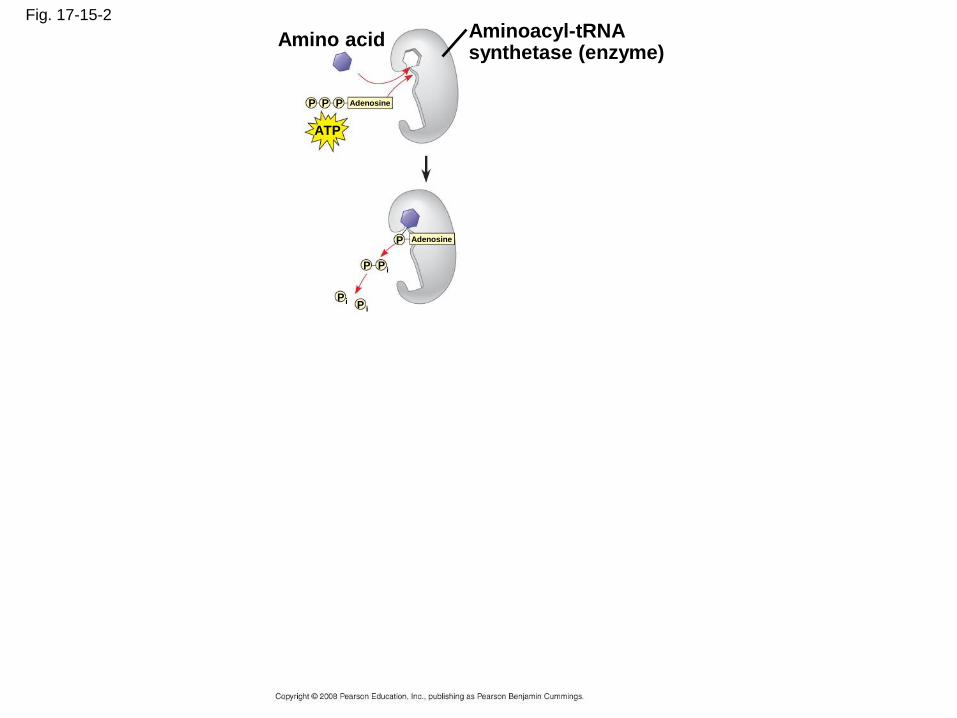
Fig. 17-15-2
Amino acid Aminoacyl-tRNAsynthetase (enzyme)
ATP
AdenosineP P P
AdenosineP
PP i
PPi
i
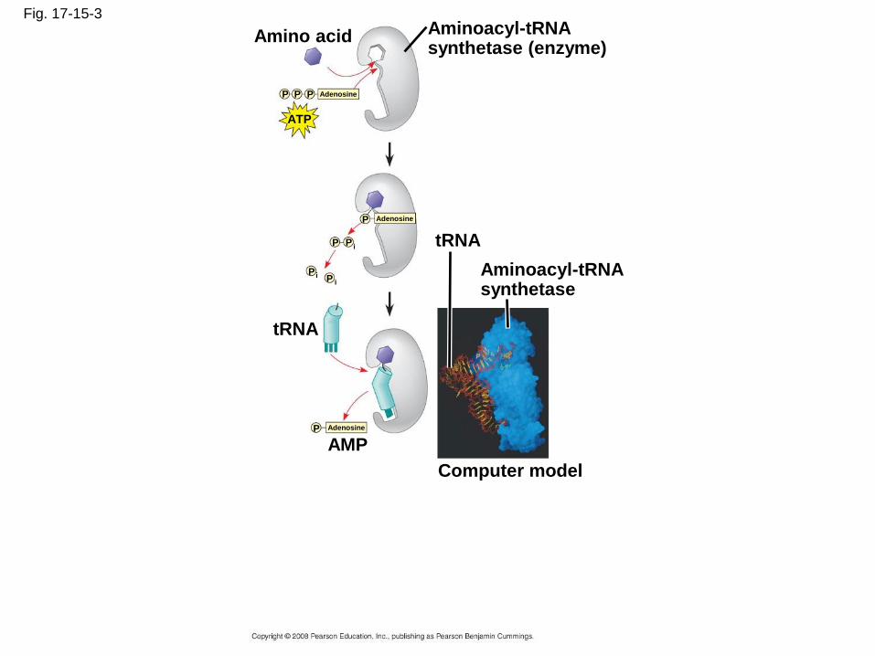
Fig. 17-15-3
Amino acid Aminoacyl-tRNAsynthetase (enzyme)
ATP
AdenosineP P P
AdenosineP
PP i
PPi
i
tRNA
tRNA
Aminoacyl-tRNAsynthetase
Computer model
AMPAdenosineP

Fig. 17-15-4
Amino acid Aminoacyl-tRNAsynthetase (enzyme)
ATP
AdenosineP P P
AdenosineP
PP i
PPi
i
tRNA
tRNA
Aminoacyl-tRNAsynthetase
Computer model
AMPAdenosineP
Aminoacyl-tRNA(“charged tRNA”)
Ribosomes
• Ribosomes facilitate specific coupling of tRNA
anticodons with mRNA codons in protein
synthesis
• The two ribosomal subunits (large and small)
are made of proteins and ribosomal RNA
(rRNA)
Copyright © 2008 Pearson Education Inc., publishing as Pearson Benjamin Cummings
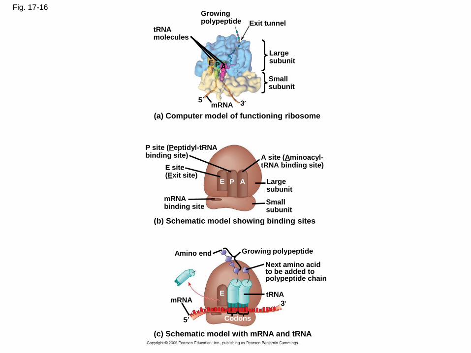
Fig. 17-16Growingpolypeptide Exit tunnel
Largesubunit
Smallsubunit
tRNAmolecules
E PA
mRNA5 3
(a) Computer model of functioning ribosome
P site (Peptidyl-tRNAbinding site)
E site(Exit site)
A site (Aminoacyl-tRNA binding site)
E P A Largesubunit
mRNAbinding site
Smallsubunit
(b) Schematic model showing binding sites
Amino end Growing polypeptide
Next amino acidto be added topolypeptide chain
mRNAtRNAE
3
5 Codons
(c) Schematic model with mRNA and tRNA

Fig. 17-16a
Growingpolypeptide Exit tunnel
tRNAmolecules
Largesubunit
Smallsubunit
(a) Computer model of functioning ribosome
mRNA
E PA
53
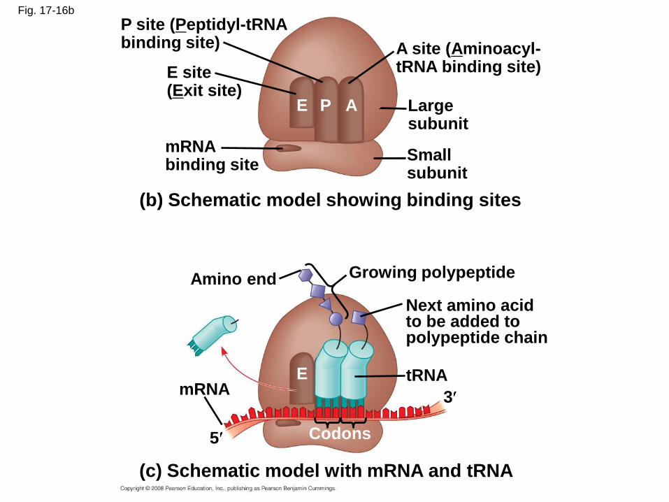
Fig. 17-16b
P site (Peptidyl-tRNAbinding site) A site (Aminoacyl-
tRNA binding site)E site(Exit site)
mRNAbinding site
Largesubunit
Smallsubunit
(b) Schematic model showing binding sites
Next amino acidto be added topolypeptide chain
Amino end Growing polypeptide
mRNAtRNA
E P A
E
Codons
(c) Schematic model with mRNA and tRNA
5
3
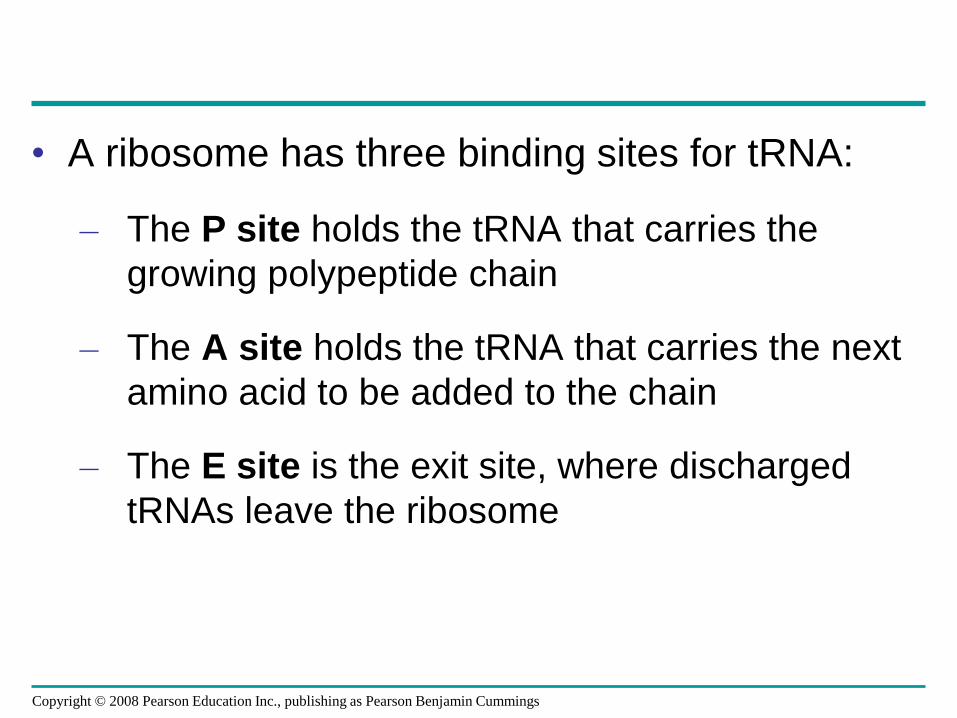
• A ribosome has three binding sites for tRNA:
– The P site holds the tRNA that carries the
growing polypeptide chain
– The A site holds the tRNA that carries the next
amino acid to be added to the chain
– The E site is the exit site, where discharged
tRNAs leave the ribosome
Copyright © 2008 Pearson Education Inc., publishing as Pearson Benjamin Cummings
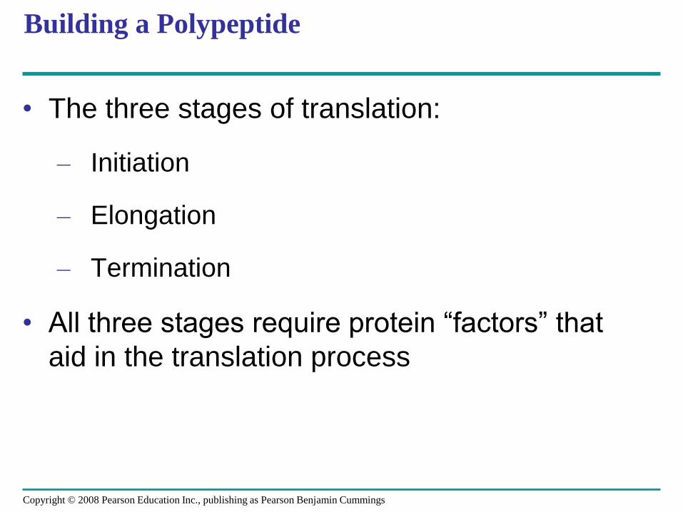
Building a Polypeptide
• The three stages of translation:
– Initiation
– Elongation
– Termination
• All three stages require protein “factors” that
aid in the translation process
Copyright © 2008 Pearson Education Inc., publishing as Pearson Benjamin Cummings
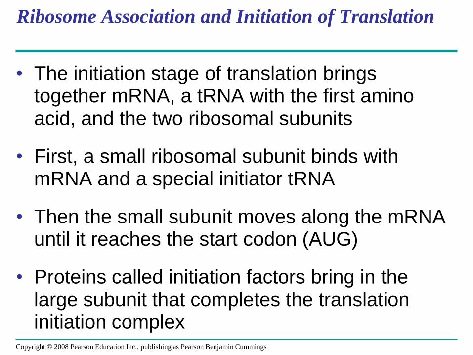
Ribosome Association and Initiation of Translation
• The initiation stage of translation brings together mRNA, a tRNA with the first amino acid, and the two ribosomal subunits
• First, a small ribosomal subunit binds with mRNA and a special initiator tRNA
• Then the small subunit moves along the mRNA until it reaches the start codon (AUG)
• Proteins called initiation factors bring in the large subunit that completes the translation initiation complex
Copyright © 2008 Pearson Education Inc., publishing as Pearson Benjamin Cummings
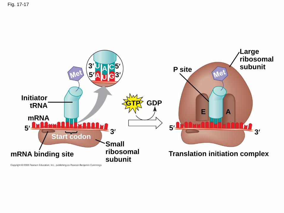
Fig. 17-17
3
35
5U
U
AA
C
G
GTP GDPInitiator
tRNA
mRNA
53
Start codon
mRNA binding site
Smallribosomalsubunit
5
P site
Translation initiation complex
3
E A
Largeribosomalsubunit
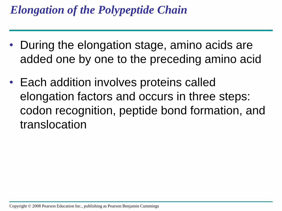
Elongation of the Polypeptide Chain
• During the elongation stage, amino acids are
added one by one to the preceding amino acid
• Each addition involves proteins called
elongation factors and occurs in three steps:
codon recognition, peptide bond formation, and
translocation
Copyright © 2008 Pearson Education Inc., publishing as Pearson Benjamin Cummings
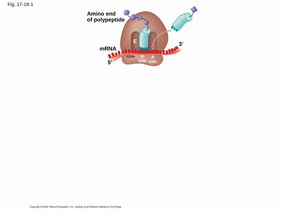
Fig. 17-18-1
Amino endof polypeptide
mRNA
5
3E
Psite
Asite
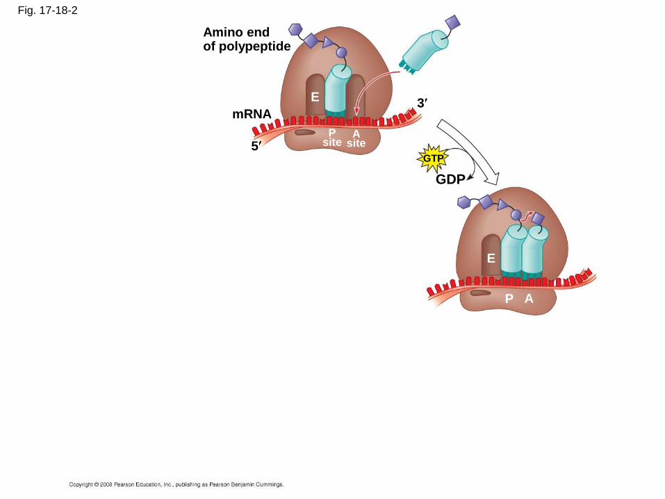
Fig. 17-18-2
Amino endof polypeptide
mRNA
5
3E
Psite
Asite
GTP
GDP
E
P A
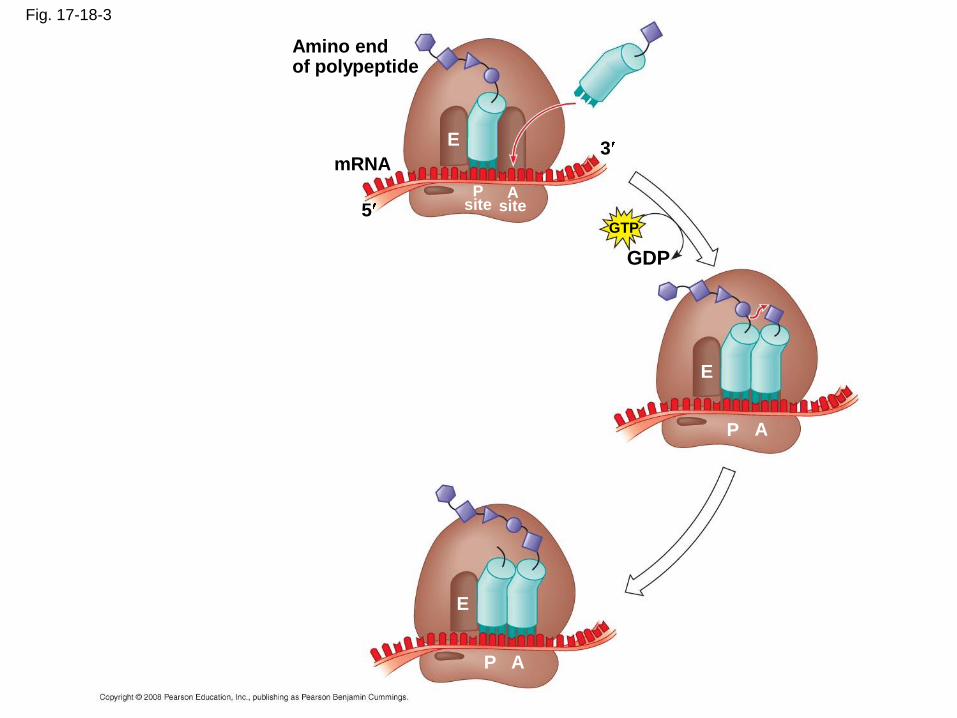
Fig. 17-18-3
Amino endof polypeptide
mRNA
5
3E
Psite
Asite
GTP
GDP
E
P A
E
P A
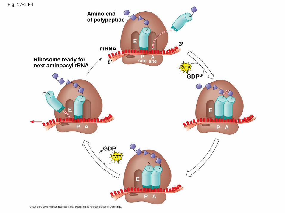
Fig. 17-18-4
Amino endof polypeptide
mRNA
5
3E
Psite
Asite
GTP
GDP
E
P A
E
P A
GDP
GTP
Ribosome ready fornext aminoacyl tRNA
E
P A
Termination of Translation
• Termination occurs when a stop codon in the
mRNA reaches the A site of the ribosome
• The A site accepts a protein called a release
factor
• The release factor causes the addition of a
water molecule instead of an amino acid
• This reaction releases the polypeptide, and the
translation assembly then comes apart
Copyright © 2008 Pearson Education Inc., publishing as Pearson Benjamin Cummings
Translation
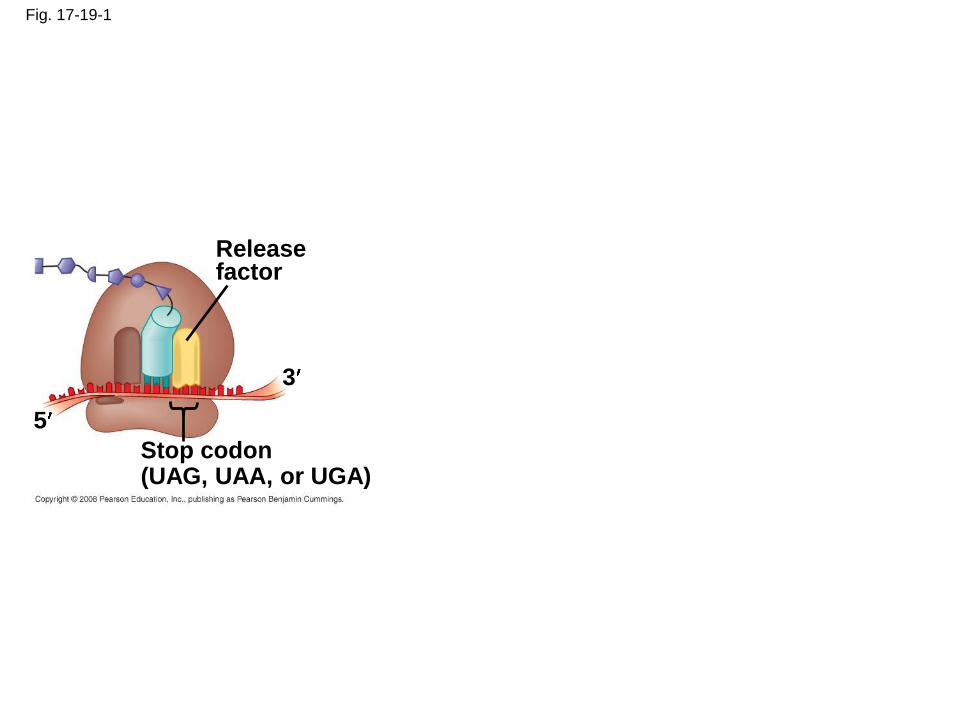
Fig. 17-19-1
Releasefactor
3
5
Stop codon(UAG, UAA, or UGA)

Fig. 17-19-2
Releasefactor
3
5
Stop codon(UAG, UAA, or UGA)
5
3
2
Freepolypeptide
2 GDP
GTP
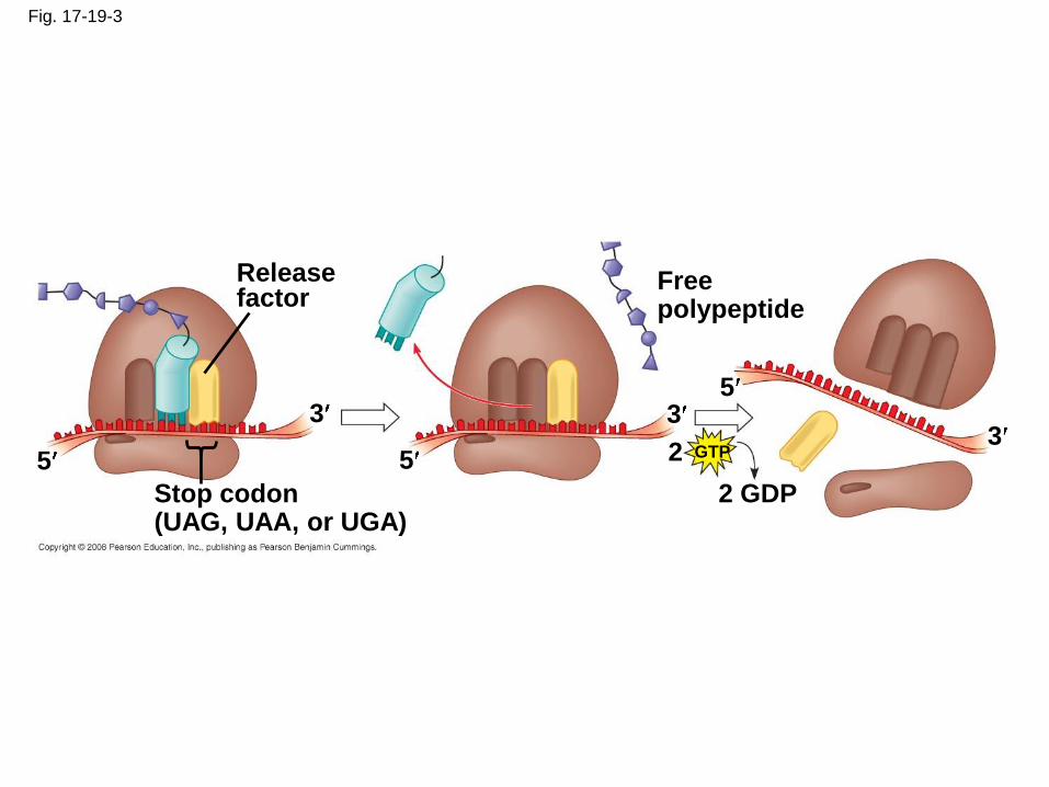
Fig. 17-19-3
Releasefactor
3
5
Stop codon(UAG, UAA, or UGA)
5
3
2
Freepolypeptide
2 GDP
GTP
5
3
Polyribosomes
• A number of ribosomes can translate a single
mRNA simultaneously, forming a
polyribosome (or polysome)
• Polyribosomes enable a cell to make many
copies of a polypeptide very quickly
Copyright © 2008 Pearson Education Inc., publishing as Pearson Benjamin Cummings
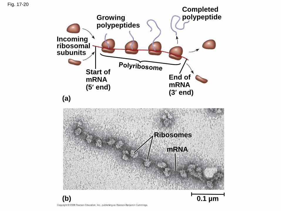
Fig. 17-20
Growingpolypeptides
Completedpolypeptide
Incomingribosomalsubunits
Start ofmRNA(5 end)
End ofmRNA(3 end)
(a)
Ribosomes
mRNA
(b) 0.1 µm
Completing and Targeting the Functional Protein
• Often translation is not sufficient to make a
functional protein
• Polypeptide chains are modified after
translation
• Completed proteins are targeted to specific
sites in the cell
Copyright © 2008 Pearson Education Inc., publishing as Pearson Benjamin Cummings
Protein Folding and Post-Translational Modifications
• During and after synthesis, a polypeptide chain
spontaneously coils and folds into its three-
dimensional shape
• Proteins may also require post-translational
modifications before doing their job
• Some polypeptides are activated by enzymes
that cleave them
• Other polypeptides come together to form the
subunits of a protein
Copyright © 2008 Pearson Education Inc., publishing as Pearson Benjamin Cummings
Targeting Polypeptides to Specific Locations
• Two populations of ribosomes are evident in cells: free ribsomes (in the cytosol) and bound ribosomes (attached to the ER)
• Free ribosomes mostly synthesize proteins that function in the cytosol
• Bound ribosomes make proteins of the endomembrane system and proteins that are secreted from the cell
• Ribosomes are identical and can switch from free to bound
Copyright © 2008 Pearson Education Inc., publishing as Pearson Benjamin Cummings
• Polypeptide synthesis always begins in the
cytosol
• Synthesis finishes in the cytosol unless the
polypeptide signals the ribosome to attach to
the ER
• Polypeptides destined for the ER or for
secretion are marked by a signal peptide
Copyright © 2008 Pearson Education Inc., publishing as Pearson Benjamin Cummings
• A signal-recognition particle (SRP) binds to
the signal peptide
• The SRP brings the signal peptide and its
ribosome to the ER
Copyright © 2008 Pearson Education Inc., publishing as Pearson Benjamin Cummings
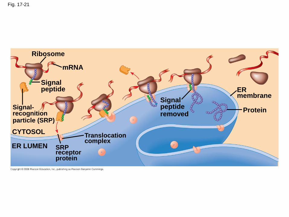
Fig. 17-21
Ribosome
mRNA
Signalpeptide
Signal-recognitionparticle (SRP)
CYTOSOLTranslocationcomplex
SRPreceptorprotein
ER LUMEN
Signalpeptideremoved
ERmembrane
Protein
Concept 17.5: Point mutations can affect protein structure and function
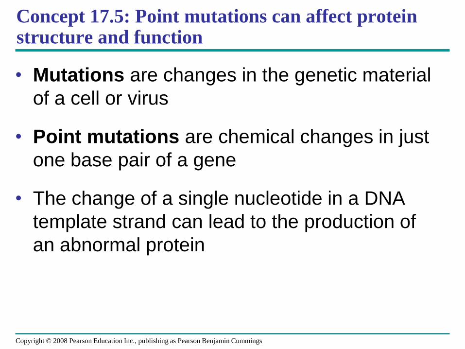
• Mutations are changes in the genetic material
of a cell or virus
• Point mutations are chemical changes in just
one base pair of a gene
• The change of a single nucleotide in a DNA
template strand can lead to the production of
an abnormal protein
Copyright © 2008 Pearson Education Inc., publishing as Pearson Benjamin Cummings

Fig. 17-22
Wild-type hemoglobin DNA
mRNA
Mutant hemoglobin DNA
mRNA
3
3
3
3
3
3
5
5
5
5
5
5
C CT T T
TG GA A A
A
A A AGG U
Normal hemoglobin Sickle-cell hemoglobin
Glu Val
Types of Point Mutations
• Point mutations within a gene can be divided
into two general categories
– Base-pair substitutions
– Base-pair insertions or deletions
Copyright © 2008 Pearson Education Inc., publishing as Pearson Benjamin Cummings
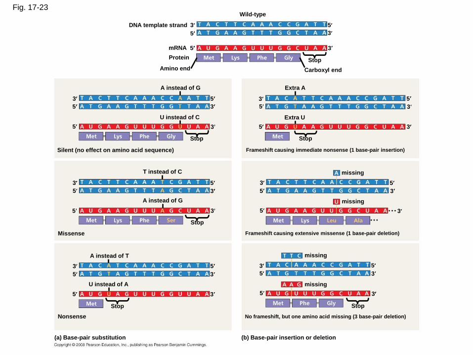
Fig. 17-23Wild-type
3DNA template strand
5
5
5
3
3
Stop
Carboxyl endAmino end
Protein
mRNA
3
3
3
5
5
5
A instead of G
U instead of C
Silent (no effect on amino acid sequence)
Stop
T instead of C
3
3
3
5
5
5
A instead of G
Stop
Missense
A instead of T
U instead of A
3
3
3
5
5
5
Stop
Nonsense No frameshift, but one amino acid missing (3 base-pair deletion)
Frameshift causing extensive missense (1 base-pair deletion)
Frameshift causing immediate nonsense (1 base-pair insertion)
5
5
53
3
3
Stop
missing
missing
3
3
3
5
5
5
missing
missing
Stop
5
5
53
3
3
Extra U
Extra A
(a) Base-pair substitution (b) Base-pair insertion or deletion
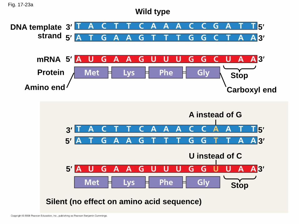
Fig. 17-23a
Wild type
3DNA templatestrand
3
35
5
5mRNA
Protein
Amino end
Stop
Carboxyl end
A instead of G
3
3
3
U instead of C
5
5
5
Stop
Silent (no effect on amino acid sequence)
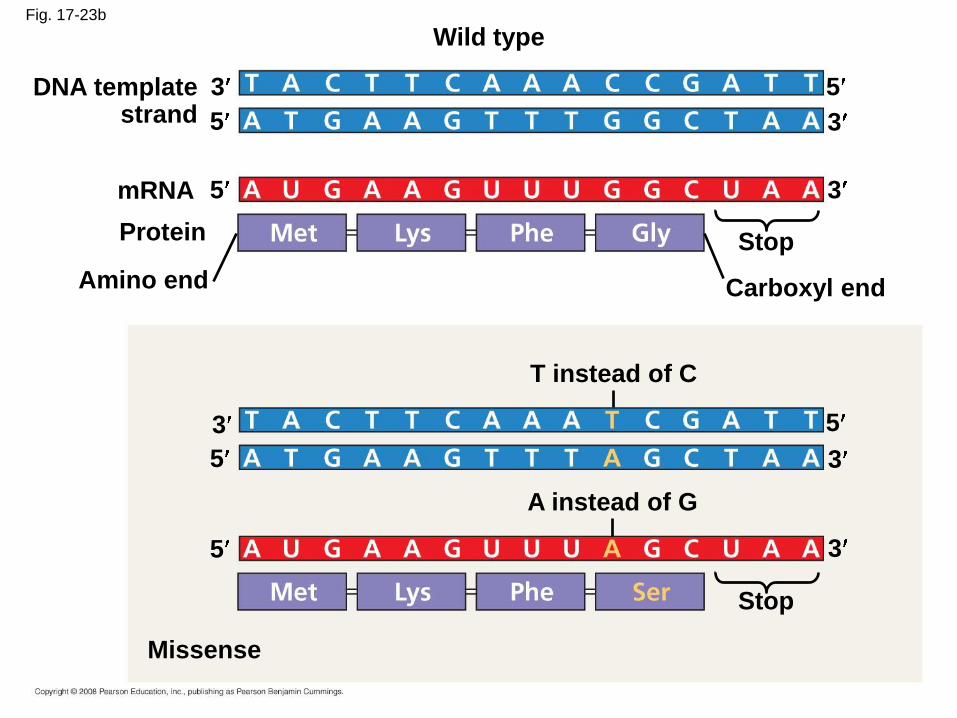
Fig. 17-23b
Wild type
DNA templatestrand
3
5
mRNA
Protein
5
Amino end
Stop
Carboxyl end
5
3
3
T instead of C
A instead of G
3
3
3
5
5
5
Stop
Missense
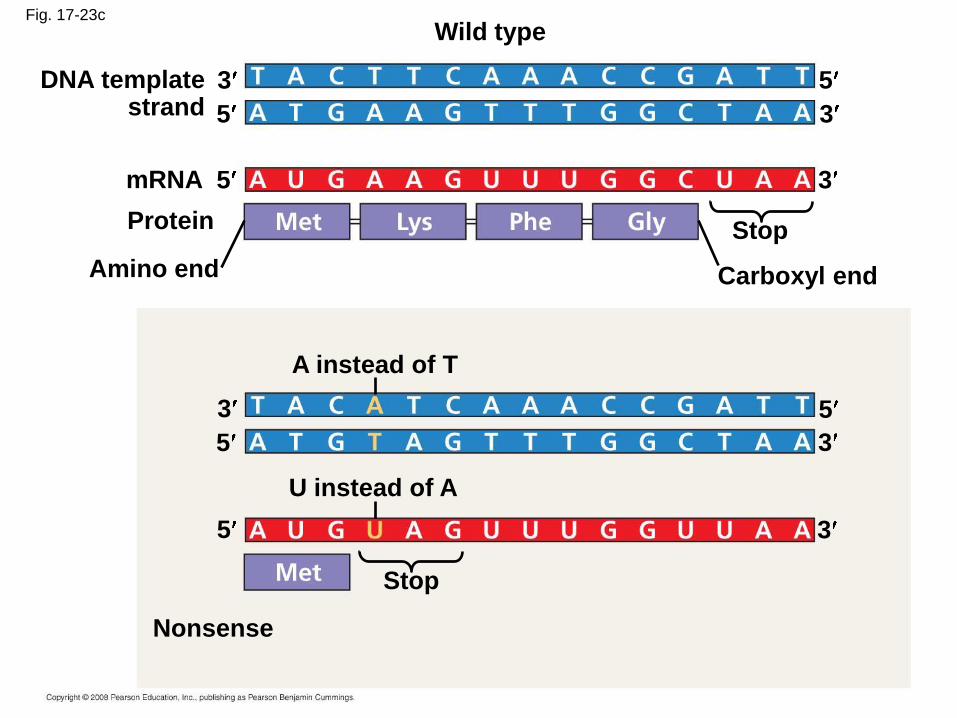
Fig. 17-23c
Wild type
DNA templatestrand
3
5
mRNA
Protein
5
Amino end
Stop
Carboxyl end
5
3
3
A instead of T
U instead of A
3
3
3
5
5
5
Stop
Nonsense
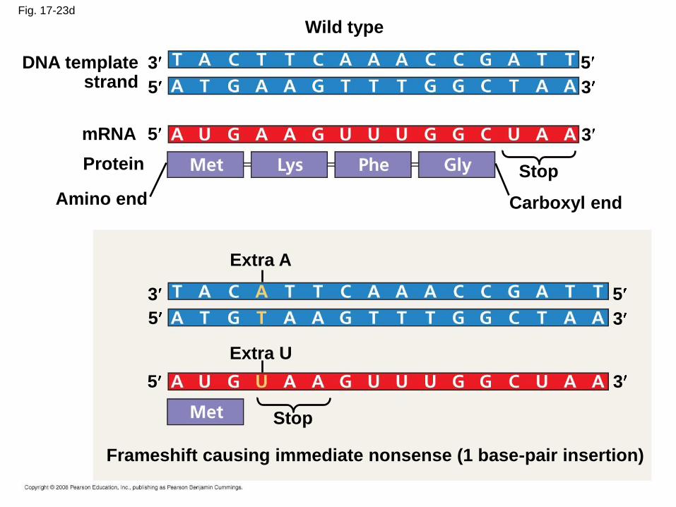
Fig. 17-23d
Wild type
DNA templatestrand
3
5
mRNA
Protein
5
Amino end
Stop
Carboxyl end
5
3
3
Extra A
Extra U
3
3
3
5
5
5
Stop
Frameshift causing immediate nonsense (1 base-pair insertion)
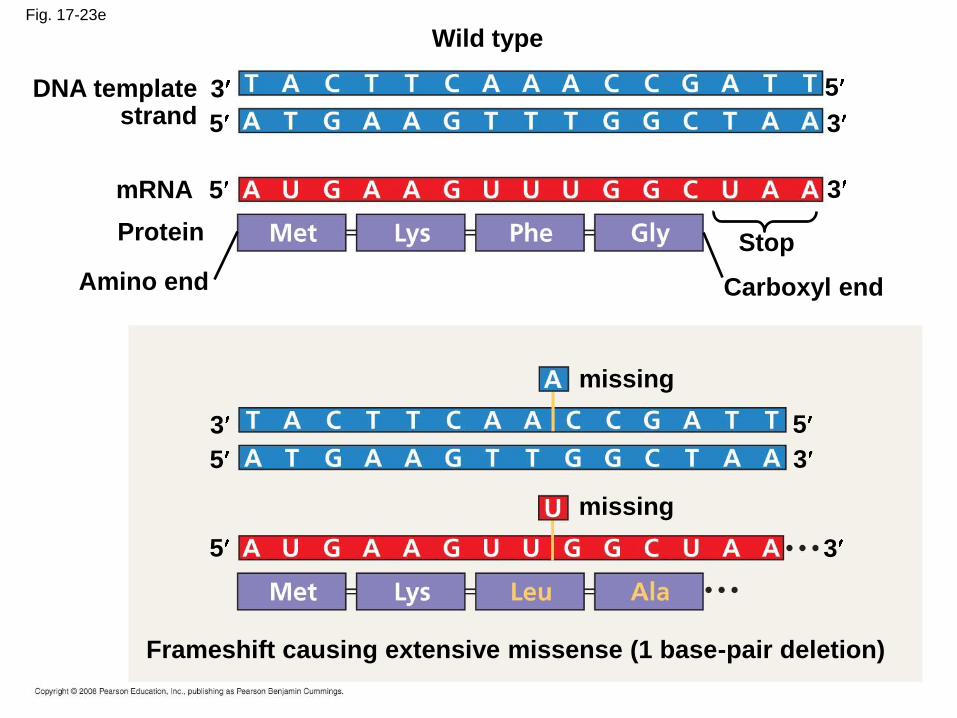
Fig. 17-23e
Wild type
DNA templatestrand
3
5
mRNA
Protein
5
Amino end
Stop
Carboxyl end
5
3
3
missing
missing
3
3
3
5
5
5
Frameshift causing extensive missense (1 base-pair deletion)

Fig. 17-23f
Wild type
DNA templatestrand
3
5
mRNA
Protein
5
Amino end
Stop
Carboxyl end
5
3
3
missing
missing
3
3
3
5
5
5
No frameshift, but one amino acid missing (3 base-pair deletion)
Stop
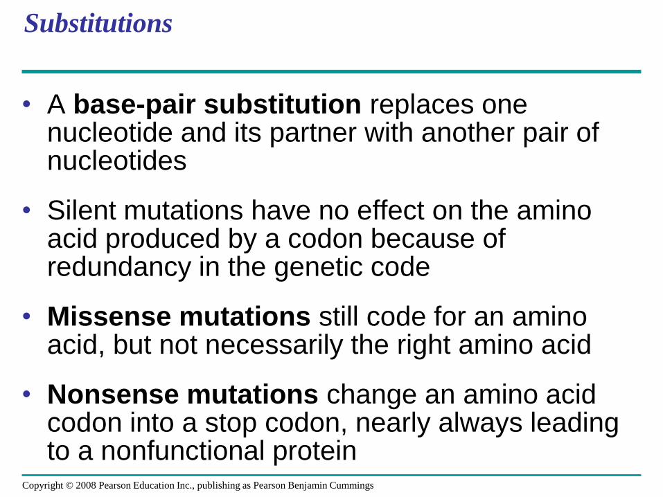
Substitutions
• A base-pair substitution replaces one nucleotide and its partner with another pair of nucleotides
• Silent mutations have no effect on the amino acid produced by a codon because of redundancy in the genetic code
• Missense mutations still code for an amino acid, but not necessarily the right amino acid
• Nonsense mutations change an amino acid codon into a stop codon, nearly always leading to a nonfunctional protein
Copyright © 2008 Pearson Education Inc., publishing as Pearson Benjamin Cummings
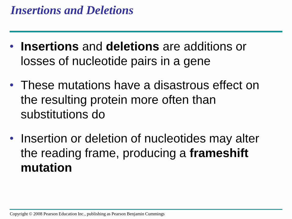
Insertions and Deletions
• Insertions and deletions are additions or
losses of nucleotide pairs in a gene
• These mutations have a disastrous effect on
the resulting protein more often than
substitutions do
• Insertion or deletion of nucleotides may alter
the reading frame, producing a frameshift
mutation
Copyright © 2008 Pearson Education Inc., publishing as Pearson Benjamin Cummings
Mutagens
• Spontaneous mutations can occur during DNA
replication, recombination, or repair
• Mutagens are physical or chemical agents that
can cause mutations
Copyright © 2008 Pearson Education Inc., publishing as Pearson Benjamin Cummings
Concept 17.6: While gene expression differs among the domains of life, the concept of a gene is universal
• Archaea are prokaryotes, but share many
features of gene expression with eukaryotes
Copyright © 2008 Pearson Education Inc., publishing as Pearson Benjamin Cummings
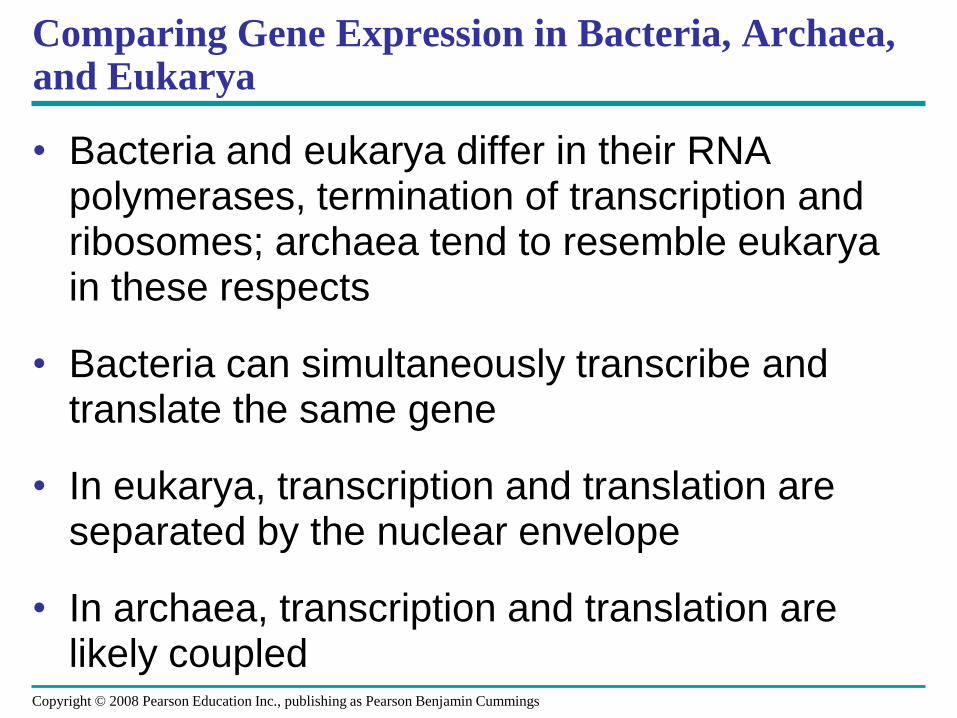
Comparing Gene Expression in Bacteria, Archaea, and Eukarya
• Bacteria and eukarya differ in their RNA polymerases, termination of transcription and ribosomes; archaea tend to resemble eukarya in these respects
• Bacteria can simultaneously transcribe and translate the same gene
• In eukarya, transcription and translation are separated by the nuclear envelope
• In archaea, transcription and translation are likely coupled
Copyright © 2008 Pearson Education Inc., publishing as Pearson Benjamin Cummings
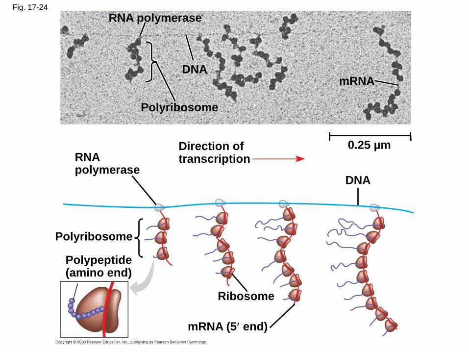
Fig. 17-24
RNA polymerase
DNA
Polyribosome
mRNA
0.25 µmDirection oftranscription
DNA
RNApolymerase
Polyribosome
Polypeptide(amino end)
Ribosome
mRNA (5 end)
What Is a Gene? Revisiting the Question
• The idea of the gene itself is a unifying concept
of life
• We have considered a gene as:
– A discrete unit of inheritance
– A region of specific nucleotide sequence in a
chromosome
– A DNA sequence that codes for a specific
polypeptide chain
Copyright © 2008 Pearson Education Inc., publishing as Pearson Benjamin Cummings
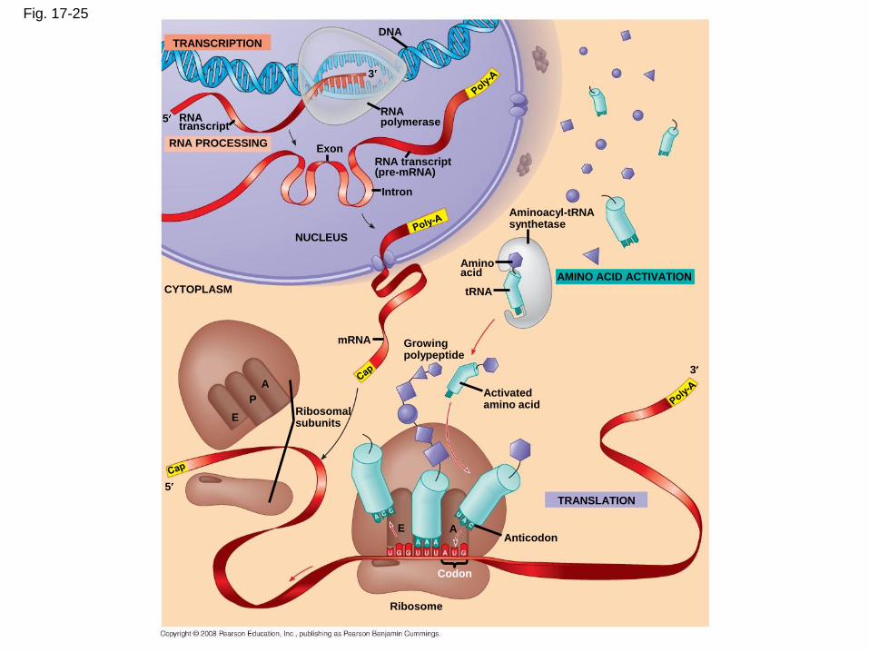
Fig. 17-25
TRANSCRIPTION
RNA PROCESSING
DNA
RNAtranscript
3
5RNApolymerase
RNA transcript(pre-mRNA)
Intron
Exon
NUCLEUS
Aminoacyl-tRNAsynthetase
AMINO ACID ACTIVATION
Aminoacid
tRNACYTOPLASM
Growingpolypeptide
3
Activatedamino acid
mRNA
TRANSLATION
Ribosomalsubunits
5
E
P
A
AAnticodon
Ribosome
Codon
E
• In summary, a gene can be defined as a region
of DNA that can be expressed to produce a
final functional product, either a polypeptide or
an RNA molecule
Copyright © 2008 Pearson Education Inc., publishing as Pearson Benjamin Cummings
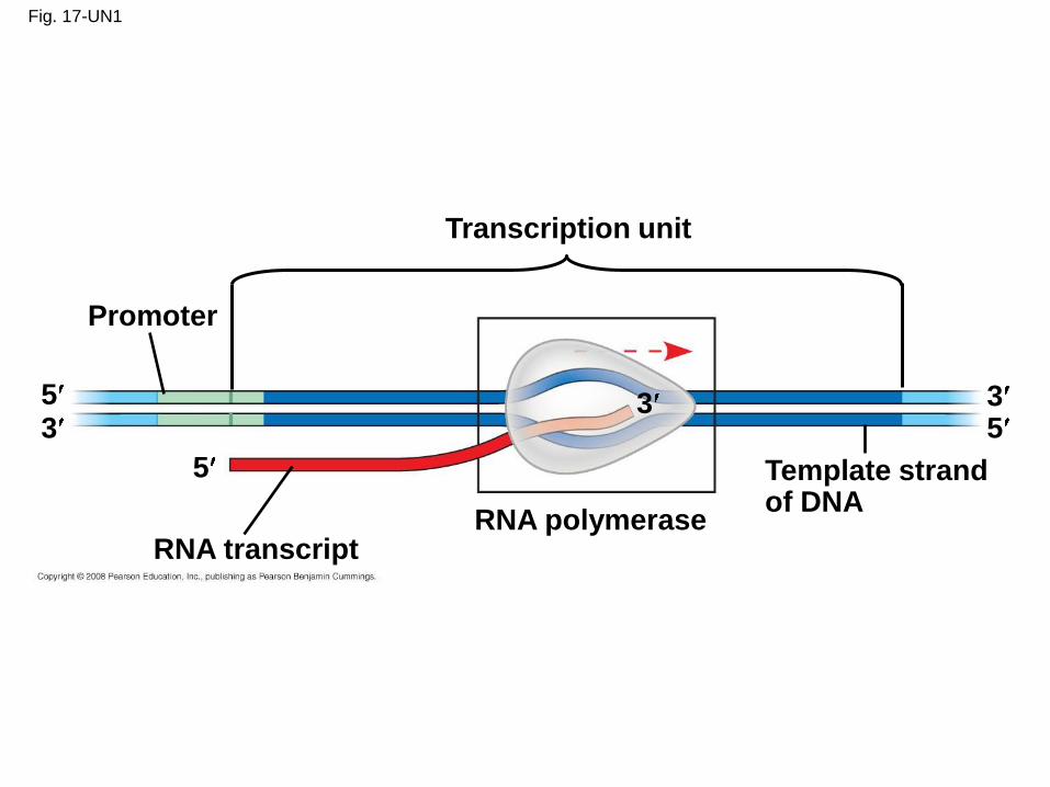
Fig. 17-UN1
Transcription unit
Promoter
RNA transcriptRNA polymerase
Template strandof DNA
5
5
533 3
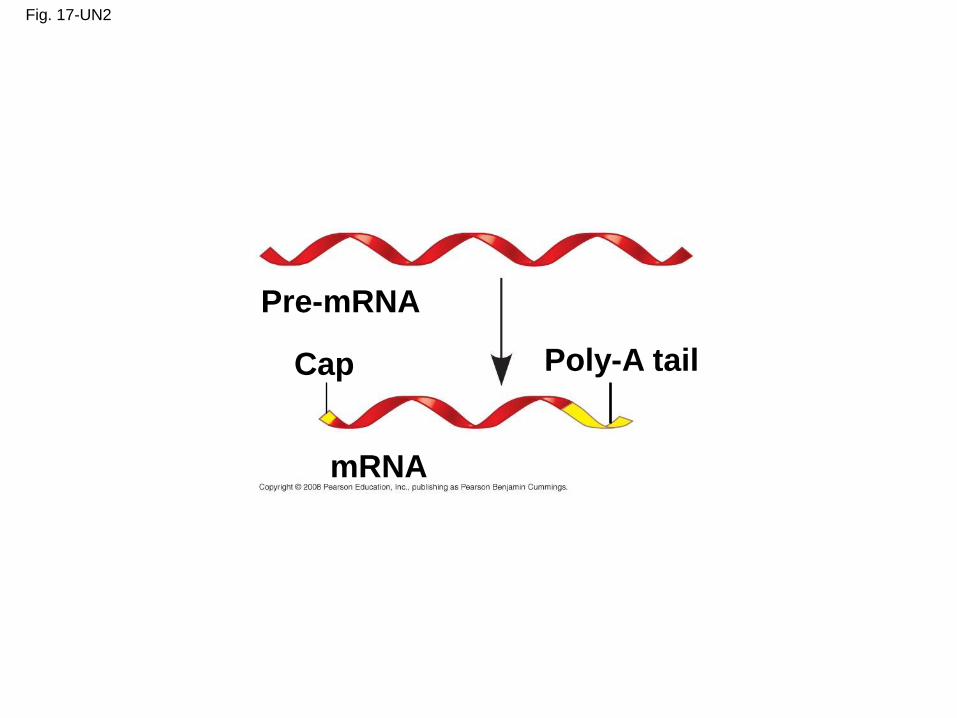
Fig. 17-UN2
Pre-mRNA
Cap
mRNA
Poly-A tail
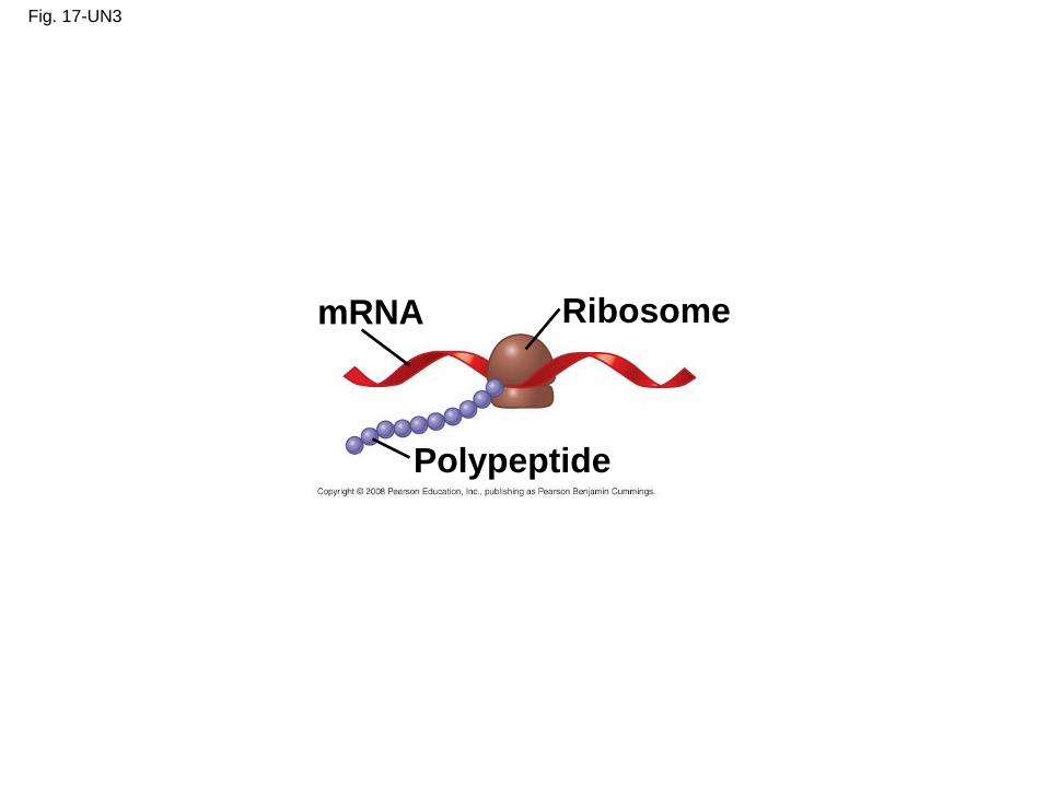
Fig. 17-UN3
mRNA Ribosome
Polypeptide
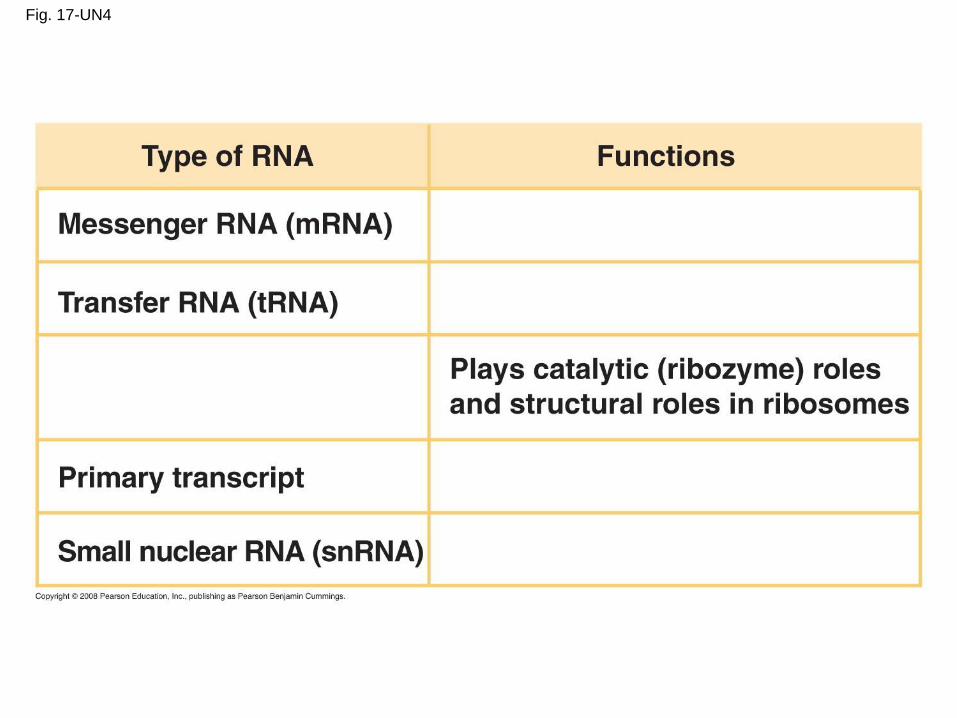
Fig. 17-UN4
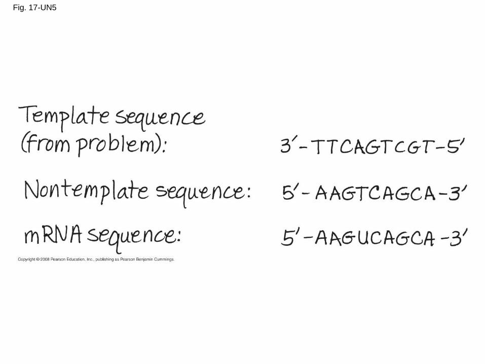
Fig. 17-UN5

Fig. 17-UN6
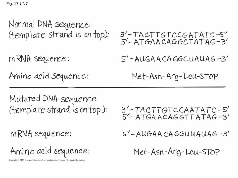
Fig. 17-UN7
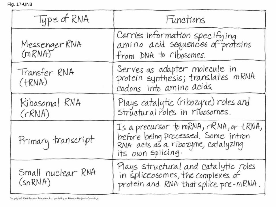
Fig. 17-UN8
You should now be able to:
1. Describe the contributions made by Garrod, Beadle,
and Tatum to our understanding of the relationship
between genes and enzymes
2. Briefly explain how information flows from gene to
protein
3. Compare transcription and translation in bacteria and
eukaryotes
4. Explain what it means to say that the genetic code is
redundant and unambiguous
Copyright © 2008 Pearson Education Inc., publishing as Pearson Benjamin Cummings
5. Include the following terms in a description of
transcription: mRNA, RNA polymerase, the promoter,
the terminator, the transcription unit, initiation,
elongation, termination, and introns
6. Include the following terms in a description of
translation: tRNA, wobble, ribosomes, initiation,
elongation, and termination
Copyright © 2008 Pearson Education Inc., publishing as Pearson Benjamin Cummings