Embed Size (px)
Citation preview
Cite as: J.-M. Kim et al., Science 10.1126/science.aat0174 (2018).
RESEARCH ARTICLES
First release: 8 November 2018 www.sciencemag.org (Page numbers not final at time of first release) 1
Nascent proteins bear the N-terminal (Nt) Met residue, en-coded by the AUG initiation codon. In bacteria and in eukar-yotic organelles mitochondria and chloroplasts, virtually all nascent proteins bear the N-terminal formyl-methionine (Nt-fMet), which is generated pretranslationally. Formyl-transferases (FMTs) use 10-formyltetrahydrofolate as a cosubstrate to Nα-terminally formylate the Met moiety of ini-tiator Met-tRNAs. Consequently, nascent bacterial proteins bear Nt-fMet (1–7). In contrast, proteins synthesized by the cytosolic ribosomes of eukaryotes bear the unformylated Nt-Met residue. This Nt-Met is often Nα-terminally acetylated by Nt-acetylases (8). We have previously shown that the co-translational Nt-acetylation of eukaryotic proteins creates specific degradation signals, termed Ac/N-degrons, that are targeted by a distinct N-end rule pathway, termed the Ac/N-end rule pathway (fig. S1F) (9–13).
The N-end rule pathways are a set of proteolytic systems whose unifying feature is their ability to recognize proteins containing degradation signals called N-degrons, thereby causing the degradation of these proteins by the proteasome or autophagy in eukaryotes and by the proteasome-like ClpAP protease in bacteria (fig. S1) (9–26). The main determinant of an N-degron is a destabilizing Nt-residue of a protein. Ini-tially, most N-degrons are pro-N-degrons. They are converted to active N-degrons either constitutively (e.g., during the emergence of a protein from a ribosome) or conditionally, via regulated steps. Among the routes to N-degrons are site-
specific cleavages of proteins by proteases such as, for exam-ple, caspases or calpains, and/or enzymatic Nt-acetylation, Nt-deamidation, Nt-arginylation or Nt-leucylation of specific proteins at the α-amino groups of their Nt-residues (10, 22).Studies over the last three decades have shown that all 20 amino acids of the genetic code can act, in cognate sequence contexts, as destabilizing Nt-residues (fig. S1). Consequently, many proteins in a cell are conditionally short-lived N-end rule substrates, either as full-length proteins or as protease-generated natural protein fragments. Recognition compo-nents of N-end rule pathways, called N-recognins, are either E3 ubiquitin (Ub) ligases or other proteins that can target N-degrons (10, 22).
Regulated degradation of proteins and/or their fragments by the N-end rule pathways mediates a multitude of pro-cesses, including the sensing of oxygen, nitric oxide, and heme; the control of subunit stoichiometries in protein com-plexes; the elimination of misfolded proteins and also of pro-teins retrotranslocated to the cytosol from other compartments; the regulation of apoptosis and repression of neurodegeneration; the regulation of DNA repair, transcrip-tion, replication, and chromosome cohesion/segregation; the regulation of chaperones, G proteins, cytoskeletal proteins, autophagy, gluconeogenesis, peptide import, meiosis, circa-dian rhythms, fat metabolism, cell migration, immunity, car-diovascular development, spermatogenesis, and neurogenesis; and the regulation of leaf development,
Formyl-methionine as an N-degron of a eukaryotic N-end rule pathway Jeong-Mok Kim1, Ok-Hee Seok1, Shinyeong Ju2,3, Ji-Eun Heo1, Jeonghun Yeom3,4, Da-Som Kim1, Joo-Yeon Yoo1, Alexander Varshavsky5, Cheolju Lee3,4,6*, Cheol-Sang Hwang1* 1Department of Life Sciences, Pohang University of Science and Technology, Pohang, Gyeongbuk 37673, Republic of Korea. 2Department of Life Science and Research Institute for Natural Sciences, Hanyang University, Seoul 04763, Republic of Korea. 3Center for Theragnosis, Korea Institute of Science and Technology, Seoul 02792, Republic of Korea. 4Division of Bio-Medical Science & Technology, KIST School, Korea University of Science and Technology, Seoul 02792, Republic of Korea. 5Division of Biology and Biological Engineering, California Institute of Technology, Pasadena, CA 91125, USA. 6Department of Converging Science and Technology, KHU-KIST, Kyung Hee University, Seoul 02447, Republic of Korea.
*Corresponding author. Email: [email protected] (C.-S.H.); [email protected] (C.L.)
In bacteria, nascent proteins bear the pretranslationally generated N-terminal (Nt) formyl-methionine (fMet) residue. Nt-fMet of bacterial proteins is a degradation signal, termed fMet/N-degron. In contrast, proteins synthesized by cytosolic ribosomes of eukaryotes were presumed to bear unformylated Nt-Met. Here we found that the yeast formyltransferase Fmt1, although imported into mitochondria, could also produce Nt-formylated proteins in the cytosol. Nt-formylated proteins were strongly up-regulated in stationary phase or upon starvation for specific amino acids. This up-regulation strictly required the Gcn2 kinase, which phosphorylates Fmt1 and mediates its retention in the cytosol. We also found that the Nt-fMet residues of Nt-formylated proteins act as fMet/N-degrons, and identified the Psh1 ubiquitin ligase as the recognition component of this eukaryotic fMet/N-end rule pathway, which destroys Nt-formylated proteins.
First release: 8 November 2018 www.sciencemag.org (Page numbers not final at time of first release) 2
senescence, and many other processes in plants [(10, 18–20, 22) and references therein].
Eukaryotes have been known to contain three N-end rulepathways. One of them is the Ac/N-end rule pathway (fig. S1F) (9–13). At least 60% and 80% of nascent proteins in, re-spectively, the yeast Saccharomyces cerevisiae and human cells are cotranslationally and irreversibly Nt-acetylated by ribosome-associated Nt-acetylases (8). Many Nt-acetylated proteins contain Ac/N-degrons, whose regulation includes their reversible steric shielding in cognate protein complexes (11–13, 27).
Another N-end rule pathway is the Arg/N-end rule path-way. It targets unacetylated Nt-residues (fig. S1G) (10, 14, 15, 28). N-terminal Arg, Lys, His, Leu, Phe, Tyr, Trp, Ile, and Met (if Met is followed by a bulky hydrophobic residue) are di-rectly recognized by N-recognins (12, 29, 30). In contrast, N-terminal Asn, Gln, Glu, and Asp (as well as Cys, under some conditions) are destabilizing owing to enzymatic deami-dation of Nt-Asn and Nt-Gln, and Nt-arginylation of Nt-Asp and Nt-Glu (25, 26, 31).
The third eukaryotic N-end rule pathway, termed the Pro/N-end rule pathway, targets proteins that bear the Nt-Pro residue or a Pro at position 2, in addition to adjoining and also required sequence motifs (fig. S1E) (20). Physiologi-cal substrates of the Pro/N-end rule pathway include gluco-neogenic enzymes, which are long-lived in cells deprived of glucose but are selectively destroyed upon return to glucose-replete conditions (20).
N-terminally formylated proteins in eukaryotic cytosol In bacteria, the formyl group of Nt-fMet is cotranslationally removed from most (though not all) nascent proteins by the ribosome-associated peptide-deformylase (PDF) (2, 3, 5). Some fungal and animal genomes, such as those of S. cerevisiae and the nematode Caenorhabditis elegans, do not encode proteins that are sequelogous [similar in se-quence (32)] to PDFs (6). In contrast, the FMTs, which pro-duce fMet-tRNA from Met-tRNA, are encoded by all examined eukaryotic genomes, in addition to being universal among bacteria. Mitochondrial FMTs, specified by nuclear DNA, are imported from the cytosol to the inner matrix of mitochondria. Consequently, 8 proteins that are encoded by the S. cerevisiae mitochondrial DNA, are pretranslationally Nt-formylated at the start of their synthesis in the matrix, and retain the formyl group of their Nt-fMet, in agreement with the (inferred) absence of PDF in yeast (6).
Nt-acetylation of eukaryotic proteins creates Ac/N-degrons (fig. S1F) (9–13). The chemical similarity of the formyl and acetyl groups, and their identical locations in, re-spectively, Nt-formylated and Nt-acetylated proteins led us to suggest (9), and later to show (7), that the fMet residues at the N-termini of nascent bacterial proteins can act as
bacterial N-degrons, termed fMet/N-degrons (fig. S1C). Given this function of Nt-fMet in bacteria (7), and also be-
cause a eukaryotic FMT enzyme, before its import into mito-chondria, is transiently cytosolic, we began this study by expressing E. coli FMT (EcFMT) in yeast. Such an expression was previously shown to convert up to 70% of yeast Met-tRNA to fMet-tRNA and to inhibit growth, suggesting that Nt-formylated proteins might be generated in the yeast cytosol (4). Our initial aim was to address this possibility directly, and to determine the metabolic fate of Nt-formylated pro-teins if they could be produced outside mitochondria.
EcFMT or vector alone were expressed in yeast from the PGAL1 promoter. Wild-type EcFMT (but not its inactive EcFMTR43L mutant) retarded yeast growth (fig. S2B). These as-says involved SILAC (stable-isotope labeling by amino acids in cell culture), an enrichment for Nt-peptides, and capillary liquid chromatography-tandem mass spectrometry (cLC-MS/MS) (fig. S2A). We found that ~11% of nuclear DNA-encoded proteins that yielded Nt-peptides analyzable by cLC-MS/MS were Nt-formylated in yeast cells that expressed EcFMT. 42 Nt-formylated peptides were detected by cLC-MS/MS, out of 467 distinct Nt-peptides, which were derived from 357 detected proteins (fig. S2, A, C, and E, and table S1).
An unexpected result of these analyses was the outcome of controls: in yeast lacking EcFMT, ~3% of nuclear DNA-encoded non-mitochondrial proteins contained Nt-fMet (figs. S2, A, C, and D, and S3; and table S1). Thus, remarkably, pro-teins containing Nt-fMet were synthesized in the yeast cyto-sol under normal conditions as well. Additional and independent evidence for this conclusion is described below.
Nt-formylated proteins in wild-type (lacking EcFMT) yeast that were identified by cLC-MS/MS were Act1 (actin); Bos1 (a SNAP receptor); Bud27 (a bud site selector); Rps28a and Rps28b (ribosomal proteins); Leu2 (3-isopropyl malate dehydrogenase); Sup45 (a peptide chain release factor); Dyn2 (a dynein light chain); Uso1 (a vesicle transporter); and Vps52 (a vacuolar sorting-associated protein) (figs. S2D and S3). We also detected mitochondrial DNA-encoded Cox3, which is produced by mitochondrial ribosomes and thus would be ex-pected to bear Nt-fMet, in contrast to nuclear DNA-encoded proteins (figs. S2D and S3G) (6).
A parsimonious explanation of these results is that Nt-formylation of non-mitochondrial proteins in wild-type (lacking EcFMT) yeast is caused by low but significant cyto-solic levels of the nuclear DNA-encoded S. cerevisiae Fmt1 (ScFmt1) formyltransferase. In this interpretation, the trans-location of ScFmt1 from the cytosol (in which ScFmt1 is pro-duced) into the mitochondrial matrix (in which ScFmt1 normally resides) is not as efficient, even under unstressful conditions, as assumed. Consequently, enough ScFmt1 would be present in the cytosol to cause the formation of cytosolic fMet-tRNA and the pretranslational Nt-formylation of
First release: 8 November 2018 www.sciencemag.org (Page numbers not final at time of first release) 3
cytosolic proteins (fig. S2D and tables S1 and S2). This explanation was found to be correct -- the levels of
an Nt-formylated Nt-fMet reporter became negligible in fmt1Δ yeast (Fig. 1, B and D; see below for details).
Given the pretranslational synthesis of fMet-tRNA, one would expect, a priori, a uniform “partitioning” of the Nt-fMet residues among all or most proteins of a cell’s prote-ome. We do not understand why the actual MS-based results (figs. S2, D and E, and S3; and tables S1 and S2) are different from this expectation, i.e., why only some proteins were de-tected as (partially) Nt-formylated ones. Plausible explana-tions include an incomplete cLC-MS/MS-mediated coverage of the proteome, different rates of degradation of specific Nt-formylated proteins, and/or that the efficiency of transla-tion initiation with fMet-tRNA is not the same for all proteins synthesized in the cytosol.
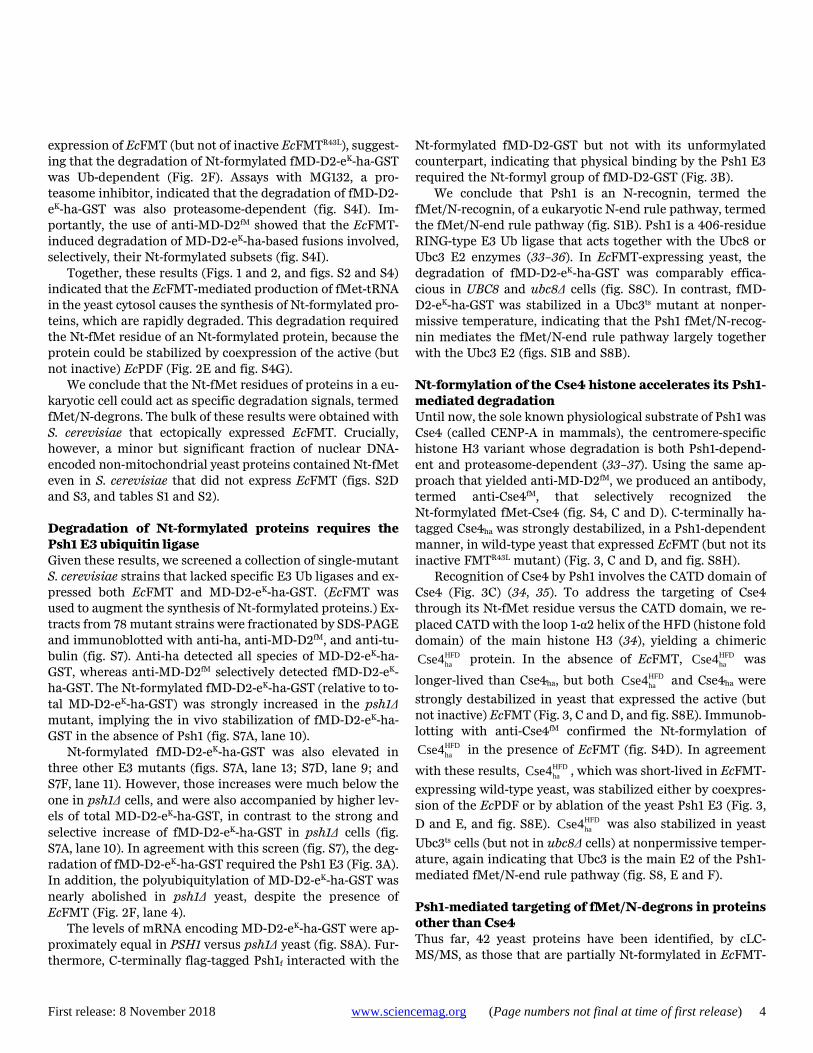
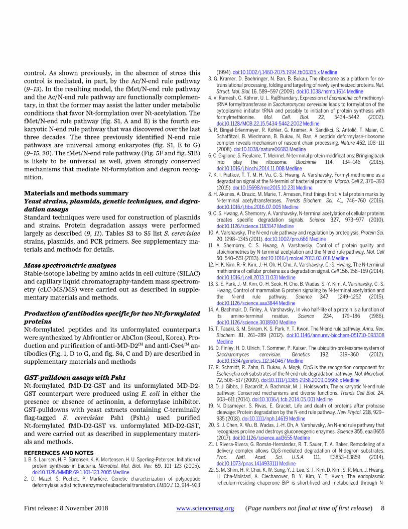
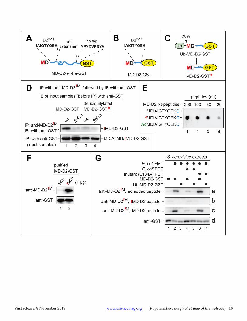
Antibody to N-terminally formylated protein reporters The 33 kDa reporter, denoted as MD-D2-eK-ha-GST, was iden-tical to our previously characterized reporter MD-D2-eK-ha-Ura3, save for the glutathione transferase (GST) moiety, used alternately with Ura3 (7). The reporter comprised the Nt-se-quence MDIAIGTYQEK [denoted as MD-D2 (7)], followed by a 44-residue sequence eK [extension (e) containing lysine (K)], by the ha epitope (YPYDVPDYA), by the AFLGQ linker (9), and by either GST or Ura3 (Figs. 1A and 2B). The eK seg-ment is often used in reporters, because it is unstructured (disordered) while lacking degrons in E. coli and yeast (10). MD-D2-GST was identical to MD-D2-eK-ha-GST save for the absence of the eK-ha segment (Fig. 1B).
Cycloheximide (CHX)-chases with MD-D2-eK-ha-Ura3 or MD-D2-eK-ha-GST showed that these reporters were short-lived in wild-type S. cerevisiae but were nearly completely stable in double-mutant naa20Δ ubr1Δ cells that lacked both the NatB (Naa20) Nt-acetylase and the Arg/N-end rule path-way (owing to the absence of Ubr1) (fig. S4, A and B). The naa20Δ mutation abrogated the otherwise expected Nt-acet-ylation, by NatB, of MD-D2-eK-ha-based reporters and thereby precluded their degradation by the Ac/N-end rule pathway (8–13). Given the stability of MD-D2-eK-ha-based re-porters in naa20Δ ubr1Δ cells (fig. S4, A and B), most degra-dation assays were performed in this genetic background.
To detect Nt-fMet by a method independent of mass spec-trometry, the synthetic Nt-formylated peptide fMDIAIGTYQEK (the fMD-D2 moiety; Fig. 1, A and B) was used to produce an affinity-purified rabbit antibody, termed anti-MD-D2fM, which recognized the above Nt-formylated peptide and also fMD-D2-GST, but did not recognize their un-formylated or Nt-acetylated counterparts (Fig. 1E).
Nt-formylated and unformylated MD-D2-GST were pro-duced in E. coli that were either incubated or not incubated with actinonin, an inhibitor of PDF-mediated deformylation
(5). The presence of Nt-fMet or unformylated Nt-Met in MD-D2-GST was verified using both mass spectrometry and anti-MD-D2fM antibody (Fig. 1F and fig. S5).
SDS-PAGE and immunoblotting with anti-MD-D2fM de-tected Nt-formylated fMD-D2-GST in extracts from EcFMT-expressing yeast (Fig. 1, B and G). More sensitive immunopre-cipitation-immunoblotting assays could also detect fMD-D2-GST in extracts from yeast that lacked EcFMT (Fig. 1D). The specificity of anti-MD-D2fM (Fig. 1, E and F) was also con-firmed by observing the quenching of immunoblotting signal by the Nt-formylated fMD-D2 peptide but not by its un-formylated counterpart (Fig. 1G).
In cells expressing MD-D2-GST, the Nt-fMet residue of fMD-D2-GST was generated pretranslationally (see Introduc-tion) (Fig. 1B). In contrast, MD-D2-GST was generated co-translationally from Ub-MD-D2-GST (bearing the Nt-Ub moiety), upon the cleavage-mediated removal of Nt-Ub by deubiquitylases (Fig. 1C) (10). Whereas directly expressed MD-D2-GST could be readily detected (using anti-MD-D2fM) as the Nt-formylated fMD-D2-GST species, no Nt-fMet was detected in MD-D2-GST* that resulted from deubiquitylation of Ub-MD-D2-GST (the asterisk of MD-D2-GST* denotes its distinct origin) (Fig. 1G). Besides confirming the specificity of anti-MD-D2fM, these results indicated the pretranslational (at the level of fMet-tRNA) origin of the Nt-fMet moiety of fMD-D2-GST. In addition, coexpression of EcFMT and E. coli PDF (EcPDF) (but not of its inactive mutant) in yeast strongly down-regulated the Nt-formylated fMD-D2-GST (Fig. 1G), in agreement with the (inferred) absence of PDF in S. cerevisiae (6).
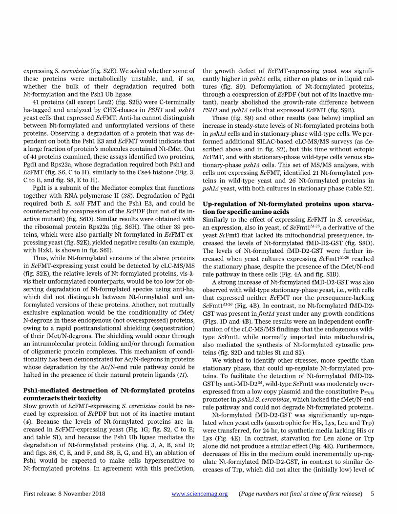
Selective degradation of Nt-formylated proteins The expression of EcFMT in yeast resulted in rapid degrada-tion of the otherwise long-lived MD-D2-eK-ha-Ura3 and MD-D2-eK-ha-GST (Fig. 2, A to D, and fig. S4, E to I). In contrast, no significant degradation of MD-D2-eK-ha-Ura3 or MD-D2-eK-ha-GST occurred upon expression of the inactive EcFMTR43L mutant or vector alone (Fig. 2, C and D, and fig. S4, E, F, and H). The EcFMT-induced degradation of MD-D2-eK-ha-GST and MD-D2-eK-ha-Ura3 in yeast was nearly abol-ished by coexpression of the EcPDF but not of its inactive EcPDFE134A mutant (Fig. 2, B and E, and fig. S4G).
Increasing the levels of EcFMT, using the incrementally stronger yeast promoters PADH1, PTEF1 and PTDH3, progressively destabilized MD-D2-eK-ha-GST, in part by decreasing its pre-chase (time-zero) levels (fig. S4H). In contrast, the expression of inactive EcFMTR43L had no effect (fig. S4H). EcFMT did not significantly alter the rate of protein synthesis in S. cerevisiae, in comparison to expression of inactive EcFMTR43L (fig. S6, A and B).
Tandem Ub-binding entity (TUBE)-pulldowns showed that MD-D2-eK-ha-GST was polyubiquitylated in yeast upon
First release: 8 November 2018 www.sciencemag.org (Page numbers not final at time of first release) 4
expression of EcFMT (but not of inactive EcFMTR43L), suggest-ing that the degradation of Nt-formylated fMD-D2-eK-ha-GST was Ub-dependent (Fig. 2F). Assays with MG132, a pro-teasome inhibitor, indicated that the degradation of fMD-D2-eK-ha-GST was also proteasome-dependent (fig. S4I). Im-portantly, the use of anti-MD-D2fM showed that the EcFMT-induced degradation of MD-D2-eK-ha-based fusions involved, selectively, their Nt-formylated subsets (fig. S4I).
Together, these results (Figs. 1 and 2, and figs. S2 and S4) indicated that the EcFMT-mediated production of fMet-tRNA in the yeast cytosol causes the synthesis of Nt-formylated pro-teins, which are rapidly degraded. This degradation required the Nt-fMet residue of an Nt-formylated protein, because the protein could be stabilized by coexpression of the active (but not inactive) EcPDF (Fig. 2E and fig. S4G).
We conclude that the Nt-fMet residues of proteins in a eu-karyotic cell could act as specific degradation signals, termed fMet/N-degrons. The bulk of these results were obtained with S. cerevisiae that ectopically expressed EcFMT. Crucially, however, a minor but significant fraction of nuclear DNA-encoded non-mitochondrial yeast proteins contained Nt-fMet even in S. cerevisiae that did not express EcFMT (figs. S2D and S3, and tables S1 and S2). Degradation of Nt-formylated proteins requires the Psh1 E3 ubiquitin ligase Given these results, we screened a collection of single-mutant S. cerevisiae strains that lacked specific E3 Ub ligases and ex-pressed both EcFMT and MD-D2-eK-ha-GST. (EcFMT was used to augment the synthesis of Nt-formylated proteins.) Ex-tracts from 78 mutant strains were fractionated by SDS-PAGE and immunoblotted with anti-ha, anti-MD-D2fM, and anti-tu-bulin (fig. S7). Anti-ha detected all species of MD-D2-eK-ha-GST, whereas anti-MD-D2fM selectively detected fMD-D2-eK-ha-GST. The Nt-formylated fMD-D2-eK-ha-GST (relative to to-tal MD-D2-eK-ha-GST) was strongly increased in the psh1Δ mutant, implying the in vivo stabilization of fMD-D2-eK-ha-GST in the absence of Psh1 (fig. S7A, lane 10).
Nt-formylated fMD-D2-eK-ha-GST was also elevated in three other E3 mutants (figs. S7A, lane 13; S7D, lane 9; and S7F, lane 11). However, those increases were much below the one in psh1Δ cells, and were also accompanied by higher lev-els of total MD-D2-eK-ha-GST, in contrast to the strong and selective increase of fMD-D2-eK-ha-GST in psh1Δ cells (fig. S7A, lane 10). In agreement with this screen (fig. S7), the deg-radation of fMD-D2-eK-ha-GST required the Psh1 E3 (Fig. 3A). In addition, the polyubiquitylation of MD-D2-eK-ha-GST was nearly abolished in psh1Δ yeast, despite the presence of EcFMT (Fig. 2F, lane 4).
The levels of mRNA encoding MD-D2-eK-ha-GST were ap-proximately equal in PSH1 versus psh1Δ yeast (fig. S8A). Fur-thermore, C-terminally flag-tagged Psh1f interacted with the
Nt-formylated fMD-D2-GST but not with its unformylated counterpart, indicating that physical binding by the Psh1 E3 required the Nt-formyl group of fMD-D2-GST (Fig. 3B).
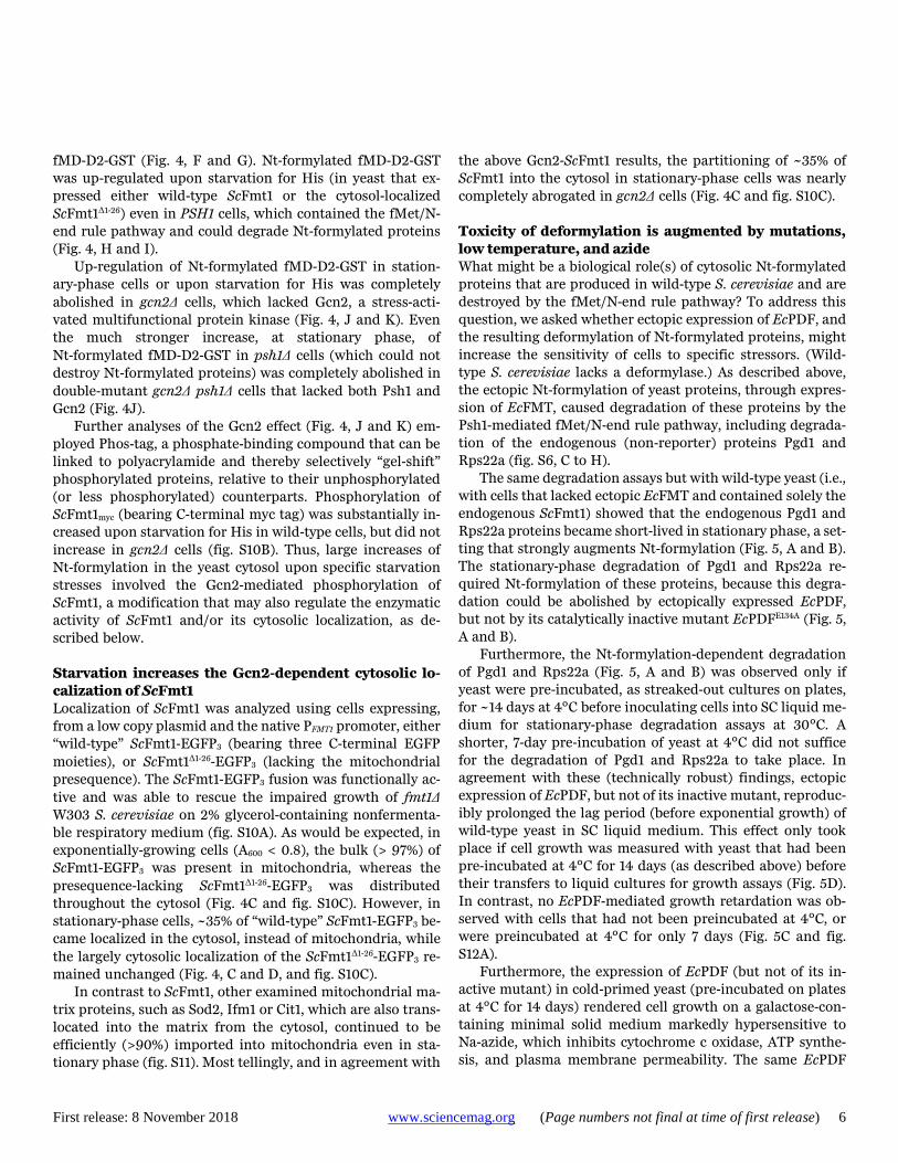
We conclude that Psh1 is an N-recognin, termed the fMet/N-recognin, of a eukaryotic N-end rule pathway, termed the fMet/N-end rule pathway (fig. S1B). Psh1 is a 406-residue RING-type E3 Ub ligase that acts together with the Ubc8 or Ubc3 E2 enzymes (33–36). In EcFMT-expressing yeast, the degradation of fMD-D2-eK-ha-GST was comparably effica-cious in UBC8 and ubc8Δ cells (fig. S8C). In contrast, fMD-D2-eK-ha-GST was stabilized in a Ubc3ts mutant at nonper-missive temperature, indicating that the Psh1 fMet/N-recog-nin mediates the fMet/N-end rule pathway largely together with the Ubc3 E2 (figs. S1B and S8B). Nt-formylation of the Cse4 histone accelerates its Psh1-mediated degradation Until now, the sole known physiological substrate of Psh1 was Cse4 (called CENP-A in mammals), the centromere-specific histone H3 variant whose degradation is both Psh1-depend-ent and proteasome-dependent (33–37). Using the same ap-proach that yielded anti-MD-D2fM, we produced an antibody, termed anti-Cse4fM, that selectively recognized the Nt-formylated fMet-Cse4 (fig. S4, C and D). C-terminally ha-tagged Cse4ha was strongly destabilized, in a Psh1-dependent manner, in wild-type yeast that expressed EcFMT (but not its inactive FMTR43L mutant) (Fig. 3, C and D, and fig. S8H).
Recognition of Cse4 by Psh1 involves the CATD domain of Cse4 (Fig. 3C) (34, 35). To address the targeting of Cse4 through its Nt-fMet residue versus the CATD domain, we re-placed CATD with the loop 1-α2 helix of the HFD (histone fold domain) of the main histone H3 (34), yielding a chimeric
HFDhaCse4 protein. In the absence of EcFMT, HFD
haCse4 was
longer-lived than Cse4ha, but both HFDhaCse4 and Cse4ha were
strongly destabilized in yeast that expressed the active (but not inactive) EcFMT (Fig. 3, C and D, and fig. S8E). Immunob-lotting with anti-Cse4fM confirmed the Nt-formylation of
HFDhaCse4 in the presence of EcFMT (fig. S4D). In agreement
with these results, HFDhaCse4 , which was short-lived in EcFMT-
expressing wild-type yeast, was stabilized either by coexpres-sion of the EcPDF or by ablation of the yeast Psh1 E3 (Fig. 3,
D and E, and fig. S8E). HFDhaCse4 was also stabilized in yeast
Ubc3ts cells (but not in ubc8Δ cells) at nonpermissive temper-ature, again indicating that Ubc3 is the main E2 of the Psh1-mediated fMet/N-end rule pathway (fig. S8, E and F). Psh1-mediated targeting of fMet/N-degrons in proteins other than Cse4 Thus far, 42 yeast proteins have been identified, by cLC-MS/MS, as those that are partially Nt-formylated in EcFMT-
First release: 8 November 2018 www.sciencemag.org (Page numbers not final at time of first release) 5
expressing S. cerevisiae (fig. S2E). We asked whether some of these proteins were metabolically unstable, and, if so, whether the bulk of their degradation required both Nt-formylation and the Psh1 Ub ligase.
41 proteins (all except Leu2) (fig. S2E) were C-terminally ha-tagged and analyzed by CHX-chases in PSH1 and psh1Δ yeast cells that expressed EcFMT. Anti-ha cannot distinguish between Nt-formylated and unformylated versions of these proteins. Observing a degradation of a protein that was de-pendent on both the Psh1 E3 and EcFMT would indicate that a large fraction of protein’s molecules contained Nt-fMet. Out of 41 proteins examined, these assays identified two proteins, Pgd1 and Rps22a, whose degradation required both Psh1 and EcFMT (fig. S6, C to H), similarly to the Cse4 histone (Fig. 3, C to E, and fig. S8, E to H).
Pgd1 is a subunit of the Mediator complex that functions together with RNA polymerase II (38). Degradation of Pgd1 required both E. coli FMT and the Psh1 E3, and could be counteracted by coexpression of the EcPDF (but not of its in-active mutant) (fig. S6D). Similar results were obtained with the ribosomal protein Rps22a (fig. S6H). The other 39 pro-teins, which were also partially Nt-formylated in EcFMT-ex-pressing yeast (fig. S2E), yielded negative results (an example, with Hxk1, is shown in fig. S6I).
Thus, while Nt-formylated versions of the above proteins in EcFMT-expressing yeast could be detected by cLC-MS/MS (fig. S2E), the relative levels of Nt-formylated proteins, vis-à-vis their unformylated counterparts, would be too low for ob-serving degradation of Nt-formylated species using anti-ha, which did not distinguish between Nt-formylated and un-formylated versions of these proteins. Another, not mutually exclusive explanation would be the conditionality of fMet/ N-degrons in these endogenous (not overexpressed) proteins, owing to a rapid posttranslational shielding (sequestration) of their fMet/N-degrons. The shielding would occur through an intramolecular protein folding and/or through formation of oligomeric protein complexes. This mechanism of condi-tionality has been demonstrated for Ac/N-degrons in proteins whose degradation by the Ac/N-end rule pathway could be halted in the presence of their natural protein ligands (11). Psh1-mediated destruction of Nt-formylated proteins counteracts their toxicity Slow growth of EcFMT-expressing S. cerevisiae could be res-cued by expression of EcPDF but not of its inactive mutant (4). Because the levels of Nt-formylated proteins are in-creased in EcFMT-expressing yeast (Fig. 1G; fig. S2, C to E; and table S1), and because the Psh1 Ub ligase mediates the degradation of Nt-formylated proteins (Fig. 3, A, B, and D; and figs. S6, C, E, and F, and S8, E, G, and H), an ablation of Psh1 would be expected to make cells hypersensitive to Nt-formylated proteins. In agreement with this prediction,
the growth defect of EcFMT-expressing yeast was signifi-cantly higher in psh1Δ cells, either on plates or in liquid cul-tures (fig. S9). Deformylation of Nt-formylated proteins, through a coexpression of EcPDF (but not of its inactive mu-tant), nearly abolished the growth-rate difference between PSH1 and psh1Δ cells that expressed EcFMT (fig. S9B).
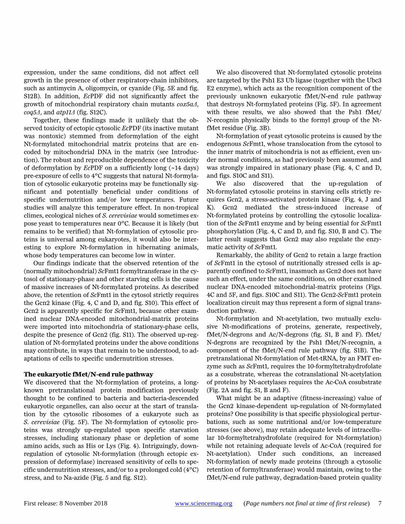
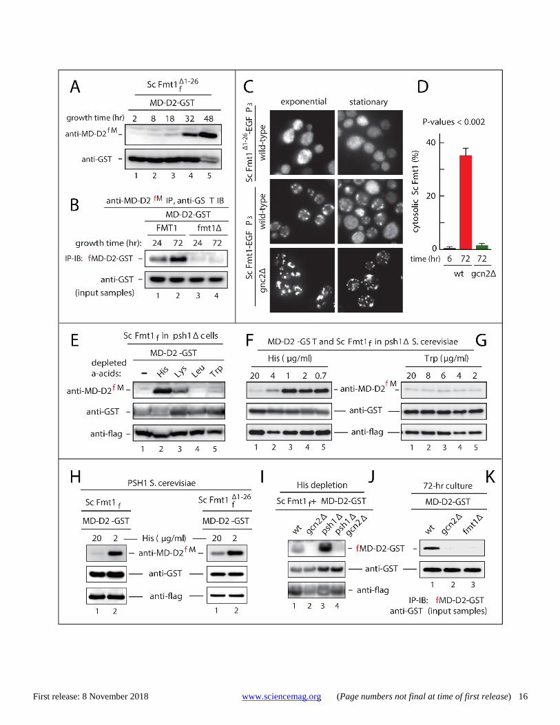
These (fig. S9) and other results (see below) implied an increase in steady-state levels of Nt-formylated proteins both in psh1Δ cells and in stationary-phase wild-type cells. We per-formed additional SILAC-based cLC-MS/MS surveys (as de-scribed above and in fig. S2), but this time without ectopic EcFMT, and with stationary-phase wild-type cells versus sta-tionary-phase psh1Δ cells. This set of MS/MS analyses, with cells not expressing EcFMT, identified 21 Nt-formylated pro-teins in wild-type yeast and 26 Nt-formylated proteins in psh1Δ yeast, with both cultures in stationary phase (table S2). Up-regulation of Nt-formylated proteins upon starva-tion for specific amino acids Similarly to the effect of expressing EcFMT in S. cerevisiae, an expression, also in yeast, of ScFmt1Δ1-26, a derivative of the yeast ScFmt1 that lacked its mitochondrial presequence, in-creased the levels of Nt-formylated fMD-D2-GST (fig. S8D). The levels of Nt-formylated fMD-D2-GST were further in-creased when yeast cultures expressing ScFmt1Δ1-26 reached the stationary phase, despite the presence of the fMet/N-end rule pathway in these cells (Fig. 4A and fig. S1B).
A strong increase of Nt-formylated fMD-D2-GST was also observed with wild-type stationary-phase yeast, i.e., with cells that expressed neither EcFMT nor the presequence-lacking ScFmt1Δ1-26 (Fig. 4B). In contrast, no Nt-formylated fMD-D2-GST was present in fmt1Δ yeast under any growth conditions (Figs. 1D and 4B). These results were an independent confir-mation of the cLC-MS/MS findings that the endogenous wild-type ScFmt1, while normally imported into mitochondria, also mediated the synthesis of Nt-formylated cytosolic pro-teins (fig. S2D and tables S1 and S2).
We wished to identify other stresses, more specific than stationary phase, that could up-regulate Nt-formylated pro-teins. To facilitate the detection of Nt-formylated fMD-D2-GST by anti-MD-D2fM, wild-type ScFmt1 was moderately over-expressed from a low copy plasmid and the constitutive PTDH3 promoter in psh1Δ S. cerevisiae, which lacked the fMet/N-end rule pathway and could not degrade Nt-formylated proteins.
Nt-formylated fMD-D2-GST was significnantly up-regu-lated when yeast cells (auxotrophic for His, Lys, Leu and Trp) were transferred, for 24 hr, to synthetic media lacking His or Lys (Fig. 4E). In contrast, starvation for Leu alone or Trp alone did not produce a similar effect (Fig. 4E). Furthermore, decreases of His in the medium could incrementally up-reg-ulate Nt-formylated fMD-D2-GST, in contrast to similar de-creases of Trp, which did not alter the (initially low) level of
First release: 8 November 2018 www.sciencemag.org (Page numbers not final at time of first release) 6
fMD-D2-GST (Fig. 4, F and G). Nt-formylated fMD-D2-GST was up-regulated upon starvation for His (in yeast that ex-pressed either wild-type ScFmt1 or the cytosol-localized ScFmt1Δ1-26) even in PSH1 cells, which contained the fMet/N-end rule pathway and could degrade Nt-formylated proteins (Fig. 4, H and I).
Up-regulation of Nt-formylated fMD-D2-GST in station-ary-phase cells or upon starvation for His was completely abolished in gcn2Δ cells, which lacked Gcn2, a stress-acti-vated multifunctional protein kinase (Fig. 4, J and K). Even the much stronger increase, at stationary phase, of Nt-formylated fMD-D2-GST in psh1Δ cells (which could not destroy Nt-formylated proteins) was completely abolished in double-mutant gcn2Δ psh1Δ cells that lacked both Psh1 and Gcn2 (Fig. 4J).
Further analyses of the Gcn2 effect (Fig. 4, J and K) em-ployed Phos-tag, a phosphate-binding compound that can be linked to polyacrylamide and thereby selectively “gel-shift” phosphorylated proteins, relative to their unphosphorylated (or less phosphorylated) counterparts. Phosphorylation of ScFmt1myc (bearing C-terminal myc tag) was substantially in-creased upon starvation for His in wild-type cells, but did not increase in gcn2Δ cells (fig. S10B). Thus, large increases of Nt-formylation in the yeast cytosol upon specific starvation stresses involved the Gcn2-mediated phosphorylation of ScFmt1, a modification that may also regulate the enzymatic activity of ScFmt1 and/or its cytosolic localization, as de-scribed below.
Starvation increases the Gcn2-dependent cytosolic lo-calization of ScFmt1 Localization of ScFmt1 was analyzed using cells expressing, from a low copy plasmid and the native PFMT1 promoter, either “wild-type” ScFmt1-EGFP3 (bearing three C-terminal EGFP moieties), or ScFmt1Δ1-26-EGFP3 (lacking the mitochondrial presequence). The ScFmt1-EGFP3 fusion was functionally ac-tive and was able to rescue the impaired growth of fmt1Δ W303 S. cerevisiae on 2% glycerol-containing nonfermenta-ble respiratory medium (fig. S10A). As would be expected, in exponentially-growing cells (A600 < 0.8), the bulk (> 97%) of ScFmt1-EGFP3 was present in mitochondria, whereas the presequence-lacking ScFmt1Δ1-26-EGFP3 was distributed throughout the cytosol (Fig. 4C and fig. S10C). However, in stationary-phase cells, ~35% of “wild-type” ScFmt1-EGFP3 be-came localized in the cytosol, instead of mitochondria, while the largely cytosolic localization of the ScFmt1Δ1-26-EGFP3 re-mained unchanged (Fig. 4, C and D, and fig. S10C).
In contrast to ScFmt1, other examined mitochondrial ma-trix proteins, such as Sod2, Ifm1 or Cit1, which are also trans-located into the matrix from the cytosol, continued to be efficiently (>90%) imported into mitochondria even in sta-tionary phase (fig. S11). Most tellingly, and in agreement with
the above Gcn2-ScFmt1 results, the partitioning of ~35% of ScFmt1 into the cytosol in stationary-phase cells was nearly completely abrogated in gcn2Δ cells (Fig. 4C and fig. S10C).
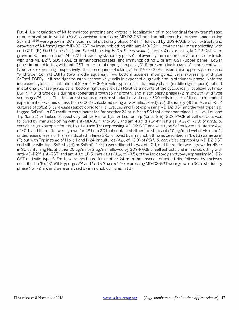
Toxicity of deformylation is augmented by mutations, low temperature, and azide What might be a biological role(s) of cytosolic Nt-formylated proteins that are produced in wild-type S. cerevisiae and are destroyed by the fMet/N-end rule pathway? To address this question, we asked whether ectopic expression of EcPDF, and the resulting deformylation of Nt-formylated proteins, might increase the sensitivity of cells to specific stressors. (Wild-type S. cerevisiae lacks a deformylase.) As described above, the ectopic Nt-formylation of yeast proteins, through expres-sion of EcFMT, caused degradation of these proteins by the Psh1-mediated fMet/N-end rule pathway, including degrada-tion of the endogenous (non-reporter) proteins Pgd1 and Rps22a (fig. S6, C to H).
The same degradation assays but with wild-type yeast (i.e., with cells that lacked ectopic EcFMT and contained solely the endogenous ScFmt1) showed that the endogenous Pgd1 and Rps22a proteins became short-lived in stationary phase, a set-ting that strongly augments Nt-formylation (Fig. 5, A and B). The stationary-phase degradation of Pgd1 and Rps22a re-quired Nt-formylation of these proteins, because this degra-dation could be abolished by ectopically expressed EcPDF, but not by its catalytically inactive mutant EcPDFE134A (Fig. 5, A and B).
Furthermore, the Nt-formylation-dependent degradation of Pgd1 and Rps22a (Fig. 5, A and B) was observed only if yeast were pre-incubated, as streaked-out cultures on plates, for ~14 days at 4°C before inoculating cells into SC liquid me-dium for stationary-phase degradation assays at 30°C. A shorter, 7-day pre-incubation of yeast at 4°C did not suffice for the degradation of Pgd1 and Rps22a to take place. In agreement with these (technically robust) findings, ectopic expression of EcPDF, but not of its inactive mutant, reproduc-ibly prolonged the lag period (before exponential growth) of wild-type yeast in SC liquid medium. This effect only took place if cell growth was measured with yeast that had been pre-incubated at 4°C for 14 days (as described above) before their transfers to liquid cultures for growth assays (Fig. 5D). In contrast, no EcPDF-mediated growth retardation was ob-served with cells that had not been preincubated at 4°C, or were preincubated at 4°C for only 7 days (Fig. 5C and fig. S12A).
Furthermore, the expression of EcPDF (but not of its in-active mutant) in cold-primed yeast (pre-incubated on plates at 4°C for 14 days) rendered cell growth on a galactose-con-taining minimal solid medium markedly hypersensitive to Na-azide, which inhibits cytochrome c oxidase, ATP synthe-sis, and plasma membrane permeability. The same EcPDF
First release: 8 November 2018 www.sciencemag.org (Page numbers not final at time of first release) 7
expression, under the same conditions, did not affect cell growth in the presence of other respiratory-chain inhibitors, such as antimycin A, oligomycin, or cyanide (Fig. 5E and fig. S12B). In addition, EcPDF did not significantly affect the growth of mitochondrial respiratory chain mutants cox5aΔ, coq5Δ, and atp11Δ (fig. S12C).
Together, these findings made it unlikely that the ob-served toxicity of ectopic cytosolic EcPDF (its inactive mutant was nontoxic) stemmed from deformylation of the eight Nt-formylated mitochondrial matrix proteins that are en-coded by mitochondrial DNA in the matrix (see Introduc-tion). The robust and reproducible dependence of the toxicity of deformylation by EcPDF on a sufficiently long (~14 days) pre-exposure of cells to 4°C suggests that natural Nt-formyla-tion of cytosolic eukaryotic proteins may be functionally sig-nificant and potentially beneficial under conditions of specific undernutrition and/or low temperatures. Future studies will analyze this temperature effect. In non-tropical climes, ecological niches of S. cerevisiae would sometimes ex-pose yeast to temperatures near 0°C. Because it is likely (but remains to be verified) that Nt-formylation of cytosolic pro-teins is universal among eukaryotes, it would also be inter-esting to explore Nt-formylation in hibernating animals, whose body temperatures can become low in winter.
Our findings indicate that the observed retention of the (normally mitochondrial) ScFmt1 formyltransferase in the cy-tosol of stationary-phase and other starving cells is the cause of massive increases of Nt-formylated proteins. As described above, the retention of ScFmt1 in the cytosol strictly requires the Gcn2 kinase (Fig. 4, C and D, and fig. S10). This effect of Gcn2 is apparently specific for ScFmt1, because other exam-ined nuclear DNA-encoded mitochondrial-matrix proteins were imported into mitochondria of stationary-phase cells, despite the presence of Gcn2 (fig. S11). The observed up-reg-ulation of Nt-formylated proteins under the above conditions may contribute, in ways that remain to be understood, to ad-aptations of cells to specific undernutrition stresses. The eukaryotic fMet/N-end rule pathway We discovered that the Nt-formylation of proteins, a long-known pretranslational protein modification previously thought to be confined to bacteria and bacteria-descended eukaryotic organelles, can also occur at the start of transla-tion by the cytosolic ribosomes of a eukaryote such as S. cerevisiae (Fig. 5F). The Nt-formylation of cytosolic pro-teins was strongly up-regulated upon specific starvation stresses, including stationary phase or depletion of some amino acids, such as His or Lys (Fig. 4). Intriguingly, down-regulation of cytosolic Nt-formylation (through ectopic ex-pression of deformylase) increased sensitivity of cells to spe-cific undernutrition stresses, and/or to a prolonged cold (4°C) stress, and to Na-azide (Fig. 5 and fig. S12).
We also discovered that Nt-formylated cytosolic proteins are targeted by the Psh1 E3 Ub ligase (together with the Ubc3 E2 enzyme), which acts as the recognition component of the previously unknown eukaryotic fMet/N-end rule pathway that destroys Nt-formylated proteins (Fig. 5F). In agreement with these results, we also showed that the Psh1 fMet/ N-recognin physically binds to the formyl group of the Nt-fMet residue (Fig. 3B).
Nt-formylation of yeast cytosolic proteins is caused by the endogenous ScFmt1, whose translocation from the cytosol to the inner matrix of mitochondria is not as efficient, even un-der normal conditions, as had previously been assumed, and was strongly impaired in stationary phase (Fig. 4, C and D, and figs. S10C and S11).
We also discovered that the up-regulation of Nt-formylated cytosolic proteins in starving cells strictly re-quires Gcn2, a stress-activated protein kinase (Fig. 4, J and K). Gcn2 mediated the stress-induced increase of Nt-formylated proteins by controlling the cytosolic localiza-tion of the ScFmt1 enzyme and by being essential for ScFmt1 phosphorylation (Fig. 4, C and D, and fig. S10, B and C). The latter result suggests that Gcn2 may also regulate the enzy-matic activity of ScFmt1.
Remarkably, the ability of Gcn2 to retain a large fraction of ScFmt1 in the cytosol of nutritionally stressed cells is ap-parently confined to ScFmt1, inasmuch as Gcn2 does not have such an effect, under the same conditions, on other examined nuclear DNA-encoded mitochondrial-matrix proteins (Figs. 4C and 5F, and figs. S10C and S11). The Gcn2-ScFmt1 protein localization circuit may thus represent a form of signal trans-duction pathway.
Nt-formylation and Nt-acetylation, two mutually exclu-sive Nt-modifications of proteins, generate, respectively, fMet/N-degrons and Ac/N-degrons (fig. S1, B and F). fMet/ N-degrons are recognized by the Psh1 fMet/N-recognin, a component of the fMet/N-end rule pathway (fig. S1B). The pretranslational Nt-formylation of Met-tRNA, by an FMT en-zyme such as ScFmt1, requires the 10-formyltetrahydrofolate as a cosubstrate, whereas the cotranslational Nt-acetylation of proteins by Nt-acetylases requires the Ac-CoA cosubstrate (Fig. 2A and fig. S1, B and F).
What might be an adaptive (fitness-increasing) value of the Gcn2 kinase-dependent up-regulation of Nt-formylated proteins? One possibility is that specific physiological pertur-bations, such as some nutritional and/or low-temperature stresses (see above), may retain adequate levels of intracellu-lar 10-formyltetrahydrofolate (required for Nt-formylation) while not retaining adequate levels of Ac-CoA (required for Nt-acetylation). Under such conditions, an increased Nt-formylation of newly made proteins (through a cytosolic retention of formyltransferase) would maintain, owing to the fMet/N-end rule pathway, degradation-based protein quality
First release: 8 November 2018 www.sciencemag.org (Page numbers not final at time of first release) 8
control. As shown previously, in the absence of stress this control is mediated, in part, by the Ac/N-end rule pathway (9–13). In the resulting model, the fMet/N-end rule pathway and the Ac/N-end rule pathway are functionally complemen-tary, in that the former may assist the latter under metabolic conditions that favor Nt-formylation over Nt-acetylation. The fMet/N-end rule pathway (fig. S1, A and B) is the fourth eu-karyotic N-end rule pathway that was discovered over the last three decades. The three previously identified N-end rule pathways are universal among eukaryotes (fig. S1, E to G) (9–15, 20). The fMet/N-end rule pathway (Fig. 5F and fig. S1B) is likely to be universal as well, given strongly conserved mechanisms that mediate Nt-formylation and degron recog-nition. Materials and methods summary Yeast strains, plasmids, genetic techniques, and degra-dation assays Standard techniques were used for construction of plasmids and strains. Protein degradation assays were performed largely as described (9, 11). Tables S3 to S5 list S. cerevisiae strains, plasmids, and PCR primers. See supplementary ma-terials and methods for details. Mass spectrometric analyses Stable-isotope labeling by amino acids in cell culture (SILAC) and capillary liquid chromatography-tandem mass spectrom-etry (cLC-MS/MS) were carried out as described in supple-mentary materials and methods. Production of antibodies specific for two Nt-formylated proteins Nt-formylated peptides and its unformylated counterparts were synthesized by Abfrontier or AbClon (Seoul, Korea). Pro-duction and purification of anti-MD-D2fM and anti-Cse4fM an-tibodies (Fig. 1, D to G, and fig. S4, C and D) are described in supplementary materials and methods GST-pulldown assays with Psh1 Nt-formylated fMD-D2-GST and its unformylated MD-D2-GST counterpart were produced using E. coli in either the presence or absence of actinonin, a deformylase inhibitor. GST-pulldowns with yeast extracts containing C-terminally flag-tagged S. cerevisiae Psh1 (Psh1f) used purified Nt-formylated fMD-D2-GST vs. unformylated MD-D2-GST, and were carried out as described in supplementary materi-als and methods.
REFERENCES AND NOTES 1. B. S. Laursen, H. P. Sørensen, K. K. Mortensen, H. U. Sperling-Petersen, Initiation of
protein synthesis in bacteria. Microbiol. Mol. Biol. Rev. 69, 101–123 (2005). doi:10.1128/MMBR.69.1.101-123.2005 Medline
2. D. Mazel, S. Pochet, P. Marlière, Genetic characterization of polypeptide deformylase, a distinctive enzyme of eubacterial translation. EMBO J. 13, 914–923
(1994). doi:10.1002/j.1460-2075.1994.tb06335.x Medline 3. G. Kramer, D. Boehringer, N. Ban, B. Bukau, The ribosome as a platform for co-
translational processing, folding and targeting of newly synthesized proteins. Nat. Struct. Mol. Biol. 16, 589–597 (2009). doi:10.1038/nsmb.1614 Medline
4. V. Ramesh, C. Köhrer, U. L. RajBhandary, Expression of Escherichia coli methionyl-tRNA formyltransferase in Saccharomyces cerevisiae leads to formylation of the cytoplasmic initiator tRNA and possibly to initiation of protein synthesis with formylmethionine. Mol. Cell. Biol. 22, 5434–5442 (2002). doi:10.1128/MCB.22.15.5434-5442.2002 Medline
5. R. Bingel-Erlenmeyer, R. Kohler, G. Kramer, A. Sandikci, S. Antolić, T. Maier, C. Schaffitzel, B. Wiedmann, B. Bukau, N. Ban, A peptide deformylase-ribosome complex reveals mechanism of nascent chain processing. Nature 452, 108–111 (2008). doi:10.1038/nature06683 Medline
6. C. Giglione, S. Fieulaine, T. Meinnel, N-terminal protein modifications: Bringing back into play the ribosome. Biochimie 114, 134–146 (2015). doi:10.1016/j.biochi.2014.11.008 Medline
7. K. I. Piatkov, T. T. M. H. Vu, C.-S. Hwang, A. Varshavsky, Formyl-methionine as a degradation signal at the N-termini of bacterial proteins. Microb. Cell 2, 376–393 (2015). doi:10.15698/mic2015.10.231 Medline
8. H. Aksnes, A. Drazic, M. Marie, T. Arnesen, First things first: Vital protein marks by N-terminal acetyltransferases. Trends Biochem. Sci. 41, 746–760 (2016). doi:10.1016/j.tibs.2016.07.005 Medline
9. C. S. Hwang, A. Shemorry, A. Varshavsky, N-terminal acetylation of cellular proteins creates specific degradation signals. Science 327, 973–977 (2010). doi:10.1126/science.1183147 Medline
10. A. Varshavsky, The N-end rule pathway and regulation by proteolysis. Protein Sci. 20, 1298–1345 (2011). doi:10.1002/pro.666 Medline
11. A. Shemorry, C. S. Hwang, A. Varshavsky, Control of protein quality and stoichiometries by N-terminal acetylation and the N-end rule pathway. Mol. Cell 50, 540–551 (2013). doi:10.1016/j.molcel.2013.03.018 Medline
12. H. K. Kim, R.-R. Kim, J.-H. Oh, H. Cho, A. Varshavsky, C.-S. Hwang, The N-terminal methionine of cellular proteins as a degradation signal. Cell 156, 158–169 (2014). doi:10.1016/j.cell.2013.11.031 Medline
13. S. E. Park, J.-M. Kim, O.-H. Seok, H. Cho, B. Wadas, S.-Y. Kim, A. Varshavsky, C.-S. Hwang, Control of mammalian G protein signaling by N-terminal acetylation and the N-end rule pathway. Science 347, 1249–1252 (2015). doi:10.1126/science.aaa3844 Medline
14. A. Bachmair, D. Finley, A. Varshavsky, In vivo half-life of a protein is a function of its amino-terminal residue. Science 234, 179–186 (1986). doi:10.1126/science.3018930 Medline
15. T. Tasaki, S. M. Sriram, K. S. Park, Y. T. Kwon, The N-end rule pathway. Annu. Rev. Biochem. 81, 261–289 (2012). doi:10.1146/annurev-biochem-051710-093308 Medline
16. D. Finley, H. D. Ulrich, T. Sommer, P. Kaiser, The ubiquitin-proteasome system of Saccharomyces cerevisiae. Genetics 192, 319–360 (2012). doi:10.1534/genetics.112.140467 Medline
17. R. Schmidt, R. Zahn, B. Bukau, A. Mogk, ClpS is the recognition component for Escherichia coli substrates of the N-end rule degradation pathway. Mol. Microbiol. 72, 506–517 (2009). doi:10.1111/j.1365-2958.2009.06666.x Medline
18. D. J. Gibbs, J. Bacardit, A. Bachmair, M. J. Holdsworth, The eukaryotic N-end rule pathway: Conserved mechanisms and diverse functions. Trends Cell Biol. 24, 603–611 (2014). doi:10.1016/j.tcb.2014.05.001 Medline
19. N. Dissmeyer, S. Rivas, E. Graciet, Life and death of proteins after protease cleavage: Protein degradation by the N-end rule pathway. New Phytol. 218, 929–935 (2018). doi:10.1111/nph.14619 Medline
20. S. J. Chen, X. Wu, B. Wadas, J.-H. Oh, A. Varshavsky, An N-end rule pathway that recognizes proline and destroys gluconeogenic enzymes. Science 355, eaal3655 (2017). doi:10.1126/science.aal3655 Medline
21. I. Rivera-Rivera, G. Román-Hernández, R. T. Sauer, T. A. Baker, Remodeling of a delivery complex allows ClpS-mediated degradation of N-degron substrates. Proc. Natl. Acad. Sci. U.S.A. 111, E3853–E3859 (2014). doi:10.1073/pnas.1414933111 Medline
22. S. M. Shim, H. R. Choi, K. W. Sung, Y. J. Lee, S. T. Kim, D. Kim, S. R. Mun, J. Hwang, H. Cha-Molstad, A. Ciechanover, B. Y. Kim, Y. T. Kwon, The endoplasmic reticulum-residing chaperone BiP is short-lived and metabolized through N-
First release: 8 November 2018 www.sciencemag.org (Page numbers not final at time of first release) 9
terminal arginylation. Sci. Signal. 11, eaan0630 (2018). doi:10.1126/scisignal.aan0630 Medline
23. J. H. Oh, J. Y. Hyun, A. Varshavsky, Control of Hsp90 chaperone and its clients by N-terminal acetylation and the N-end rule pathway. Proc. Natl. Acad. Sci. U.S.A. 114, E4370–E4379 (2017). doi:10.1073/pnas.1705898114 Medline
24. K. I. Piatkov, C. S. Brower, A. Varshavsky, The N-end rule pathway counteracts cell death by destroying proapoptotic protein fragments. Proc. Natl. Acad. Sci. U.S.A. 109, E1839–E1847 (2012). doi:10.1073/pnas.1207786109 Medline
25. B. Wadas, K. I. Piatkov, C. S. Brower, A. Varshavsky, Analyzing N-terminal arginylation through the use of peptide arrays and degradation assays. J. Biol.Chem. 291, 20976–20992 (2016). doi:10.1074/jbc.M116.747956 Medline
26. R.-G. Hu, J. Sheng, X. Qi, Z. Xu, T. T. Takahashi, A. Varshavsky, The N-end rulepathway as a nitric oxide sensor controlling the levels of multiple regulators.Nature 437, 981–986 (2005). doi:10.1038/nature04027 Medline
27. D. C. Scott, J. K. Monda, E. J. Bennett, J. W. Harper, B. A. Schulman, N-terminal acetylation acts as an avidity enhancer within an interconnected multiproteincomplex. Science 334, 674–678 (2011). doi:10.1126/science.1209307 Medline
28. C.-S. Hwang, A. Shemorry, D. Auerbach, A. Varshavsky, The N-end rule pathway ismediated by a complex of the RING-type Ubr1 and HECT-type Ufd4 ubiquitinligases. Nat. Cell Biol. 12, 1177–1185 (2010). doi:10.1038/ncb2121 Medline
29. W. S. Choi, B.-C. Jeong, Y. J. Joo, M.-R. Lee, J. Kim, M. J. Eck, H. K. Song, Structural basis for the recognition of N-end rule substrates by the UBR box of ubiquitinligases. Nat. Struct. Mol. Biol. 17, 1175–1181 (2010). doi:10.1038/nsmb.1907 Medline
30. E. Matta-Camacho, G. Kozlov, F. F. Li, K. Gehring, Structural basis of substraterecognition and specificity in the N-end rule pathway. Nat. Struct. Mol. Biol. 17, 1182–1187 (2010). doi:10.1038/nsmb.1894 Medline
31. M. K. Kim, S. J. Oh, B. G. Lee, H. K. Song, Structural basis for dual specificity ofyeast N-terminal amidase in the N-end rule pathway. Proc. Natl. Acad. Sci. U.S.A. 113, 12438–12443 (2016). doi:10.1073/pnas.1612620113 Medline
32. A. Varshavsky, ‘Spalog’ and ‘sequelog’: Neutral terms for spatial and sequencesimilarity. Curr. Biol. 14, R181–R183 (2004). doi:10.1016/j.cub.2004.02.014 Medline
33. G. Hewawasam, M. Shivaraju, M. Mattingly, S. Venkatesh, S. Martin-Brown, L.Florens, J. L. Workman, J. L. Gerton, Psh1 is an E3 ubiquitin ligase that targets the centromeric histone variant Cse4. Mol. Cell 40, 444–454 (2010).doi:10.1016/j.molcel.2010.10.014 Medline
34. P. Ranjitkar, M. O. Press, X. Yi, R. Baker, M. J. MacCoss, S. Biggins, An E3 ubiquitin ligase prevents ectopic localization of the centromeric histone H3 variant via the centromere targeting domain. Mol. Cell 40, 455–464 (2010).doi:10.1016/j.molcel.2010.09.025 Medline
35. W. C. Au, A. R. Dawson, D. W. Rawson, S. B. Taylor, R. E. Baker, M. A. Basrai, A novel role of the N terminus of budding yeast histone H3 variant Cse4 in ubiquitin-mediated proteolysis. Genetics 194, 513–518 (2013). doi:10.1534/genetics.113.149898 Medline
36. M. B. Metzger, J. L. Scales, M. F. Dunklebarger, A. M. Weissman, The ubiquitin ligase (E3) Psh1p is required for proper segregation of both centromeric and two-micron plasmids in Saccharomyces cerevisiae. G3 (Bethesda) 7, 3731–3743 (2017). Medline
37. H. Cheng, X. Bao, X. Gan, S. Luo, H. Rao, Multiple E3s promote the degradation of histone H3 variant Cse4. Sci. Rep. 7, 8565 (2017). doi:10.1038/s41598-017-08923-w Medline
38. P. J. Robinson, M. J. Trnka, D. A. Bushnell, R. E. Davis, P.-J. Mattei, A. L.Burlingame, R. D. Kornberg, Structure of a complete mediator-RNA polymerase II pre-initiation complex. Cell 166, 1411–1422.e16 (2016). doi:10.1016/j.cell.2016.08.050 Medline
39. F. M. Ausubel, R. Brent, R. E. Kingston, D. D. Moore, J. G. Seidman, J. A. Smith, D. Struhl, Eds., Current Protocols in Molecular Biology (Wiley, 2010).40. F. Sherman, Getting started with yeast. Methods Enzymol. 194, 3–21 (1991).doi:10.1016/0076-6879(91)94004-V Medline
41. M. S. Longtine, A. McKenzie 3rd, D. J. Demarini, N. G. Shah, A. Wach, A. Brachat, P. Philippsen, J. R. Pringle, Additional modules for versatile and economical PCR-based gene deletion and modification in Saccharomyces cerevisiae. Yeast 14, 953–961 (1998). doi:10.1002/(SICI)1097-0061(199807)14:10<953:AID-YEA293>3.0.CO;2-U Medline
42. C. Janke, M. M. Magiera, N. Rathfelder, C. Taxis, S. Reber, H. Maekawa, A. Moreno-Borchart, G. Doenges, E. Schwob, E. Schiebel, M. Knop, A versatile toolbox forPCR-based tagging of yeast genes: New fluorescent proteins, more markers and promoter substitution cassettes. Yeast 21, 947–962 (2004).doi:10.1002/yea.1142 Medline
43. S. Spector, J. M. Flynn, B. Tidor, T. A. Baker, R. T. Sauer, Expression of N-formylated proteins in Escherichia coli. Protein Expr. Purif. 32, 317–322 (2003).doi:10.1016/j.pep.2003.08.004 Medline
44. J. Cox, M. Mann, MaxQuant enables high peptide identification rates,individualized p.p.b.-range mass accuracies and proteome-wide protein quantification. Nat. Biotechnol. 26, 1367–1372 (2008). doi:10.1038/nbt.1511 Medline
45. J. Yeom, S. Ju, Y. Choi, E. Paek, C. Lee, Comprehensive analysis of human protein N-termini enables assessment of various protein forms. Sci. Rep. 7, 6599 (2017).doi:10.1038/s41598-017-06314-9 Medline
46. B. Westermann, W. Neupert, Mitochondria-targeted green fluorescent proteins:Convenient tools for the study of organelle biogenesis in Saccharomyces cerevisiae. Yeast 16, 1421–1427 (2000). doi:10.1002/1097-0061(200011)16:15<1421:AID-YEA624>3.0.CO;2-U Medline
47. L. Vial, P. Gomez, M. Panvert, E. Schmitt, S. Blanquet, Y. Mechulam, Mitochondrial methionyl-tRNAfMet formyltransferase from Saccharomyces cerevisiae: Genedisruption and tRNA substrate specificity. Biochemistry 42, 932–939 (2003).doi:10.1021/bi026901x Medline
48. W. L. Lee, J. R. Oberle, J. A. Cooper, The role of the lissencephaly protein Pac1during nuclear migration in budding yeast. J. Cell Biol. 160, 355–364 (2003).doi:10.1083/jcb.200209022 Medline
ACKNOWLEDGMENTS
We thank W.-K. Huh (Seoul National University, Seoul, Korea) for yeast deletion library mutants, S. Biggins (Hutchinson Cancer Center, Seattle, WA, USA) for pSB1535 and pSB1541, and R. Sauer (MIT, Cambridge, MA, USA) for AG110A(DE) E. coli. We also thank the present and former members of the Hwang laboratory for their assistance and advice. Funding: Supported by grants from the Samsung Science & Technology Foundation (SSTF-BA1401-17) and the BK21 plus program (C.-S.H.), by the NRF grants of the Korean Government (MSIP) NRF-2017M3A9F9030559 (C.L.) and NRF-2017R1A5A1015366 (J.-Y.Y.), and by NIH grants R01GM031530 and R01DK039520 (A.V.). Author contributions: C.-S.H., J.-M.K., J.-Y.Y., C.L., A.V., and other coauthors designed research. J.-M.K., O.-H.S., S.J., J.-E.H., J.Y., D.-S.K. and C.-S.H. performed research, and all coauthors analyzed data. C.-S.H., J.-M.K., C.L. and A.V. wrote the paper. Competing interests: All coauthors declare no competing interests. Data and materials availability: All mass spectrometric data of this study are available in PRIDE database (accession number: PXD010780). All (other) data needed to evaluate the conclusions in the paper are present in the paper or the Supplementary Materials.
SUPPLEMENTARY MATERIALS www.sciencemag.org/cgi/content/full/science.aat0174/DC1 Materials and Methods Figs. S1 to S12 Tables S1 to S5 References (39–48)
16 January 2018; resubmitted 24 August 2018 Accepted 24 October 2018 Published online 8 November 2018 10.1126/science.aat0174
First release: 8 November 2018 www.sciencemag.org (Page numbers not final at time of first release) 10
First release: 8 November 2018 www.sciencemag.org (Page numbers not final at time of first release) 11
Fig. 1. Antibody specific for a set of N-terminally formylated reporters. (A) MD-D2-eK-ha-GST and some of its amino acid sequences. GST, eK: see the main text. (B) MD-D2-GST, same fusion as in (A) but lacking the eK-ha segment. (C) Same fusion as in (B) but bearing, in addition, the Nt-Ub moiety. (D) Lane 1, upper panel: MD-D2-GST was expressed in wild-type (FMT1) S. cerevisiae, and was immunoprecipitated from yeast extracts with anti-MD-D2fM antibody (which selectively recognized Nt-formylated fMD-D2-GST), followed by SDS-PAGE and immunoblotting with anti-GST. Lane 2, same as lane 1 but with fmt1Δ yeast. Lane 3, same as lane 1 but with Ub-MD-D2-GST. Lane 4, same as lane 3 but with fmt1Δ yeast. Lower panel, inputs; immunoblotting with anti-GST. (E) Dot immunoblotting with anti-MD-D2fM antibody versus decreasing amounts of the unmodified MDIAIGTYQEKC peptide and either its Nt-formylated or Nt-acetylated counterparts. (F) Purified unformylated MD-D2-GST (lane 1) and Nt-formylated fMD-D2-GST (lane 2) were subjected to SDS-PAGE and immunoblotting with either anti-MD-D2fM (upper panel) or anti-GST (lower panel). (G) Pretranslational Nt-formylation of MD-D2-GST. The upper three panels show immunoblots, using anti-MD-D2fM, of SDS-PAGE-fractionated extracts from S. cerevisiae that expressed (or did not express) specific proteins indicated above the panels (see the main text). Immunoblots a, b, and c with anti-MD-D2fM were performed in the absence or presence of the indicated peptides. Immunoblot d is the same as immunoblot a but with anti-GST.
First release: 8 November 2018 www.sciencemag.org (Page numbers not final at time of first release) 12
First release: 8 November 2018 www.sciencemag.org (Page numbers not final at time of first release) 13
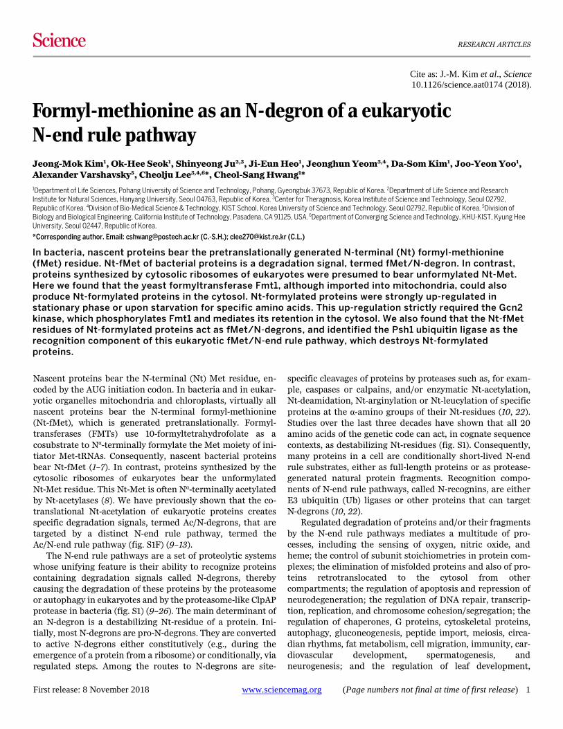
Fig. 2. Selective in vivo degradation of Nt-formylated proteins. (A) A diagram of the fMet-mediated cytosolic synthesis and degradation of a GST-based or Ura3-based Nt-formylation reporter (relevant to Figs. 2, B to F, and 3A, and to figs. S4, E to I). (B) 1) A high copy plasmid expressing EcFMT in S. cerevisiae from the PGAL1 promoter, 2) a high copy plasmid expressing EcFMT and EcPDF from the bidirectional PGAL1,10 promoter, and 3) a low copy plasmid expressing MD-D2-eK-ha-Ura3 (or MD-D2-eK-ha-GST) from the PCUP1 promoter. (C) Cycloheximide (CHX)-chases with MD-D2-eK-ha-Ura3 in naa20Δ ubr1Δ S. cerevisiae (see the main text) that expressed either vector alone (lanes 1-4) or EcFMT (lanes 5-8). Immunoblotting with anti-ha. (D) Same as in (C) but with MD-D2-eK-ha-GST and cells that expressed either EcFMT (lanes 1-4) or its inactive EcFMTR43L mutant (lanes 5-8). The graphs show quantification of data (three independent pairs of CHX-chases), with mean ± standard error (SE). (E) CHX-chases, using anti-ha, with MD-D2-eK-ha-Ura3 in naa20Δ ubr1Δ S. cerevisiae that expressed wild-type EcFMT and also expressed either wild-type EcPDF (lanes 3-4) or its inactive EcPDFE134A mutant (lanes 1-2). (F) Tandem Ub-binding entity (TUBE)-pulldowns with S. cerevisiae expressing (or not expressing) either MD-D2-eK-ha-GST, or EcFMT, or its EcFMTR43L mutant, with anti-GST immunoblotting, after TUBE-pulldowns. Lower panel: inputs; immunoblotting with anti-GST.
First release: 8 November 2018 www.sciencemag.org (Page numbers not final at time of first release) 14
First release: 8 November 2018 www.sciencemag.org (Page numbers not final at time of first release) 15
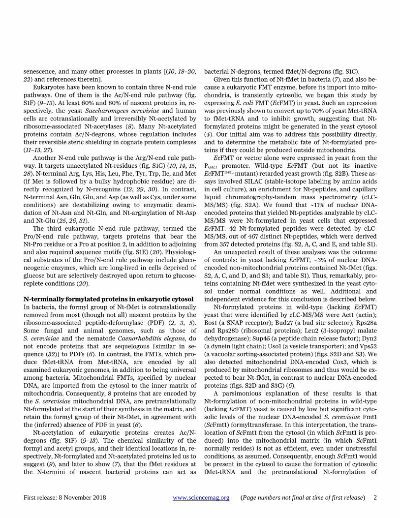
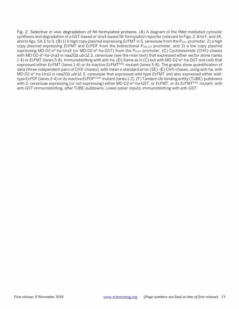
Fig. 3. The Psh1 E3 ubiquitin ligase as an fMet/N-recognin. (A) CHX-chases with MD-D2-eK-ha-GST in naa20Δ ubr1Δ S. cerevisiae (see the main text) that expressed EcFMT and either contained (lanes 1-4) or lacked (lanes 5-8) the Psh1 E3. Immunoblotting with anti-fMD-D2fM. The graphs show quantification of data (three independent pairs of CHX-chases), with mean ± standard error (SE). (B) GST-pulldowns with purified Nt-formylated fMD-D2-GST vs. unformylated MD-D2-GST and extracts of S. cerevisiae that expressed Psh1f (see Materials and methods). Detection with anti-flag (specific for Psh1f), anti-GST, and anti-fMD-D2fM (specific for Nt-formylated fMD-D2-GST). Lanes 1-3 are described in (B). Note specific binding of Psh1f to fMD-D2-GST. (C) Wild-type ha-tagged Cse4ha, histone H3, and “hybrid” HFD
haCse4 (see the main text). (D) CHX-chases with Cse4ha (lanes 1-4) and HFDhaCse4 (lanes 5-10) in S. cerevisiae
expressing EcFMT or its EcFMTR43L mutant. Lanes 9-10, same as lanes 5-6, but in psh1Δ yeast. Immunoblotting with anti-ha. (E) CHX-chases of HFD
haCse4 in EcFMT-expressing S. cerevisiae that also expressed EcPDF (lanes 1-3) or its inactive EcPDFE134A mutant (lanes 4-6). The graphs show quantification of data (three independent pairs of CHX-chases), with mean ± standard error (SE).
First release: 8 November 2018 www.sciencemag.org (Page numbers not final at time of first release) 16
First release: 8 November 2018 www.sciencemag.org (Page numbers not final at time of first release) 17
Fig. 4. Up-regulation of Nt-formylated proteins and cytosolic localization of mitochondrial formyltransferase upon starvation in yeast. (A) S. cerevisiae expressing MD-D2-GST and the mitochondrial presequence-lacking ScFmt1f Δ1-26 were grown in SC medium until stationary phase (48 hr), followed by SDS-PAGE of cell extracts and detection of Nt-formylated fMD-D2-GST by immunoblotting with anti-MD-D2fM. Lower panel, immunoblotting with anti-GST. (B) FMT1 (lanes 1-2) and ScFmt1-lacking fmt1Δ S. cerevisiae (lanes 3-4) expressing MD-D2-GST were grown in SC medium from 24 to 72 hr (reaching stationary phase), followed by immunoprecipitation of cell extracts with anti-MD-D2fM, SDS-PAGE of immunoprecipitates, and immunoblotting with anti-GST (upper panel). Lower panel: immunoblotting with anti-GST, but of total (input) samples. (C) Representative images of fluorescent wild-type cells expressing, respectively, the presequence-lacking ScFmt1Δ1-26-EGFP3 fusion (two upper squares) and “wild-type” ScFmt1-EGFP3 (two middle squares). Two bottom squares show gcn2Δ cells expressing wild-type ScFmt1-EGFP3. Left and right squares, respectively: cells in exponential growth and in stationary phase. Note the increased cytosolic localization of ScFmt1-EGFP3 in wild-type cells in stationary phase (middle right square) but not in stationary-phase gcn2Δ cells (bottom right square). (D) Relative amounts of the cytosolically localized ScFmt1-EGFP3 in wild-type cells during exponential growth (6-hr growth) and in stationary-phase (72-hr growth) wild-type versus gcn2Δ cells. The data are shown as means ± standard deviations; ~300 cells in each of three independent experiments. P-values of less than 0.002 (calculated using a two-tailed t-test). (E) Stationary (48 hr; A600 of ~3.5) cultures of psh1Δ S. cerevisiae (auxotrophic for His, Lys, Leu and Trp) expressing MD-D2-GST and the wild-type flag-tagged ScFmt1f in SC medium were incubated for another 24 hr in fresh SC that either contained His, Lys, Leu and Trp (lane 1) or lacked, respectively, either His, or Lys, or Leu, or Trp (lanes 2-5). SDS-PAGE of cell extracts was followed by immunoblotting with anti-MD-D2fM, anti-GST, and anti-flag. (F) 24-hr cultures (A600 of ~3.0) of psh1Δ S. cerevisiae (auxotrophic for His, Lys, Leu and Trp) expressing MD-D2-GST and wild-type ScFmt1f were diluted to A600 of ~0.1, and thereafter were grown for 48 hr in SC that contained either the standard (20 μg/ml) level of His (lane 1) or decreasing levels of His, as indicated in lanes 2-5, followed by immunoblotting as described in (E). (G) Same as in (F) but with Trp instead of His. (H and I) 24-hr cultures (A600 of ~3.0) of PSH1 S. cerevisiae expressing MD-D2-GST and either wild-type ScFmt1f (H) or ScFmt1f Δ1-26 (I) were diluted to A600 of ~0.1, and thereafter were grown for 48 hr in SC containing His at either 20 μg/ml or 2 μg/ml, followed by SDS-PAGE of cell extracts and immunoblotting with anti-MD-D2fM, anti-GST, and anti-flag. (J) S. cerevisiae (A600 of ~3.5), of the indicated genotypes, expressing MD-D2-GST and wild-type ScFmt1f, were incubated for another 24 hr in the absence of added His, followed by analyses described in (E). (K) Wild-type, gcn2Δ and fmt1Δ S. cerevisiae expressing MD-D2-GST were grown in SC to stationary phase (for 72 hr), and were analyzed by immunoblotting as in (B).
First release: 8 November 2018 www.sciencemag.org (Page numbers not final at time of first release) 18
Fig. 5. Toxicity of deformylase is augmented by mutations, low temperature, and azide. (A) CHX-chases for 0, 3 and 6 hr with C-terminally ha-tagged Pgd1ha and 48-hr culture (stationary phase) of wild-type S. cerevisiae that expressed either EcPDF or its catalytically inactive EcPDFE134A mutant. Before these assays in liquid cultures, cells were kept as streaked-out cultures on plates for ~14 days at 4°C (see the main text). The graphs show quantification of data (three independent pairs of CHX-chases), with mean ± standard error (SE). (B) Same as in (A), but CHX-chases for 0, 6 and 9 hr with Rps22aha. (C) Growth (A600) of wild-type yeast in SC medium, measured in 96-well microplates using orbital shaker and Epoch 2 microplate spectrophotometer (BioTek, Winooski, VT). S. cerevisiae expressed either vector alone, or EcPDF or its inactive EcPDFE134A mutant, and were not preincubated at 4°C. Note indistinguishable rates of growth, irrespective of expression of or EcPDF. (D) Same as in (C), but yeast were kept as streaked-out cultures on plates for 14 days at 4°C [see (A) and the main text] before growth assays in liquid culture. Note a significantly slower growth, with pre-incubation at 4°C, of cells that expressed EcPDF (but not its inactive EcPDFE134A mutant). In (C) and (D), each point of a curve also shows ± standard error (SE) of A600 values (at 10-min intervals), with measurements carried out independently six times. (E) Wild-type S. cerevisiae that expressed either vector alone, or EcPDF, or its inactive EcPDFE134A mutant were serially diluted (5-fold) and spotted on galactose-containing minimal medium (SGal) plates with or without either 20 μM NaN3 or 20 nM antimycin A. The plates were incubated at 30°C for 3 days. As in (A) to (D), yeast cells were kept for ~14 days at 4°C before a 16-hr liquid-culture growth and spot assays. (F) A partial summary of main results. Upon specific nutritional stresses (including stationary phase), the ScFmt1 formyltransferase is substantially retained in the cytosol, an alteration that strictly requires the Gcn2 kinase, which phosphorylates ScFmt1 and might also increase its enzymatic activity. The effect of Gcn2 on the cytosolic retention of ScFmt1 appears to be confined to ScFmt1, i.e., it does not extend to other nuclear DNA-encoded mitochondrial-matrix proteins. The increased cytosolic localization of ScFmt1 increases production of cytosolic fMet-tRNAi, and thereby up-regulates cytosolic Nt-formylated proteins. The latter are targeted for degradation by the Psh1 Ub ligase, the fMet/N-recognin of the proteasome-mediated fMet/N-end rule pathway.


![Dietary supplementation with free methionine or methionine … · 2019. 6. 27. · with MHA or DL-methionine in heat stress-exposed broilers [23, 24]. In this study, we hypothesize](https://img.dokumen.tips/doc/110x75/60e337666b3f9a31a45a96d1/dietary-supplementation-with-free-methionine-or-methionine-2019-6-27-with-mha.jpg)