Embed Size (px)
Citation preview
10
International Journal of Pharmaceutical Science and Research ISSN: 2455-4685 www.pharmacyresearchjournal.com Volume 1; Issue 1; January 2016; Page No. 10-26
Formulation and evaluation of anti-emetic microspheres for nasal drug delivery system 1 Jaya Nigam, 1 Preeti Pandey, 1 Manoj Kumar Mishra, 1 Anupam Singh Bhadouriya, 2 Pramod Kumar Biswal
1 Shambhunath Institute of Pharmacy, Jhalwa, Allahabad, Uttar Pradesh. 2 Kunwar Haribansh Singh College of Pharmacy, Jaunpur, Uttar Pradesh.
Abstract The microspheres, A natural biodegradable polymer, is of great interest in pharmacy research due to its excellent properties including bioavailability, no toxicity, high charge density and mucoadhesivity, which creates immense potential for various pharmaceutical applications. It has been studied as a promising carrier system for mucoadhesive drug delivery via nasal route to enhanced bioavailability. Preparing microspheres which will help in releasing small quantity of drug and thereby reduces the intensity of adverse effect associated with the drug, slowly dissolving polymer for sustaining the release may be suitable for long term therapy is controlled alleviation of clinical manifestation. Microspheres are evaluated by percentage yield, determination of percentage drug content, entrapment efficiency and in-vitro dissolution studies. The purpose of this research work was to develop optimized and systematically evaluate performances of microspheres of antiemetic drug ondansetron. Alginate microspheres coated with polymer pectin, starch corn were prepared by ionotropic- gelation technique utilizing various cross linking agent like calcium chloride (CaCl2) and (BaCl2), depending upon the variability in the concentration of alginate, percentage of cross linking agent, time of curing, the factors like particle size, incorporation efficiency and release rate of microspheres varies. The microspheres obtained were discrete, spherical and free flowing. Aim of our present study was to formulate the Alginate Microsphere loaded with Ondansetron by using Ionic- gelation technique for control prolonged release of drug. Ondansetron Microspheres were formulated by using drug with Sodium alginate, Pectin and Starch in 9 formulations was f1, f2, f3, f4, f5, f6, f7, f8, f9. All the formulation were investigation for various evaluation parameters like Particle Size (360 to 280), Bulk Density (0.55 to 0.46 gm/ml) , Flow Behavior, Entrapment Efficiency (72 to 85%), Percentage yield (55 to78%), and In-vitro drug release etc. All the formulation showed good flow behavior. SEM Study revealed that the spheres were almost spherical in shape with smooth surface. In-vitro drug release study showed that by increasing the polymer concentration the drug release of all the formulation were gradually decreased and the optimized formulation (f3) was able to sustain the drug release for 12 hours. So, It was Concluded that Alginate Microspheres loaded with Ondansetron can be prepared by Ionic-gelation technique and used for sustaining the drug release for prolong period of time. Keywords: Tuberculosis, Drugs, Herbs, Synthetic drugs. 1. Introduction 1.1 Microsphere Microspheres are solid spherical particles ranging in size 1 to 1000 mm. They are the spherical free flowing particle. The microspheres are free flowing powders consisting of proteins or synthetic polymers, which are biodegradable in nature. Microcapsules are those in which entrapped substance is added at the time of preparation. Distinctly surrounded by distinct capsule wall and micro matrices in which entrapped substance is dispersing throughout the microspheres matrix. Solid biodegradable involves microspheres incorporating a drug dispersed or dissolved through particle matrix have the potential for the controlled release of drug. They are made up polymeric, waxy, or other protective materials, that is, biodegradable synthetic polymers and modified natural products [1]. Nasal drug delivery may be used for either local or systemic effects. Low molecular weight drugs which are rapidly absorbed through nasal mucosa. The main reasons for this are the high permeability, fairly wide absorption area, porous and thin endothelial basement membrane of the nasal epithelium [2]. Microspheres received much attention not only for prolonged release, but also for targeting of anticancer drugs. In future by
combining various other strategies, microspheres will find the central place in novel drug delivery, particularly in diseased cell sorting, diagnostics, gene & genetic materials, safe, targeted and effective in vivo delivery and supplements as miniature versions of diseased organ and tissues in the body [3]. Anti-emetics are used to prevent nausea and vomiting caused by cancer chemotherapy, radiation therapy, and surgery. Ondansetron, a highly selective 5-HT3 receptor antagonist, is used as an anti-emetic and under goes extensive first pass metabolism [4]. Ondansetron hydrochloride (OND) is a 5HT3 serotonin antagonist used in the prevention of nausea and vomiting associated with emetogenic cancer chemotherapy [5]. OND is well absorbed and undergoes first-pass metabolism. Oral bioavailability of OND is almost 59%, and peak plasma about 0.03–0.04 μg/ml is obtained after 1.5 to 2 h of administration [6]. As administering drug by buccal route avoids hepatic first-pass metabolism, thus, delivery of OND to the systemic circulation via the buccal route would improve its bioavailability. Buccal delivery involves the administration of the drug through the buccal mucosal membrane lining of the oral cavity [7].
11
1.2 Microspheres as Nasal drug delivery system Morphologically, there are two general structures: microcapsules and microspheres1. A microcapsule is a reservoir-type system with regular or irregular shapes that contains a well-defined core and envelope. The core can be solid, liquid, or gas and the envelope are made of a continuous, porous or nonporous, polymeric phase created by one or more polymers. Alternatively, a microsphere is a homogeneous structure made of a continuous phase of one or more miscible polymers in which particulate drug is dispersed throughout the matrix, at either the macroscopic (particulates) or molecular (dissolution) level. However, the main difference between the two systems-microcapsules and microspheres- are the nature of the microsphere matrix, in which no well-defined envelope or wall exists [8]
1.3 Salient features of Microspheres Microspheres are made of polymeric, waxy, or other protective materials that may be synthetic polymers or modified natural products such as starch, waxes etc. The natural polymers include albumin and gelatin and the synthetic polymers include polylactic acid and polyglycolic acid. The microspheres have been used in pharmaceutical industry since 1960s. For the following applications 1. Taste and odour masking 2. Protection of drugs against the environmental factors
(moisture, light, heat and/or oxidation) and protection of body from adverse effects of drugs (prevention of pain on injection) [22].
3. For ease of handling (e.g. conversion of oils and other liquids to solids)
4. Improvement of flow properties of powders 5. Safe handling of toxic substances 6. Prevention or delay of volatilisation 7. Separation of incompatible materials (other drugs or
excipients such as buffers) 8. For dispersing water-insoluble substances in aqueous
media, and 9. Production of sustained- release, controlled-release, and
targeted medications [23-26 ]
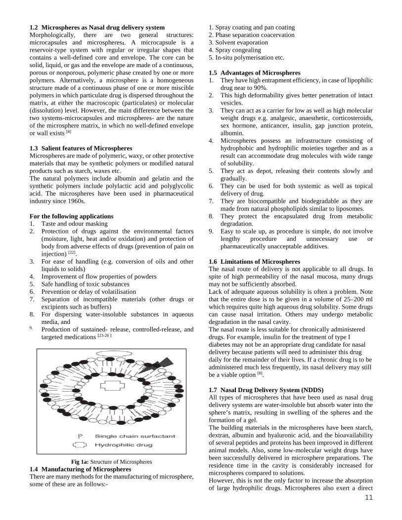
Fig 1a: Structure of Microspheres 1.4 Manufacturing of Microspheres There are many methods for the manufacturing of microsphere, some of these are as follows:-
1. Spray coating and pan coating 2. Phase separation coacervation 3. Solvent evaporation 4. Spray congealing 5. In-situ polymerisation etc. 1.5 Advantages of Microspheres 1. They have high entrapment efficiency, in case of lipophilic
drug near to 90%. 2. This high deformability gives better penetration of intact
vesicles. 3. They can act as a carrier for low as well as high molecular
weight drugs e.g. analgesic, anaesthetic, corticosteroids, sex hormone, anticancer, insulin, gap junction protein, albumin.
4. Microspheres possess an infrastructure consisting of hydrophobic and hydrophilic moieties together and as a result can accommodate drug molecules with wide range of solubility.
5. They act as depot, releasing their contents slowly and gradually.
6. They can be used for both systemic as well as topical delivery of drug.
7. They are biocompatible and biodegradable as they are made from natural phospholipids similar to liposomes.
8. They protect the encapsulated drug from metabolic degradation.
9. Easy to scale up, as procedure is simple, do not involve lengthy procedure and unnecessary use or pharmaceutically unacceptable additives.
1.6 Limitations of Microspheres The nasal route of delivery is not applicable to all drugs. In spite of high permeability of the nasal mucosa, many drugs may not be sufficiently absorbed. Lack of adequate aqueous solubility is often a problem. Note that the entire dose is to be given in a volume of 25–200 ml which requires quite high aqueous drug solubility. Some drugs can cause nasal irritation. Others may undergo metabolic degradation in the nasal cavity. The nasal route is less suitable for chronically administered drugs. For example, insulin for the treatment of type I diabetes may not be an appropriate drug candidate for nasal delivery because patients will need to administer this drug daily for the remainder of their lives. If a chronic drug is to be administered much less frequently, its nasal delivery may still be a viable option [8].
1.7 Nasal Drug Delivery System (NDDS) All types of microspheres that have been used as nasal drug delivery systems are water-insoluble but absorb water into the sphere’s matrix, resulting in swelling of the spheres and the formation of a gel. The building materials in the microspheres have been starch, dextran, albumin and hyaluronic acid, and the bioavailability of several peptides and proteins has been improved in different animal models. Also, some low-molecular weight drugs have been successfully delivered in microsphere preparations. The residence time in the cavity is considerably increased for microspheres compared to solutions. However, this is not the only factor to increase the absorption of large hydrophilic drugs. Microspheres also exert a direct
12
effect on the mucosa, resulting in the opening of tight junctions between the epithelial cells. The nasal route for systemic drug delivery has mainly been investigated with large hydrophilic peptides and proteins in mind, although other type of drugs has also been investigated.
Different types of absorption enhancers have been used to avoid the problem of low absorption. On the basis absorptions two type of the polymers used in microspheres 1. Mucoadhesive, 2. Bioadhesive [9]
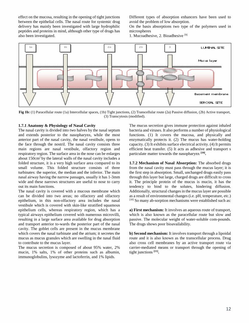
Fig 1b: (1) Paracellular route (1a) Intercellular spaces, (1b) Tight junctions, (2) Transcellular route (2a) Passive diffusion, (2b) Active transport, (3) Transcytosis (modified).
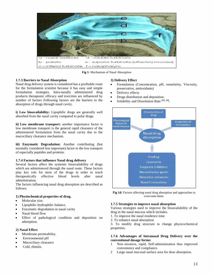
1.7.1 Anatomy & Physiology of Nasal Cavity The nasal cavity is divided into two halves by the nasal septum and extends posterior to the nasopharynx, while the most anterior part of the nasal cavity, the nasal vestibule, opens to the face through the nostril. The nasal cavity consists three main regions are nasal vestibule, olfactory region and respiratory region. The surface area in the nose can be enlarges about 150cm2 by the lateral walls of the nasal cavity includes a folded structure, it is a very high surface area compared to its small volume. This folded structure consists of three turbinates: the superior, the median and the inferior. The main nasal airway having the narrow passages, usually it has 1-3mm wide and these narrows structures are useful to nose to carry out its main functions. The nasal cavity is covered with a mucous membrane which can be divided into two areas; no olfactory and olfactory epithelium, in this non-olfactory area includes the nasal vestibule which is covered with skin-like stratified squamous epithelium cells, whereas respiratory region, which has a typical airways epithelium covered with numerous microvilli, resulting in a large surface area available for drug absorption and transport anterior to-wards the posterior part of the nasal cavity. The goblet cells are present in the mucus membrane which covers the nasal turbinate and the atrium; it secretes the mucus as mucus granules which are swelling in the nasal fluid to contribute to the mucus layer. The mucus secretion is composed of about 95% water, 2% mucin, 1% salts, 1% of other proteins such as albumin, immunoglobulins, lysozyme and lactoferrin, and 1% lipids.
The mucus secretion gives immune protection against inhaled bacteria and viruses. It also performs a number of physiological functions. (1) It covers the mucosa, and physically and enzymatically protects it. (2) The mucus has water-holding capacity. (3) It exhibits surface electrical activity. (4) It permits efficient heat transfer. (5) It acts as adhesive and transport s particulate matter towards the nasopharynx [10]. 1.7.2 Mechanism of Nasal Absorption: The absorbed drugs from the nasal cavity must pass through the mucus layer; it is the first step in absorption. Small, unchanged drugs easily pass through this layer but large, charged drugs are difficult to cross it. The principle protein of the mucus is mucin, it has the tendency to bind to the solutes, hindering diffusion. Additionally, structural changes in the mucus layer are possible as a result of environmental changes (i.e. pH, temperature, etc.) [10] So many ab-sorption mechanisms were established such as: a) First mechanism: It involves an aqueous route of transport, which is also known as the paracellular route but slow and passive. The molecular weight of water-soluble com-pounds. The drugs shows poor bioavailability. b) Second mechanism: It involves transport through a lipoidal route and it is also known as the transcellular process. Drug also cross cell membranes by an active transport route via carrier-mediated means or transport through the opening of tight junctions [11].
13
Fig 1: Mechanism of Nasal Absorption
1.7.3 Barriers to Nasal Absorption Nasal drug delivery system is considered has a profitable route for the formulation scientist because it has easy and simple formulation strategies. Intra-nasally administered drug products therapeutic efficacy and toxicities are influenced by number of factors Following factors are the barriers to the absorption of drugs through nasal cavity. i) Low bioavailability: Lipophilic drugs are generally well absorbed from the nasal cavity compared to polar drugs. ii) Low membrane transport: another importance factor is low membrane transport is the general rapid clearance of the administered formulation from the nasal cavity due to the mucociliary clearance mechanism. iii) Enzymatic Degradation: Another contributing (but normally considered less important) factor to the low transport of especially peptides and proteins. 1.7.4 Factors that influence Nasal drug delivery Several factors affect the systemic bioavailability of drugs which are administered through the nasal route. These factors play key role for most of the drugs in order to reach therapeutically effective blood levels after nasal administration. The factors influencing nasal drug absorption are described as follows: 1) Physiochemical properties of drug. • Molecular size. • Lipophilic-hydrophilic balance. • Enzymatic degradation in nasal cavity • Nasal blood flow • Effect of pathological condition and deposition on
adsorption.
2) Nasal Effect • Membrane permeability. • Environmental pH • Mucociliary clearance • Cold, rhinitis.
3) Delivery Effect • Formulation (Concentration, pH, osmolarity, Viscosity,
preservative, antioxidants) • Delivery effects • Drugs distribution and deposition. • Solubility and Dissolution Rate [12, 13].
Fig 1d: Factors affecting nasal drug absorption and approaches to overcome them.
1.7.5 Strategies to improve nasal absorption Various strategies used to improve the bioavailability of the drug in the nasal mucosa which includes. 1. To improve the nasal residence time 2. To enhance nasal absorption 3. To modify drug structure to change physicochemical properties. 1.7.6 Advantages of Intranasal Drug Delivery over the conventional dosage forms: 1 Non–invasive, rapid, Self-administration thus improved
convenience and compliance. 2 Large nasal mucosal surface area for dose absorption.
14
3 Bypasses the BBB (Blood Brain Barrier) and targets the CNS, reducing systemic exposure and thus systemic exposure and thus systemic side effects.
4 Minimal aftertaste. 5 Does not require nay modification of the therapeutic agent
being delivered neurological and psychiatric disorders. 6 Rich vasculature and highly permeable structure of the
nasal mucosa greatly enhance drug absorption. 7 Problem of degradation of peptide drugs in minimized up
to a certain extent. 8 Easy accessibility to blood capillaries. 1.7.7 Disadvantages of Intranasal Drug Delivery • Concentration achievable in different regions of the brain
and spinal cord varies with each agent. • Delivery is expected to decrease with increasing molecular
weight of drug. • Some therapeutic agents may be susceptible to partial
degradation in the nasal mucosa or may cause irritation to the mucosa.
• Nasal congestion due to cold or allergies may interfere with this method of delivery.
• Frequent use of this route may result in mucosal damage.
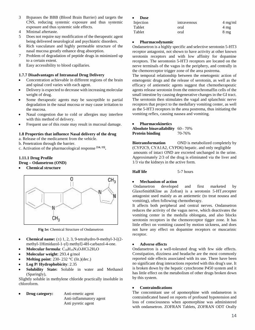
1.8 Properties that influence Nasal delivery of the drug a. Release of the medicament from the vehicle. b. Penetration through the barrier. c. Activation of the pharmacological response [14, 15]. 1.11.1 Drug Profile Drug – Odansetron (OND) • Chemical structure
Fig 1e: Chemical Structure of Ondansetron
• Chemical name: (±) 1, 2, 3, 9-tetrahydro-9-methyl-3-[(2-methyl-1Himidazol-1-yl) methyl]-4H-carbazol-4-one.
• Molecular formula: C18H19N3O•HCl•2H2O • Molecular weight: 293.4 g/mol • Melting point: 230- 232 °C (lit.)(dec.) • Log P/ Hydrophobicity: 2.35 • Solubility State: Soluble in water and Methanol
(Sparingly), Slightly soluble in methylene chloride practically insoluble in chloroform. • Drug category: Anti-emetic agent
Anti-inflammatory agent Anti pyretic agent
• Dose Injection intravenous 4 mg/ml Tablet oral 4 mg Tablet oral 8 mg • Pharmacodynamic Ondansetron is a highly specific and selective serotonin 5-HT3 receptor antagonist, not shown to have activity at other known serotonin receptors and with low affinity for dopamine receptors. The serontonin 5-HT3 receptors are located on the nerve terminals of the vagus in the periphery, and centrally in the chemoreceptor trigger zone of the area postrema. The temporal relationship between the emetogenic action of emetogenic drugs and the release of serotonin, as well as the efficacy of antiemetic agents suggest that chemotherapeutic agents release serotonin from the enterochromaffin cells of the small intestine by causing degenerative changes in the GI tract. The serotonin then stimulates the vagal and splanchnic nerve receptors that project to the medullary vomiting center, as well as the 5-HT3 receptors in the area postrema, thus initiating the vomiting reflex, causing nausea and vomiting. • Pharmacokinetics Absolute bioavailability 60– 70% Protein binding 70-76% Biotransformation OND is metabolized completely by (CYP2C9, CYA1A2, CYPD6) hepatic. and only negligible amounts of intact OND are excreted unchanged in the urine. Approximately 2/3 of the drug is eliminated via the liver and 1/3 via the kidneys in the active form. Half life 5-7 hours • Mechanism of action Ondansetron developed and first marketed by GlaxoSmithKline as Zofran) is a serotonin 5-HT3recepter antagonist used mainly as an antiemetic (to treat nousea and vomiting), often following chemotherapy. It affects both peripheral and central nerves. Ondansetron reduces the activity of the vagus nerve, which deactivates the vomiting center in the medulla oblongata, and also blocks serotonin receptors in the chemoreceptor tigger zone. It has little effect on vomiting caused by motion sickness, and does not have any effect on dopamine receptors or muscarinic receptor. • Adverse effects Ondansetron is a well-tolerated drug with few side effects. Constipation, dizziness and headache are the most commonly reported side effects associated with its use. There have been no significant drug interactions reported with this drug's use. It is broken down by the hepatic cytochrome P450 system and it has little effect on the metabolism of other drugs broken down by this system. • Contraindications The concomitant use of apomorphine with ondansetron is contraindicated based on reports of profound hypotension and loss of consciousness when apomorphine was administered with ondansetron. ZOFRAN Tablets, ZOFRAN ODT Orally
15
Disintegrating Tablets, and ZOFRAN Oral Solution are contraindicated for patients known to have hypersensitivity to the drug. • Drug Interactions Ondansetron does not itself appear to induce or inhibit the cytochrome P-450 drug-metabolizing enzyme system of the live. Because ondansetron is metabolized by hepatic cytochrome P-450 drug-metabolizing enzymes (CYP3A4, CYP2D6, CYP1A2), inducers or inhibitors of these enzymes may change the clearance and, hence, the half-life of ondansetron. Polymers commonly used for the preparation of microspheres is, 1. Sodium alginate 2. Pectin 3. Starch
2. Preformulation study
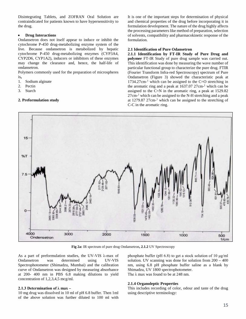
It is one of the important steps for determination of physical and chemical properties of the drug before incorporating it in formulation development. The nature of the drug highly affects the processing parameters like method of preparation, selection of solvents, compatibility and pharmacokinetic response of the formulation. 2.1 Identification of Pure Odansetron 2.1.1 Identification by FT-IR Study of Pure Drug and polymer FT-IR Study of pure drug sample was carried out. This identification was done by measuring the wave number of particular functional group to characterize the pure drug. FTIR (Fourier Transform Infra-red Spectroscopy) spectrum of Pure Ondansetron (Figure 3) showed the characteristic peak at 1734.27cm-1 which can be assigned to the C=O stretching in the aromatic ring and a peak at 1637.07 27cm-1 which can be assigned to the C=N in the aromatic ring, a peak at 1529.82 27cm-1 which can be assigned to the N-H stretching and a peak at 1279.87 27cm-1 which can be assigned to the stretching of C-C in the aromatic ring.
Fig 2a: IR spectrum of pure drug Ondansetron, 2.1.2 UV Spectroscopy As a part of preformulation studies, the UV-VIS λ-max of Ondansetron was determined using UV-VIS Spectrophotometer (Shimadzu, Mumbai) and the calibration curve of Ondansetron was designed by measuring absorbance at 200- 400 nm in PBS 6.8 making dilutions to yield concentration of 1,2,3,4,5 mcg/ml. 2.1.3 Determination of λ max – 10 mg drug was dissolved in 10 ml of pH 6.8 buffer. Then 1ml of the above solution was further diluted to 100 ml with
phosphate buffer (pH 6.8) to get a stock solution of 10 μg/ml solution. UV scanning was done for solution from 200 – 400 nm, using 6.8 pH phosphate buffer saline as a blank by Shimadzu, UV 1800 spectrophotometer. The λ max was found to be at 248 nm. 2.1.4 Organoleptic Properties This includes recording of color, odour and taste of the drug using descriptive terminology:
16
• Color- stability problems, improve appearance by including dye in body or coating.
• Taste- palatability problems, flavor and excipients may be added.
Odor- degradation products, stable form of drug to be used, flavors and excipients may be used.
Table 2A: Identification test of Ondansetron
Test
Ondansetron Specification Observation
Physical appearance White to off-white powder White powder Taste Tasteless Tasteless Odour Nil Nil
Melting Point 216-219 °C 218 °C
Infra-red spectra Sample IR spectrum should comply with standard IR Spectrum Sample IR spectrum complies with standard IR spectrum.
2.2 Melting Point Melting point is the temperature at which the solid phase is at equilibrium with the liquid phase. It was determined by using the digital melting point instrument. Melting point of Odansetron- 218 oC. 2.3 Solubility Study The solubility test is an important parameter before formulation development. The solubility of solute means the maximum quantity of solute that can be dissolved in certain quantity of solvent or quantity of solution at a specified temperature. Solubility is usually determined in variety of commonly used solvents. The solubility of material is usually determined by the equilibrium solubility method.
Table 2B: Solubility of Drug in different Solvents
S. No. Solvent Solubility 1 Water Soluble 2 Methanol free soluble 3 Chloroform Slightly soluble 4 Dichloromethane Sparingly soluble 5 Methylene chloride Slightly soluble
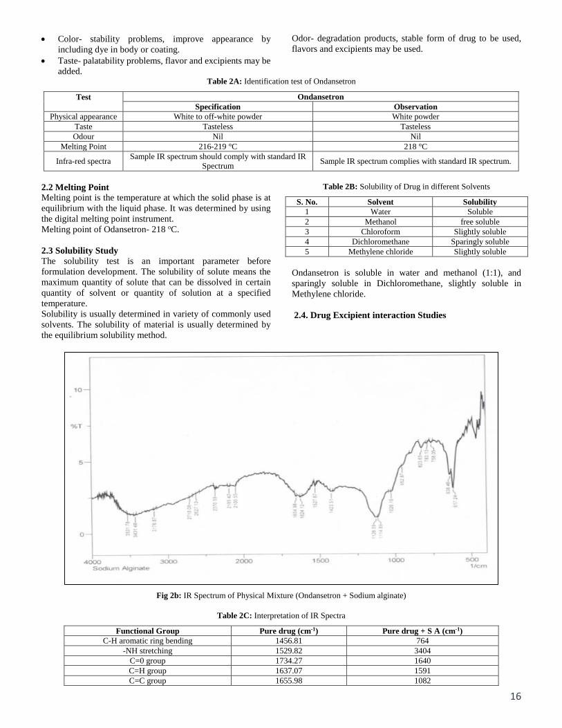
Ondansetron is soluble in water and methanol (1:1), and sparingly soluble in Dichloromethane, slightly soluble in Methylene chloride. 2.4. Drug Excipient interaction Studies
Fig 2b: IR Spectrum of Physical Mixture (Ondansetron + Sodium alginate)
Table 2C: Interpretation of IR Spectra
Functional Group Pure drug (cm-1) Pure drug + S A (cm-1) C-H aromatic ring bending 1456.81 764
-NH stretching 1529.82 3404 C=0 group 1734.27 1640 C=H group 1637.07 1591 C=C group 1655.98 1082
17
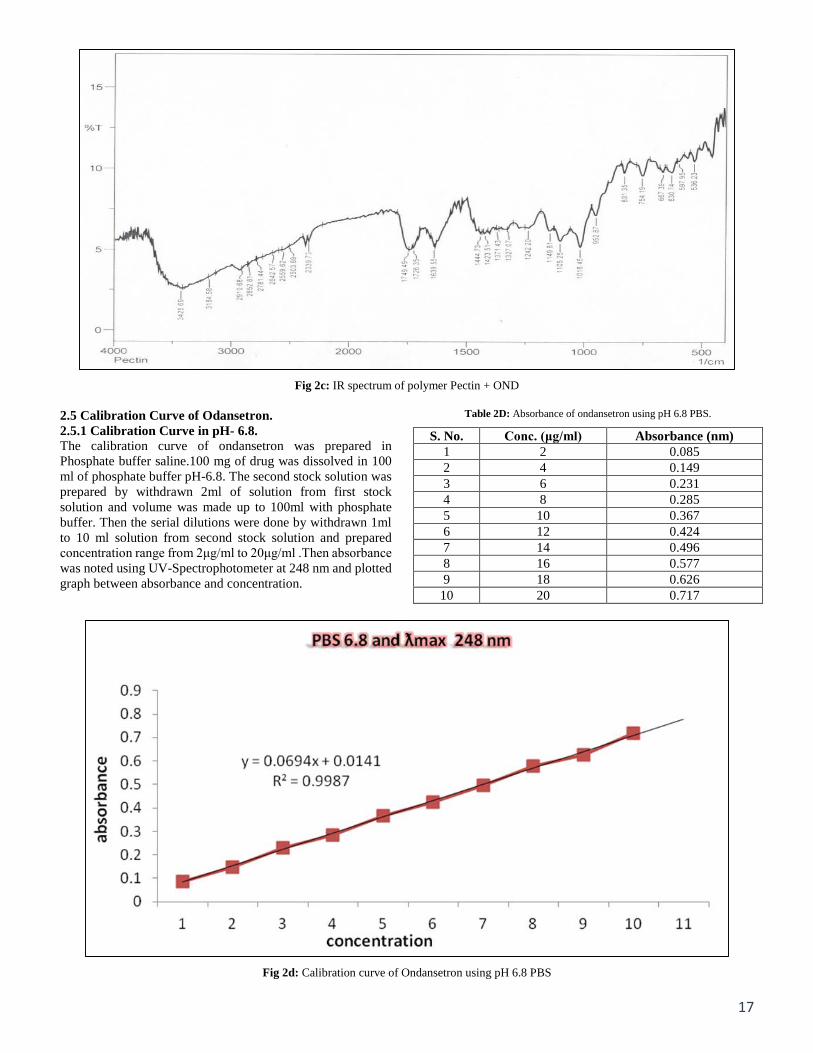
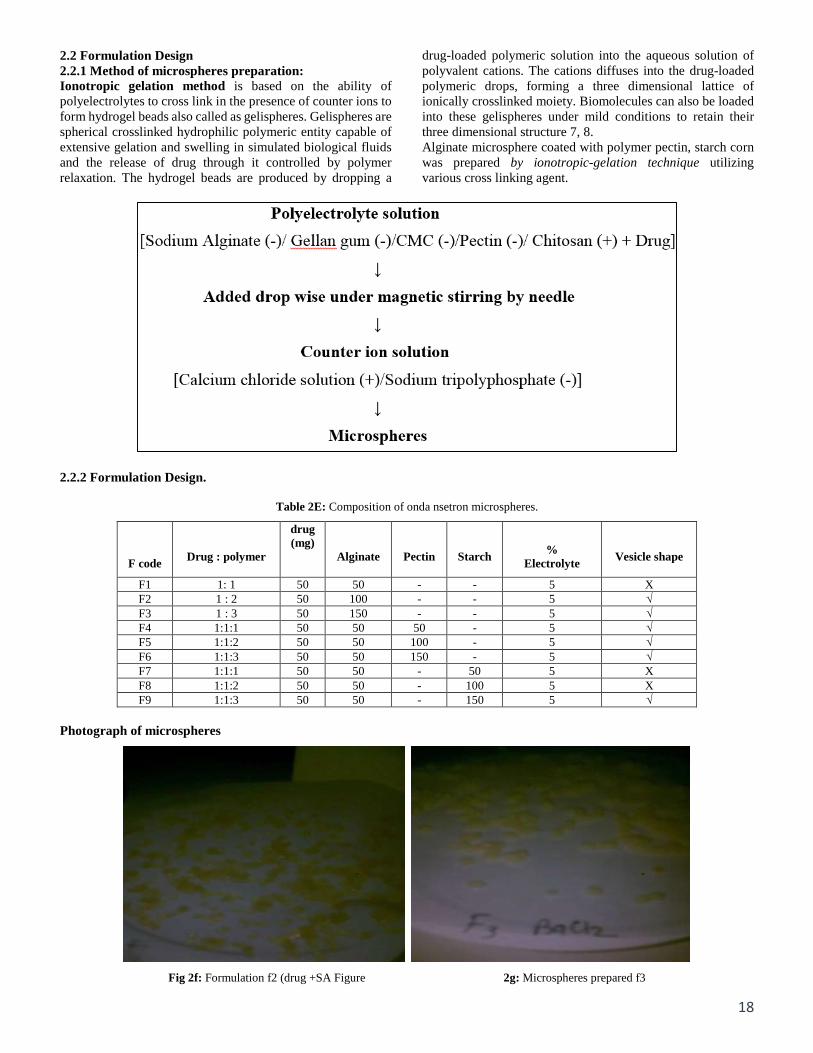
Fig 2c: IR spectrum of polymer Pectin + OND 2.5 Calibration Curve of Odansetron. 2.5.1 Calibration Curve in pH- 6.8. The calibration curve of ondansetron was prepared in Phosphate buffer saline.100 mg of drug was dissolved in 100 ml of phosphate buffer pH-6.8. The second stock solution was prepared by withdrawn 2ml of solution from first stock solution and volume was made up to 100ml with phosphate buffer. Then the serial dilutions were done by withdrawn 1ml to 10 ml solution from second stock solution and prepared concentration range from 2μg/ml to 20μg/ml .Then absorbance was noted using UV-Spectrophotometer at 248 nm and plotted graph between absorbance and concentration.
Table 2D: Absorbance of ondansetron using pH 6.8 PBS.
S. No. Conc. (μg/ml) Absorbance (nm) 1 2 0.085 2 4 0.149 3 6 0.231 4 8 0.285 5 10 0.367 6 12 0.424 7 14 0.496 8 16 0.577 9 18 0.626
10 20 0.717
Fig 2d: Calibration curve of Ondansetron using pH 6.8 PBS
18
2.2 Formulation Design 2.2.1 Method of microspheres preparation: Ionotropic gelation method is based on the ability of polyelectrolytes to cross link in the presence of counter ions to form hydrogel beads also called as gelispheres. Gelispheres are spherical crosslinked hydrophilic polymeric entity capable of extensive gelation and swelling in simulated biological fluids and the release of drug through it controlled by polymer relaxation. The hydrogel beads are produced by dropping a
drug-loaded polymeric solution into the aqueous solution of polyvalent cations. The cations diffuses into the drug-loaded polymeric drops, forming a three dimensional lattice of ionically crosslinked moiety. Biomolecules can also be loaded into these gelispheres under mild conditions to retain their three dimensional structure 7, 8. Alginate microsphere coated with polymer pectin, starch corn was prepared by ionotropic-gelation technique utilizing various cross linking agent.
2.2.2 Formulation Design.
Table 2E: Composition of onda nsetron microspheres.
F code
Drug : polymer
drug (mg)
Alginate
Pectin
Starch
%
Electrolyte
Vesicle shape
F1 1: 1 50 50 - - 5 X F2 1 : 2 50 100 - - 5 √ F3 1 : 3 50 150 - - 5 √ F4 1:1:1 50 50 50 - 5 √ F5 1:1:2 50 50 100 - 5 √ F6 1:1:3 50 50 150 - 5 √ F7 1:1:1 50 50 - 50 5 X F8 1:1:2 50 50 - 100 5 X F9 1:1:3 50 50 - 150 5 √
Photograph of microspheres
Fig 2f: Formulation f2 (drug +SA Figure 2g: Microspheres prepared f3
19
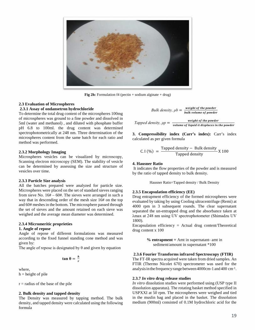
Fig 2h: Formulation f4 (pectin + sodium alginate + drug)
2.3 Evaluation of Microspheres 2.3.1 Assay of ondansetron hydrochloride To determine the total drug content of the microspheres 100mg of microspheres was ground to a fine powder and dissolved in 5ml (water and methanol) , and diluted with phosphate buffer pH 6.8 to 100ml. the drug content was determined spectrophotometrically at 248 nm. Three determination of the microspheres content from the same batch for each ratio and method was performed. 2.3.2 Morphology Imaging Microspheres vesicles can be visualized by microscopy, Scanning electron microscopy (SEM). The stability of vesicle can be determined by assessing the size and structure of vesicles over time. 2.3.3 Particle Size analysis All the batches prepared were analyzed for particle size. Microspheres were placed on the set of standard sieves ranging from sieve No. 16# – 60#. The sieves were arranged in such a way that in descending order of the mesh size 16# on the top and 60# meshes in the bottom. The microsphere passed through the set of sieves and the amount retained on each sieve was weighed and the average mean diameter was determined. 2.3.4 Micromeritic proprieties 1. Angle of repose Angle of repose of different formulations was measured according to the fixed funnel standing cone method and was given by: The angle of repose is designated by θ and given by equation
tan θ = 𝒉𝒉𝒓𝒓
where, h = height of pile r = radius of the base of the pile 2. Bulk density and tapped density The Density was measured by tapping method. The bulk density, and tapped density were calculated using the following formula
Bulk density, ρb = 𝒘𝒘𝒘𝒘𝒘𝒘𝒘𝒘𝒉𝒉𝒘𝒘 𝒐𝒐𝒐𝒐 𝒘𝒘𝒉𝒉𝒘𝒘 𝒑𝒑𝒐𝒐𝒘𝒘𝒑𝒑𝒘𝒘𝒓𝒓
𝒃𝒃𝒃𝒃𝒃𝒃𝒃𝒃 𝒗𝒗𝒐𝒐𝒃𝒃𝒃𝒃𝒗𝒗𝒘𝒘 𝒐𝒐𝒐𝒐 𝒑𝒑𝒐𝒐𝒘𝒘𝒑𝒑𝒘𝒘𝒓𝒓
Tapped density, ρp = 𝒘𝒘𝒘𝒘𝒘𝒘𝒘𝒘𝒉𝒉𝒘𝒘 𝒐𝒐𝒐𝒐 𝒘𝒘𝒉𝒉𝒘𝒘 𝒑𝒑𝒐𝒐𝒘𝒘𝒑𝒑𝒘𝒘𝒓𝒓
𝒗𝒗𝒐𝒐𝒃𝒃𝒃𝒃𝒗𝒗𝒘𝒘 𝒐𝒐𝒐𝒐 𝒃𝒃𝒘𝒘𝒍𝒍𝒃𝒃𝒘𝒘𝒑𝒑 𝒘𝒘𝒘𝒘 𝒑𝒑𝒘𝒘𝒅𝒅𝒑𝒑𝒃𝒃𝒅𝒅𝒅𝒅𝒘𝒘𝒅𝒅 𝒘𝒘𝒊𝒊 𝒘𝒘𝒉𝒉𝒘𝒘 𝒑𝒑𝒐𝒐𝒘𝒘𝒑𝒑𝒘𝒘𝒓𝒓
3. Compressibility index (Carr’s index): Carr’s index calculated as per given formula
C. I (%) = Tapped density − Bulk density
Tapped densityX 100
4. Hausner Ratio It indicates the flow properties of the powder and is measured by the ratio of tapped density to bulk density.
Hausner Ratio=Tapped density / Bulk Density 2.3.5 Encapsulation efficiency (EE) Drug entrapment efficiency of the formed microspheres were evaluated by taking by using Cooling ultracentrifuge (Remi) at 4000 rpm in 3 subsequent rounds. The clear supernatant separated the un-entrapped drug and the absorbance taken at λmax at 248 nm using UV spectrophotometer (Shimadzu UV 1800). Encapsulation efficiency = Actual drug content/Theoretical drug content x 100
% entrapment = Amt in supernatant- amt in sediment/amount in supernatant *100
2.3.6 Fourier Transforms infrared Spectroscopy (FTIR) The FT‐IR spectra acquired were taken from dried samples. An FTIR (Thermo Nicolet 670) spectrometer was used for the analysis in the frequency range between 4000cm‐1 and 400 cm‐1. 2.3.7 In vitro drug release studies In vitro dissolution studies were performed using (USP type II dissolution apparatus). The rotating basket method specified in USPXXI at 50 rpm. The microspheres were weighed and tied in the muslin bag and placed in the basket. The dissolution medium (900ml) consisted of 0.1M hydrochloric acid for the
20
first 2 hours and then changed to phosphate buffer pH 6.8 from the 3rd hour. The temperature was maintained at 37 °C. An aliquot of (5ml) sample was withdrawn at specified time interval and replaced with an equivalent volume of dissolution fluid. Drug content was determined by UV‐Visible spectrophotometer (Schimazdu UV 1700 E 23) at 248 nm. The release studies were conducted in triplicate. 2.3.8 Kinetic treatment of release data The obtained dissolution data were fitted to zero order (Najib and Suleiman 1985), first order (Desai et al. 1966), Higuchi (Higuchi 1963), Korsmeyer-Peppas models to determine the mechanism of CAP release from the prepared microspheres. 2.3.9 Stability studies The success of an effective formulation was evaluated only through the stability studies. The purpose of stability testing was to obtain a stable product which assures its safety and efficacy up to the end of shelf life. In this study, stability study was done for atonditions like Room temp. (RT), 30 ºC & 60% RH, 40 ºC & 75% RH. The samples were assayed for drug content at regular intervals for two weeks. 2.3.10 Scanning Electron Microscopy (SEM) For SEM, one drop of Microspheres were mounted on the stab covered with clean glass and coated with gold and were observed under the scanning electron microscope at an accelerating voltage of 20KV and photomicrographs of suitable magnification was obtained. 2.3.11 Physical Stability Studies Physical stability tests of the prepared vesicles were carried out to investigate the aggregation of vesicles and leakage of drug from them during storage. The prepared drug vesicles were stored in transparent vials covered with plastic cap at room temperature for one month. The physical stability was evaluated by vesicle size, EE% and over a one month period. Samples from each vesicle were
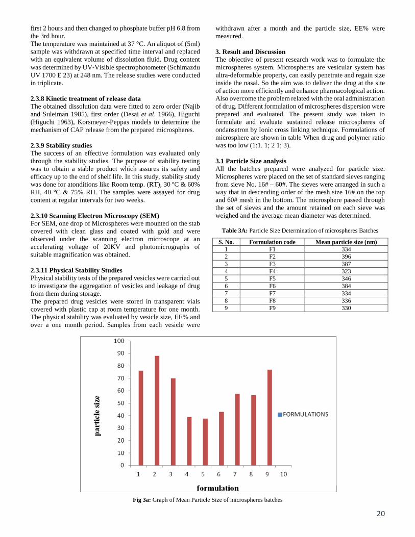
withdrawn after a month and the particle size, EE% were measured. 3. Result and Discussion The objective of present research work was to formulate the microspheres system. Microspheres are vesicular system has ultra-deformable property, can easily penetrate and regain size inside the nasal. So the aim was to deliver the drug at the site of action more efficiently and enhance pharmacological action. Also overcome the problem related with the oral administration of drug. Different formulation of microspheres dispersion were prepared and evaluated. The present study was taken to formulate and evaluate sustained release microspheres of ondansetron by Ionic cross linking technique. Formulations of microsphere are shown in table When drug and polymer ratio was too low (1:1. 1; 2 1; 3). 3.1 Particle Size analysis All the batches prepared were analyzed for particle size. Microspheres were placed on the set of standard sieves ranging from sieve No. 16# – 60#. The sieves were arranged in such a way that in descending order of the mesh size 16# on the top and 60# mesh in the bottom. The microsphere passed through the set of sieves and the amount retained on each sieve was weighed and the average mean diameter was determined.
Table 3A: Particle Size Determination of microspheres Batches
S. No. Formulation code Mean particle size (nm) 1 F1 334 2 F2 396 3 F3 387 4 F4 323 5 F5 346 6 F6 384 7 F7 334 8 F8 336 9 F9 330
Fig 3a: Graph of Mean Particle Size of microspheres batches
21
3.2 Micromeritic properties of the microspheres Angle of repose of microspheres was in the range of 28º12΄. Shown excellent flow ability as represented in term of angle of repose (<40°).
Table 3B: The angle repose values of Microspheres
S. No Formulation code Angle of repose Comments 1 F1 20o.56 Good flow 2 F2 28o.44’ Good flow 3 F3 28o.61’ Good flow 4 F4 24o.57’ Good flow 5 F5 26o.41’ Good flow 6 F6 26o.58’ Good flow 7 F7 28o.44’ Good flow 8 F8 28o.61’ Good flow 9 F9 24o.57’ Good flow
Bulk density values ranged from 0.312 to 0.365 gm/cm3Tapped density was determined by the tapping method.
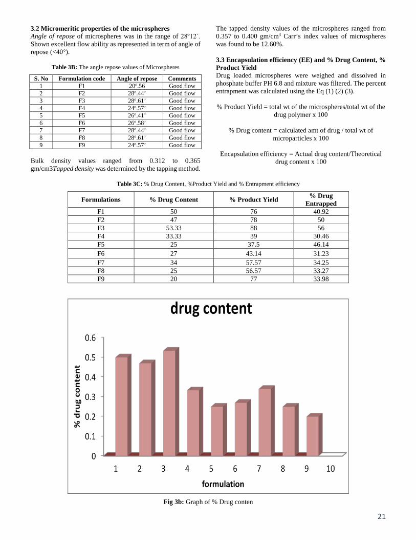
The tapped density values of the microspheres ranged from 0.357 to 0.400 gm/cm3 Carr’s index values of microspheres was found to be 12.60%. 3.3 Encapsulation efficiency (EE) and % Drug Content, % Product Yield Drug loaded microspheres were weighed and dissolved in phosphate buffer PH 6.8 and mixture was filtered. The percent entrapment was calculated using the Eq (1) (2) (3). % Product Yield = total wt of the microspheres/total wt of the
drug polymer x 100
% Drug content = calculated amt of drug / total wt of microparticles x 100
Encapsulation efficiency = Actual drug content/Theoretical
drug content x 100
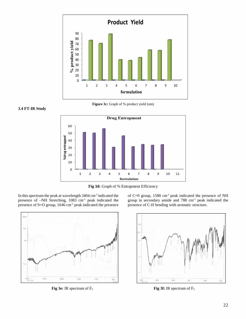
Table 3C: % Drug Content, %Product Yield and % Entrapment efficiency
Formulations % Drug Content % Product Yield % Drug Entrapped
F1 50 76 40.92 F2 47 78 50 F3 53.33 88 56 F4 33.33 39 30.46 F5 25 37.5 46.14 F6 27 43.14 31.23 F7 34 57.57 34.25 F8 25 56.57 33.27 F9 20 77 33.98
Fig 3b: Graph of % Drug conten
22
Figure 3c: Graph of % product yield (nm)
3.4 FT-IR Study
Fig 3d: Graph of % Entrapment Efficiency
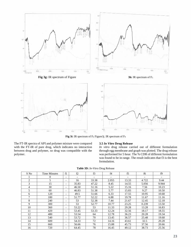
In this spectrum the peak at wavelength 3404 cm-1 indicated the presence of –NH Stretching, 1083 cm-1 peak indicated the presence of S=O group, 1646 cm-1 peak indicated the presence
of C=0 group, 1588 cm-1 peak indicated the presence of NH group in secondary amide and 788 cm-1 peak indicated the presence of C-H bending with aromatic structure.
Fig 3e: IR spectrum of F2 Fig 3f: IR spectrum of F3
23
Fig 3g: IR spectrum of Figure 3h: IR spectrum of F5
Fig 3i: IR spectrum of F6 Figure3j. IR spectrum of F9
The FT-IR spectra of API and polymer mixture were compared with the FT-IR of pure drug, which indicates no interaction between drug and polymer, so drug was compatible with the polymer.
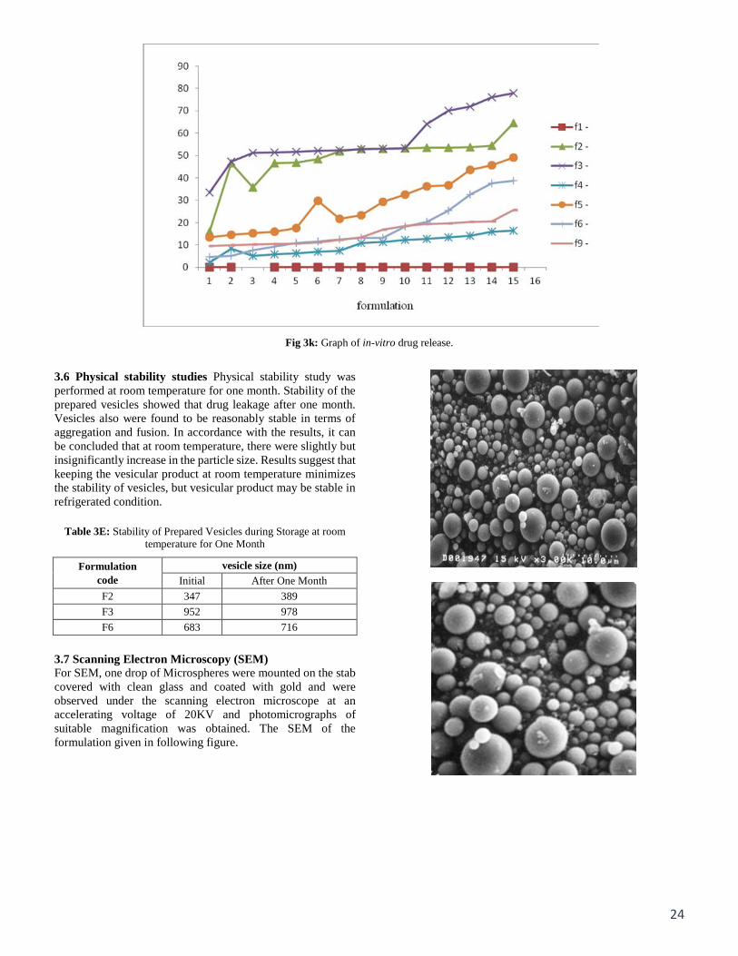
3.5 In Vitro Drug Release In vitro drug release carried out of different formulation through egg membrane and graph was plotted. The drug release was performed for 5 hour. The % CDR of different formulation was found to be in range. The result indicates that f3 is the best formulation.
Table 3D: In-Vitro Drug Release
S No Time Minutes f1 f2 f3 f4 f5 f6 f9 1 0 - - - - - - - 2 5 - 16 33.38 2.055 13.33 4.722 9.44 3 15 - 35.83 47.22 8.45 14.55 5.056 9.944 4 30 - 46.50 51.16 5.22 15.16 7.56 10.23 5 60 - 46.83 51.38 5.77 15.83 9.27 10.50 6 120 - 49.5 51.66 6.33 17.55 10.95 10.68 7 180 - 51.77 52.22 6.88 19.78 11.67 11.16 8 240 - 53 52.38 7.44 21.67 12.45 12.18 9 300 - 53 52.77 10.77 23.25 0.239 13.50 10 360 - 53.22 53 11.33 29.28 13.28 16.83 11 420 - 53.45 53.33 12.24 32.56 18.17 18.55 12 480 - 53.54 64 12.78 36.23 20.28 19.34 13 540 - 53.72 70 13.45 36.57 25.48 19.68 14 600 - 54.33 72 14.16 43.61 32.5 20.28 15 660 - 56.66 76 15.88 45.73 37.56 20.66 16 720 - 64.45 78 16.45 49.12 38.73 25.56
24
Fig 3k: Graph of in-vitro drug release.
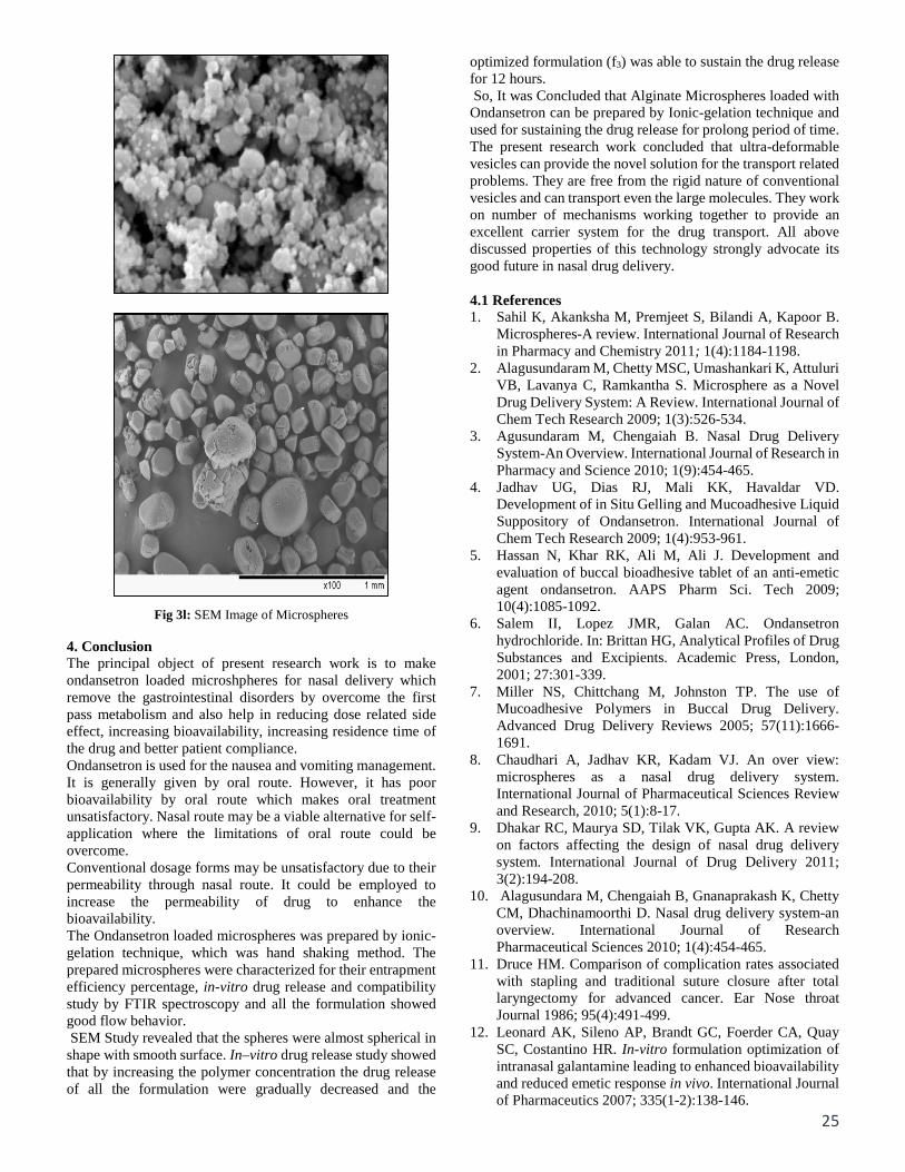
3.6 Physical stability studies Physical stability study was performed at room temperature for one month. Stability of the prepared vesicles showed that drug leakage after one month. Vesicles also were found to be reasonably stable in terms of aggregation and fusion. In accordance with the results, it can be concluded that at room temperature, there were slightly but insignificantly increase in the particle size. Results suggest that keeping the vesicular product at room temperature minimizes the stability of vesicles, but vesicular product may be stable in refrigerated condition.
Table 3E: Stability of Prepared Vesicles during Storage at room temperature for One Month
Formulation code
vesicle size (nm) Initial After One Month
F2 347 389 F3 952 978 F6 683 716
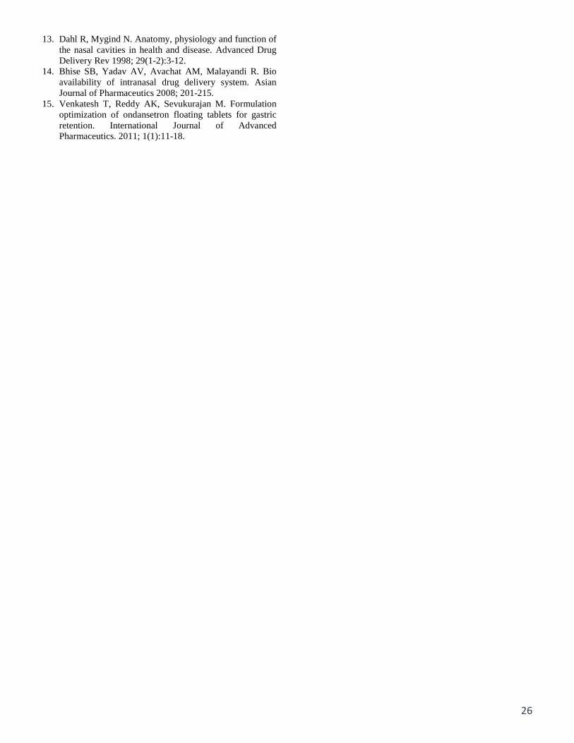
3.7 Scanning Electron Microscopy (SEM) For SEM, one drop of Microspheres were mounted on the stab covered with clean glass and coated with gold and were observed under the scanning electron microscope at an accelerating voltage of 20KV and photomicrographs of suitable magnification was obtained. The SEM of the formulation given in following figure.
25
Fig 3l: SEM Image of Microspheres 4. Conclusion The principal object of present research work is to make ondansetron loaded microshpheres for nasal delivery which remove the gastrointestinal disorders by overcome the first pass metabolism and also help in reducing dose related side effect, increasing bioavailability, increasing residence time of the drug and better patient compliance. Ondansetron is used for the nausea and vomiting management. It is generally given by oral route. However, it has poor bioavailability by oral route which makes oral treatment unsatisfactory. Nasal route may be a viable alternative for self-application where the limitations of oral route could be overcome. Conventional dosage forms may be unsatisfactory due to their permeability through nasal route. It could be employed to increase the permeability of drug to enhance the bioavailability. The Ondansetron loaded microspheres was prepared by ionic-gelation technique, which was hand shaking method. The prepared microspheres were characterized for their entrapment efficiency percentage, in-vitro drug release and compatibility study by FTIR spectroscopy and all the formulation showed good flow behavior. SEM Study revealed that the spheres were almost spherical in shape with smooth surface. In–vitro drug release study showed that by increasing the polymer concentration the drug release of all the formulation were gradually decreased and the
optimized formulation (f3) was able to sustain the drug release for 12 hours. So, It was Concluded that Alginate Microspheres loaded with Ondansetron can be prepared by Ionic-gelation technique and used for sustaining the drug release for prolong period of time. The present research work concluded that ultra-deformable vesicles can provide the novel solution for the transport related problems. They are free from the rigid nature of conventional vesicles and can transport even the large molecules. They work on number of mechanisms working together to provide an excellent carrier system for the drug transport. All above discussed properties of this technology strongly advocate its good future in nasal drug delivery. 4.1 References 1. Sahil K, Akanksha M, Premjeet S, Bilandi A, Kapoor B.
Microspheres-A review. International Journal of Research in Pharmacy and Chemistry 2011; 1(4):1184-1198.
2. Alagusundaram M, Chetty MSC, Umashankari K, Attuluri VB, Lavanya C, Ramkantha S. Microsphere as a Novel Drug Delivery System: A Review. International Journal of Chem Tech Research 2009; 1(3):526-534.
3. Agusundaram M, Chengaiah B. Nasal Drug Delivery System-An Overview. International Journal of Research in Pharmacy and Science 2010; 1(9):454-465.
4. Jadhav UG, Dias RJ, Mali KK, Havaldar VD. Development of in Situ Gelling and Mucoadhesive Liquid Suppository of Ondansetron. International Journal of Chem Tech Research 2009; 1(4):953-961.
5. Hassan N, Khar RK, Ali M, Ali J. Development and evaluation of buccal bioadhesive tablet of an anti-emetic agent ondansetron. AAPS Pharm Sci. Tech 2009; 10(4):1085-1092.
6. Salem II, Lopez JMR, Galan AC. Ondansetron hydrochloride. In: Brittan HG, Analytical Profiles of Drug Substances and Excipients. Academic Press, London, 2001; 27:301-339.
7. Miller NS, Chittchang M, Johnston TP. The use of Mucoadhesive Polymers in Buccal Drug Delivery. Advanced Drug Delivery Reviews 2005; 57(11):1666-1691.
8. Chaudhari A, Jadhav KR, Kadam VJ. An over view: microspheres as a nasal drug delivery system. International Journal of Pharmaceutical Sciences Review and Research, 2010; 5(1):8-17.
9. Dhakar RC, Maurya SD, Tilak VK, Gupta AK. A review on factors affecting the design of nasal drug delivery system. International Journal of Drug Delivery 2011; 3(2):194-208.
10. Alagusundara M, Chengaiah B, Gnanaprakash K, Chetty CM, Dhachinamoorthi D. Nasal drug delivery system-an overview. International Journal of Research Pharmaceutical Sciences 2010; 1(4):454-465.
11. Druce HM. Comparison of complication rates associated with stapling and traditional suture closure after total laryngectomy for advanced cancer. Ear Nose throat Journal 1986; 95(4):491-499.
12. Leonard AK, Sileno AP, Brandt GC, Foerder CA, Quay SC, Costantino HR. In-vitro formulation optimization of intranasal galantamine leading to enhanced bioavailability and reduced emetic response in vivo. International Journal of Pharmaceutics 2007; 335(1-2):138-146.
26
13. Dahl R, Mygind N. Anatomy, physiology and function of the nasal cavities in health and disease. Advanced Drug Delivery Rev 1998; 29(1-2):3-12.
14. Bhise SB, Yadav AV, Avachat AM, Malayandi R. Bio availability of intranasal drug delivery system. Asian Journal of Pharmaceutics 2008; 201-215.
15. Venkatesh T, Reddy AK, Sevukurajan M. Formulation optimization of ondansetron floating tablets for gastric retention. International Journal of Advanced Pharmaceutics. 2011; 1(1):11-18.