Embed Size (px)
Citation preview
41
FOREHEAD, TEMPLE AND SCALP RECONSTRUCTION
H.D. Vuyk
INTRODUCTION
The forehead is a gently rounded, often smooth, area presenting certain reconstructivechallenges. A large number of variables determine the method of reconstruction aswell the final outcome. These variables include defect characteristics (etiology, locationand size) and additional patient factors such as specific individual forehead anatomy,as well as health status and patients’ expectation.
In order to appropriately prioritise these multiple factors, the 3 main goals of recon-struction should be kept foremost in mind23.1. Preservation of motor function (frontalis branch of facial nerve) and, if possible,
sensory nerve function.2. Maintenance of the normal boundaries of the forehead temporal esthetic unit,
including position and symmetry of the brow as well as frontal and temporal hairlines.3. Optimal scar camouflage by placement of scars in relaxed skin tension lines or adjacent
to the hairline or brow whenever possible.In this chapter anatomy and principles of forehead and temporal reconstruction willbe reviewed. Practical and reconstructive suggestions will be made for each forehead/temporal subunit.
PERTINENT ANATOMY
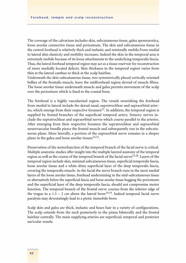
Anatomic knowledge is a basic pre-requisite to optimise the reconstructiveprocedure. The forehead temporal region may be considered an estheticunit running from the anterior hairlinesuperiorly, zygoma laterally and thebrow inferiorly. Forehead reconstructionmay be further conceptualised by dividingthis region into a midline forehead,paramedian forehead, lateral forehead,temporal and glabella and brow region(Fig. 1).
The area from the midline to the midbrow of the forehead tends to be convex, whilethe lateral forehead is flatter, smoothly blending into the more concave temporal region.The eyebrows are subunits onto themself with hairbairing skin.
Fig. 1. Aesthetic units of forehead and temple region.
The coverage of the calvarium includes skin, subcutaneous tissue, galea aponeurotica,loose areolar connective tissue and periosteum. The skin and subcutaneous tissue inthe central forehead is relatively thick and inelastic and minimally mobile.From medialto lateral skin elasticity and mobility increases. Indeed the skin in the temporal area isextremely mobile because of its loose attachments to the underlying temporalis fascia.Thus, the lateral forehead temporal region may act as a tissue reservoir for reconstructionof more medially located defects. Skin thickness in the temporal region varies fromthin at the lateral canthus to thick at the scalp hairline.Underneath the skin-subcutaneous tissue, two symmetrically placed vertically orientedbellies of the frontalis muscle, leave the midforehead region devoid of muscle fibres.The loose areolar tissue underneath muscle and galea permits movement of the scalpover the periostium which is fixed to the cranial bone.
The forehead is a highly vascularised region. The vessels nourishing the foreheadfrom medial to lateral include the dorsal nasal, supratrochlear and supraorbital arter-ies, which emerge from their respective foramen22. In addition, the temporal region issupplied by frontal branches of the superficial temporal artery. Sensory nerves in-clude the supratrochlear and supraorbital nerves which course parallel to the arteries.After emerging from their respective foramen the supratrochlear and supraorbitalneurovascular bundle pierce the frontal muscle and subsequently run in the subcuta-neous plane. More laterally, a portion of the supraorbital nerve remains in a deeperplane in the galea and loose areolar tissues14,15.
Preservation of the motorfunction of the temporal branch of the facial nerve is critical.Multiple anatomic studies offer insight into the multiple layered anatomy of the temporalregion as well as the course of the temporal branch of the facial nerve25,28. Layers of thetemporal region include skin, minimal subcutaneous tissue, superficial temporalis fascia,loose areolar tissue and a white shiny superficial layer of the deep temporalis fascia,covering the temporalis muscle. As the facial the nerve branch runs in the most mediallayers of the loose areolar tissue, forehead undermining in the mid-subcutaneous tissueor alternatively below the superficial fascia and loose areolar tissue hugging the periosteumand the superficial layer of the deep temporalis fascia, should not compromise motorfunction. The temporal branch of the frontal nerve courses from the inferior edge ofthe tragus to a 1.5 – 2 cm above the lateral brow10,19. Indeed temporal facial nerveparalysis may devestatingly lead to a ptotic immobile brow.
Scalp skin and galea are thick, inelastic and bears hair in a variety of configurations.The scalp extends from the neck posteriorly to the pinna bilaterally and the frontalhairline centrally. The main supplying arteries are superficial, temporal and posteriorauricular vessels.
42
Forehead , t emp le and s ca lp re cons t ruc t i on
RECONSTRUCTIVE PRINCIPLES
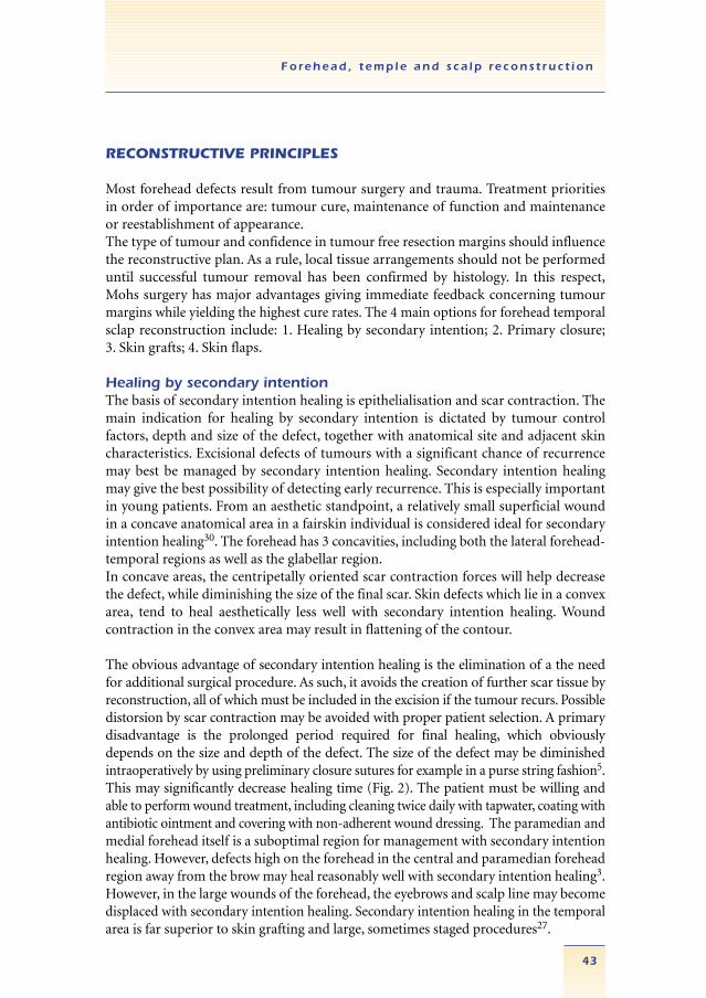
Most forehead defects result from tumour surgery and trauma. Treatment prioritiesin order of importance are: tumour cure, maintenance of function and maintenanceor reestablishment of appearance.The type of tumour and confidence in tumour free resection margins should influencethe reconstructive plan. As a rule, local tissue arrangements should not be performeduntil successful tumour removal has been confirmed by histology. In this respect,Mohs surgery has major advantages giving immediate feedback concerning tumourmargins while yielding the highest cure rates. The 4 main options for forehead temporalsclap reconstruction include: 1. Healing by secondary intention; 2. Primary closure;3. Skin grafts; 4. Skin flaps.
Healing by secondary intention The basis of secondary intention healing is epithelialisation and scar contraction. Themain indication for healing by secondary intention is dictated by tumour control factors, depth and size of the defect, together with anatomical site and adjacent skincharacteristics. Excisional defects of tumours with a significant chance of recurrencemay best be managed by secondary intention healing. Secondary intention healingmay give the best possibility of detecting early recurrence. This is especially importantin young patients. From an aesthetic standpoint, a relatively small superficial woundin a concave anatomical area in a fairskin individual is considered ideal for secondaryintention healing30. The forehead has 3 concavities, including both the lateral forehead-temporal regions as well as the glabellar region.In concave areas, the centripetally oriented scar contraction forces will help decreasethe defect, while diminishing the size of the final scar. Skin defects which lie in a convexarea, tend to heal aesthetically less well with secondary intention healing. Wound contraction in the convex area may result in flattening of the contour.
The obvious advantage of secondary intention healing is the elimination of a the needfor additional surgical procedure. As such, it avoids the creation of further scar tissue byreconstruction, all of which must be included in the excision if the tumour recurs. Possibledistorsion by scar contraction may be avoided with proper patient selection. A primarydisadvantage is the prolonged period required for final healing, which obviously depends on the size and depth of the defect. The size of the defect may be diminishedintraoperatively by using preliminary closure sutures for example in a purse string fashion5.This may significantly decrease healing time (Fig. 2). The patient must be willing andable to perform wound treatment, including cleaning twice daily with tapwater, coating withantibiotic ointment and covering with non-adherent wound dressing. The paramedian andmedial forehead itself is a suboptimal region for management with secondary intentionhealing. However, defects high on the forehead in the central and paramedian foreheadregion away from the brow may heal reasonably well with secondary intention healing3.However, in the large wounds of the forehead, the eyebrows and scalp line may becomedisplaced with secondary intention healing. Secondary intention healing in the temporalarea is far superior to skin grafting and large, sometimes staged procedures27.
43
Forehead , t emp le and s ca lp re cons t ruc t i on
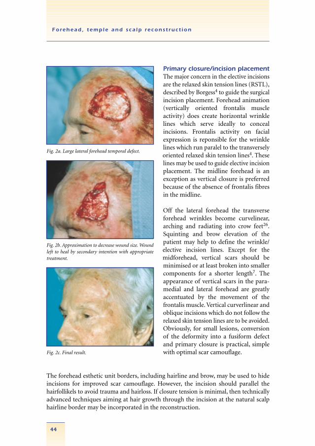
Primary closure/incision placementThe major concern in the elective incisionsare the relaxed skin tension lines (RSTL),described by Borgess4 to guide the surgicalincision placement. Forehead animation(vertically oriented frontalis muscle activity) does create horizontal wrinklelines which serve ideally to conceal incisions. Frontalis activity on facial expression is reponsible for the wrinklelines which run paralel to the transverselyoriented relaxed skin tension lines4. Theselines may be used to guide elective incisionplacement. The midline forehead is anexception as vertical closure is preferredbecause of the absence of frontalis fibresin the midline.
Off the lateral forehead the transverseforehead wrinkles become curvelinear,arching and radiating into crow feet26.Squinting and brow elevation of the patient may help to define the wrinkle/elective incision lines. Except for the midforehead, vertical scars should beminimised or at least broken into smallercomponents for a shorter length7. Theappearance of vertical scars in the para-medial and lateral forehead are greatlyaccentuated by the movement of thefrontalis muscle. Vertical curverlinear andoblique incisions which do not follow therelaxed skin tension lines are to be avoided.Obviously, for small lesions, conversionof the deformity into a fusiform defectand primary closure is practical, simplewith optimal scar camouflage.
The forehead esthetic unit borders, including hairline and brow, may be used to hideincisions for improved scar camouflage. However, the incision should parallel thehairfollikels to avoid trauma and hairloss. If closure tension is minimal, then technicallyadvanced techniques aiming at hair growth through the incision at the natural scalphairline border may be incorporated in the reconstruction.
44
Forehead , t emp le and s ca lp re cons t ruc t i on
Fig. 2c. Final result.
Fig. 2a. Large lateral forehead temporal defect.
Fig. 2b. Approximation to decrease wound size. Woundleft to heal by secondary intention with appropriatetreatment.
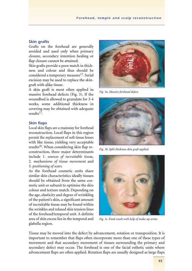
Skin graftsGrafts on the forehead are generallyavoided and used only when primaryclosure, secondary intention healing orflap closure cannot be attained.Skin grafts provide a poor match in thick-ness and colour and thus should be considered a temporary measure17. Serialexcision may be used to replace the skin-graft with alike tissue.A skin graft is most often applied inmassive forehead defects (Fig. 3). If thewoundbed is allowed to granulate for 3-4weeks, some additional thickness in covering may be obtained with adequateresults17.
Skin flapsLocal skin flaps are a mainstay for foreheadreconstruction. Local flaps in this regionpermit the replacement of soft tissue losseswith like tissue, yielding very acceptableresults20. When considering skin flap re-construction, three major determinantsinclude 1. sources of recruitable tissue,2. mechanisms of tissue movement and 3. positioning of scars.As the forehead cosmetic units sharesimilar skin characteristics ideally tissuesshould be obtained from the same cos-metic unit or subunit to optimise the skincolour and texture match. Depending onthe age, elasticity and degree of wrinklingof the patient’s skin, a significant amountof recruitable tissue may be found withinthe wrinkles and relaxed skin tension linesof the forehead/temporal unit. A definitearea of skin excess lies in the temporal andglabella region.
Tissue may be moved into the defect by advancement, rotation or transposition. It isimportant to remember that flaps often incorporate more than one of these types ofmovement and that secondary movement of tissues surrounding the primary andsecondary defect may occur. The forehead is one of the facial esthetic units where advancement flaps are often applied. Rotation flaps are usually designed as large flaps
45
Forehead , t emp le and s ca lp re cons t ruc t i on
Fig. 3c. Final result with help of make-up artist.
Fig. 3a. Massive forehead defect.
Fig. 3b. Split thickness skin graft applied.
with lengthy incisions, which may run diagonal to the horizontal crease of the forehead.Rotation flaps are preferably used when defects are at the border of the esthetic units.Transposition flaps may be designed within the forehead unit or in order to moveglabellar or temporal skin into the defect. Transposition flaps play a lesser role in foreheadrepairs, because of the resulting complex scars.
REGIONAL FOREHEAD RECONSTRUCTION
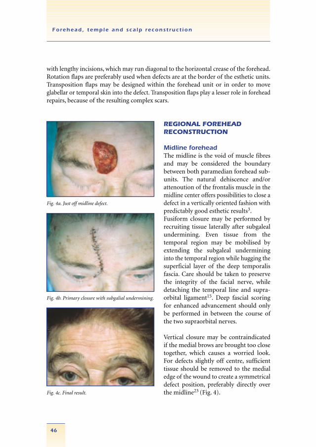
Midline foreheadThe midline is the void of muscle fibresand may be considered the boundary between both paramedian forehead sub-units. The natural dehiscence and/or attenoution of the frontalis muscle in themidline center offers possibilities to close adefect in a vertically oriented fashion withpredictably good esthetic results3.Fusiform closure may be performed byrecruiting tissue laterally after subgalealundermining. Even tissue from the temporal region may be mobilised by extending the subgaleal undermininginto the temporal region while hugging thesuperficial layer of the deep temporalisfascia. Care should be taken to preservethe integrity of the facial nerve, while detaching the temporal line and supra-orbital ligament15. Deep fascial scoringfor enhanced advancement should onlybe performed in between the course ofthe two supraorbital nerves.
Vertical closure may be contraindicatedif the medial brows are brought too closetogether, which causes a worried look.For defects slightly off centre, sufficienttissue should be removed to the medialedge of the wound to create a symmetricaldefect position, preferably directly overthe midline23 (Fig. 4).
46
Forehead , t emp le and s ca lp re cons t ruc t i on
Fig. 4c. Final result.
Fig. 4a. Just off midline defect.
Fig. 4b. Primary closure with subgalial undermining.
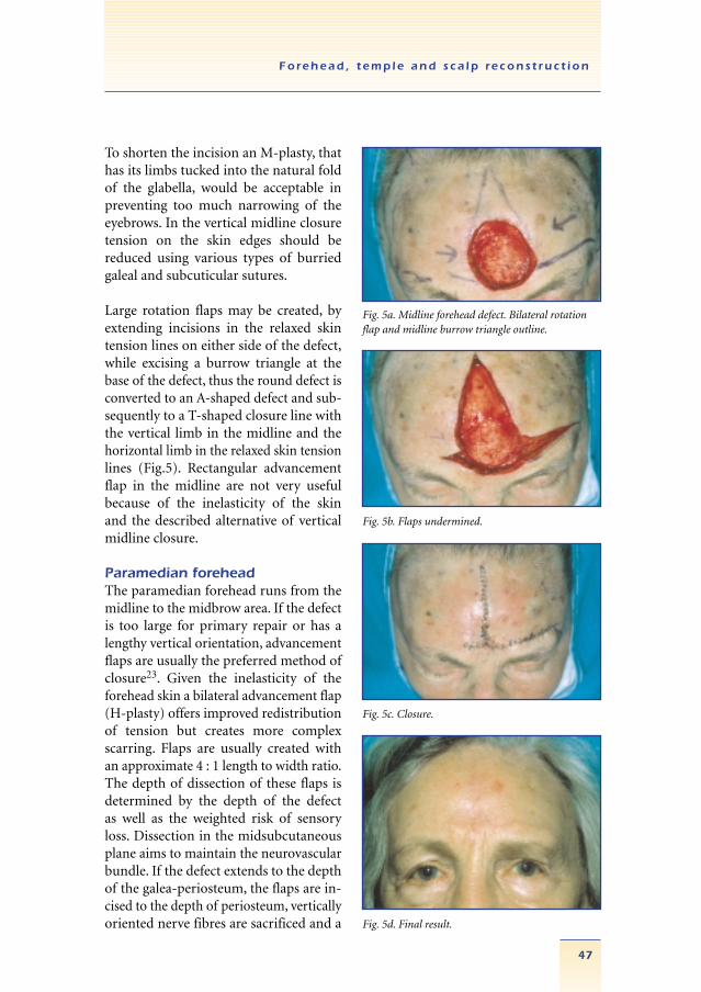
To shorten the incision an M-plasty, thathas its limbs tucked into the natural foldof the glabella, would be acceptable inpreventing too much narrowing of theeyebrows. In the vertical midline closuretension on the skin edges should be reduced using various types of burriedgaleal and subcuticular sutures.
Large rotation flaps may be created, byextending incisions in the relaxed skintension lines on either side of the defect,while excising a burrow triangle at thebase of the defect, thus the round defect isconverted to an A-shaped defect and sub-sequently to a T-shaped closure line withthe vertical limb in the midline and thehorizontal limb in the relaxed skin tensionlines (Fig.5). Rectangular advancementflap in the midline are not very usefulbecause of the inelasticity of the skinand the described alternative of verticalmidline closure.
Paramedian foreheadThe paramedian forehead runs from themidline to the midbrow area. If the defectis too large for primary repair or has alengthy vertical orientation, advancementflaps are usually the preferred method ofclosure23. Given the inelasticity of theforehead skin a bilateral advancement flap(H-plasty) offers improved redistributionof tension but creates more complexscarring. Flaps are usually created withan approximate 4 : 1 length to width ratio.The depth of dissection of these flaps isdetermined by the depth of the defect as well as the weighted risk of sensoryloss. Dissection in the midsubcutaneousplane aims to maintain the neurovascularbundle. If the defect extends to the depthof the galea-periosteum, the flaps are in-cised to the depth of periosteum, verticallyoriented nerve fibres are sacrificed and a
47
Forehead , t emp le and s ca lp re cons t ruc t i on
Fig. 5c. Closure.
Fig. 5a. Midline forehead defect. Bilateral rotationflap and midline burrow triangle outline.
Fig. 5b. Flaps undermined.
Fig. 5d. Final result.
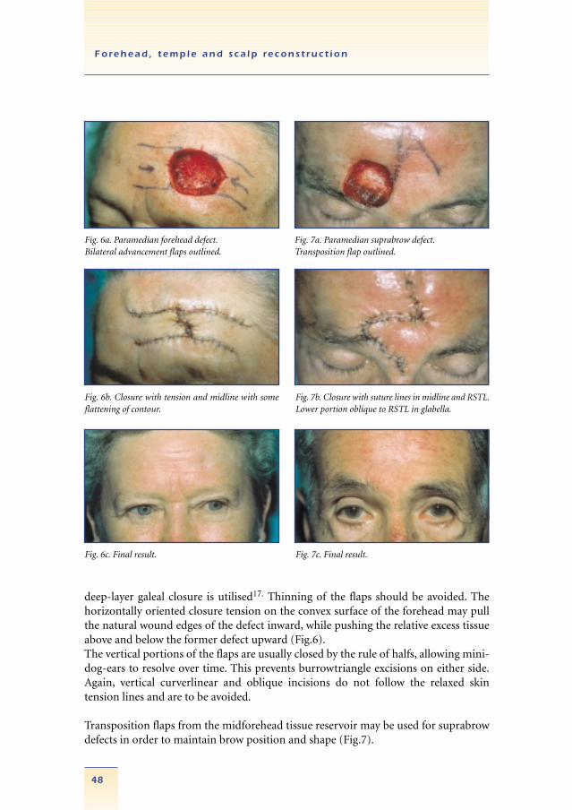
deep-layer galeal closure is utilised17. Thinning of the flaps should be avoided. Thehorizontally oriented closure tension on the convex surface of the forehead may pullthe natural wound edges of the defect inward, while pushing the relative excess tissueabove and below the former defect upward (Fig.6).The vertical portions of the flaps are usually closed by the rule of halfs, allowing mini-dog-ears to resolve over time. This prevents burrowtriangle excisions on either side.Again, vertical curverlinear and oblique incisions do not follow the relaxed skin tension lines and are to be avoided.
Transposition flaps from the midforehead tissue reservoir may be used for suprabrowdefects in order to maintain brow position and shape (Fig.7).
48
Forehead , t emp le and s ca lp re cons t ruc t i on
Fig. 6c. Final result.
Fig. 6a. Paramedian forehead defect.Bilateral advancement flaps outlined.
Fig. 6b. Closure with tension and midline with someflattening of contour.
Fig. 7c. Final result.
Fig. 7a. Paramedian suprabrow defect.Transposition flap outlined.
Fig. 7b. Closure with suture lines in midline and RSTL.Lower portion oblique to RSTL in glabella.
Defects at the border of the anterior hairline may be closed with rotation flaps, mak-ing use of the curve of the forehead while the additional incision comes to lie in theesthetic junction of the forehead and hairline. Bilateral rotation flaps may be used inan A-T fashion.
Lateral foreheadThe lateral forehead begins at the midbrow and extends to the lateral brow where itjoins the upper temple.
The enhanced elasticity of the lateral forehead compared to the central part of theforehead yields a number of reconstructive alternatives. Primary closure may be possible.The flat surface of the lateral forehead may yield reasonable results with secondary intention healing. Elevation of the brow by scar contraction must be prevented. Forexample the brow may be tacked down by sutures to the supraorbital rim.
Multiple types of transpositions flaps may be oriented so that donor site closure takesadvantage of temporal laxity latero-inferiorly.Designing skin flaps in the lateral forehead temporal region must take the course andintegrity of the temporal branch of the facial nerve as an extremely important landmark.Subsequently dissection should be in a subcutaneous plane to prevent injury to the motornerve of the forehead. Furthermore, flaps should be designed in order to prevent dis-torsion of the eyebrow. A burrow wedged advancement flap is created by extending an incision unilaterally along the base of the triangular defect and creat-ing a smaller triangular-burrow excision on the other side of the incision opposite thedefect. The lateral burrow triangle may be hidden in the crowfeet. The wound mayeven be extended to the brow margin in order to camouflage the releasing incision inthe suprabrow transion zone. A number of other variations such as O-Z repair mayyield satisfactory results in the concave surface of the lateral forehead.
Temporal reconstructionThe eyebrow, scalp hairline and lateral canthus as well as zygoma compromise the es-thetic boundaries of the temporal region.When excisions and flap reconstruction are performed in the temporal region, thefrontal branch of the facial nerve which courses very superficial, may be at risk. More-over, the skin and subcutaneous soft tissues over the zygoma are very thin. In the later-al forehead proper, the nerve is better protected by the frontalis muscle.The temporal region is an ideal indication for secondary intension healing30 (Fig. 2).Given its skin laxity primary closure may definitively be an option. A variety of skinflaps are useful to close smaller defects within the esthetic subunit or to recruit tissuefor a larger defect. For example, tissue may be moved over a larger area using single orbilobed transposition flaps (anteriorly or posteriorly based)26.
49
Forehead , t emp le and s ca lp re cons t ruc t i on
drawback because of the considerable temporary deformity of the forehead duringthe expansion process2. However, relatively large defects may be closed after preoperative progressive tissue expansion. For example the contralateral forehead tissue may be expanded and transposed to achieve ipsilateral tissue replacement (sail flap)12.Central and paramedian forehead defects may be amendable to closure after intraoperativeexpansion of the scalp and forehead skin11. A 30 mm Foly catheter is placed subgaleallyand inflated until the tissue blanches. Two or three cycles of 3 minutes volume maintenanceand 3 minutes decompression yield additional tissue stretch. The enhanced tissue mobility often results in less wound-closure tension. By placing the balloon in a deeplayer, vascular compromise of the skin is very unlikely (Fig.8). Alternatively mechanicalcreep (distension) may be promoted, either by multiple skin retraction1 or technicallyadvanced devices, such as the sure-closure skin stretching system20.
SCALP
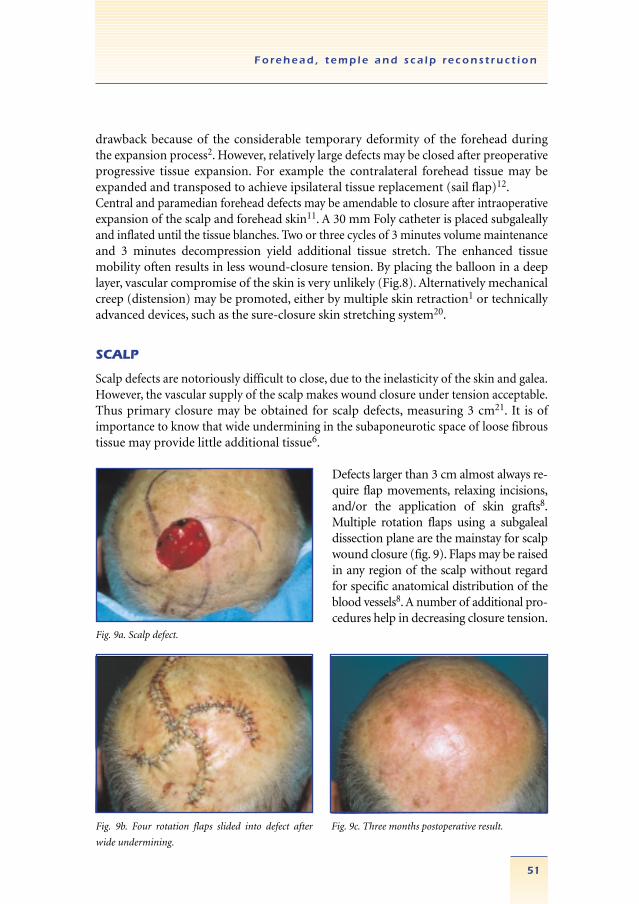
Scalp defects are notoriously difficult to close, due to the inelasticity of the skin and galea.However, the vascular supply of the scalp makes wound closure under tension acceptable.Thus primary closure may be obtained for scalp defects, measuring 3 cm21. It is ofimportance to know that wide undermining in the subaponeurotic space of loose fibroustissue may provide little additional tissue6.
Defects larger than 3 cm almost always re-quire flap movements, relaxing incisions,and/or the application of skin grafts8.Multiple rotation flaps using a subgalealdissection plane are the mainstay for scalpwound closure (fig. 9). Flaps may be raisedin any region of the scalp without regardfor specific anatomical distribution of theblood vessels8. A number of additional pro-cedures help in decreasing closure tension.
51
Forehead , t emp le and s ca lp re cons t ruc t i on
Fig. 9a. Scalp defect.
Fig. 9b. Four rotation flaps slided into defect after
wide undermining.
Fig. 9c. Three months postoperative result.
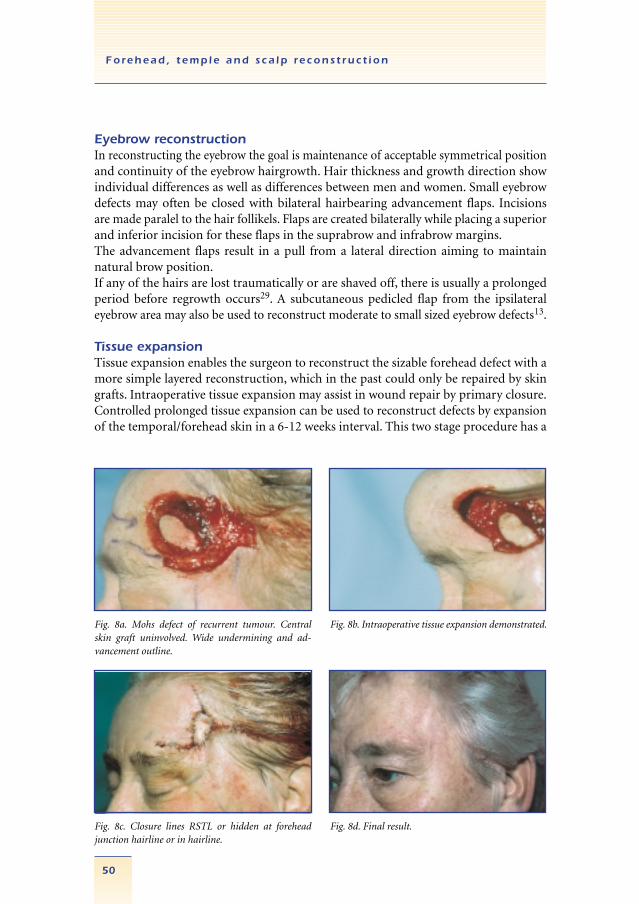
Eyebrow reconstructionIn reconstructing the eyebrow the goal is maintenance of acceptable symmetrical positionand continuity of the eyebrow hairgrowth. Hair thickness and growth direction showindividual differences as well as differences between men and women. Small eyebrowdefects may often be closed with bilateral hairbearing advancement flaps. Incisionsare made paralel to the hair follikels. Flaps are created bilaterally while placing a superiorand inferior incision for these flaps in the suprabrow and infrabrow margins.The advancement flaps result in a pull from a lateral direction aiming to maintainnatural brow position.If any of the hairs are lost traumatically or are shaved off, there is usually a prolongedperiod before regrowth occurs29. A subcutaneous pedicled flap from the ipsilateraleyebrow area may also be used to reconstruct moderate to small sized eyebrow defects13.
Tissue expansionTissue expansion enables the surgeon to reconstruct the sizable forehead defect with amore simple layered reconstruction, which in the past could only be repaired by skingrafts. Intraoperative tissue expansion may assist in wound repair by primary closure.Controlled prolonged tissue expansion can be used to reconstruct defects by expansionof the temporal/forehead skin in a 6-12 weeks interval. This two stage procedure has a
50
Forehead , t emp le and s ca lp re cons t ruc t i on
Fig. 8d. Final result.Fig. 8c. Closure lines RSTL or hidden at foreheadjunction hairline or in hairline.
Fig. 8a. Mohs defect of recurrent tumour. Centralskin graft uninvolved. Wide undermining and ad-vancement outline.
Fig. 8b. Intraoperative tissue expansion demonstrated.
Increased incision length may allow easier closure under less tension and short incisions8.Galeal closure should be performed with deeply burried sutures. Scalp skin may becombined with forehead and neck of large size defects16. Galeal incisions at 5-10 mminterval allow increased flap stretching. However, there is a significant increase risk ofnecrosis. Pre- and intraoperative tissue expansion are options to be considered. Rotationpuckers developing as a result of a rotational movement should usually be left intact,with focal undermining of the area allowing significant postoperative adjustment andsmoothing8.
Large size wounds may heal by secondary intention healing, even with exposed facial-scalp bone24. Fenestration of the exposed bone, outer table of the calvarian bone, mayhelp to stimulate wound healing18. However, healing by secondary intention is lesssatisfactory in a scalpwound than in other areas, forming thin atrophic scars, susceptibleto minor trauma17. But, in older patients with extensive wounds, secondary intentionhealing is a definite option despite of the time needed for postoperative care. Skingrafts may also be a viable alternative in a large defect. However, the esthetic deficiencyif used on hair bearing skin is considered to be similar to secondary intention healing.
SUMMARY
Several characteristics inherent to the forehead/temporal scalp unit provide uniquereconstructive challenges. These include maintenance of motor and sensory nervefunction as well as maintenance or reestablishment of the esthetic boundaries byrecreating a camouflaged surface and optimal surgical scar.Only with a thorough knowledge of forehead anatomy, function and principles of tissuemovement can the optimal reconstructive procedure be designed and performed.
52
Forehead , t emp le and s ca lp re cons t ruc t i on
References1. Auletta M.J., Matarasso S.L., Glogau R.G., Tromowitz T.A. (1993) Comparison of skin hooks and foley
catheter for immediate tissue expansion. J Dermat Surg Oncol 19: 1084-1088.2. Baker S.R., Swanson N.A. (1990) Clinical applications of tissue expansion in head and neck surgery.
Laryngoscope 100: 313-319.3. Baker S.R. Editorial comments, pp. 439-441. In: Local flaps in facial reconstruction . S.R. Baker, N.A.
Swanson, Mosby 1995.4. Borgess A.F. (1973) Elective incisions and scar revision. Little Brown company, Boston.5. Brady J.G., Grande D.J. Katz, A.E (1992) The purse string suture in facial reconstruction. J. Dermatol.
Surg. Oncol. 18: 8-12-816.6. Cupp C.L., Larrabee W.F. (1992) Reconstruction of the forehead and scalp. Operative techniques in
otolaryngology/head & Neck surgery. Vol. 4, 1, 93, pp 11-17.7. Dzubow L.M. (1990) Facial flaps. Biomechanics and regional application. Appleton and Lange.8. Field L.M. (1991) Scalps flaps. J. Dermatol. Surg. Oncol. 17: 190-199.9. Frodel J.L., Marentette L.J. (1993) The coronal approach. Anatomic and technical considerations and
morbidity. Arch. Otolaryngol Head & Neck Surg. 119: 201-207.10. Gosain A.K., Sewall S.R., Yousif N.J. (1997) Plast. Reconstr. Surg. 99: 1224-1233.11. Greenbaum S.S. (1990) Intraoperative tissue expension using a foly catheter. J. Dermatol. Surg. Oncol. 19:
12:1079-1083.12. Iwahira Y, Maruyama Y. (1993) Expanded unilateral forehead flap (sail flap) for coverage of opposite
forehead defect Plast. Rec. Surg, 92: 1052-1056.13. Kasai K. & Ogawa Y. (1990) Partial eyebrow reconstruction using subcutaneous pedicle flaps to preserve
the natural hair direction. Ann. Plast. Surg. 24, no. 2, 117-125.14. Knize D.M. (1995) A study of the supraorbital nerve. Plast. Reconstr. Surg. 96, no. 3:564-569.15. Knize D.M. (1999) Limited incision foreheadplasy. Plast. Reconstr. Surg. 103. no. 1.271-284.16. Kroll S.S., Margolis R.(1993). Scalp flap reconstruction with primary donor site closure. Annals Plast. Surg.
30, nr. 5, 452-455.17. Larrabee W.F., Sherris D.A.(1995) Principles of facial reconstruction. Lippincott, Raven Publ.18. Latenser J., Snow S.N. Mohs F.E., Weltman R., Hruza G (1991). Power drills to fenestrate exposed bone to
stimulate wound healing. J. Dermatol. Surg. Oncol. 17:265-170.19. Liebman E.P., Webster R.C., Berger A.S. Della Vecchia M. (1982) Arch. Otolaryngol 108: 232-235.20. Ling E.H., Wang T.D. (1996) Local flaps in forehead and temporal reconstruction. Facial Plast. Surg. Clin.
North Amer. 4, no. 4: 469-479.21. Minor L., Panje W.B. (1993) Malignant neoplasms of the scalp. Otolaryngol. Clin. North Amer. 26, no.
2:279-293.22. Shumrick K.A. & Smith T.L (1992) The anatomic basis for the design of forehead flaps in nasal reconstruction.
Arch Otolaryngol Head & Neck Surg 118:373-379.23. Siegle R.J. (1995) Reconstruction of the forehead. Chapter 20, pp. 421-439. In: Local flaps in facial recon-
struction. S.R. Baker, N.A. Swanson, Mosby 1995.24. Snow S.N. Stiff, M.A., Bullen R., Mohs F.E., Wei Hsiung Chao (1994). Second intention healing of exposed
facial-scalp bone after Mohs surgery for skin cancer: review of ninety-one cases. J.Amer. Acad. Dermatol.31., no 3. Part I, pp. 450-454.
25. Stuzin J.M., Wagstrom L., Kawamoto H.K., Wolfe S.A. (1989) Plast. And Reconstr. Surg. 83:265-271.26. Sutton A.W. Quatela V.S. (1992) Bilobed flap reconstruction of the temporal forehead.27. Swanson N.A. (1995) Editorial comments. Chapter 20, Reconstruction of the forehead, pp. 441-442. In:
,Local flaps in facial reconstruction.28. Tolhurst D.E., Carstens M.H., Greco R.J., Hurwitz D.J. (1991) Plast. Reconstr. Surg. 87:603-612.29. Tromovitch T.A., Stegman S.J., Glogau R.G. (1989) Flaps and grafts in dermatologic surgery. Yearbook
Medical publishers.30. Zitelli J.A. (1983) Wound healing by secondary intention: a cosmetic appraisal. J. Amer. Acad. Dermatol. 9:
407-15.
53
Forehead , t emp le and s ca lp re cons t ruc t i on

54