Embed Size (px)
Citation preview

Focus OnCervical Myelopathy
Cervical myelopathy is a condition caused by narrowing of the spinal canal leading to cord dysfunction.1 The most common causes are congenital stenosis and degenerative stenosis caused by spondylosis (degenerative osteoarthritis).2 When it is caused by spondylosis it is commonly referred to as cervical spondylotic myelopathy (CSM). Rheumatoid arthritis (RA) is a common condition affecting around 1% of the population. Around 90% of patients with RA have cervical spine involvement with between 11% and 58% of these patients having neurological involvement.3,4 Many of these patients have cervical myelopathy. Whatever the underlying disease process, the compression is usually progressive and will often require surgical intervention to prevent further disability. Many patients experience significant improvement in symptoms after surgery, so operative intervention should be considered for almost all patients.
EpidemiologyCervical myelopathy is more common in men and tends to present earlier than in women. Radiologically, the condition is present in 13% of men in the third decade and almost 100% of men over the age of 70. In women the disease presents later, with 5% showing radiographic changes in the fourth decade going up to 96% in women over the age of 70.5 Changes are more common in patients with RA where 85% of those with moderate to severe disease will have x-ray changes.6
When patients with cervical myelopathy present in the third or fourth decade of life it is usually secondary to congenital stenosis. If it presents later in life, degenerative spondylosis is usually the underlying cause. Patients who have a congenitally narrow canal are predisposed to the effects of spondylotic changes earlier, as there is less space in the canal to accommodate the compressing lesions.
Pathological processThe underlying cause of the condition is compression of the long tracts in the spinal cord. The normal diameter of the cervical spinal canal is between 17 mm and 18 mm. When this diameter falls below 12 mm to 14 mm for any reason this is likely to cause stenosis and myelopathic symptoms. The average diameter of the spinal cord in the cervical spine is 10 mm. The common pathological processes underlying cervical myelopathy are outlined below:
Disc Herniation. Discogenic disease may cause myelopathy in the acute setting as a large central soft disc herniation causing cord compression. Disc disease is also often seen as part of the compressive lesion in spondylotic disease.
Congenital. Myelopathy due to congenital stenosis does not have a specific underlying lesion. It is caused by a canal diameter which is narrower from birth. It is often not symptomatic until secondary degeneration further narrows the canal.
Spondylosis. CSM is the result of degenerative changes which develop with age, including ligamentum flavum hypertrophy or buckling, facet joint hypertrophy, disc protrusion and posterior spondylotic ridges. One or all of these changes contribute to an overall reduction in canal diameter which may result in cord compression. Spondylolisthesis usually occurs in the lower cervical spine. It is caused by arthrosis of the facet joints combined with disc degeneration leading to instability.7
Post traumatic myelopathy. Trauma may induce myelopathy or precipitate symptoms of an underlying stenosis of the spinal canal. Smaller diameter canals have an increased chance of neurological injury in trauma.8
Ossification of the posterior longitudinal ligament (OPLL). This is a common feature in patients with cervical myelopathy with up to 25% being affected. It is particularly common in patients from the far east. It is seen on imaging as areas of ossification behind the vertebral bodies. The extent is best defined by CT.9
Myelopathy due to tumour expansion. Intraspinal tumours are a relatively uncommon cause of cervical myelopathy but must always be considered given the potentially catastrophic consequences of the diagnosis being missed. Tumours may originate in the spinal cord (intramedullary tumours) or compress from outside (extramedullary tumours). Metastatic deposits are usually slow growing with gradual onset of symptoms.
SymptomsPatients may present with a range of symptoms and many of these are non-specific. It is important to remember that although cervical myelopathy is a disease of the cervical spine it may manifest with lower as well as upper limb symptoms. The classical presentation is loss of balance with poor coordination, decreased dexterity, weakness, numbness and in severe cases paralysis. Pain is a symptom in many patients but it is important to remember that it may be absent which often leads to a delay in diagnosis. In older patients it often manifests with a rapid deterioration of gait and hand function. Cervical lesions in the region of C3 - C6 cause a loss of manual dexterity with difficulties in writing and nonspecific alteration in arm weakness and sensation. Cervical lesions from C6 - C8 tend
©2012 British Editorial Society of Bone and Joint Surgery 1
BONE AND JOINT
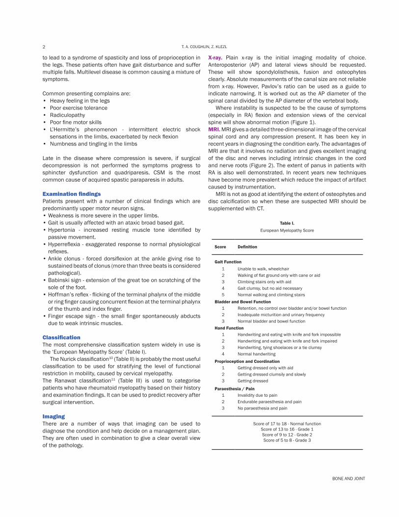
2 T. A. COUGHLIN, Z. KLEZL
to lead to a syndrome of spasticity and loss of proprioception in the legs. These patients often have gait disturbance and suffer multiple falls. Multilevel disease is common causing a mixture of symptoms.
Common presenting complains are:• Heavy feeling in the legs• Poor exercise tolerance• Radiculopathy• Poor fine motor skills• L’Hermitte’s phenomenon - intermittent electric shock
sensations in the limbs, exacerbated by neck flexion• Numbness and tingling in the limbs
Late in the disease where compression is severe, if surgical decompression is not performed the symptoms progress to sphincter dysfunction and quadriparesis. CSM is the most common cause of acquired spastic paraparesis in adults.
Examination findingsPatients present with a number of clinical findings which are predominantly upper motor neuron signs. • Weakness is more severe in the upper limbs. • Gait is usually affected with an ataxic broad based gait.• Hypertonia - increased resting muscle tone identified by
passive movement.• Hyperreflexia - exaggerated response to normal physiological
reflexes.• Ankle clonus - forced dorsiflexion at the ankle giving rise to
sustained beats of clonus (more than three beats is considered pathological).
• Babinski sign - extension of the great toe on scratching of the sole of the foot.
• Hoffman’s reflex - flicking of the terminal phalynx of the middle or ring finger causing concurrent flexion at the terminal phalynx of the thumb and index finger.
• Finger escape sign - the small finger spontaneously abducts due to weak intrinsic muscles.
ClassificationThe most comprehensive classification system widely in use is the ‘European Myelopathy Score’ (Table I). The Nurick classification10 (Table II) is probably the most useful classification to be used for stratifying the level of functional restriction in mobility, caused by cervical myelopathy.The Ranawat classification11 (Table III) is used to categorise patients who have rheumatoid myelopathy based on their history and examination findings. It can be used to predict recovery after surgical intervention.
ImagingThere are a number of ways that imaging can be used to diagnose the condition and help decide on a management plan. They are often used in combination to give a clear overall view of the pathology.
X-ray. Plain x-ray is the initial imaging modality of choice. Anteroposterior (AP) and lateral views should be requested. These will show spondylolisthesis, fusion and osteophytes clearly. Absolute measurements of the canal size are not reliable from x-ray. However, Pavlov’s ratio can be used as a guide to indicate narrowing. It is worked out as the AP diameter of the spinal canal divided by the AP diameter of the vertebral body. Where instability is suspected to be the cause of symptoms (especially in RA) flexion and extension views of the cervical spine will show abnormal motion (Figure 1). MRI. MRI gives a detailed three-dimensional image of the cervical spinal cord and any compression present. It has been key in recent years in diagnosing the condition early. The advantages of MRI are that it involves no radiation and gives excellent imaging of the disc and nerves including intrinsic changes in the cord and nerve roots (Figure 2). The extent of panus in patients with RA is also well demonstrated. In recent years new techniques have become more prevalent which reduce the impact of artifact caused by instrumentation. MRI is not as good at identifying the extent of osteophytes and disc calcification so when these are suspected MRI should be supplemented with CT.
Table I.
European Myelopathy Score
Score Definition
Gait Function1 Unable to walk, wheelchair2 Walking of flat ground only with cane or aid3 Climbing stairs only with aid4 Gait clumsy, but no aid necessary5 Normal walking and climbing stairs
Bladder and Bowel Function1 Retention, no control over bladder and/or bowel function2 Inadequate micturition and urinary frequency3 Normal bladder and bowel function
Hand Function1 Handwriting and eating with knife and fork impossible2 Handwriting and eating with knife and fork impaired3 Handwriting, tying shoelaces or a tie clumsy4 Normal handwriting
Proprioception and Coordination1 Getting dressed only with aid2 Getting dressed clumsily and slowly3 Getting dressed
Paraesthesia / Pain1 Invalidity due to pain2 Endurable paraesthesia and pain 3 No paraesthesia and pain
Score of 17 to 18 - Normal functionScore of 13 to 16 - Grade 1Score of 9 to 12 - Grade 2Score of 5 to 8 - Grade 3

3CERVICAL MYELOPATHY
©2012 British Editorial Society of Bone and Joint Surgery
Table II.
Nurick’s Functional Scale
Grade Level of Neurological Involvement
Grade I No difficulty in walking
Grade II Mild gait involvement not interfering with employment
Grade III Gait abnormality preventing employment
Grade IV Able to walk only with assistance
Grade V Chairbound or bedridden
Table III.
Ranawat Classification of Neurological Deficit
Class Level of Neurological Involvement
Class I No neural deficit
Class II Subjective weakness, dysaesthesia and hyperreflexia
Class III A Objective weakness and long tract signs; patient ambulatory
Class III B Objective weakness and long tract signs; patient no longer ambulatory
Fig. 1.
Atlantoaxial subluxation in flexion.
CT. There are two main uses for CT scanning in the diagnosis of cervical myelopathy. There are some patients who are unable to undergo MRI imaging, often because of an implanted cardiac device. For these patients a CT myelogram offers an imaging option which is almost as sensitive as MRI.12 In general CT can also give additional information on the presence of osteophytes or other bony compressive lesions which are underestimated on MRI scans (Figure 3).
Fig. 2.
A stenotic segment between C4 and C6 on a T2 weighted saggital reconstruction.
Fig. 3.
A saggital section of a spondylotic c-spine with clear protrusion of osteophyte into the canal.
CT myelography requires a cervical or lumbar puncture and the introduction of contrast. Patients require inpatient monitoring for a period of time, as reactions to the contrast can present late. CT myelography has become rare in practice now due to the widespread availability of MRI.
BONE AND JOINT
4 T. A. COUGHLIN, Z. KLEZL
Conservative managementConservative management may be considered in many cases, provided patients are closely followed up in clinic. Cervical myelopathy is predominantly progressive and only a small number of patients experience regression in their symptoms. Some patients are medically co-morbid making surgery high risk. In these patients a trial of conservative management should certainly be considered. Non-operative management should involve regular analgesia. Gabapentin should be considered as an adjunct to simple analgesics for patients who have significant pain from radicular symptoms. A soft collar may be used, especially when instability is a significant cause of symptoms which is often the case in patients with RA. Physiotherapy may be of use in patients who can tolerate it which can involve traction, heat and ultrasound therapy. Activity modification can be a useful way of limiting factors which provoke symptoms. Extension of the cervical spine and restriction of heavy lifting can be particularly helpful. The use of epidural steroid injection is controversial. They are primarily used in patients who have significant radicular symptoms. Their efficacy has been shown in some studies to the similar to caudal epidural injection for radicular symptoms in the lumbar spine.13
Patients who suffer a failure of at least three months conservative management should be considered for operative intervention. Particularly if symptoms impair activities of daily living surgical intervention should be considered. Any patient who exhibits progressive neurological deficit should undergo operative intervention unless the risk of surgery makes this impossible. It should be made clear to the patient that the main aim of operative intervention is to prevent progression of the neurological symptoms and that recovery is unpredictable and cannot be guaranteed.
Surgical managementThe primary goal of any surgery is to restore the diameter of the spinal canal such that compression of the spinal cord is relieved. The choice of operation depends to a degree on the surgeon’s experience with any particular procedure. However, the location of stenosis and the vertebral body alignment will guide whether an anterior or posterior approach is suitable. Although controversial, there have been studies which show that even when a patient is bedridden because of cervical myelopathy, there is a benefit to operative intervention. One study showed that in 55 patients with a Nurick score of 5 (chair bound or bedridden), two thirds improved one point after decompression returning the patient to walking, albeit with assistance.14
Surgical interventions can be divided into two anatomical areas; the upper (C0-C2) and lower cervical spine (C3-C7) and two approaches; anterior and posterior. When considering approach, the decision making process has to consider a number of factors:
• Main site of cord compression • Whether compression comes from the front or the back • the number of levels involved • Bone quality • Deformity (mainly kyphosis)• Instability (stepladder deformity in RA patients - multiple
sequential levels of subluxation) • Presence of fused segments • Patient wishes and expectations
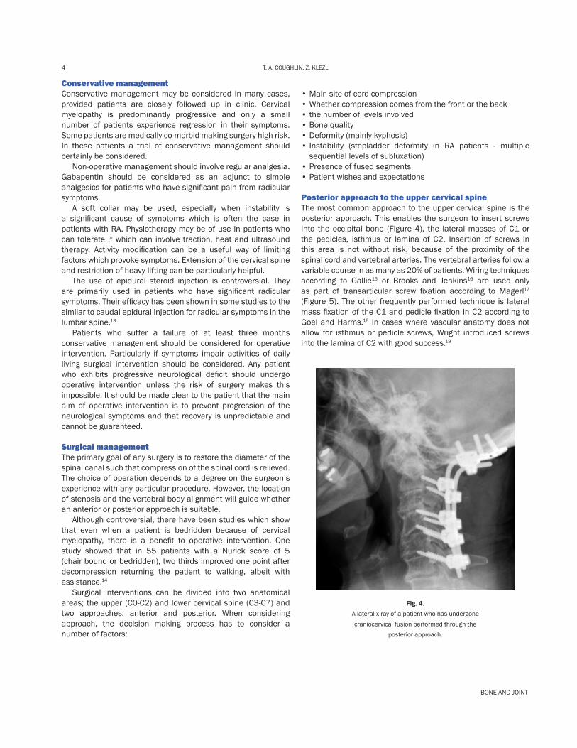
Posterior approach to the upper cervical spineThe most common approach to the upper cervical spine is the posterior approach. This enables the surgeon to insert screws into the occipital bone (Figure 4), the lateral masses of C1 or the pedicles, isthmus or lamina of C2. Insertion of screws in this area is not without risk, because of the proximity of the spinal cord and vertebral arteries. The vertebral arteries follow a variable course in as many as 20% of patients. Wiring techniques according to Gallie15 or Brooks and Jenkins16 are used only as part of transarticular screw fixation according to Magerl17
(Figure 5). The other frequently performed technique is lateral mass fixation of the C1 and pedicle fixation in C2 according to Goel and Harms.18 In cases where vascular anatomy does not allow for isthmus or pedicle screws, Wright introduced screws into the lamina of C2 with good success.19
Fig. 4.
A lateral x-ray of a patient who has undergone craniocervical fusion performed through the
posterior approach.

5CERVICAL MYELOPATHY
©2012 British Editorial Society of Bone and Joint Surgery
Fig. 5.
A patient who has undergone atlantoaxial screw fixation for subluxation with Magerl’s technique.17
Anterior approach to the upper cervical spineThe anterior approach to the upper cervical spine is rarely used now. Historically it was used for resection of inflammatory panus in patients with RA. It has since been shown that panus resorbs with rigid posterior stabilization so the approach is now infrequently indicated.
Anterior approach to the lower cervical spineThe Smith-Robinson approach20 is the most common anterior approach to the lower cervical spine. It is most frequently used to perform discectomy. It allows disc material to be excised from the distracted disc space with curettes and pituitary rongeurs. In order to make sure the decompression is adequate, the accessible posterior longitudinal ligament (PLL) and posterior osteophytes are resected and the foramina are widened (Figure 6).
Fig. 6.
A patient following a C5/6 discectomy and cage fusion. Access to posterior osteophytes or OPLL is limited with discectomy alone. When these structures cause significant compression, corpectomy (vertebral body resection) should be performed (Figure 7). Most commonly one level is resected but resection of
two or even three vertebral bodies has to been performed. One study demonstrated an improvement in the Nurick score of over two grades following the procedure.21 More extensive resection leads to suboptimal stability of the cervical spine which should be addressed with the addition of posterior stabilisation. Autologous bone graft harvested from the iliac crest has been the benchmark procedure for many years when providing graft for fusion procedures. Because there is up to 20% morbidity associated with harvesting iliac crest graft, the vast majority of spinal surgeons now insert synthetic bone graft with or without a cage. Following discectomy a cage is inserted into the disc space to maintain or restore the disc space and return any lost cervical lordosis.
Fig. 7.
A patient following C5 and C6 corpectomy and fusion with a cage and plate.
Different cages are used in filling corpectomy defects. These are usually a titanium or polyether-etherketone (PEEK) mesh, filled with bone graft harvested locally during the corpectomy. Plates are then used to restore the stability of the cervical spine. There are various designs, with most being unicortical locking plates allowing placement of fixed angle or variable angle screws. Some plates allow for minimal subsidence of the graft/cage. Plates are designed to be low profile to prevent irritation of the overlying oesophagus and should be contoured to match the cervical lordosis whenever possible.
Posterior approach to the lower cervical spineThe posterior approach is mainly used to relieve compression from posterior structures. It is also used in lordotic spines with multilevel anterior compressions, the most common cause being ossification of the PLL. Identification of the midline can be difficult and is usually done in the area of C6-7, it significantly reduces blood loss and trauma to the surrounding paraspinal musculature. The procedures most commonly performed

BONE AND JOINT
6 T. A. COUGHLIN, Z. KLEZL
through the posterior approach are laminectomy (with or without instrumented fusion) and laminoplasty. Laminectomy extensively decompresses the spinal canal allowing for decompression of the foramina when needed. Instrumentation is usually performed to prevent post-laminectomy kyphosis and frequently to restore the normal lordosis of the c-spine (Figure 8). The screws are inserted most frequently into the lateral masses, they are bicortical and give good stability. Pedicle screw fixation has been gaining popularity recently, with the introduction of spinal navigation.
Fig. 8.
A patient who has undergone posterior decompression and instrumented fusion of C2 - C5.
Laminoplasty is an excellent option in lordotic spines, in younger patients, where fusion is undesirable and who can retain at least limited cervical spine mobility following the procedure. Laminoplasty techniques come from the Far East, mainly because of the high incidence of ossification of the PLL in the region.22 There are two main laminoplasty techniques, the French door and the open door. They are technically demanding procedures which lead to significant enlargement of the cervical spine diameter. Mini-plates have been designed to support the opened laminae (Figure 9). Laminoplasty with mini-plates has been shown to be a reliable technique in multilevel disease.23 However, neurological recovery has been shown to be worse in patients over 75 years of age than in younger patients.24
Fig. 9.
A patient who has undergone instrumented laminoplasty of C4 - C7.
Combined approach to the cervical spineIn complex cases, especially with a combination of compression from both anterior and posterior structures associated with instability, both approaches to the cervical spine have to be utilised (Figure 10). These cases may require extension down to the upper thoracic spine or up high as the occiput. Patients have to be made aware of the significant functional restrictions resulting from these demanding procedures.
Fig. 10.
A patient who has undergone posterior laminectomy and instrumented fusion of C4 - C7 combined with anterior corpectomy and fusion of C3 - C5.
SummaryCervical myelopathy is a relatively common condition affecting a spectrum of patients across different age ranges. There are many underlying causes for the condition but in most cases the condition is progressive. Conservative management has a place in some patients who present a high surgical risk or where symptoms seem to be stable and compatible with normal life. However, many patients ultimately require surgical intervention. Surgical treatment of cervical spondylotic myelopathy is very demanding and not without risk. Obviously timing of surgical treatment, before the onset of severe myelopathy, would seem ideal. Unfortunately we frequently do not have that option. Approach and extent of surgical intervention has to be carefully selected and ideally should match the patient’s expectations whilst minimising risk. Intraoperative and postoperative complications are common, especially in elderly patients with a wide range of medical co-morbidities. According to our results mirrored by many other centres, surgery is rewarding, because it stops progression of the cord damage and in most cases leads to improvement in the functional status of our patients.

7CERVICAL MYELOPATHY
©2012 British Editorial Society of Bone and Joint Surgery
T. A. Coughlin BM BS BMedSci MRCSZ. Klezl MD PhD
Royal Derby HospitalUttoxeter RoadDerbyDE22 3NE
E-mail: [email protected]
References
1. Payne EE, Spillane J. The cervical spine; an anatomico-pathological study of 70 specimens (using a special technique) with particular reference to the problem of cervical spondylosis. Brain 1957;80:571-96. 2. Bernhardt M, Hynes RA, Blume HW, White AA 3rd. Cervical spondylotic myelopathy. J Bone Joint Surg [Am] 1993;75-A:119-28. 3. Conaty JP, Mongan ES. Cervical fusion in rheumatoid arthritis. J Bone Joint Surg [Am] 1981;63-A:1218-27.4. Goel A, Laheri V. Re: Harms J, Melcher P. Posterior C1-C2 fusion with polyaxial screw and rod fixation. Spine 2002;27:1589-90.5. Irvine DH, Foster JB, Newell DJ, Klukvin BN. Prevalence of cervical spondylosis in a general practice. Lancet 1965;14:1089-92.6. Bland JH. Rheumatoid arthritis of the cervical spine. J Rheumatol 1974;1:319-42.7. Woiciechowsky C, Thomale UW, Kroppenstedt SN. Degenerative spondylolisthesis of the cervical spine: symptoms and surgical strategies depending on disease progress. Eur Spine J 2004;13:680-4.8. Eismont FJ, Clifford S, Goldberg M, Green B. Cervical sagittal canal size in spinal injury. Spine 1984;9:663-6. 9. Epstein N. Ossification of the cervical posterior longitudinal ligament: a review. Neurosurg Focus 2002;13:ECP1.
10. Nurick S. The pathogenesis of spinal cord disorder associated with cervical spondylosis. Brain 1972;95:87-10011. Ranawat CS, O’Leary P, Pellicci P, et al. Cervical spine fusion in rheumatoid arthritis. J Bone Joint Surg [Am] 1979;61-A:1003-10.12. Pressman BD, Mink JH, Turner RM, Rothman BJ. Low-dose metrizamide spinal computed tomography in outpatients. J Comput Assist Tomogr 1987;10:817-21.13. Lin EL, Lieu V, Halevi L, Shamie AN, Wang JC. Cervical steroid injections for symptomatic disc herniations. J Spinal Disord Tech 2006;19:183-6.14. Scardino FB, Rocha LP, Barcelos ACES, Rotta JM, Botelho RV. Is there a benefit to operating on patients (bedridden or in wheelchairs) with advanced stage cervical spondylotic myelopathy? Eur Spine J 2010;19:699-705.15. Gallie WE. Fractures and dislocations of the cervical spine. Am J Surg 1939;46:495-9.16. Brooks AL, Jenkins EB. Atlanto-axial arthrodesis by the wedge compression method. J Bone Joint Surg [Am] 1978;60-A:279-84.17. Grob D. Atlantoaxial screw fixation (Magerl’s technique). Rev Ortp Traumatol 2008;52:243-9.18. Harms J, Melcher RP. Posterior C1–C2 fusion with poly-axial screw and rod fixation. Spine (Phila Pa 1976) 2001;26:2467-71.19. Wright NM. Posterior C2 fixation using bilateral, crossing C2 laminar screws: case series and technical note. J Spinal Disord Tech 2004;17:158-62.20. Southwick WO, Robinson RA. Surgical approaches to the vertebral bodies in the cervical and lumbar regions. J Bone and Joint Surg [Am] 1957;39-A:631-44.21. Williams KE, Paul R, Dewan Y. Functional outcome of corpectomy in cervical spondylotic myelopathy. Indian J Orthop 2009;43:205-9.22. Wu JC, Liu L, Chen YC, et al. Ossification of the posterior longitudinal ligament in the cervical spine: an 11-year comprehensive national epidemiological study. Neurosurg Focus 2011;30:E523. Dimar JR II, Bratcher KR, Brock DC, et al. Instrumented open-door laminoplasty as treatment for cervical myelopathy in 104 patients. Am J Orthop 2009;38:123-8.24. Matsuda Y, Shibata T, Oki S, et al. Outcomes of surgical treatment for cervical myelopathy in patients more than 75 years of age. Spine 1999;24:529-34.