Embed Size (px)
Citation preview
Fluorescence imaging of dendriticspines of Golgi-Cox-stained neuronsusing brightening background
Min AiHanqing XiongTao YangZhenhua ShangMuqing ChenXiuli LiuShaoqun Zeng
Downloaded From: https://www.spiedigitallibrary.org/journals/Journal-of-Biomedical-Optics on 6/6/2018 Terms of Use: https://www.spiedigitallibrary.org/terms-of-use
Fluorescence imagingof dendritic spines ofGolgi-Cox-stainedneurons usingbrightening background
Min Ai,a,b,† Hanqing Xiong,a,b,† Tao Yang,a,bZhenhua Shang,a,b Muqing Chen,c Xiuli Liu,a,b,* andShaoqun Zenga,b
aHuazhong University of Science and Technology, Britton ChanceCenter for Biomedical Photonics, Wuhan National Laboratory forOptoelectronics, Wuhan 430074, ChinabHuazhong University of Science and Technology, Department ofBiomedical Engineering, Key Laboratory of Biomedical Photonics ofMinistry of Education, Wuhan 430074, ChinacHubei University of Education, School of Physics and ElectronicInformation, Wuhan 430205, China
Abstract. We report a novel fluorescence imagingapproach to imaging nonfluorescence-labeled biologicaltissue samples. The method was demonstrated by imag-ing neurons in Golgi-Cox-stained and epoxy-resin-embedded samples through the excitation of the back-ground fluorescence of the specimens. The dark neuronsstood out clearly against background fluorescence in theimages, enabling the tracing of a single dendritic spineusing both confocal and wide-field fluorescence micros-copy. The results suggest that the reported fluorescenceimaging method would provide an effective alternative sol-ution to image nonfluorescence-labeled samples, andit allows tracing the dendritic spine structure of neurons.© The Authors. Published by SPIE under a Creative Commons Attribution
3.0 Unported License. Distribution or reproduction of this work in whole or in
part requires full attribution of the original publication, including its DOI.
[DOI: 10.1117/1.JBO.20.1.010501]
Keywords: fluorescence imaging; Golgi-Cox staining; backgroundfluorescence; dendritic spine.
Paper 140775LR received Nov. 24, 2014; accepted for publicationDec. 23, 2014; published online Jan. 13, 2015.
Golgi-Cox staining has been recognized as one of the mostelegant and effective procedures for studying the morphologyof neurons.1 The neurons stained using the Golgi-Cox methodappear as black deposits, enabling the visualization of the den-dritic branching pattern and dendritic spines using light micros-copy and a tracing algorithm.2,3 With the optical absorptioncharacterization of black deposits, one can image the neuronmorphology via reflection or transmission wide-field imagingtechniques.4–6 Generally, nonfluorescence-labeled biologicaltissues are sliced, and Golgi-Cox-stained biological samplesare imaged through the optical absorption approach. A slice im-aging depth of less than several tens of micrometers is difficult
to achieve. During sample preparation, as the embedded Spurrresin has strong fluorescence, we can utilize the backgroundfluorescence to image the black stained samples. In thisstudy, to demonstrate the fluorescence imaging method, weshow the fluorescence data of mouse brain slices stainedusing the Golgi-Cox method. By using this method, theGolgi-Cox-stained tissues could be imaged using currentlyavailable fluorescence imaging techniques. To our knowledge,this is the first report of imaging Golgi-Cox-stained neuronsusing the contrast between bright-background fluorescenceand dark-labeled neurons.
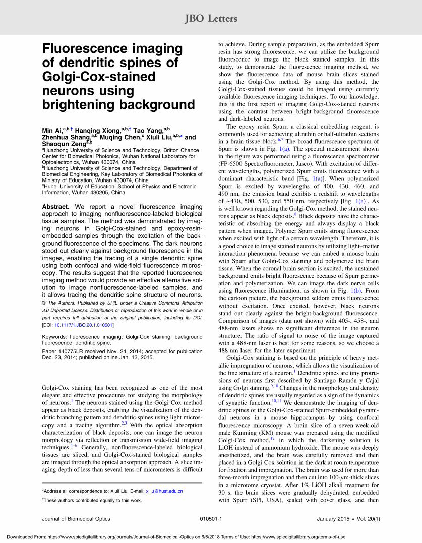
The epoxy resin Spurr, a classical embedding reagent, iscommonly used for achieving ultrathin or half-ultrathin sectionsin a brain tissue block.6,7 The broad fluorescence spectrum ofSpurr is shown in Fig. 1(a). The spectral measurement shownin the figure was performed using a fluorescence spectrometer(FP-6500 Spectrofluorometer, Jasco). With excitation of differ-ent wavelengths, polymerized Spurr emits fluorescence with adominant characteristic band [Fig. 1(a)]. When polymerizedSpurr is excited by wavelengths of 400, 430, 460, and490 nm, the emission band exhibits a redshift to wavelengthsof ∼470, 500, 530, and 550 nm, respectively [Fig. 1(a)]. Asis well known regarding the Golgi-Cox method, the stained neu-rons appear as black deposits.8 Black deposits have the charac-teristic of absorbing the energy and always display a blackpattern when imaged. Polymer Spurr emits strong fluorescencewhen excited with light of a certain wavelength. Therefore, it isa good choice to image stained neurons by utilizing light–matterinteraction phenomena because we can embed a mouse brainwith Spurr after Golgi-Cox staining and polymerize the braintissue. When the coronal brain section is excited, the unstainedbackground emits bright fluorescence because of Spurr perme-ation and polymerization. We can image the dark nerve cellsusing fluorescence illumination, as shown in Fig. 1(b). Fromthe cartoon picture, the background seldom emits fluorescencewithout excitation. Once excited, however, black neuronsstand out clearly against the bright-background fluorescence.Comparison of images (data not shown) with 405-, 458-, and488-nm lasers shows no significant difference in the neuronstructure. The ratio of signal to noise of the image capturedwith a 488-nm laser is best for some reasons, so we choose a488-nm laser for the later experiment.
Golgi-Cox staining is based on the principle of heavy met-allic impregnation of neurons, which allows the visualization ofthe fine structure of a neuron.1 Dendritic spines are tiny protru-sions of neurons first described by Santiago Ramón y Cajalusing Golgi staining.9,10 Changes in the morphology and densityof dendritic spines are usually regarded as a sign of the dynamicsof synaptic function.10,11 We demonstrate the imaging of den-dritic spines of the Golgi-Cox-stained Spurr-embedded pyrami-dal neurons in a mouse hippocampus by using confocalfluorescence microscopy. A brain slice of a seven-week-oldmale Kunming (KM) mouse was prepared using the modifiedGolgi-Cox method,12 in which the darkening solution isLiOH instead of ammonium hydroxide. The mouse was deeplyanesthetized, and the brain was carefully removed and thenplaced in a Golgi-Cox solution in the dark at room temperaturefor fixation and impregnation. The brain was used for more thanthree-month impregnation and then cut into 100-μm-thick slicesin a microtome cryostat. After 1% LiOH alkali treatment for30 s, the brain slices were gradually dehydrated, embeddedwith Spurr (SPI, USA), sealed with cover glass, and then
*Address all correspondence to: Xiuli Liu, E-mail: [email protected]
†These authors contributed equally to this work.
Journal of Biomedical Optics 010501-1 January 2015 • Vol. 20(1)
JBO Letters
Downloaded From: https://www.spiedigitallibrary.org/journals/Journal-of-Biomedical-Optics on 6/6/2018 Terms of Use: https://www.spiedigitallibrary.org/terms-of-use
kept in an oven at 60°C for 36 h. After the polymerized brainwas cooled to room temperature, it was kept dry in the dark untildata acquisition. Neuron morphology in the subcortex wasimaged using a 488-nm laser (5-mW output power) and 20×objective (dry, N.A. 0.8); the laser power at the specimenwill be lowered to 20 to 30% of the output power. Imageswere acquired using inverted confocal fluorescence microscopy(LSM710, Zeiss). With Spurr fluorescence illumination, we canobserve clear soma and dendrite structures in the entire scanningarea [Fig. 2(a)]. An apical dendrite and its spines were imagedusing a 488-nm laser (15-mWoutput power) and a 63× objective(water immersion, N.A. 1.20), and are shown at 3× magnifica-tion in Fig. 2(b) [corresponding to the red box in Fig. 2(a)]. Theoutput power of the 488-nm laser in the confocal microscopy is5 mW when imaging with a 20× objective and 15 mW whenimaging with a 63× objective. Depending on the morphology,spines can be classified as follows: stubby (short without neck),thin (thin with a small head and a long neck), mushroom (bul-bous head with a narrow neck), and cup-shaped or branching
(one neck protruding from dendritic shaft and splitting intotwo subnecks, and one small head for each subneck).10 Inorder to show the clear spine morphology of the apical dendrite,in Figs. 2(c) and 2(d), we acquired images of the part shown inthe boxes in Fig. 2(b) at ∼4× magnification by using a 63×water objective with color the inverted using Photoshop soft-ware. We can clearly observe the stubby (arrow head), thin(arrow), mushroom (star), and branched (diamond) spines inFigs. 2(c) and 2(d). The results of confocal fluorescence imagingof the stained neurons show that this method is suitable for trac-ing fine spine structures of pyramidal neurons in the hippocam-pus. Through the comparison of the fluorescence imagingapproach with a conventional Golgi-staining imaging method,the image from the fluorescence imaging approach demonstratesa better, at least comparative, ability to reveal the fine structureof Golgi-stained neurons.
We can also record many dendritic spines of Golgi-Cox-stained cortical pyramidal neurons of a mouse-brain coronal sec-tion with wide-field fluorescence imaging. For this purpose, abrain of a seven-week-old male KM mouse was preparedusing the modified Golgi-Cox method described above.12 Ahome-made bright-field line-scan imaging system with ultrami-crotome sectioning of Spurr-embedded tissuewas used for wide-field fluorescence imaging.6,13 All the images were obtainedusing laser illumination (488 nm, continuous-wave mode,Sapphire) with ∼30-mW output through an objective lens(LUMPlanFLN, 40×, N.A. 0.8, water immersion, Olympus)and using the strategy of imaging first and cutting-off later.The axial serial imaging was performed using a diamond
Fig. 1 Fluorescence imaging diagram of Golgi-Cox-stained neurons.(a) Fluorescence spectrum of polymerized Spurr with excitation from400 to 490 nm. When polymerized Spurr interacts with light of wave-lengths 400, 430, 460, and 490 nm, the emission peak appears at∼470, 500, 530, and 550 nm. (b) The polymerized Spurr-embeddedspecimen before excitation (left). When the surface of the specimen isexcited, the section absorbs incident light and emits bright fluores-cence, while dark neurons absorb incident light and appear asblack patterns (right). The pseudo color of polymerized Spurr fluores-cence is green.
Fig. 2 Confocal fluorescence imaging of pyramidal neurons of Golgi-Cox-stained Spurr-embedded slices of a mouse brain hippocampus.(a) Neuron morphology in the subcortex. Scale bar: 50 μm. (b) Spinesalong partial neuron dendrites at 3× magnification. Scale bar: 10 μm.(c) and (d) Dendritic spines in the boxes in (b) with inverted color at 4×magnification. The arrow head, arrow, star, and diamond indicate den-dritic spines of stained neurons. Scale bar: 2 μm.
Journal of Biomedical Optics 010501-2 January 2015 • Vol. 20(1)
JBO Letters
Downloaded From: https://www.spiedigitallibrary.org/journals/Journal-of-Biomedical-Optics on 6/6/2018 Terms of Use: https://www.spiedigitallibrary.org/terms-of-use
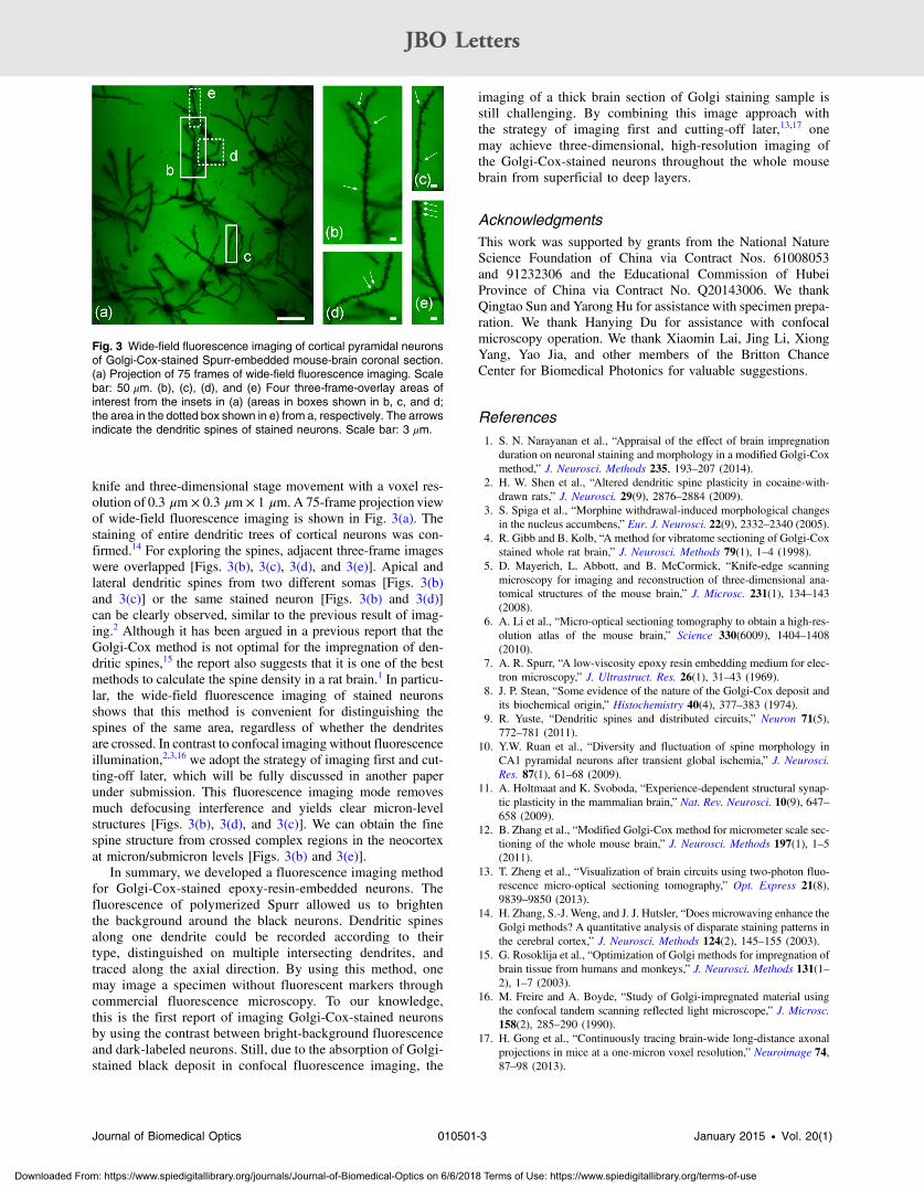
knife and three-dimensional stage movement with a voxel res-olution of 0.3 μm × 0.3 μm × 1 μm. A 75-frame projection viewof wide-field fluorescence imaging is shown in Fig. 3(a). Thestaining of entire dendritic trees of cortical neurons was con-firmed.14 For exploring the spines, adjacent three-frame imageswere overlapped [Figs. 3(b), 3(c), 3(d), and 3(e)]. Apical andlateral dendritic spines from two different somas [Figs. 3(b)and 3(c)] or the same stained neuron [Figs. 3(b) and 3(d)]can be clearly observed, similar to the previous result of imag-ing.2 Although it has been argued in a previous report that theGolgi-Cox method is not optimal for the impregnation of den-dritic spines,15 the report also suggests that it is one of the bestmethods to calculate the spine density in a rat brain.1 In particu-lar, the wide-field fluorescence imaging of stained neuronsshows that this method is convenient for distinguishing thespines of the same area, regardless of whether the dendritesare crossed. In contrast to confocal imaging without fluorescenceillumination,2,3,16 we adopt the strategy of imaging first and cut-ting-off later, which will be fully discussed in another paperunder submission. This fluorescence imaging mode removesmuch defocusing interference and yields clear micron-levelstructures [Figs. 3(b), 3(d), and 3(c)]. We can obtain the finespine structure from crossed complex regions in the neocortexat micron/submicron levels [Figs. 3(b) and 3(e)].
In summary, we developed a fluorescence imaging methodfor Golgi-Cox-stained epoxy-resin-embedded neurons. Thefluorescence of polymerized Spurr allowed us to brightenthe background around the black neurons. Dendritic spinesalong one dendrite could be recorded according to theirtype, distinguished on multiple intersecting dendrites, andtraced along the axial direction. By using this method, onemay image a specimen without fluorescent markers throughcommercial fluorescence microscopy. To our knowledge,this is the first report of imaging Golgi-Cox-stained neuronsby using the contrast between bright-background fluorescenceand dark-labeled neurons. Still, due to the absorption of Golgi-stained black deposit in confocal fluorescence imaging, the
imaging of a thick brain section of Golgi staining sample isstill challenging. By combining this image approach withthe strategy of imaging first and cutting-off later,13,17 onemay achieve three-dimensional, high-resolution imaging ofthe Golgi-Cox-stained neurons throughout the whole mousebrain from superficial to deep layers.
AcknowledgmentsThis work was supported by grants from the National NatureScience Foundation of China via Contract Nos. 61008053and 91232306 and the Educational Commission of HubeiProvince of China via Contract No. Q20143006. We thankQingtao Sun and Yarong Hu for assistance with specimen prepa-ration. We thank Hanying Du for assistance with confocalmicroscopy operation. We thank Xiaomin Lai, Jing Li, XiongYang, Yao Jia, and other members of the Britton ChanceCenter for Biomedical Photonics for valuable suggestions.
References1. S. N. Narayanan et al., “Appraisal of the effect of brain impregnation
duration on neuronal staining and morphology in a modified Golgi-Coxmethod,” J. Neurosci. Methods 235, 193–207 (2014).
2. H. W. Shen et al., “Altered dendritic spine plasticity in cocaine-with-drawn rats,” J. Neurosci. 29(9), 2876–2884 (2009).
3. S. Spiga et al., “Morphine withdrawal-induced morphological changesin the nucleus accumbens,” Eur. J. Neurosci. 22(9), 2332–2340 (2005).
4. R. Gibb and B. Kolb, “A method for vibratome sectioning of Golgi-Coxstained whole rat brain,” J. Neurosci. Methods 79(1), 1–4 (1998).
5. D. Mayerich, L. Abbott, and B. McCormick, “Knife-edge scanningmicroscopy for imaging and reconstruction of three-dimensional ana-tomical structures of the mouse brain,” J. Microsc. 231(1), 134–143(2008).
6. A. Li et al., “Micro-optical sectioning tomography to obtain a high-res-olution atlas of the mouse brain,” Science 330(6009), 1404–1408(2010).
7. A. R. Spurr, “A low-viscosity epoxy resin embedding medium for elec-tron microscopy,” J. Ultrastruct. Res. 26(1), 31–43 (1969).
8. J. P. Stean, “Some evidence of the nature of the Golgi-Cox deposit andits biochemical origin,” Histochemistry 40(4), 377–383 (1974).
9. R. Yuste, “Dendritic spines and distributed circuits,” Neuron 71(5),772–781 (2011).
10. Y.W. Ruan et al., “Diversity and fluctuation of spine morphology inCA1 pyramidal neurons after transient global ischemia,” J. Neurosci.Res. 87(1), 61–68 (2009).
11. A. Holtmaat and K. Svoboda, “Experience-dependent structural synap-tic plasticity in the mammalian brain,” Nat. Rev. Neurosci. 10(9), 647–658 (2009).
12. B. Zhang et al., “Modified Golgi-Cox method for micrometer scale sec-tioning of the whole mouse brain,” J. Neurosci. Methods 197(1), 1–5(2011).
13. T. Zheng et al., “Visualization of brain circuits using two-photon fluo-rescence micro-optical sectioning tomography,” Opt. Express 21(8),9839–9850 (2013).
14. H. Zhang, S.-J. Weng, and J. J. Hutsler, “Does microwaving enhance theGolgi methods? A quantitative analysis of disparate staining patterns inthe cerebral cortex,” J. Neurosci. Methods 124(2), 145–155 (2003).
15. G. Rosoklija et al., “Optimization of Golgi methods for impregnation ofbrain tissue from humans and monkeys,” J. Neurosci. Methods 131(1–2), 1–7 (2003).
16. M. Freire and A. Boyde, “Study of Golgi-impregnated material usingthe confocal tandem scanning reflected light microscope,” J. Microsc.158(2), 285–290 (1990).
17. H. Gong et al., “Continuously tracing brain-wide long-distance axonalprojections in mice at a one-micron voxel resolution,” Neuroimage 74,87–98 (2013).
Fig. 3 Wide-field fluorescence imaging of cortical pyramidal neuronsof Golgi-Cox-stained Spurr-embedded mouse-brain coronal section.(a) Projection of 75 frames of wide-field fluorescence imaging. Scalebar: 50 μm. (b), (c), (d), and (e) Four three-frame-overlay areas ofinterest from the insets in (a) (areas in boxes shown in b, c, and d;the area in the dotted box shown in e) from a, respectively. The arrowsindicate the dendritic spines of stained neurons. Scale bar: 3 μm.
Journal of Biomedical Optics 010501-3 January 2015 • Vol. 20(1)
JBO Letters
Downloaded From: https://www.spiedigitallibrary.org/journals/Journal-of-Biomedical-Optics on 6/6/2018 Terms of Use: https://www.spiedigitallibrary.org/terms-of-use


![8 The Dendritic Cytoskeleton as a Computational …296 Avner Priel, Jack A. Tuszynski, and Horacion F. Cantiello stability of dendritic spines [28, 79, 63, 29]. Twitching of dendritic](https://img.dokumen.tips/doc/110x75/5f8dcd062227ba1c7c5790dc/8-the-dendritic-cytoskeleton-as-a-computational-296-avner-priel-jack-a-tuszynski.jpg)