Embed Size (px)
Citation preview
FluorescenceFluorescence and and
ChemiluminescenceChemiluminescence
Skoumalová, Vytášek, Srbová
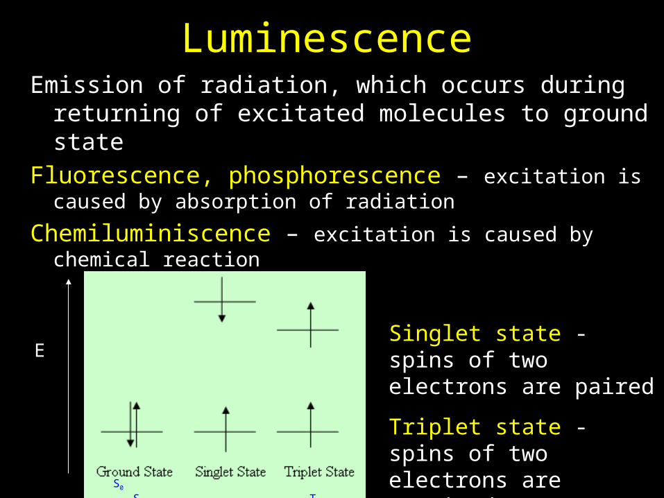
LuminescenceEmission of radiation, which occurs during returning of
excitated molecules to ground state
Fluorescence, phosphorescence – excitation is caused by absorption of radiation
Chemiluminiscence – excitation is caused by chemical reaction
S0 S1 T1
ESinglet state - spins of two electrons are paired
Triplet state - spins of two electrons are unpaired

Fluorescence and fosforescence

Energy level diagram for photoluminescent molecules
Radiationless transitions:
VR –vibrational relaxation
IC- internal conversion
ISC –intersystem crossing
E
Radiation transitions:
Fluorescence - transition to the ground state with the same multiplicity S1S0
probability of fluorescence is higher than phosphorescence
PhosphorescencePhosphorescence – – transition between states with different multiplicity Ttransition between states with different multiplicity T11SS00
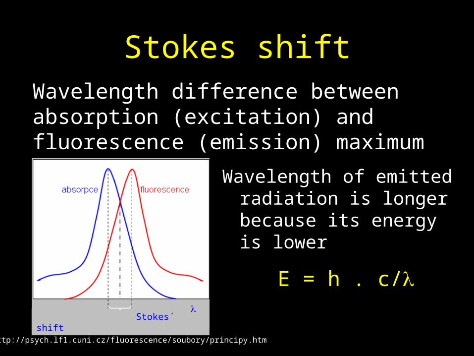
Stokes shift
Wavelength of emitted radiation is longer because its energy is lower
E = h . c/
Stokes´ shift
Wavelength difference between absorption (excitation) and fluorescence (emission) maximum
http://psych.lf1.cuni.cz/fluorescence/soubory/principy.htm

Quantitative fluorescent measurement I0 It
If
intensity of fluorescence If
intensity of absorption Ia
f = =
Ia = I0 - It
sample
Fluorescence efficiency (f ) is the fraction of the incident radiation which is emitted as fluorescence f < 1
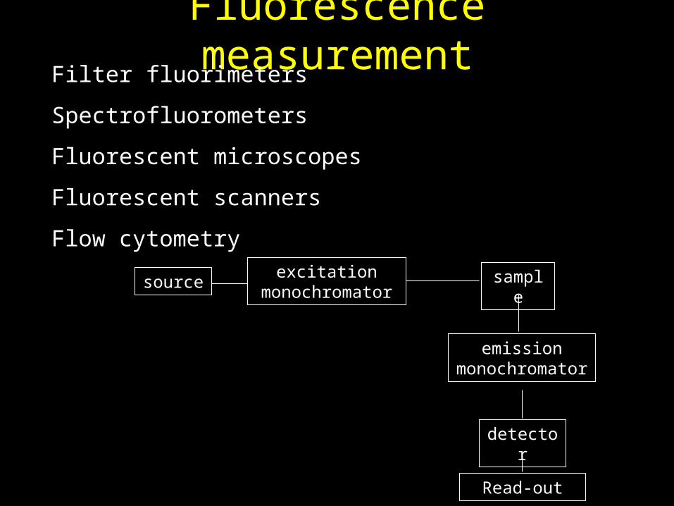
Fluorescence measurement
sample
emission monochromator
detector
sourceexcitation
monochromator
Read-out
Filter fluorimeters
Spectrofluorometers
Fluorescent microscopes
Fluorescent scanners
Flow cytometry

Spectrofluorometer
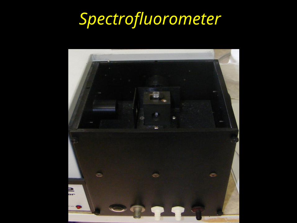
Spectrofluorometer

Analysis of the unknown sample
Erythrocytes (patients with Alzheimer´s disease)
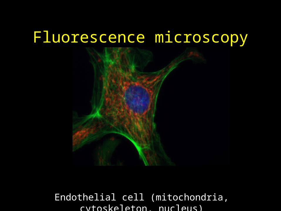
Fluorescence microscopy
Endothelial cell (mitochondria, cytoskeleton, nucleus)
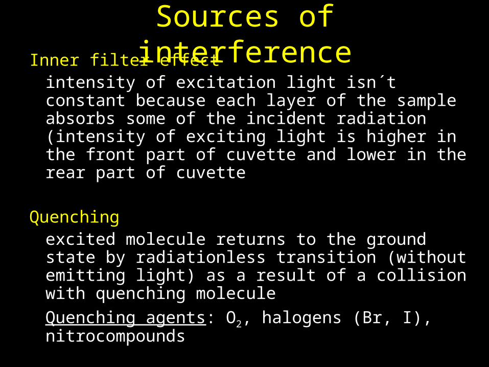
Sources of interferenceInner filter effect
intensity of excitation light isn´t constant because each layer of the sample absorbs some of the incident radiation (intensity of exciting light is higher in the front part of cuvette and lower in the rear part of cuvette
Quenching excited molecule returns to the ground state by radiationless transition (without emitting light) as a result of a collision with quenching molecule
Quenching agents: O2, halogens (Br, I), nitrocompounds
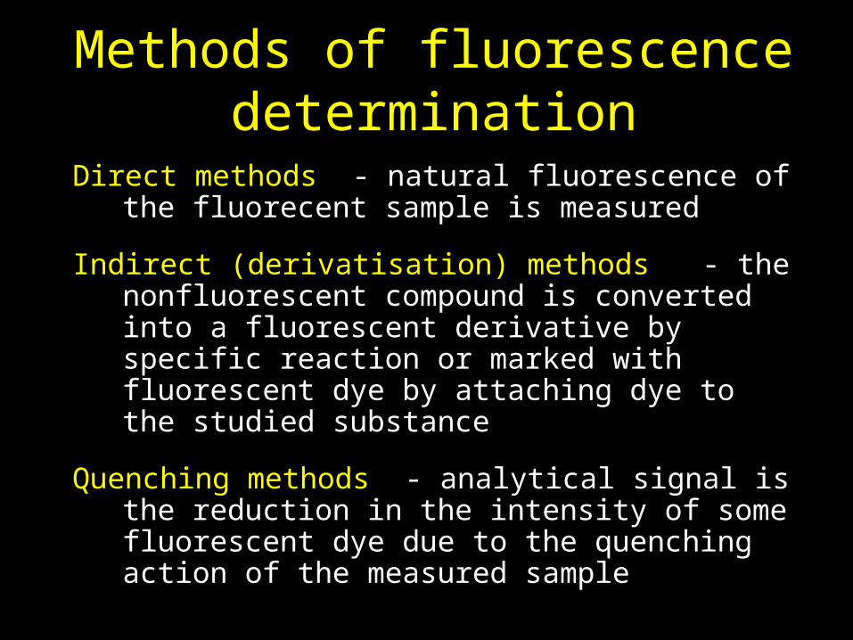
Methods of fluorescence determination
Direct methods - natural fluorescence of the fluorecent sample is measured
Indirect (derivatisation) methods - the nonfluorescent compound is converted into a fluorescent derivative by specific reaction or marked with fluorescent dye by attaching dye to the studied substance
Quenching methods - analytical signal is the reduction in the intensity of some fluorescent dye due to the quenching action of the measured sample

Natural fluorophores
• Polyaromatic hydrocarbons• Vitamin A, E• Coenzymes (FAD, FMN, NADH)• Carotenes• Quinine• Steroids• Aromatic aminoacids• Nucleotides• Fluorescent proteins –GFP (green fluorescent
protein)
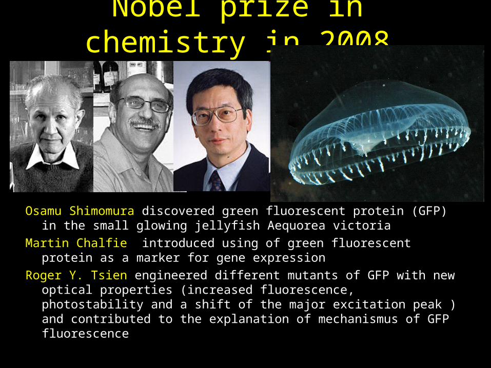
Nobel prize in chemistry in 2008
Osamu Shimomura discovered green fluorescent protein (GFP) in the small glowing jellyfish Aequorea victoria
Martin Chalfie introduced using of green fluorescent protein as a marker for gene expression
Roger Y. Tsien engineered different mutants of GFP with new optical properties (increased fluorescence, photostability and a shift of the major excitation peak ) and contributed to the explanation of mechanismus of GFP fluorescence
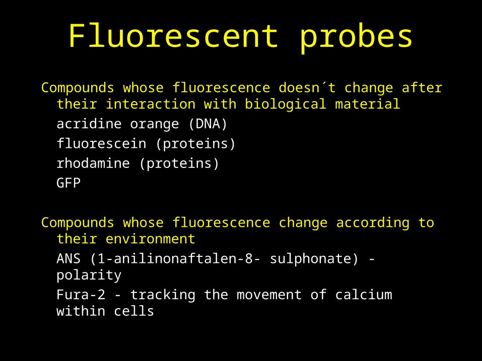
Fluorescent probes
Compounds whose fluorescence doesn´t change after their interaction with biological material
acridine orange (DNA)
fluorescein (proteins)
rhodamine (proteins)
GFP
Compounds whose fluorescence change according to their environment
ANS (1-anilinonaftalen-8- sulphonate) - polarity
Fura-2 - tracking the movement of calcium within cells

Some applications of fluorescence detection
• Protein conformation• Membrane potential• Membrane transport• Membrane viscosity• Enzymatic reactions• DNA analysis• Genetic engineering (manipulations)• Immunochemical methods• Cell proliferation and apoptosis
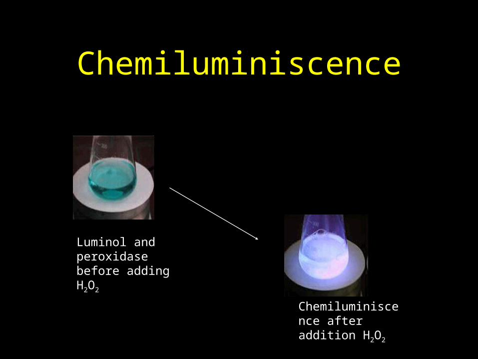
Chemiluminiscence
Luminol and peroxidase before adding H2O2
Chemiluminiscence after addition H2O2
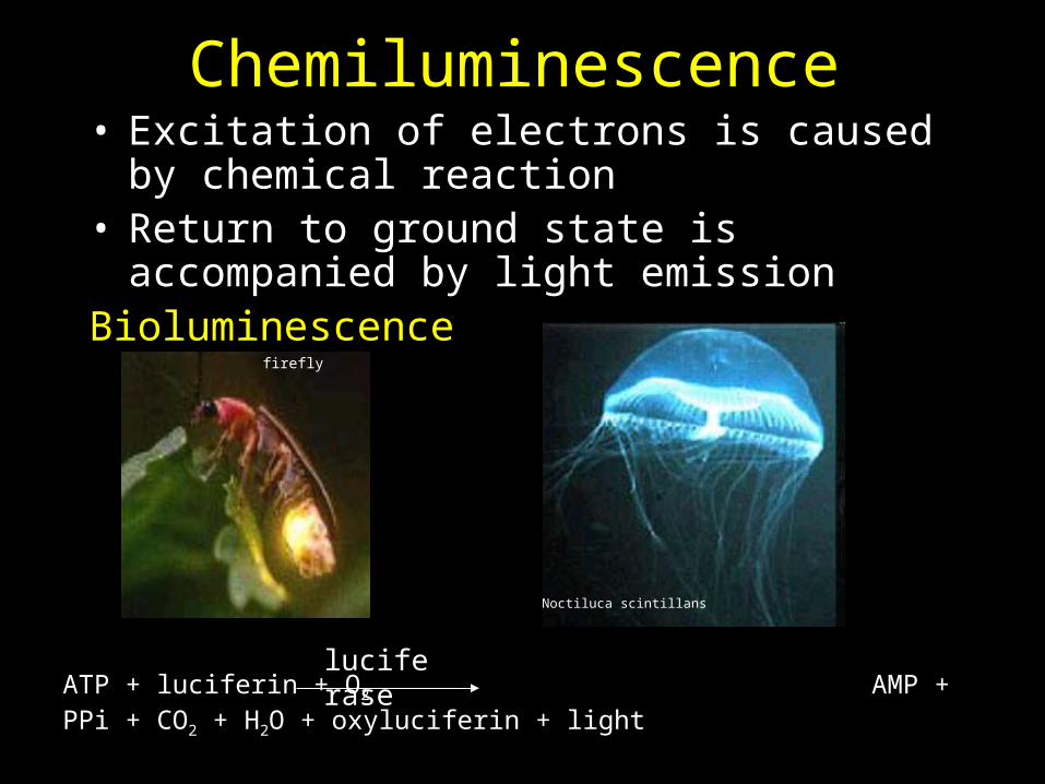
Chemiluminescence• Excitation of electrons is caused by chemical
reaction • Return to ground state is accompanied by light
emissionBioluminescence
firefly
Noctiluca scintillans
ATP + luciferin + O2 AMP + PPi + CO2 + H2O + oxyluciferin + lightluciferase

Application of chemiluminescence detection
• NO assay
NO + O3 NO2* + O2
NO2* NO2 + light
• H2O2 assay, peroxidase activity assay, immunochemical assays
Luminol + H2O2 3-aminoftalate + lightperoxidase

Summary:
1. The principle of fluorescence
2. Applications of fluorescence in medicine - examples
3. Chemiluminescence - applications